Báo cáo y học: "Variation in HIV-1 R5 macrophage-tropism correlates with sensitivity to reagents that block envelope: CD4 interactions but not with sensitivity to other entry inhibitors" pps
Bạn đang xem bản rút gọn của tài liệu. Xem và tải ngay bản đầy đủ của tài liệu tại đây (413.86 KB, 17 trang )
BioMed Central
Page 1 of 17
(page number not for citation purposes)
Retrovirology
Open Access
Research
Variation in HIV-1 R5 macrophage-tropism correlates with
sensitivity to reagents that block envelope: CD4 interactions but
not with sensitivity to other entry inhibitors
Paul J Peters
1
, Maria J Duenas-Decamp
1
, W Matthew Sullivan
1
,
Richard Brown
2
, Chiambah Ankghuambom
2
, Katherine Luzuriaga
3
,
James Robinson
4
, Dennis R Burton
5
, Jeanne Bell
6
, Peter Simmonds
7
,
Jonathan Ball
2
and Paul R Clapham*
1
Address:
1
Center for AIDS Research, Program in Molecular Medicine and Department of Molecular Genetics and Microbiology, 373 Plantation
Street, University of Massachusetts Medical School, Worcester, MA 01605, USA,
2
Microbiology and Infectious Diseases, Institute of Infection,
Immunity and Inflammation, The University of Nottingham, Queen's Medical Centre, Nottingham NG7 2UH, UK,
3
Center for AIDS Research,
Program in Molecular Medicine and Department of Pediatrics, 373 Plantation Street, University of Massachusetts Medical School, Worcester, MA
01605, USA,
4
Department of Pediatrics, Tulane University School of Medicine, 1430 Tulane Avenue, New Orleans, LA 70112, USA,
5
The Scripps
Research Institute, Departments of Immunology and Molecular Biology, IMM2, La Jolla, CA 92037, USA,
6
Department of Neuropathology,
Western General Hospital, Crewe Road, Edinburgh, EH4 2XU, UK and
7
Centre for Infectious Diseases, University of Edinburgh, Summerhall,
Edinburgh, EH9 1QH, UK
Email: Paul J Peters - ; Maria J Duenas-Decamp - ; W
Matthew Sullivan - ; Richard Brown - ;
Chiambah Ankghuambom - ; Katherine Luzuriaga - ;
James Robinson - ; Dennis R Burton - ; Jeanne Bell - ;
Peter Simmonds - ; Jonathan Ball - ;
Paul R Clapham* -
* Corresponding author
Abstract
Background: HIV-1 R5 viruses cause most of the AIDS cases worldwide and are preferentially
transmitted compared to CXCR4-using viruses. Furthermore, R5 viruses vary extensively in
capacity to infect macrophages and highly macrophage-tropic variants are frequently identified in
the brains of patients with dementia. Here, we investigated the sensitivity of R5 envelopes to a
range of inhibitors and antibodies that block HIV entry. We studied a large panel of R5 envelopes,
derived by PCR amplification without culture from brain, lymph node, blood and semen. These R5
envelopes conferred a wide range of macrophage tropism and included highly macrophage-tropic
variants from brain and non-macrophage-tropic variants from lymph node.
Results: R5 macrophage-tropism correlated with sensitivity to inhibition by reagents that inhibited
gp120:CD4 interactions. Thus, increasing macrophage-tropism was associated with increased
sensitivity to soluble CD4 and to IgG-CD4 (PRO 542), but with increased resistance to the anti-
CD4 monoclonal antibody (mab), Q4120. These observations were highly significant and are
consistent with an increased affinity of envelope for CD4 for macrophage-tropic envelopes. No
overall correlations were noted between R5 macrophage-tropism and sensitivity to CCR5
antagonists or to gp41 specific reagents. Intriguingly, there was a relationship between increasing
Published: 18 January 2008
Retrovirology 2008, 5:5 doi:10.1186/1742-4690-5-5
Received: 9 November 2007
Accepted: 18 January 2008
This article is available from: />© 2008 Peters et al; licensee BioMed Central Ltd.
This is an Open Access article distributed under the terms of the Creative Commons Attribution License ( />),
which permits unrestricted use, distribution, and reproduction in any medium, provided the original work is properly cited.
Retrovirology 2008, 5:5 />Page 2 of 17
(page number not for citation purposes)
macrophage-tropism and increased sensitivity to the CD4 binding site mab, b12, but decreased
sensitivity to 2G12, a mab that binds a glycan complex on gp120.
Conclusion: Variation in R5 macrophage-tropism is caused by envelope variation that
predominantly influences sensitivity to reagents that block gp120:CD4 interactions. Such variation
has important implications for therapy using viral entry inhibitors and for the design of envelope
antigens for vaccines.
Introduction
HIV-1 infection is triggered by interactions between the
viral envelope glycoprotein and cell surface receptor CD4
and either of the coreceptors; CCR5 or CXCR4. These
interactions induce the fusion of viral and cellular mem-
branes and viral entry into cells. CCR5-using (R5) viruses
are mainly transmitted [1], while CXCR4-using (X4) vari-
ants can be isolated from up to 50% of AIDS patients in
subtype B infections and correlate with a more rapid loss
of CD4
+
T-cells and faster disease progression [2-5].
Among T-cells, CCR5 expression is mainly restricted to
memory T-cells [6,7], while CXCR4 is more widely
expressed on various CD4
+
T-cell populations including
naïve T-cells [6]. R5 viruses therefore target CCR5
+
mem-
ory T-cell populations and in the acute phase of replica-
tion, decimate the populations of CD4
+
memory cells in
lymphoid tissue associated with the gut and other mucosa
[8-10]. CCR5 is also expressed on macrophage lineage
cells [7] in non-lymphoid tissues e.g. the brain [11], and
R5 viruses predominantly target these cells in neural tis-
sues [12-14]. When CXCR4-using viruses emerge in late
disease, they colonize naïve T-cell populations that were
not infected by R5 viruses [15,16]. Nonetheless, CD4
depletion and AIDS occur in patients from which only
CCR5-using viruses can be isolated [17,18]. In clade C
infections, CXCR4-using variants have been detected in
far fewer individuals in the late stages of disease [17,19-
22]. Thus, AIDS and death presumably occurs in the
absence of CXCR4-using variants for a substantial number
of HIV
+
patients and is caused directly by R5 viruses.
R5 viruses are frequently regarded as macrophage-tropic.
However, several groups have reported considerable vari-
ation in the cell tropism of R5 viruses [23-25]. We
reported that primary HIV-1 R5 isolates varied in their
capacity to infect primary macrophage cultures by over
1000-fold [25] and we first described a subset of HIV-1 R5
isolates that could infect CD4
+
T-cell lines via trace
amounts of CCR5 [23]. More recently, we described R5
envelopes amplified from brain and lymph node tissue of
AIDS patients that also differed markedly in tropism prop-
erties [26,27]. Thus R5 envelopes from brain tissue were
highly macrophage-tropic and were able to exploit low
amounts of CD4 and/or CCR5 for infection. They con-
trasted considerably with R5 envelopes from immune tis-
sue (lymph node) that conferred inefficient macrophage
infection and required high amounts of CD4 for infec-
tion. Moreover, these non-macrophage-tropic envelopes
were more prevalent (than macrophage-tropic envelopes)
amplified from immune tissue, blood or semen [27].
These results generally support earlier reports that
described a small number of highly macrophage-tropic R5
virus isolates made from brain tissue [28]. Others have
confirmed that envelopes amplified from brain tissue can
infect cells via low CD4 levels [29,30]. However, Thomas
et al. reported less compartmentalized variation of R5
macrophage tropism, with macrophage-tropic R5 enve-
lopes present in both lymphoid and brain tissue [30]. The
capacity of highly macrophage-tropic envelopes to use
low amounts of CD4 and/or CCR5 suggests that such var-
iants could also confer a broader tropism among CD4
+
T-
cells (that express low amounts of these receptors) and
contribute to CD4
+
T-cell depletion late in disease if they
are present in immune tissue.
Several groups have also reported differences in the prop-
erties of R5 virus isolates made from blood. Thus, virus
isolates from late disease were reported to be more macro-
phage-tropic than those from earlier stages [31-33]. In
addition, Repits et al. described late disease isolates with
increased replicative capacity and reduced sensitivity to
entry inhibitors including TAK779 (CCR5 antagonist)
and T20 (gp41 inhibitor) [34]. However, they did not test
whether these late isolates could exploit low CD4 or infect
macrophages. It is unclear whether the highly macro-
phage-tropic envelopes that we have amplified from brain
tissue and other sites, correspond to the late isolates
described by other groups [31-34].
Recently, Dunfee et al. described a polymorphism in the
C2 region of the CD4 binding site on gp120. Thus, 41%
of their envelope sequences from brain tissue of patients
with dementia carried an asparagine at residue 283 com-
pared with 8% of envelopes from patients without
dementia [35]. We also reported a predominance of N283
in highly macrophage-tropic brain envelopes compared
to lymph node, blood and semen [27]. N283 was shown
to increase the affinity of monomeric gp120 for CD4 [35].
More recently, the loss of a glycosylation site (N386) close
to the CD4 binding loop on gp120 was reported to occur
more frequently in HIV in the brain and was shown to
contribute to increased R5 macrophage-tropism [36], an
Retrovirology 2008, 5:5 />Page 3 of 17
(page number not for citation purposes)
observation that we have recently confirmed (Duenas-
Decamp et al. Personal communication).
How variation in R5 tropism impacts on the sensitivity of
HIV-1 to neutralizing antibodies and entry inhibitors is
unclear. We, and others have reported that R5 macro-
phage-tropism correlated with increased resistance to
anti-CD4 monoclonal antibodies (mabs), consistent with
an increased affinity between gp120 and CD4. However,
there was no correlation with sensitivity to the CCR5
antagonist, TAK779 [26,29]. Here, we have extensively
analyzed the sensitivity of thirty-six envelopes from brain,
LN, blood and semen to a range of reagents that block
HIV-1 entry. All these envelopes were derived from
patient material by PCR without culture and have there-
fore not been altered by viral isolation procedures. Rea-
gents tested for inhibition included soluble CD4 (sCD4)
and tetrameric IgG-CD4 (PRO 542), BMS-378806; a small
molecule that targets a site deep in the cleft that binds
CD4, mabs to CD4 and CCR5, CCR5 antagonists, T20 and
human mabs that recognize conserved neutralization
epitopes on gp120 and gp41.
Our results strongly suggest that R5 macrophage-tropism
is primarily modulated by changes in the CD4 binding
site on gp120 and in its affinity for CD4. Such changes
impact on sensitivity to the CD4bs mab, b12 and may be
driven by the presence or absence of neutralizing antibod-
ies in vivo that target the CD4bs or proximal sites. If highly
macrophage-tropic R5 variants are preferentially transmit-
ted, then vaccines that generate antibodies to the CD4bs
may be particularly effective at preventing viral transmis-
sion.
Results
Macrophage-tropism of brain and lymph node envelopes
Envelopes used here have been described previously
[26,27] with the addition of SQ43 380.4. They are all R5,
predominantly using CCR5 as a coreceptor [26,27]. Table
1 shows macrophage infectivity as a percentage of the titer
recorded on HeLa TZM-BL cells as described previously
[27]. Macrophage infectivity was highly variable. Enve-
lopes that conferred macrophage infectivity of >0.5% of
infectivity for HeLa TZM-BL cells were designated as mac-
rophage-tropic and are shown by bold script in Table 1.
This arbitrary designation allows for easy identification of
these envelopes as grey symbols in subsequent figures. All
but one brain envelope conferred macrophage infection.
None of the env
+
pseudovirions carrying lymph node
envelopes conferred significant macrophage infection.
Table 1: Macrophage tropism of R5 envelopes studied.
Patient Number Envelope Macrophage Infectivity (%)
a
Patient Number Envelope Macrophage Infectivity (%)
a
NA20 B59 16.9
b
P1114 C95-65 0.029
B76 0.179 C96-26 0.097
B501 51.6 C98-15 32.4
LN3 <0.001 C98-18 2.21
LN8 <0.001 C98-27 0.144
LN10 0.030 C98-28 0.004
LN14 0.025 C98-67 0.003
LN16 0.036 P3 Q3 164 1.4 0.002
NA420 B13 0.335 Q3 180 6.4 0.003
B33 3.35 SQ3 196 10.1 0.012
B42 0.559 SQ3 197 9.3 0.338
LN40 0.009 SQ3 199 8.5 0.003
LN85 0.026 P31 Q31 350.1 0.05
NA118 B12 0.006 Q31 351.6 0.02
LN27 0.023 SQ31 308.2 0.02
LN33 0.023 P43 Q43 378.2 0.03
NA176 B93 8.2 SQ43 380.1 0.6
NA353 B27 12.6 SQ43 380.4 9.63
Controls AD8 4.60
SF162 6.25
YU2 6.36
JRFL 3.27
JRCSF 0.011
a
Macrophage infectivity as a percent of infectivity recorded on HeLa TZM-BL cells. Most of this data is derived from that presented in Peters et al.
[27] with the addition of SQ43 380.4.
b
Bolded percentages indicate envelopes that were designated as macrophage-tropic i.e. >0.5% of infectivity for HeLa TZM-BL cells.
Retrovirology 2008, 5:5 />Page 4 of 17
(page number not for citation purposes)
Macrophage-tropic R5 envelopes were amplified less fre-
quently from blood and semen of adults and in plasma of
infants.
The effect of variation in R5 envelope tropism on
sensitivity to entry inhibitors and neutralizing antibodies
In immune tissue where there are high levels of neutraliz-
ing antibodies, the HIV-1 envelope may evolve to protect
critical sites (e.g. the CD4bs) from antibodies. In contrast,
the brain is enclosed by the blood brain barrier, which
usually restricts immunoglobulin from entering [37,38].
HIV-1 variants replicating in the brain may therefore
evolve stronger interactions with CD4 and/or CCR5
resulting from enhanced exposure of the CD4 and/or
CCR5 binding sites, but become more vulnerable to anti-
body neutralization. We tested the sensitivity of our panel
of brain, LN, blood and semen envelopes to a range of
entry inhibitors and monoclonal antibodies. The entry
inhibitors specifically block interactions of envelope with
CD4 or CCR5, or prevent gp41 conformational changes
required for fusion, while monoclonal antibodies steri-
cally inhibit infection by binding conserved envelope sites
on virions.
Inhibitors and antibodies that interfere with envelope:CD4
interactions
Figure 1A shows that macrophage-tropic envelopes were
more resistant to inhibition by the CD4 mab, Q4120,
which binds domain 1 of CD4 and competes with enve-
lope for binding to CD4. In contrast, the same macro-
phage-tropic envelopes were more sensitive to soluble
CD4 (sCD4) (Figure 1B) and to the more potent tetra-
meric IgG-CD4 construct (PRO 542) (Figure 1C). We used
two-tailed non-parametric Spearman analyses to evaluate
whether macrophage-tropism correlated with sensitivity
to these reagents. Importantly, such analyses do not rely
on our arbitrary designation of macrophage-tropism but
simply compare macrophage infectivity titers (Table 1)
with IC50s for each inhibitor. Our results showed highly
significant correlations between increasing macrophage-
tropism and increased sensitivity to sCD4 and PRO 542 as
well as with an increased resistance to Q4120 (Table 2).
These results are consistent with an increased affinity of
R5 macrophage-tropic gp120s for binding to CD4,
although alternative explanations should also be consid-
ered (see below). Statistical evaluations of correlations
between R5 macrophage-tropism and sensitivity to differ-
ent inhibitors are discussed more fully below and p values
are shown in Table 2.
We also tested the small molecule, BMS-378806, which
was reported to inhibit gp120 binding to CD4 [39-41]
and subsequent conformational changes [42]. BMS-
378806 is believed to bind into a deep hydrophobic chan-
nel of unliganded gp120 close to and underneath the sites
that bind to CD4. Thus, BMS-378806 may directly inhibit
CD4 binding and also act to stabilize the unliganded form
of the gp120 [43]. There was also a highly significant cor-
relation between R5 macrophage-tropism and BMS-
378806 sensitivity (Table 2, see below). However, in con-
trast to sCD4 and tetrameric IgG-CD4, BMS-378806 sen-
sitivity decreased with increasing macrophage-tropism.
We next tested envelope sensitivity to the CD4bs mab,
b12 (Figure 2). All but one macrophage-tropic env con-
ferred sensitivity to b12 neutralization, while many non-
macrophage-tropic envelopes were resistant at 50 μg/ml
antibody. These results indicate that there is also a strong
relationship between b12 sensitivity and R5 envelope tro-
pism, although this did not result in a statistically signifi-
cant overall correlation (Table 2).
Sensitivity of R5 envelopes to reagents that target
envelope:CCR5 interactions
The mouse mab 17b binds to a conserved CD4-induced
epitope on gp120 that overlaps the conserved part of the
coreceptor binding site (not shown). None of the patient
envelopes were inhibited by 17b, suggesting that this site
is not more exposed on macrophage-tropic envelopes.
However, 17b did neutralize T-cell line adapted HIV-1 iso-
lates NL4.3 and HXBc2 (not shown).
In contrast, both CCR5 antagonists TAK779 and
SCH350581 inhibited all the envelopes regardless of their
tropism for macrophages (Figures 3A and 3B). As
expected SCH350581 was a substantially more potent
inhibitor compared to TAK779. In contrast to the strong
correlations observed between macrophage-tropism and
reagents that inhibited gp120:CD4 interactions, overall
correlations with sensitivity to CCR5 antagonists were not
significant (Table 2). CCR5 antagonists bind to a cavity in
between the transmembrane domains of CCR5. It is
believed that these reagents confer a CCR5 structure that
is no longer recognized by the HIV envelope [44,45].
Thus, although CCR5 antagonists compete with HIV for
binding CCR5, they are not competing for the same site.
It was possible that CCR5-specific inhibitors that compete
directly with HIV for binding the extracellular regions of
CCR5 may confer a different pattern of envelope sensitiv-
ity. We therefore tested the anti-CCR5 monoclonal anti-
body, 2D7, which binds ECL2 of CCR5, a region that
interacts with sites on the V3 loop of envelope. Due to
limiting amounts of 2D7, we tested only brain and LN
envelopes from patients NA420 and NA20, with JRFL and
JRCSF as controls. Figure 3C shows a trend of brain mac-
rophage-tropic envelopes being more sensitive to 2D7
compared to LN envelopes, although this did not reach
statistical significance (p = 0.0839). NA20 LN14 was a
clear 'outlier' from other LN envelopes and was among the
Retrovirology 2008, 5:5 />Page 5 of 17
(page number not for citation purposes)
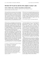
Sensitivity of HIV-1 R5 envelopes to reagents that interfere with gp120:CD4 interactionsFigure 1
Sensitivity of HIV-1 R5 envelopes to reagents that interfere with gp120:CD4 interactions. Pseudovirions carrying envelopes
encoded by envelope genes amplified from patient samples were tested for sensitivity to inhibition by (A) anti-CD4 mab,
Q4120, (B) sCD4, (C) PRO 542 and (D) BMS-378806. Macrophage-tropic envelopes (light symbols) were more sensitive to
sCD4 and PRO 542 compared to non-macrophage-tropic envelopes (dark symbols) but were more resistant to the anti-CD4
mab, Q4120.
$'
1$%
1$%
1$%
1$%
1$%
1$%
1$/1
1$/1
1$/1
1$/1
1$/1
1$/1
-5&6)
6)
-5)/
1$%
1$%
1$%
1$/1
1$/1
1$/1
<8
&
&
&
&
&
&
&
4
4
4
4
4
64
64
64
64
64
64
!
1$%
1$%
1$%
1$%
1$%
1$%
1$%
1$%
1$%
1$/1
1$/1
1$/1
1$/1
1$/1
1$/1
1$/1
1$/1
1$/1
&
&
&
&
&
&
&
4
4
4
4
4
64
64
64
64
<8
-5)/
-5&6)
$'
6)
1$%
1$%
1$%
1$%
1$%
1$%
1$/1
1$/1
1$/1
1$/1
&
&
&
&
&
&
&
4
4
4
4
64
64
64
64
-5&6)
$'
<8
6)
-5)/
!
1$/1
1$/1
1$/1
1$/1
1$/1
64
64
4
1$%
1$%
1$%
64
64
1$%
1$%
1$%
1$%
1$%
1$%
1$%
1$%
1$%
1$/1
1$/1
1$/1
1$/1
1$/1
1$/1
1$/1
1$/1
&
&
&
&
&
&
4
4
4
4
4
64
64
64
64
<8
-5)/
-5&6)
$'
6)
64
64
1$/1
&
!
%UDLQ /1 3HGLDWULF
SODVPD
$GXOW
EORRG
$GXOW
VHPHQ
&RQWUROV
PJPO
PJPOPJPO
Q0
,&V
$4
%V&'
&352
'%06
Retrovirology 2008, 5:5 />Page 6 of 17
(page number not for citation purposes)
envelopes most sensitive to all three CCR5 inhibitors (see
discussion below).
Inhibition by human mab, 2G12 that targets gp120
glycosylation groups
The human monoclonal antibody, 2G12, neutralizes HIV-
1 isolates mainly from clade B via relatively conserved gly-
cosylation structures on gp120 [46,47]. Clear variation in
sensitivity to 2G12 was noted, with most envelopes sensi-
tive, while some were resistant (Figure 4). Of note, several
brain-derived envelopes were resistant including NA420
envelopes B13, B33 and B42 as well as NA353 B27 and
YU2. A significant correlation between macrophage-tro-
pism and decreased 2G12 sensitivity was noted. Table 3
lists the presence or absence of glycosylation sites previ-
ously reported to be important for 2G12 binding [46,47].
All five of the NA420 envelopes lacked the critical poten-
tial glycosylation site at N339, while B13 and B33 also
lacked N386. The loss of these glycosylation sites likely
contributes to 2G12 resistance for some of these enve-
lopes. However, LN40 is sensitive to 2G12 despite lacking
N339, and NA353 B27 is resistant even though all the
Table 2: Non-parametric two-tailed Spearman analysis for correlations between R5 envelope macrophage-tropism and sensitivity to
entry inhibitors.
Inhibitor/Antibody Target of reagent Stage of entry blocked
3
. Correlation with Macrophage-tropism (p Values)
Q4120 CD4 env: CD4 interactions <0.0001**
sCD4 gp120, CD4bs env: CD4 interactions <0.0001**
PRO 542 (IgG-CD4) gp120, CD4bs env: CD4 interactions <0.0001**
BMS-378806 gp120, CD4bs channel
1
. env: CD4 interactions 0.0002**
b12 gp120, overlapping CD4bs
2
. 0.6843
TAK779 CCR5 env: CCR5 interactions 0.7964
SCH350581 CCR5 env: CCR5 interactions 0.7587
2D7 CCR5 env: CCR5 interactions
2G12 gp120 glycan env: CCR5 interactions 0.0138*
T20 gp41 conformational changes gp41 conformational changes 0.7061
2F5 gp41 membrane proximal region gp41 conformational changes
4
. 0.3741
4E10 gp41 membrane proximal region gp41 conformational changes
4
. 0.3502
1. BMS-378806 binds to a hydrophobic channel deep in the channel targeted by CD4 [42].
2. Mab b12 binds an epitope that overlaps the CD4bs [57].
3. Mab 2G12 binds to a glycan on gp120. 2G12 blocks env:CCR5 interactions but may also block earlier stages of entry [73].
4. Mabs 2F5 and 4E10 block gp41 conformational changes but may also block earlier stages of entry [73].
* Significant (p ≤ 0.05).
** Highly significant (p ≤ 0.01)
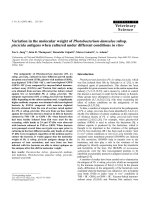
Sensitivity of HIV-1 R5 envelopes to the CD4bs mab, b12Figure 2
Sensitivity of HIV-1 R5 envelopes to the CD4bs mab, b12. Pseudovirions carrying envelopes encoded by envelope genes ampli-
fied from patient samples were tested for sensitivity to inhibition by b12. All but one of the macrophage-tropic envelopes (light
symbols) were sensitive to b12, while many non-macrophage-tropic envelopes (dark symbols) were resistant.
%UDLQ /1 3HGLDWULF
SODVPD
$GXOW
EORRG
$GXOW
VHPHQ
&RQWUROV
!
1$%
1$%
1$%
1$%
1$%
1$%
1$%
1$%
1$%
1$/1
1$/1
1$/1
1$/1
1$/1
1$/1
1$/1
1$/1
1$/1
&
&
&
&
&
&
&
4
4
4
4
4
64
64
64
-5&6)
$'
<8
6)
-5)/
64
64
64
,&PJPO
Retrovirology 2008, 5:5 />Page 7 of 17
(page number not for citation purposes)
2G12-implicated glycosylation sites are present. The
determinants for 2G12 resistance and sensitivity for these
envelopes are therefore unclear and will require further
investigation to define precisely.
Inhibition by mabs 4E10 and 2F5 that bind membrane
proximal epitopes on gp41
Figures 5A, 5B and Table 2 show that there was also no
clear correlation between macrophage-tropism and sensi-
tivity to the mabs 4E10 and 2F5 that bind conserved
membrane proximal epitopes on gp41. Of the envelopes
that conferred 2F5 resistance, only NA420 B42 (ELD-
NWA) did not contain the core ELDKWA epitope associ-
ated with 2F5 sensitivity [48-50].
Inhibition by T20 that inhibits formation of the gp41 6-
helix bundle required for fusion
All envelopes tested were sensitive to T20 (Figure 5C).
However, no overall correlation was observed between
T20 sensitivity and R5 macrophage-tropism. The envelope
determinants of resistance and sensitivity to T20 shown
here are unclear. All envelopes carried the GIV 36–38
motif in HR1, the site where resistance mutations fre-
quently appear [51,52].
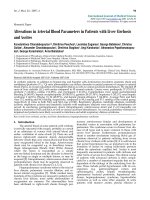
Sensitivity of HIV-1 R5 envelopes to reagents that interfere with gp120:CCR5 interactionsFigure 3
Sensitivity of HIV-1 R5 envelopes to reagents that interfere with gp120:CCR5 interactions. Pseudovirions carrying envelopes
encoded by envelope genes amplified from patient samples were tested for sensitivity to inhibition by (A) TAK779, (B)
SCH350581 and (C) anti-CCR5, 2D7. Macrophage-tropic envelopes (light symbols) and non-macrophage-tropic envelopes
(dark symbols) were examined. Statistical analysis showed no overall correlation between macrophage-tropism and sensitivity
to TAK779 or SCH350581 (Table 2).
1$%
1$%
1$%
1$%
1$%
1$%
1$%
1$%
1$%
1$/1
1$/1
1$/1
1$/1
1$/1
1$/1
1$/1
1$/1
1$/1
&
&
&
&
&
&
&
4
4
4
4
4
64
64
64
64
<8
6)
-5)/
1$%
1$%
1$%
1$%
1$%
1$%
1$%
1$%
1$%
1$/1
1$/1
1$/1
1$/1
1$/1
1$/1
1$/1
1$/1
1$/1
&
&
&
&
&
&
&
4
4
4
4
4
64
64
64
64
-5&6)
$'
<8
6)
-5)/
%UDLQ /1
3HGLDWULF
SODVPD
$GXOW
EORRG
$GXOW
VHPHQ
&RQWUROV
,&Q0
64
64
1$%
1$%
1$%
1$%
1$%
1$%
1$/1
1$/1
1$/1
1$/1
1$/1
1$/1
1$/1
-5)/
-5&6)
%UDLQ /1 &RQWUROV
64
64
-5&6)
$'
,&PJPO ,&P0
$7$.
%6&+
&'
Retrovirology 2008, 5:5 />Page 8 of 17
(page number not for citation purposes)
Summary of correlations between macrophage-tropism
and sensitivity to inhibitors
Table 2 and Figure 6 show that R5 macrophage-tropism
correlates with sensitivity to inhibitors that interfere with
gp120:CD4 interactions. There was also a significant cor-
relation between increased macrophage-tropism and with
decreased sensitivity to 2G12 neutralization. No overall
correlation was noted between macrophage-tropism and
sensitivity to the gp41 mabs or T20. In summary, R5 mac-
rophage-tropism correlated with sensitivity to reagents
that interfere with gp120:CD4 binding but not with
inhibitors that prevent gp120, CCR5 interactions or gp41
conformational changes.
Intrapatient variation in sensitivity to b12, and CCR5
antagonists
Although all but one of the macrophage-tropic brain
envelopes were sensitive to b12 and most non-macro-
phage-tropic envelopes were resistant, there was not a sig-
nificant correlation between macrophage-tropism and
b12 sensitivity. However, Figure 7 shows dose dependent
b12 neutralization profiles for brain and lymph node
envelopes from patients NA20 and NA420. For both
patients, all macrophage-tropic brain envelopes were
more sensitive to b12, while non-macrophage-tropic LN
envelopes were resistant.
Figure 7 also shows dose dependent variation in TAK779
and SCH350581 for envelopes from patients NA20 and
NA420. For both patients, the macrophage-tropic brain
envelopes were more sensitive to TAK779 and
SCH350581 compared to most or all of the non-macro-
phage-tropic LN envelopes. These results do not support
an increase in envelope: CCR5 affinity for highly macro-
phage-tropic brain envelopes as suggested by an earlier
study [28].
Together these results show clear intrapatient and tissue
modulation of envelope sensitivity to b12 and to TAK779
and SCH350581. Similar tissue specific sensitivity was
also observed for the NA20 and NA420 envelopes with
PRO 542 and Q4120 (Figure 7), sCD4 (not shown), and
2D7 (Figure 3C).
Discussion
For the majority of HIV
+
patients, AIDS and death result
from replication by HIV-1 R5 viruses in the absence of
detectable CXCR4-using variants. The mechanisms of
CD4
+
T-cell loss and immune destruction conferred by R5
viruses are unclear. Whether R5 variants with increased
virulence emerge in late disease and contribute to CD4
+
T-
cell loss remains an open question. Several groups have
reported the presence of R5 variants in late disease that are
highly macrophage-tropic [31-33]. The capacity of highly
macrophage-tropic R5 viruses to infect cells with low lev-
els of CD4 and/or CCR5 may confer a broader tropism for
CD4
+
T-cells and exacerbate their depletion late in disease.
Our previous studies have highlighted the variation of R5
viruses at different tissue sites [26,27], showing that
highly macrophage-tropic R5 envelopes predominated in
brain tissue but were less prevalent in immune tissue
(lymph node), blood and semen.
In this study we have examined the sensitivity of enve-
lopes amplified from these different sites to a range of
inhibitors and antibodies that target CD4, CCR5, or vari-
Sensitivity of HIV-1 R5 envelopes to 2G12Figure 4
Sensitivity of HIV-1 R5 envelopes to 2G12. Pseudovirions carrying envelopes encoded by envelope genes amplified from
patient samples were tested for sensitivity to inhibition by 2G12. Macrophage-tropic envelopes (light symbols) and non-macro-
phage-tropic envelopes (dark symbols) were examined. Statistical analysis showed a significant correlation between macro-
phage-tropism and sensitivity to 2G12.
1$%
1$%
1$%
1$%
1$%
1$%
1$%
1$%
1$%
!
1$/1
1$/1
1$/1
1$/1
1$/1
1$/1
1$/1
1$/1
1$/1
,&PJPO
&
&
&
&
&
&
&
4
4
4
4
4
64
64
64
<8
6)
-5)/
64
64
64
$'
-5&6)
%UDLQ /1 3HGLDWULF
SODVPD
$GXOW
EORRG
$GXOW
VHPHQ
&RQWUROV
Retrovirology 2008, 5:5 />Page 9 of 17
(page number not for citation purposes)
ous sites on the HIV envelope and block different stages in
the entry process. We focused entirely on R5 envelopes
and did not include R5X4 or X4 envs. We evaluated
whether the variation in macrophage-tropism estimated
for all R5 envelopes correlated with sensitivity to each of
these reagents using a two-tailed, non-parametric Spear-
man test with 95% confidence limits. Care must be taken
in interpreting these analyses since the panel of envelopes
evaluated included several sets that originated from indi-
vidual subjects i.e. thirty-six envelopes from nine subjects.
Table 3: R5 envelopes sensitivity to 2G12 neutralization and conservation of critical potential N-linked glycosylation sites.
Envelope 2G12 sensitivity N295 N332 N339 N386 N392 N448
NA20 B59 +/- ++++++
B76 + ++++++
B501 +/- ++++++
LN3 + ++++++
LN8 + ++++++
LN10 + ++++++
LN14 + ++++++
LN16 + ++++++
NA420 B13 - + + - - + +
B33 - + + - - + +
B42 +/- ++- +++
LN40 + ++ - +++
LN85 - ++ - +++
NA118B12 + ++++++
LN27 + ++++++
LN33 + + + + - + +
NA176B93 + ++++++
NA353B27 - ++++++
P-1114 C95-65 +/- + + - + + +
C96-26 +/- ++++++
C98-15 + ++++++
C98-18 + ++++++
C98-27 + ++++++
C98-28 + ++++++
C98-67 + ++++++
P3 Q3 164 1.4 + ++++++
Q3 180 6.4 +/- ++++++
SQ3 196 10.1 + ++++++
SQ3 197 9.3 + ++++++
SQ3 199 8.5 + ++++++
P31 Q31 350.1 +/- ++++++
Q31 351.6 +/- ++++++
SQ31 308.2 +/- ++++++
P43 Q43 378.2 - +++++ -
SQ43 380.1 - +++++ -
SQ43 380.4 - +++++ -
ControlsAD8 +/- ++++++
SF162 + ++++++
YU2 - ++- +++
JRFL + ++++++
JRCSF + ++++++
For 2G12 sensitivity; -, IC50 > 50 μg/ml; +/-, IC50 20–50 μg/ml; +, IC50 < 20 μg/ml.
Retrovirology 2008, 5:5 />Page 10 of 17
(page number not for citation purposes)
Thus, it is possible that envelopes with a particular pheno-
type may be predominant in an individual due to a
founder effect or other extenuating circumstances and
shift the statistical significance in its favor. Nonetheless,
envelope sensitivity to reagents that block CD4: gp120
interactions (sCD4, IgG-CD4 and Q4120) correlated with
R5 macrophage-tropism with very high significance. Thus,
our data strongly indicates that R5 macrophage tropism
predominantly correlates with sensitivity to reagents that
interfere with envelope binding to CD4. Macrophage-
tropic R5 viruses were more sensitive to sCD4 and tetrav-
alent IgG-CD4 (PRO 542), but more resistant to inhibi-
tion by the CD4 mab, Q4120. These data are consistent
with an increased envelope affinity for CD4, although
there are other potential mechanisms e.g. gp120 shed-
ding, that could explain different sensitivities to sCD4 and
PRO 542. An increased envelope affinity for CD4 could
result from gp120 substitutions that that result in tighter
binding to CD4, in better exposure of the CD4 binding
site, or both. Certainly brain-derived envelopes are more
likely to carry the N283 in the C2 CD4 binding site as
reported by Dunfee et al. [35] and confirmed by our group
Sensitivity of HIV-1 R5 envelopes to reagents that target gp41 and inhibit conformational changes in gp41 required for fusionFigure 5
Sensitivity of HIV-1 R5 envelopes to reagents that target gp41 and inhibit conformational changes in gp41 required for fusion.
Pseudovirions carrying envelopes encoded by envelope genes amplified from patient samples were tested for sensitivity to inhi-
bition by (A) mab 2F5, (B) mab 4E10 and (C) T20. Macrophage-tropic envelopes (light symbols) and non-macrophage-tropic
envelopes (dark symbols) were examined. Statistical analysis showed no overall correlation between macrophage-tropism and
sensitivity to 2F5, 4E10 or T20. However, when just brain and lymph node envelopes were evaluated, a correlation between
macrophage-tropism and increased sensitivity to T20 was nearly reached (p = 0.0658).
%UDLQ /1 3HGLDWULF
SODVPD
$GXOW
EORRG
$GXOW
VHPHQ
&RQWUROV
1$%
1$%
1$%
1$%
1$%
1$%
1$%
1$%
1$/1
1$/1
1$/1
1$/1
1$/1
1$/1
1$/1
1$/1
1$/1
&
&
&
&
&
&
&
4
4
4
4
4
64
64
64
64
<8
6)
-5)/
1$%
64
64
$'
-5&6)
1$%
1$%
1$%
1$%
1$%
1$%
1$%
1$%
1$/1
1$/1
1$/1
1$/1
1$/1
1$/1
1$/1
,&PJPO
&
&
&
&
&
&
&
4
4
4
4
4
64
64
64
64
<8
6)
-5)/
64
$'
-5&6)
1$%
1$/1
1$/1
64
1$%
1$%
1$%
1$%
1$%
1$%
1$%
1$%
1$/1
1$/1
1$/1
1$/1
1$/1
1$/1
1$/1
&
&
&
&
&
&
&
4
4
4
4
4
64
64
64
64
<8
6)
-5)/
64
$'
-5&6)
1$%
1$/1
1$/1
64
!
!
$)
%(
&7
Retrovirology 2008, 5:5 />Page 11 of 17
(page number not for citation purposes)
[27]. N283 appears to confer a higher affinity for CD4 by
facilitating the formation of a hydrogen bond between
N283 on envelope gp120 and Q40 on CD4 [35]. We also
tested envelope sensitivity to BMS-378806, a reagent
reported to inhibit gp120:CD4 interactions [39,40] and
gp120 conformational changes [42]. Since BMS-378806 is
a small molecule, binding to gp120 will not be restricted
by variable loops or glycan residues. Intriguingly, decreas-
ing sensitivity to BMS-378806 correlated with increasing
R5 macrophage-tropism. There was only minimal varia-
tion in the amino acids implicated in BMS-378806 bind-
ing which did not associate with sensitivity (not shown)
[43]. The variation in BMS-378806 sensitivity must there-
fore be due to other mechanisms but could be explained
by changes in envelope: CD4 affinities.
Protection of the CD4 binding site may be conferred by
V1V2 shielding or by glycan groups [53-57]. Recently,
Dunfee et al. reported that a glycosylation site at N386
may protect the proximal CD4 binding loop from neutral-
izing antibodies while also compromising env:CD4 inter-
actions [36]. Curiously, N386 is a contact residue for b12
in the reported structure for b12 complexed with the
HXBc2 envelope [58]. We have recently confirmed a role
of N386 in protecting some envelopes from b12 (Duenas-
Decamp et al. Personal communication). However, N386
contributed only modestly to the lack of macrophage
infection conferred by a non-macrophage-tropic R5 enve-
lope. Rather, we showed that residues on the N-terminal
flank of the CD4 binding loop had a more significant
effect on R5 macrophage-tropism and may influence the
extent to which this loop is exposed (Duenas-Decamp et
al. Personal communication).
Enhanced macrophage-tropism of HIV in brain tissue may
result from an adaptation for infection of macrophage-
lineage cells, while HIV-1 replicating in immune tissue
may have adapted for replication in CD4
+
T-cells. How-
Sensitivity of HIV-1 R5 envelopes to inhibition by Q4120 and PRO 542 correlates with macrophage-tropismFigure 6
Sensitivity of HIV-1 R5 envelopes to inhibition by Q4120 and PRO 542 correlates with macrophage-tropism. HIV-1 R5 macro-
phage-tropism correlated with an increased sensitivity to PRO 542 but decreased sensitivity to Q4120 and BMS378806. HIV-1
R5 macrophage-tropism also correlated with sensitivity to 2G12 but not with b12, TAK779, SCH350581 or T20 (see complete
list of p values in Table 2).
4PJPO
352PJPO
3 3
EPJPO
3
0DFURSKDJHWURSLVP
7$.P0
7PJPO
6&+Q0
3 3 3
%06Q0
3
*PJPO
3
Retrovirology 2008, 5:5 />Page 12 of 17
(page number not for citation purposes)
Intrapatient variation of HIV-1 R5 envelope macrophage-tropism and sensitivity to reagents that inhibit gp120 interactions with CD4 and CCR5Figure 7
Intrapatient variation of HIV-1 R5 envelope macrophage-tropism and sensitivity to reagents that inhibit gp120 interactions with
CD4 and CCR5. Brain-derived envelopes (light symbols) from patient NA420 (left panels) and NA20 (right panels) were more
sensitive to PRO 542, b12, TAK779 and SCH350581, but more resistant to anti-CD4, Q4120, compared to LN-derived enve-
lopes (dark symbols). NA420 envelopes tested were B13 (light squares), B33 (light triangles), B42 (light diamonds), LN85 (dark
circles) and LN40 (dark triangles). NA20 envelopes tested were B59 (light squares), B76 (light triangles), B501 (light diamonds),
LN3 (crosses), LN8 (dark circles), LN10 (dark diamonds), LN14 (dark triangles) and LN16 (dark squares).
352PJP/
EPJP/
7$.P0
4PJP/
6&+Q0
3HUFHQW5HVLGXDO,QIHFWLRQ
Retrovirology 2008, 5:5 />Page 13 of 17
(page number not for citation purposes)
ever, it is unclear to what extent neutralizing antibodies in
immune tissue act to modulate these different tropisms by
selecting for envelopes that protect the critical envelope
sites e.g. the CD4 binding site. The brain is protected by
the blood brain barrier, which usually limits penetration
by antibodies [37,59], although the barrier may become
compromised in late disease [60,61]. We failed to show
an overall significant correlation between R5 macro-
phage-tropism and sensitivity to any of the neutralizing
mabs tested except for 2G12. The increased resistance of
brain macrophage-tropic envelopes to 2G12 is not likely
to be due to the presence of 2G12-like antibodies in brain
tissue. Rather, 2G12 resistance may be a side effect of the
evolution of variants that are less protected by glycosyla-
tion and thus lack N-linked glycosylation sites that are
critical for 2G12 sensitivity. Such variants therefore may
have evolved in response to the absence of neutralizing
antibodies in the brain. The carbohydrate epitope for
2G12 is comprised of a cluster of α1–2 mannose residues
on the outer face of gp120, which are associated with
potential glycosylation sites at N295, N332, N339, N386,
N392 and N448. Of these, N295 and N332 are the most
important sites [46,47]. Overall, three of nine brain enve-
lopes were resistant to 2G12, while only one of nine LN
envelope were resistant. For patient NA420, all five enve-
lopes, including those from brain and LN, lack the critical
2G12 glycosylation site at N339 [46,47] (Table 3), yet one
of these envelopes (LN40) retains sensitivity to 2G12. All
other envelopes retain the critical N295 and N332 resi-
dues indicating that the determinants of 2G12 sensitivity
and resistance are unclear but must include other determi-
nants in addition to these glycosylation sites.
For the CD4bs mab, b12, the lack of a correlation with
macrophage-tropism is intriguing. A trend of increased
b12 sensitivity for brain envelopes was observed, with all
but one of the brain envelopes sensitive, while most LN-
derived envelopes were resistant. The sensitivity of enve-
lopes to b12 may also depend on whether their host
patient carried antibodies that bound epitopes close to or
overlapping the b12 binding site and that acted as a selec-
tive force. For example, since three non-macrophage-
tropic envelopes from subject NA118 were sensitive to
b12, it may be that this person did not develop such anti-
bodies. In contrast, for patients NA20 and NA420, brain-
derived envelopes were substantially more sensitive to
b12 compared to LN-derived envelopes. Together, these
results are consistent with selection by neutralizing anti-
bodies that target the CD4bs or proximal epitopes in
immune lymphoid tissue but not in brain. So, it seems
probable that neutralizing antibodies present in immune
tissue play an important role in selecting for envelopes
that protect the CD4bs via variable loops, glycosylation or
other mechanisms. Such envelopes may evade neutraliza-
tion by antibodies, but appear to be compromised in their
interactions with CD4 and limited to infection of cells
that carry high amounts of CD4 e.g. CD4
+
T-cells. Curi-
ously increased R5 macrophage-tropism may have
resulted in increased resistance to 2G12 but increased sen-
sitivity to b12. Thus a vaccine designed to induce both
b12-like and 2G12-like neutralizing antibodies may pro-
tect against the entire range of macrophage-tropic and
non-macrophage-tropic R5 viruses.
An earlier study using HIV-1 viral isolates from brain tis-
sue suggested that their envelopes conferred a higher
affinity for both CD4 and CCR5 [28]. However, our data
do not support this conclusion. R5 envelopes from the
brain tended to be more sensitive to CCR5 inhibitors
compared to non-macrophage-tropic R5 envelopes from
other sites (Figure 7), although this was not statistically
significant. Thus, an increase in affinity for CD4 may
reduce the requirement for a high affinity for CCR5, as
suggested by Platt et al. [62]. The modulation in sensitiv-
ity to CCR5 inhibitors is clearly observed in dose depend-
ent inhibition curves that show the majority of brain-
derived envelopes from patients NA20 and NA420 are
more sensitive to both TAK779 and SCH350581 (Figure
7), and the anti-CCR5 mab, 2D7 (data not shown), com-
pared to LN-derived envelopes. NA20 LN14 is an excep-
tion that was more sensitive to CCR5 antagonists than
other brain and LN-derived envelopes tested. However,
LN14 carries the N283 motif present in the C2 CD4 bind-
ing site that has been associated with enhanced macro-
phage-tropism in the brain and increased gp120:CD4
affinity, even though this envelope was non-macrophage-
tropic.
Although this study has concentrated on brain and LN
envelopes, we have also included envelopes that were
amplified from blood and semen. Previously, we reported
that most of these additional R5 envelopes were non-mac-
rophage tropic, although several macrophage-tropic enve-
lopes were detected. These included C98-15 and C98-18
from the same pediatric plasma sample and the semen-
derived envelopes SQ43 380.1 and 380.4. These macro-
phage-tropic R5 envelopes conferred increased resistance
to Q4120 and enhanced sensitivity to sCD4 and PRO 542
indicating that the association of macrophage-tropism
with sensitivity to reagents that interfere with enve-
lope:CD4 interactions, holds true, regardless of envelope
tissue origin.
In summary, we have studied how variation in HIV-1 R5
macrophage-tropism relates to sensitivity to neutralizing
antibodies that target conserved envelope epitopes and to
reagents that inhibit virus entry. We have investigated
HIV-1 envelopes amplified directly from patient material
without culture. Such envelopes are expected to represent
those in vivo and have a distinct advantage over primary
Retrovirology 2008, 5:5 />Page 14 of 17
(page number not for citation purposes)
isolates that will have been altered by culture. Our data
demonstrate considerable phenotypic variation conferred
by R5 envelopes that impacts on macrophage-tropism
and sensitivity to entry inhibitors including the CD4
binding site mab, b12. It is currently unclear whether this
variation affects the capacity of R5 viruses to transmit.
Regardless, our results strongly indicate that macrophage-
tropism is modulated by changes in gp120 that predomi-
nantly impact on the CD4 binding site consistent with an
increased gp120:CD4 affinity. Our results have relevance
for therapies that target HIV entry and for the design of
vaccines that aim to induce neutralizing antibodies.
Methods
Patients and HIV-1 envelopes
The subjects and envelopes described in this study have
been reported previously. Subjects are summarized in
Table 4. Envelopes and their tropism for macrophages are
listed in Table 1. NA20 B76, NA420 LN40 and LN85 car-
ried determinants in gp41 that compromised envelope
assembly onto virus particles and conferred only low lev-
els of infectivity. So, NA420 LN40 and LN85 envelopes
used here carried gp41 sequences of NA420 B33, while
NA20 B76 carried gp41 from NA20 B59. The capacity of
each envelope to infect primary macrophages is described
as a percent of infectivity for HeLa TZM-BL cells as
reported previously [27]. Envelope sequences were PCR
amplified from tissue DNA using the Expand™ High Fidel-
ity DNA polymerase system (Roche Inc.) or KOD XL DNA
polymerase (Toyobo/Novagen), both of which contain
proof reading capacity. Envelopes were subcloned into
pSVIIIenv via conserved Kpn I restriction enzyme sites for
pseudovirion production.
Neutralizing monoclonal antibodies and entry inhibitors
Human monoclonal antibodies (mabs) used here recog-
nize conserved envelope epitopes and included b6, b12
(CD4 binding site, CD4bs) [63], 17b (CD4-induced,
CD4i) [64], 2G12 (carbohydrate-dependent) [65] and
gp41-specific, 2F5 [66] and 4E10 [67,68]. 2G12, 2F5 and
4E10 were obtained from the NIH AIDS Research & Refer-
ence Reagent Program and from Polymun Scientific Inc.
(Austria).
Entry inhibitors included mouse anti-CD4 mab, Q4120
(specific for the N-terminal domain of CD4) [69] (The
Centre for AIDS Reagents; EU Programme EVA/AVIP), sol-
uble CD4 (derived from Chinese Hamster Ovary cells)
[70], tetrameric IgG: CD4 (PRO 542 from Progenics Inc.),
mouse anti-CCR5 mab, 2D7 (specific for the second extra-
cellular loop of CCR5) (BD Biosciences Inc.), CCR5
antagonists (small organic molecules), TAK779 [71] (NIH
AIDS Research & Reference Reagent Program) and
SCH350581 [72] (Schering Plough Inc.), BMS-378806, a
small molecule that binds a cavity deep in the gp120 cleft
targeted by CD4 [43] (New England Peptide Inc.) and the
gp41-specific fusion inhibitor, T20 peptide (Roche Inc.).
Preparation and titration of envelope
+
pseudovirion
viruses
Envelope
+
pSVIIIenv was cotransfected into 293T cells
with env
-
pNL43. Env
+
pseudovirions were harvested after
48 hours, clarified by low speed centrifugation and frozen
as aliquots at -152°C. Pseudovirions were titrated on
HeLa TZM-BL cells (HeLa/CD4/CCR5) cells, which carry
β-galactosidase and luciferase reporter genes controlled by
an HIV LTR promoter. Briefly, 500 μl HeLa TZM-BL cells
(10
4
cells/ml) were seeded into 48-well trays 24 hours
before infection with serially diluted pseudovirus. Env
+
pseudovirus infectivity was evaluated 48 hours after infec-
Table 4: Details of patients studied.
Patient Age Status Disease stage
a
Neurological involvement Samples
P3 Adult Homosexual B2 No Blood Semen
P31 Adult Homosexual C3 No Blood Semen
P43 Adult Homosexual A1 No Blood Semen
P-1114 Neonate MTCT
b
A2 No Plasma
A2 No Plasma
C3 Yes Plasma
NA118 Adult IVDU
c
C3 Yes Frontal lobe, Lymph node
NA420 Adult Heterosexual C3 Yes Frontal lobe, Lymph node
NA20 Adult Hemophiliac C3 Yes Frontal lobe, Lymph node
NA176 Adult IVDU C3 Yes Frontal lobe
NA353 Adult IVDU C3 Yes Frontal lobe
a
Disease stage from the CDC and WHO staging system.
b
Mother to Child Transmission.
c
Intravenous Drug User.
Retrovirology 2008, 5:5 />Page 15 of 17
(page number not for citation purposes)
tion as focus forming units (FFU) following staining for β-
galactosidase activity. Infected HeLa TZM-BL cells were
washed in phosphate buffered saline, fixed in 0.5% gluter-
aldehyde and washed twice more in PBS. β-galactosidase
substrate [26] was added to the fixed cells and infected
cells stained blue. Since env
+
pseudovirions are only capa-
ble of a single round of replication, individual cells or
small groups of divided cells were counted as foci.
Neutralization and inhibition assays
HeLa TZM-BL cells were seeded into 96-well trays 24
hours before infection. For neutralization and inhibition
assays using antibodies or inhibitors that target the HIV
envelope, 200 FFU of env
+
pseudovirions was mixed with
twofold serial dilutions of antibody or inhibitor in 50 μl.
After 1 hour of incubation at 37°C, the virus/antibody
mixture was added to target cells and incubated for a fur-
ther 3–18 hours at 37°C. Then, the virus/antibody mix-
ture was removed, growth medium added, and infected
cells were incubated at 37°C for a total of 48 hours.
Medium was then removed and 100 μl of medium with-
out phenol red added. Cells were then fixed and solubi-
lized by adding 100 μl of Beta-Glo (Promega Inc.).
Luminescence was then read in a BioTek Clarity lumi-
nometer.
For inhibitors that target cell surface receptors (anti-CD4
Q4120, anti-CCR5 2D7, and CCR5 antagonists, TAK779,
SCH350581), cells were first treated for 30 minutes with
twofold serial dilutions of inhibitor or antibody in 50 μl,
before adding an equal volume of env
+
pseudovirus con-
taining 200 FFU. After 3–18 hours of incubation, the virus
was removed. Growth medium containing the appropri-
ate concentration of inhibitor was replenished and the
infected cells were incubated for a total of 48 hours before
fixing for luminescence measurements as described
above.
IC50s and correlations
IC50s and correlations were calculated using Prism 4.0c
software for Macintosh. IC50s were calculated using a
non-linear regression analysis. In some cases where inhi-
bition did not completely eliminate infectivity, IC50s
were estimated manually from an Excel plot. Correlations
were calculated using a two-tailed, non-parametric Spear-
man test with 95% confidence limits.
Competing interests
The author(s) declare that they have no competing inter-
ests.
Authors' contributions
PJP carried out the viral infectivity and inhibition assays
and contributed to the planning of experiments, overall
approach and generation of the manuscript. MJD-D pro-
vided intellectual input, discussion and pertinent infor-
mation from unpublished experiments. WMS provided
sequence information and discussion on envelopes
amplified from pediatric cases. KL provided information,
discussion and details on pediatric patients. RB, CA and JB
provided sequence and essential patient information as
well as discussion on envelopes amplified from blood and
semen from the same patients. PS and JB provided essen-
tial patient information and discussion for envelopes
amplified from brain and lymph node tissue of individu-
als with neurological complications. JR provided 17b
antibody and contributed important advice on the use of
this reagent in the experiments described. DB provided
the b12 antibody and contributed important advice and
discussion on the experiments performed and their inter-
pretation. PRC planned the study and wrote the manu-
script with the help of PJP. All authors read and approved
the final manuscript.
Acknowledgements
The authors wish to thank William Olson (Progenics Inc.), Julie Strizki
(Schering-Plough Inc.) and Sabine Hadulco (Roche Inc.) and their companies
for supplying PRO 542, SCH350581 and T20 respectively. Thanks also to
Pin-Fang Lin (Bristol-Myers Squibb Inc.) for advice on the use of BMS378806
and on the manuscript. Q4120 was provided by the Centre for AIDS Rea-
gents (EU Programme EVA/AVIP). TAK779 and other reagents were pro-
vided by the NIH AIDS Reagent and Reference Program. This study was
supported by NIH grants AI062514, MH064408 and HD049273.
References
1. Carrington M, Dean M, Martin MP, O'Brien SJ: Genetics of HIV-1
infection: chemokine receptor CCR5 polymorphism and its
consequences. Hum Mol Genet 1999, 8:1939-1945.
2. Asjo B, Morfeldt Manson L, Albert J, Biberfeld G, Karlsson A, Lidman
K, Fenyo EM: Replicative capacity of human immunodefi-
ciency virus from patients with varying severity of HIV infec-
tion. Lancet 1986, 2:660-662.
3. Connor RI, Ho DD: Human immunodeficiency virus type 1 var-
iants with increased replicative capacity develop during the
asymptomatic stage before disease progression. J Virol 1994,
68:4400-4408.
4. Scarlatti G, Tresoldi E, Bjorndal A, Fredriksson R, Colognesi C, Deng
HK, Malnati MS, Plebani A, Siccardi AG, Littman DR, et al.: In vivo
evolution of HIV-1 co-receptor usage and sensitivity to
chemokine-mediated suppression. Nat Med 1997, 3:1259-1265.
5. Tersmette M, Lange JM, de Goede RE, de Wolf F, Eeftink-Schat-
tenkerk JK, Schellekens PT, Coutinho RA, Huisman JG, Goudsmit J,
Miedema F: Association between biological properties of
human immunodeficiency virus variants and risk for AIDS
and AIDS mortality. Lancet 1989, 1:983-985.
6. Bleul CC, Wu L, Hoxie JA, Springer TA, Mackay CR: The HIV core-
ceptors CXCR4 and CCR5 are differentially expressed and
regulated on human T lymphocytes. Proc Natl Acad Sci USA
1997, 94:1925-1930.
7. Lee B, Sharron M, Montaner LJ, Weissman D, Doms RW: Quantifi-
cation of CD4, CCR5, and CXCR4 levels on lymphocyte sub-
sets, dendritic cells, and differentially conditioned
monocyte-derived macrophage. Proc Natl Acad Sci USA 1999,
96:5215-5220.
8. Brenchley JM, Schacker TW, Ruff LE, Price DA, Taylor JH, Beilman GJ,
Nguyen PL, Khoruts A, Larson M, Haase AT, Douek DC: CD4+ T
cell depletion during all stages of HIV disease occurs pre-
dominantly in the gastrointestinal tract. J Exp Med 2004,
200:749-759.
9. Veazey RS, DeMaria M, Chalifoux LV, Shvetz DE, Pauley DR, Knight
HL, Rosenzweig M, Johnson RP, Desrosiers RC, Lackner AA: Gas-
Retrovirology 2008, 5:5 />Page 16 of 17
(page number not for citation purposes)
trointestinal tract as a major site of CD4+ T cell depletion
and viral replication in SIV infection. Science 1998,
280:427-431.
10. Brenchley JM, Price DA, Douek DC: HIV disease: fallout from a
mucosal catastrophe? Nat Immunol 2006, 7:235-239.
11. Vallat AV, De Girolami U, He J, Mhashilkar A, Marasco W, Shi B, Gray
F, Bell J, Keohane C, Smith TW, Gabuzda D: Localization of HIV-
1 co-receptors CCR5 and CXCR4 in the brain of children
with AIDS. Am J Pathol 1998, 152:167-178.
12. Rostad SW, Sumi SM, Shaw CM, Olson K, McDougall JK: Human
immunodeficiency virus (HIV) infection in brains with AIDS-
related leukoencephalopathy. AIDS Res Hum Retroviruses 1987,
3:363-373.
13. Lane JH, Sasseville VG, Smith MO, Vogel P, Pauley DR, Heyes MP,
Lackner AA: Neuroinvasion by simian immunodeficiency virus
coincides with increased numbers of perivascular macro-
phages/microglia and intrathecal immune activation. J Neuro-
virol 1996, 2:423-432.
14. An SF, Groves M, Giometto B, Beckett AA, Scaravilli F: Detection
and localisation of HIV-1 DNA and RNA in fixed adult AIDS
brain by polymerase chain reaction/in situ hybridisation
technique. Acta Neuropathol (Berl) 1999, 98:481-487.
15. Blaak H, van't Wout AB, Brouwer M, Hooibrink B, Hovenkamp E,
Schuitemaker H: In vivo HIV-1 infection of CD45RA(+)CD4(+)
T cells is established primarily by syncytium-inducing vari-
ants and correlates with the rate of CD4(+) T cell decline.
Proc Natl Acad Sci USA 2000, 97:1269-1274.
16. Ostrowski MA, Chun TW, Justement SJ, Motola I, Spinelli MA,
Adelsberger J, Ehler LA, Mizell SB, Hallahan CW, Fauci AS: Both
Memory and CD45RA+/CD62L+ Naive CD4(+) T Cells Are
Infected in Human Immunodeficiency Virus Type 1-Infected
Individuals. J Virol 1999, 73:6430-6435.
17. Cecilia D, Kulkarni SS, Tripathy SP, Gangakhedkar RR, Paranjape RS,
Gadkari DA: Absence of coreceptor switch with disease pro-
gression in human immunodeficiency virus infections in
India. Virology 2000, 271:253-258.
18. de Roda Husman AM, van Rij RP, Blaak H, Broersen S, Schuitemaker
H: Adaptation to promiscuous usage of chemokine receptors
is not a prerequisite for human immunodeficiency virus type
1 disease progression.
J Infect Dis 1999, 180:1106-1115.
19. Peeters M, Vincent R, Perret JL, Lasky M, Patrel D, Liegeois F, Courg-
naud V, Seng R, Matton T, Molinier S, Delaporte E: Evidence for dif-
ferences in MT2 cell tropism according to genetic subtypes
of HIV-1: syncytium-inducing variants seem rare among sub-
type C HIV-1 viruses. J Acquir Immune Defic Syndr Hum Retrovirol
1999, 20:115-121.
20. Abebe A, Demissie D, Goudsmit J, Brouwer M, Kuiken CL, Pollakis
G, Schuitemaker H, Fontanet AL, Rinke de Wit TF: HIV-1 subtype
C syncytium- and non-syncytium-inducing phenotypes and
coreceptor usage among Ethiopian patients with AIDS. AIDS
1999, 13:1305-1311.
21. Bjorndal A, Sonnerborg A, Tscherning C, Albert J, Fenyo EM: Pheno-
typic characteristics of human immunodeficiency virus type
1 subtype C isolates of Ethiopian AIDS patients. AIDS Res Hum
Retroviruses 1999, 15:647-653.
22. Morris L, Cilliers T, Bredell H, Phoswa M, Martin DJ: CCR5 is the
major coreceptor used by HIV-1 subtype C isolates from
patients with active tuberculosis. AIDS Res Hum Retroviruses
2001, 17:697-701.
23. Dejucq N, Simmons G, Clapham PR: Expanded tropism of pri-
mary human immunodeficiency virus type 1 R5 strains to
CD4(+) T-cell lines determined by the capacity to exploit
low concentrations of CCR5. J Virol 1999, 73:7842-7847.
24. Koyanagi Y, Miles S, Mitsuyasu RT, Merrill JE, Vinters HV, Chen IS:
Dual infection of the central nervous system by AIDS viruses
with distinct cellular tropisms. Science 1987, 236:819-822.
25. Simmons G, Wilkinson D, Reeves JD, Dittmar MT, Beddows S,
Weber J, Carnegie G, Desselberger U, Gray PW, Weiss RA, Clapham
PR: Primary, syncytium-inducing human immunodeficiency
virus type 1 isolates are dual-tropic and most can use either
Lestr or CCR5 as coreceptors for virus entry. J Virol 1996,
70:8355-8360.
26. Peters PJ, Bhattacharya J, Hibbitts S, Dittmar MT, Simmons G, Bell J,
Simmonds P, Clapham PR: Biological analysis of human immun-
odeficiency virus type 1 R5 envelopes amplified from brain
and lymph node tissues of AIDS patients with neuropathol-
ogy reveals two distinct tropism phenotypes and identifies
envelopes in the brain that confer an enhanced tropism and
fusigenicity for macrophages. J Virol 2004, 78:6915-6926.
27. Peters PJ, Sullivan WM, Duenas-Decamp MJ, Bhattacharya J, Ankghua-
mbom C, Brown R, Luzuriaga K, Bell J, Simmonds P, Ball J, Clapham
PR: Non-macrophage-tropic human immunodeficiency virus
type 1 R5 envelopes predominate in blood, lymph nodes, and
semen: implications for transmission and pathogenesis. J Virol
2006, 80:6324-6332.
28. Gorry PR, Taylor J, Holm GH, Mehle A, Morgan T, Cayabyab M, Far-
zan M, Wang H, Bell JE, Kunstman K, et al.: Increased CCR5 affinity
and reduced CCR5/CD4 dependence of a neurovirulent pri-
mary human immunodeficiency virus type 1 isolate. J Virol
2002, 76:6277-6292.
29. Martin-Garcia J, Cao W, Varela-Rohena A, Plassmeyer ML, Gonzalez-
Scarano F: HIV-1 tropism for the central nervous system:
Brain-derived envelope glycoproteins with lower CD4
dependence and reduced sensitivity to a fusion inhibitor.
Virology 2006, 346:169-179.
30. Thomas ER, Dunfee RL, Stanton J, Bogdan D, Taylor J, Kunstman K,
Bell JE, Wolinsky SM, Gabuzda D: Macrophage entry mediated
by HIV Envs from brain and lymphoid tissues is determined
by the capacity to use low CD4 levels and overall efficiency
of fusion. Virology 2007, 360:105-119.
31. Gray L, Sterjovski J, Churchill M, Ellery P, Nasr N, Lewin SR, Crowe
SM, Wesselingh SL, Cunningham AL, Gorry PR: Uncoupling core-
ceptor usage of human immunodeficiency virus type 1 (HIV-
1) from macrophage tropism reveals biological properties of
CCR5-restricted HIV-1 isolates from patients with acquired
immunodeficiency syndrome. Virology 2005, 337:384-398.
32. Li S, Juarez J, Alali M, Dwyer D, Collman R, Cunningham A, Naif HM:
Persistent CCR5 utilization and enhanced macrophage tro-
pism by primary blood human immunodeficiency virus type
1 isolates from advanced stages of disease and comparison
to tissue-derived isolates. J Virol 1999, 73:9741-9755.
33. Tuttle DL, Anders CB, Aquino-De Jesus MJ, Poole PP, Lamers SL,
Briggs DR, Pomeroy SM, Alexander L, Peden KW, Andiman WA, et
al.: Increased replication of non-syncytium-inducing HIV type
1 isolates in monocyte-derived macrophages is linked to
advanced disease in infected children. AIDS Res Hum Retroviruses
2002, 18:353-362.
34. Repits J, Oberg M, Esbjornsson J, Medstrand P, Karlsson A, Albert J,
Fenyo EM, Jansson M: Selection of human immunodeficiency
virus type 1 R5 variants with augmented replicative capacity
and reduced sensitivity to entry inhibitors during severe
immunodeficiency. J Gen Virol 2005, 86:2859-2869.
35. Dunfee RL, Thomas ER, Gorry PR, Wang J, Taylor J, Kunstman K,
Wolinsky SM, Gabuzda D: The HIV Env variant N283 enhances
macrophage tropism and is associated with brain infection
and dementia. Proc Natl Acad Sci USA 2006, 103:15160-15165.
36. Dunfee RL, Thomas ER, Wang J, Kunstman K, Wolinsky SM, Gabuzda
D: Loss of the N-linked glycosylation site at position 386 in
the HIV envelope V4 region enhances macrophage tropism
and is associated with dementia. Virology 2007, 367:222-234.
37. Triguero D, Buciak JB, Yang J, Pardridge WM: Blood-brain barrier
transport of cationized immunoglobulin G: enhanced deliv-
ery compared to native protein. Proc Natl Acad Sci USA 1989,
86:4761-4765.
38. Kuang F, Wang BR, Zhang P, Fei LL, Jia Y, Duan XL, Wang X, Xu Z,
Li GL, Jiao XY, Ju G: Extravasation of blood-borne immu-
noglobulin G through blood-brain barrier during adrenaline-
induced transient hypertension in the rat. Int J Neurosci 2004,
114:575-591.
39. Guo Q, Ho HT, Dicker I, Fan L, Zhou N, Friborg J, Wang T, McAuliffe
BV, Wang HG, Rose RE, et al.: Biochemical and genetic charac-
terizations of a novel human immunodeficiency virus type 1
inhibitor that blocks gp120-CD4 interactions. J Virol 2003,
77:10528-10536.
40. Wang T, Zhang Z, Wallace OB, Deshpande M, Fang H, Yang Z, Zad-
jura LM, Tweedie DL, Huang S, Zhao F, et al.: Discovery of 4-ben-
zoyl-1-[(4-methoxy-1H-pyrrolo[2,3-b]pyridin-3-
yl)oxoacetyl]-2-(R)-methylpiperazine (BMS-378806): a novel
HIV-1 attachment inhibitor that interferes with CD4-gp120
interactions. J Med Chem 2003, 46:4236-4239.
41. Ho HT, Fan L, Nowicka-Sans B, McAuliffe B, Li CB, Yamanaka G,
Zhou N, Fang H, Dicker I, Dalterio R, et al.: Envelope conforma-
Retrovirology 2008, 5:5 />Page 17 of 17
(page number not for citation purposes)
tional changes induced by human immunodeficiency virus
type 1 attachment inhibitors prevent CD4 binding and
downstream entry events. J Virol 2006, 80:4017-4025.
42. Madani N, Perdigoto AL, Srinivasan K, Cox JM, Chruma JJ, LaLonde J,
Head M, Smith AB 3rd, Sodroski JG: Localized changes in the
gp120 envelope glycoprotein confer resistance to human
immunodeficiency virus entry inhibitors BMS-806 and #155.
J Virol 2004, 78:3742-3752.
43. Chen B, Vogan EM, Gong H, Skehel JJ, Wiley DC, Harrison SC:
Structure of an unliganded simian immunodeficiency virus
gp120 core. Nature 2005, 433:834-841.
44. Westby M, Smith-Burchnell C, Mori J, Lewis M, Mosley M, Stockdale
M, Dorr P, Ciaramella G, Perros M: Reduced maximal inhibition
in phenotypic susceptibility assays indicates that viral strains
resistant to the CCR5 antagonist maraviroc utilize inhibitor-
bound receptor for entry. J Virol 2006, 81:2359-2371.
45. Pugach P, Marozsan AJ, Ketas TJ, Landes EL, Moore JP, Kuhmann SE:
HIV-1 clones resistant to a small molecule CCR5 inhibitor
use the inhibitor-bound form of CCR5 for entry. Virology 2006,
361:212-228.
46. Scanlan CN, Pantophlet R, Wormald MR, Ollmann Saphire E, Stanfield
R, Wilson IA, Katinger H, Dwek RA, Rudd PM, Burton DR: The
broadly neutralizing anti-human immunodeficiency virus
type 1 antibody 2G12 recognizes a cluster of alpha1 > 2
mannose residues on the outer face of gp120. J Virol 2002,
76:7306-7321.
47. Sanders RW, Venturi M, Schiffner L, Kalyanaraman R, Katinger H,
Lloyd KO, Kwong PD, Moore JP: The mannose-dependent
epitope for neutralizing antibody 2G12 on human immuno-
deficiency virus type 1 glycoprotein gp120. J Virol 2002,
76:7293-7305.
48. Manrique A, Rusert P, Joos B, Fischer M, Kuster H, Leemann C, Nie-
derost B, Weber R, Stiegler G, Katinger H, et al.: In vivo and in vitro
escape from neutralizing antibodies 2G12, 2F5, and 4E10. J
Virol 2007, 81:8793-8808.
49. Nakowitsch S, Quendler H, Fekete H, Kunert R, Katinger H, Stiegler
G: HIV-1 mutants escaping neutralization by the human anti-
bodies 2F5, 2G12, and 4E10: in vitro experiments versus clin-
ical studies. AIDS 2005, 19:1957-1966.
50. Purtscher M, Trkola A, Grassauer A, Schulz PM, Klima A, Dopper S,
Gruber G, Buchacher A, Muster T, Katinger H: Restricted anti-
genic variability of the epitope recognized by the neutraliz-
ing gp41 antibody 2F5. AIDS 1996, 10:587-593.
51. Rimsky LT, Shugars DC, Matthews TJ: Determinants of human
immunodeficiency virus type 1 resistance to gp41-derived
inhibitory peptides. J Virol 1998, 72:986-993.
52. Wei X, Decker JM, Liu H, Zhang Z, Arani RB, Kilby JM, Saag MS, Wu
X, Shaw GM, Kappes JC: Emergence of resistant human immu-
nodeficiency virus type 1 in patients receiving fusion inhibi-
tor (T-20) monotherapy. Antimicrob Agents Chemother 2002,
46:1896-1905.
53. Fox DG, Balfe P, Palmer CP, May JC, Arnold C, McKeating JA:
Length polymorphism within the second variable region of
the human immunodeficiency virus type 1 envelope glyco-
protein affects accessibility of the receptor binding site. J Virol
1997, 71:759-765.
54. Ly A, Stamatatos L: V2 loop glycosylation of the human immu-
nodeficiency virus type 1 SF162 envelope facilitates interac-
tion of this protein with CD4 and CCR5 receptors and
protects the virus from neutralization by anti-V3 loop and
anti-CD4 binding site antibodies. J Virol 2000, 74:6769-6776.
55. Koch M, Pancera M, Kwong PD, Kolchinsky P, Grundner C, Wang L,
Hendrickson WA, Sodroski J, Wyatt R: Structure-based, targeted
deglycosylation of HIV-1 gp120 and effects on neutralization
sensitivity and antibody recognition. Virology 2003,
313:387-400.
56. Wei X, Decker JM, Wang S, Hui H, Kappes JC, Wu X, Salazar-
Gonzalez JF, Salazar MG, Kilby JM, Saag MS, et al.: Antibody neutral-
ization and escape by HIV-1. Nature 2003, 422:307-312.
57. Teeraputon S, Louisirirojchanakul S, Auewarakul P: N-linked glyco-
sylation in C2 region of HIV-1 envelope reduces sensitivity to
neutralizing antibodies. Viral Immunol 2005, 18:343-353.
58. Zhou T, Xu L, Dey B, Hessell AJ, Van Ryk D, Xiang SH, Yang X, Zhang
MY, Zwick MB, Arthos J, et al.: Structural definition of a con-
served neutralization epitope on HIV-1 gp120. Nature 2007,
445:732-737.
59. Bullard DE, Bourdon M, Bigner DD: Comparison of various
methods for delivering radiolabeled monoclonal antibody to
normal rat brain. J Neurosurg
1984, 61:901-911.
60. Dallasta LM, Pisarov LA, Esplen JE, Werley JV, Moses AV, Nelson JA,
Achim CL: Blood-brain barrier tight junction disruption in
human immunodeficiency virus-1 encephalitis. Am J Pathol
1999, 155:1915-1927.
61. Petito CK, Cash KS: Blood-brain barrier abnormalities in the
acquired immunodeficiency syndrome: immunohistochemi-
cal localization of serum proteins in postmortem brain. Ann
Neurol 1992, 32:658-666.
62. Platt EJ, Madani N, Kozak SL, Kabat D: Infectious properties of
human immunodeficiency virus type 1 mutants with distinct
affinities for the CD4 receptor. J Virol 1997, 71:883-890.
63. Saphire EO, Parren PW, Pantophlet R, Zwick MB, Morris GM, Rudd
PM, Dwek RA, Stanfield RL, Burton DR, Wilson IA: Crystal struc-
ture of a neutralizing human IGG against HIV-1: a template
for vaccine design. Science 2001, 293:1155-1159.
64. Kwong PD, Wyatt R, Robinson J, Sweet RW, Sodroski J, Hendrickson
WA: Structure of an HIV gp120 envelope glycoprotein in
complex with the CD4 receptor and a neutralizing human
antibody. Nature 1998, 393:648-659.
65. Trkola A, Purtscher M, Muster T, Ballaun C, Buchacher A, Sullivan N,
Srinivasan K, Sodroski J, Moore JP, Katinger H: Human monoclonal
antibody 2G12 defines a distinctive neutralization epitope on
the gp120 glycoprotein of human immunodeficiency virus
type 1. J Virol 1996, 70:1100-1108.
66. Conley AJ, Kessler JAn, Boots LJ, Tung JS, Arnold BA, Keller PM, Shaw
AR, Emini EA: Neutralization of divergent human immunode-
ficiency virus type 1 variants and primary isolates by IAM-41-
2F5, an anti-gp41 human monoclonal antibody. Proc Natl Acad
Sci USA 1994, 91:3348-3352.
67. Stiegler G, Kunert R, Purtscher M, Wolbank S, Voglauer R, Steindl F,
Katinger H: A potent cross-clade neutralizing human mono-
clonal antibody against a novel epitope on gp41 of human
immunodeficiency virus type 1. AIDS Res Hum Retroviruses 2001,
17:1757-1765.
68. Zwick MB, Labrijn AF, Wang M, Spenlehauer C, Saphire EO, Binley
JM, Moore JP, Stiegler G, Katinger H, Burton DR, Parren PW:
Broadly neutralizing antibodies targeted to the membrane-
proximal external region of human immunodeficiency virus
type 1 glycoprotein gp41. J Virol
2001, 75:10892-10905.
69. Healey D, Dianda L, Moore JP, McDougal JS, Moore MJ, Estess P, Buck
D, Kwong PD, Beverley PC, Sattentau QJ: Novel anti-CD4 mono-
clonal antibodies separate human immunodeficiency virus
infection and fusion of CD4+ cells from virus binding. J Exp
Med 1990, 172:1233-1242.
70. Clapham PR, Weber JN, Whitby D, McIntosh K, Dalgleish AG, Mad-
don PJ, Deen KC, Sweet RW, Weiss RA: Soluble CD4 blocks the
infectivity of diverse strains of HIV and SIV for T cells and
monocytes but not for brain and muscle cells. Nature 1989,
337:368-370.
71. Baba M, Nishimura O, Kanzaki N, Okamoto M, Sawada H, Iizawa Y,
Shiraishi M, Aramaki Y, Okonogi K, Ogawa Y, et al.: A small-mole-
cule, nonpeptide CCR5 antagonist with highly potent and
selective anti-HIV-1 activity. Proc Natl Acad Sci USA 1999,
96:5698-5703.
72. Tsamis F, Gavrilov S, Kajumo F, Seibert C, Kuhmann S, Ketas T,
Trkola A, Palani A, Clader JW, Tagat JR, et al.: Analysis of the
mechanism by which the small-molecule CCR5 antagonists
SCH-351125 and SCH-350581 inhibit human immunodefi-
ciency virus type 1 entry. J Virol 2003, 77:5201-5208.
73. Crooks ET, Moore PL, Richman D, Robinson J, Crooks JA, Franti M,
Schulke N, Binley JM: Characterizing anti-HIV monoclonal anti-
bodies and immune sera by defining the mechanism of neu-
tralization. Hum Antibodies 2005, 14:101-113.