Báo cáo y học: "Apoptosis resistance in HIV-1 persistently-infected cells is independent of active viral replication and involves modulation of the apoptotic mitochondrial pathway" pot
Bạn đang xem bản rút gọn của tài liệu. Xem và tải ngay bản đầy đủ của tài liệu tại đây (1.08 MB, 12 trang )
BioMed Central
Page 1 of 12
(page number not for citation purposes)
Retrovirology
Open Access
Research
Apoptosis resistance in HIV-1 persistently-infected cells is
independent of active viral replication and involves modulation of
the apoptotic mitochondrial pathway
Pablo N Fernández Larrosa*
1
, Diego O Croci
2
, Diego A Riva
3
, Mariel Bibini
1
,
Renata Luzzi
1
, Mónica Saracco
1
, Susana E Mersich
3
, Gabriel A Rabinovich
2
and Liliana Martínez Peralta
1
Address:
1
National Reference Center for AIDS, Department of Microbiology, School of Medicine, University of Buenos Aires, Buenos Aires,
Argentina,
2
Laboratory of Immunopathology, Institute of Biology and Experimental Medicine (IBYME), CONICET, Buenos Aires, Argentina and
3
Laboratory of Virology, Department of Biochemistry, School of Exact and Natural Sciences, University of Buenos Aires, Buenos Aires, Argentina
Email: Pablo N Fernández Larrosa* - ; Diego O Croci - ; Diego A Riva - ;
Mariel Bibini - ; Renata Luzzi - ; Mónica Saracco - ;
Susana E Mersich - ; Gabriel A Rabinovich - ; Liliana Martínez Peralta -
* Corresponding author
Abstract
Background: HIV triggers the decline of CD4
+
T cells and leads to progressive dysfunction of cell-
mediated immunity. Although an increased susceptibility to cell death occurs during the acute phase
of HIV infection, persistently-infected macrophages and quiescent T-cells seem to be resistant to
cell death, representing a potential reservoir for virus production.
Results: Lymphoid (H9/HTLVIII
B
and J1.1) and pro-monocytic (U1) HIV-1 persistently-infected cell
lines were treated with hydrogen peroxide (H
2
O
2
) and staurosporine (STS) for 24 h, and
susceptibility to apoptosis was evaluated and compared with uninfected counterparts (H9, Jurkat
and U937 respectively). When exposed to different pro-apoptotic stimuli, all persistently-infected
cell lines showed a dramatic reduction in the frequency of apoptotic cells in comparison with
uninfected cells. This effect was independent of the magnitude of viral replication, since the
induction of viral production in lymphoid or pro-monocytic cells by exposure to TNF-α or PMA
did not significantly change their susceptibility to H
2
O
2
- or STS-induced cell death. A mechanistic
analysis revealed significant diferences in mitochondrial membrane potential (MMP) and caspase-3
activation between uninfected and persistently-infected cells. In addition, Western blot assays
showed a dramatic reduction of the levels of pro-apototic Bax in mitochondria of persistently-
infected cells treated with H
2
O
2
or STS, but not in uninfected cells.
Conclusion: This study represents the first evidence showing that resistance to apoptosis in
persistently-infected lymphoid and monocytic cells is independent of active viral production and
involves modulation of the mitochondrial pathway. Understanding this effect is critical to specifically
target the persistence of viral reservoirs, and provide insights for future therapeutic strategies in
order to promote complete viral eradication.
Published: 8 February 2008
Retrovirology 2008, 5:19 doi:10.1186/1742-4690-5-19
Received: 11 October 2007
Accepted: 8 February 2008
This article is available from: />© 2008 Fernández Larrosa et al; licensee BioMed Central Ltd.
This is an Open Access article distributed under the terms of the Creative Commons Attribution License ( />),
which permits unrestricted use, distribution, and reproduction in any medium, provided the original work is properly cited.
Retrovirology 2008, 5:19 />Page 2 of 12
(page number not for citation purposes)
Background
Apoptosis represents a type of programmed cell death
(PCD) occurring in various physiological and pathology-
cal processes. The ability of a cell to undergo or resist
apoptosis in response to viral infection is crucial in deter-
mining the clinical outcome of the disease and its thera-
peutic oportunities [1,2]. Human imunodeficiency virus
(HIV) is the causative agent of acquired immunodefi-
ciency syndrome (AIDS), which triggers the decline of
CD4
+
T cells and leads to immune system dysfunction
[3,4]. During HIV-1 infection, most apoptotic events pre-
dominantly occur in uninfected bystander T cells through
indirect mechanisms, such as the Fas/Fas ligand and
CXCR4/CD4-mediated pathways [5,6]. However, acutely-
infected CD4
+
T cells are susceptible to dying by apopto-
sis, by direct cell cytotoxicity induced by HIV replication,
superantigen-induced cell death, immune-mediated kill-
ing involving cytotoxic T-lymphocytes (CTL), antibody-
dependent cell cytotoxicity (ADCC) or syncytia formation
[7].
However, in some circumstances, HIV-infected cells do
not seem to undergo apoptosis following infection and
these cells have been proposed to play an important role
as viral reservoirs. Persistently-infected pro-monocytic,
but not lymphoid cell lines have been shown to be less
sensitive to several apoptotic stimuli when compared with
their uninfected counterparts [8]. Besides, chronically-
infected macrophages and quiescent T cells seem to be
resistant to cell death, thus representing a potential reser-
voir for viral production which might favor viral spread to
other susceptible target cells [5,9,10]. The survival of pro-
ductively-infected CD4
+
lymphocytes or T cell lines was
found to be influenced by viral proteins when exposed to
apoptotic stimuli [11-13].
However, in spite of the relevance of these reservoir cells
in the control of viral persistence, the mechanisms
responsible of apoptosis resistance of persistently-infected
cells are not well understood. In particular, it is still
unclear whether resistance of infected cells to apoptotic
stimuli involves modulation of active viral replication. In
the present study, persistently-infected pro-monocytic
and T-cell lines and their uninfected counterparts were
treated with H
2
O
2
or STS. These apoptotic stimuli were
selected according to their ability to induce apoptosis via
reactive oxygen species (ROS) [14] and protein kinase C
(PKC) inhibition [15], which lead to an increase of oxida-
tive stress. These stimuli generate a cell state which resem-
bles the typical phenotype of cells undergoing active viral
replication and antiretroviral treatment [16,17]. When
treated, all persistently-infected cells showed significantly
lower frequency of apoptotic cells when compared with
those uninfected, independently of the magnitude of viral
production. In addition, resistance to apoptosis induced
by HIV involved modulation of mitochondrial Bax
expression in persistently-infected cells.
Results
HIV-1 persistently-infected cell lines are resistant to
apoptosis induced by H
2
O
2
and STS
Uninfected H9 and persistently-infected H9/HTLVIII
B
cells were cultured with RPMI 1640 complete medium in
a humidified atmosphere (5% CO
2
in air) at 37°C and
incubated with different concentrations of H
2
O
2
and STS.
Simultaneously, cells were incubated with medium alone
and used as controls. Cells were collected 24 h post-treat-
ment, and apoptosis was evaluated by annexin-V/propid-
ium iodide (PI) and APO-BrdU staining. Treatment with
10 μM H
2
O
2
induced 35% of annexin-V
+
/PI
-
H9 cells, and
15% of annexin-V
+
/PI
-
infected H9/HTLVIII
B
cells (P <
0.01), whereas 20 μM H
2
O
2
induced massive death in
both cell lines, characterized by predominant necrosis
(60–65%) and lower levels of apoptosis (18–20%) (Fig-
ure 1A). On the other hand, treatment with 0.1 μM STS
induced 43% of apoptotic H9 cells, whereas the frequency
of annexin-V
+
/PI
-
cells was only 15% in the infected H9/
HTLVIII
B
cells (P < 0.01). These differences were also
observed when concentrations of 1 μM STS were used to
promote cell death (Figure 1A). Furthermore, differences
in the levels of apoptosis between infected and uninfected
cells were confirmed when cells were exposed to 10 μM
H
2
O
2
or 0.1 μM STS and stained with APO-BrdU and
Hoechst 33324 (Figure 1B).
Cell viability was assessed by the MTT assay and absorb-
ances were measured at 540 nm, normalized against con-
trols (Ctrl) and expressed as percentages. The percentage
of viable cells was found to be 34% when H9 cells were
treated with 10 μM H
2
O
2
, but reached percentages of 50%
in H9/HTLVIII
B
cells. Furthermore, treatment with 0.1 μM
STS showed a decrease of MTT levels up to 48% and 64%
in H9 and H9/HTLVIII
B
cells respectively (data not
shown). MTT was also assessed in both cell lines treated
with 0.1 μM STS for 24, 48 and 72 h, indicating differ-
ences in cell viability of both cell lines that were still sig-
nificant until day 3 post-treatment (Figure 1C).
In order to investigate the association between apoptosis
and viral production in H9/HTLVIII
B
cells, p24 antigen,
viral load and production of infectious viral particles were
quantified. The magnitude of decrease of p24 antigen pro-
duction observed was 80% (119 ng/ml), 75% (189 ng/
ml), 78% (312 ng/ml) and 23% (114 ng/ml), when H9/
HTLBIII
B
cells were treated with 10 μM H
2
O
2
, 20 μM
H
2
O
2
, 0.1 μM STS and 1 μM STS respectively and com-
pared with controls (H
2
O
2
: 254 ng/ml; STS: 398 ng/ml)
(Figure 1D). Viral load values were similar to p24 antigen
levels (data not shown). Infectious virus titres were also in
Retrovirology 2008, 5:19 />Page 3 of 12
(page number not for citation purposes)
agreement with p24 levels when cells were treated with
apoptosis inducers (Figure 1D).
To examine whether apoptosis resistance in persistently-
infected cells was dependent of the nature of the cell lines
tested, experiments were carried out using the lymphoid
Jurkat T-cell line and its infected counterpart (J1.1), or in
the pro-monocytic U937 cell line and its infected U1
counterpart. The percentage of annexin-V
+
/PI
-
cells was
30% and 47% when Jurkat T cells were treated with 10 μM
H
2
O
2
and 0.1 μM STS, respectively, and only 8% and 6%
for J1.1 cells exposed to these apoptotic stimuli (Figure
2A). Regarding the pro-monocytic U937 cell line and its
infected counterpart U1, an important increase was
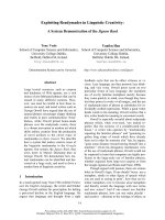
Apoptosis resistance in HIV persistently-infected H9/HTLVIII
B
cells in comparison with non-infected H9 cellsFigure 1
Apoptosis resistance in HIV persistently-infected H9/HTLVIII
B
cells in comparison with non-infected H9 cells.
A) H9 and H9/HTLVIII
B
cells were treated with different concentrations of H
2
O
2
or STS or complete medium as control. After
24 h, cells were harvested and annexin-V/PI staining was performed. The percentages of annexin-V
+
, PI
-
or PI
+
cells are shown.
B) (a) Analysis by APO-BrdU labeling by flow cytometry. The corresponding histograms and the percentages of APO-BrdU
+
cells are shown; (b) Analysis of apoptosis with Hoechst 33324 by fluorescence microscopy. Micrographs (100×) of predomi-
nant Hoechst stained nuclei are depicted. C) H9 and H9/HTLVIII
B
cells were treated with 0.1 μM STS or complete medium as
control, and cells were harvested 24, 48 and 72 h post-treatment. Cell viabillity was analyzed by the MTT assay. Absorbances
from treated samples were normalized to 100% of untreated controls. D) Cells treated with H
2
O
2
or STS or complete
medium for 24 h were pelleted and the supernatants were used to quantify infective viral (grey bar) and p24 antigen (red line)
production.
Retrovirology 2008, 5:19 />Page 4 of 12
(page number not for citation purposes)
observed in the percentage of annexin-V
+
/PI
-
cells (45%)
in U937 cells treated with 0.1 μM STS, but only 8.2% in
U1 infected cells. Remarkably, no significant differences
were observed in the frequency of apoptosis when cells
were treated with 10 μM H
2
O
2
(Figure 2B). However, a
higher concentration of H
2
O
2
(50 μM) was capable of
inducing 34% of annexin-V
+
U937 cells, and only 16.5%
of dead cells in the infected U1 cell lines (data not
shown). This result could be explained by the fact that
pro-monocytic cells are substantially less susceptible to
experience damage by ROS [18]. Thus, lymphoid and pro-
monocytic HIV-1 persistently-infected cell lines are less
susceptible to apoptosis induced by H
2
O
2
or STS treat-
ment compared with their uninfected counterparts.
Apoptosis resistance of HIV-infected cell lines is
independent of the magnitude of viral production
Unlike H9/HTLVIII
B
, J1.1 and U1 cell lines are non-pro-
ductive cells unless treated with a viral activator. To inves-
tigate the differential sensitivity to apoptosis of infected
cells under conditions of active viral replication, Jurkat
and J1.1 cells were treated with 1000 U/ml tumor necrosis
factor-α (TNF-α) for 48 h and U937 and U1 cells were
treated with 100 ng/ml phorbol-12-myristate-13-acetate
(PMA) for 24 h. Active viral production was confirmed by
determining the p24 antigen at different days post-treat-
ment. TNF-α treatment induced 100-fold viral reactiva-
tion at 48 h with respect to untreated cells, while U1 cells
showed 50-fold and 200-fold increase of viral production
at 24 h and 48 h, respectively, when cultured with PMA
(Table I). Under these conditions, the percentage of
annexin-V
+
/PI
-
cells was 48% and 52% when Jurkat cells
were treated with 10 μM H
2
O
2
and 0.1 μM STS, respec-
tively, and only 12% for J1.1 cells exposed to both apop-
totic stimuli (Figure 2A).
Regarding the pro-monocytic cell lines, when these cells
were pre-incubated with PMA, the frequency of early
apoptotic cells was significantly increased in both cell
lines treated with STS: 72% in U937 and 30% in U1 cells
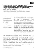
Apoptosis resistance is independent of the magnitude of viral replicationFigure 2
Apoptosis resistance is independent of the magnitude of viral replication. A) Jurkat and J1.1 cells were incubated in
the presence or absence of 1000 U/ml TNF-α for 48 h, and then treated with 10 μM H
2
O
2
or 0.1 μM STS. The percentages of
annexin-V
+
, PI
-
or PI
+
cells are shown. B) U937 and U1 cells were incubated in the presence or absence of 100 ng/ml PMA for
24 h, and then exposed to 10 μM H
2
O
2
or 0.1 μM STS. The percentages of annexin-V
+
, PI
-
or PI
+
cells are shown.
Table 1: P24 production in HIV-1 persistently-infected cell lines exposed to TNF-α or PMA
Cell line and treatment 0 h 24 h 48 h
J1.1 Cells 14.07 ± 0.01 14.14 ± 0.05 18.26 ± 0.80
J1.1 Cells + TNF α 14.08 ± 0.01 12.86 ± 0.90 152.04 ± 1.50
U1 Cells 1.06 ± 0.03 1.60 ± 0.03 4.18 ± 1.11
U1 Cells + PMA 1.05 ± 0.03 51.96 ± 9.20 191.76 ± 0.48
HIV-1 persistently-infected lymphoid J1.1 and monocytic U1 cells were treated with TNF-α and PMA respectively in order to stimulate viral
replication. P24 antigen was determined at 0, 24 and 48 h in cell supernatants by ELISA (HIVAG-1 Monoclonal, Abbot Laboratories). Values
correspond to p24 antigen per 200,000 cells, expressed in J1.1 as pg/ml and in U1 Cells as ng/ml.
Retrovirology 2008, 5:19 />Page 5 of 12
(page number not for citation purposes)
(Figure 2B). Control cells showed also higher levels of
apoptosis when pre-incubated with PMA. It should be
emphasized that PMA, independently of its ability to
stimulate viral replication, can also induce cell differenti-
ation, an effect which can influence the susceptibility to
apoptosis [19]. These data suggest that apoptosis resist-
ance in persistently-infected cell lines is independent of
the magnitude of viral replication.
Apoptosis resistance of HIV persistenly-infected cell lines
involves modulation of the mitochondrial pathway
In order to dissect the mechanisms involved in this pro-
tective effect, uninfected or persistently-infected cell lines
treated or not with apoptotic stimuli were used to analyze
different apoptotic pathways. First, the anti-Fas activating
antibody CH11 was used to induce apoptosis by the
extrinsic pathway in H9 and H9/HTLVIII
B
cells, and Jurkat
and J1.1 cells. No significant differences were observed
between uninfected and persistently-infected cells (Figure
3A–B). As Fas/CD95 expression was found to be modu-
lated by HIV-1 [5], we examined cell surface expression of
Fas antigen in order to check whether our results could be
due to differential expression of this receptor. Flow cytom-
etry analysis revealed no significant differences of Fas
expression among all cell lines tested (Figure 3C). Thus,
HIV-1 persistent infection does not seem to modulate the
susceptibility to apoptosis by controlling the extrinsic
pathway.
To gain insight into the mechanistic basis of this effect, we
next analyzed events associated with the execution of
apoptosis. When procaspase-3 expression was evaluated
by Western blot analysis in H9 and H9/HTLVIII
B
, or Jurkat
and J1.1 cells, no significant differences were observed in
untreated controls. However, when treated with the pro-
apoptotic agents, a decrease of procaspase-3 was observed
in all the cases (Figure 4A). When cells were analyzed by
flow cytometry, H9 cells were 57% and 47% positive for
Fas-mediated apoptosis in uninfected and HIV persistently-infected cellsFigure 3
Fas-mediated apoptosis in uninfected and HIV persistently-infected cells. H9 and H9/HTLVIII
B
(A) and Jurkat and
J1.1 (B) cells were incubated with 20 g/ml or 40 ng/ml of CH11, an anti-Fas activating antibody. After 24 h, cells were washed
and stained with annexin-V/PI. The percentages of annexin-V
+
, PI
-
or PI
+
cells are shown. C) Fas/CD95 and CD4 cell surface
expression was analyzed by flow cytometry on H9, H9/HTLVIII
B
, Jurkat and J1.1 cells.
Retrovirology 2008, 5:19 />Page 6 of 12
(page number not for citation purposes)
Caspase-3 activation by H
2
O
2
and STS treatment in uninfected and HIV persistently-infected lymphoid cell linesFigure 4
Caspase-3 activation by H
2
O
2
and STS treatment in uninfected and HIV persistently-infected lymphoid cell
lines. A) H9, H9/HTLVIII
B
, Jurkat and J1.1 cells were exposed to H
2
O
2
and STS. After 24 h, cells were washed and lysed with
RIPA buffer. Equal amounts of protein (30 μg/sample) were separated on a 10% SDS-PAGE and blotted onto nitrocellulose
membranes. Blots were probed with anti-procaspase-3 for 1 h and revealed with a peroxidase-conjugated anti-IgG antibody
and ECL (enhanced chemoluminiscence) Equal loading was checked by analyzing β-actin expression (data not shown). B) H9
and H9/HTLVIII
B
cells were exposed to 10μM H
2
O
2
, 0.1 μM STS or complete medium for 24 h and collected to evaluate active
caspase-3 by PE-conjugated monoclonal anti-active caspase-3 antibody by flow cytometry. C) Jurkat and J1.1 cells were
exposed to 10 μM H
2
O
2
, 0.1 μM STS, 40 ng/ml CH11 or complete medium for 24 h and collected to evaluate active caspase-3
by PE-conjugated monoclonal anti-active caspase-3 antibody by flow cytometry as described in Materials and Methods
Retrovirology 2008, 5:19 />Page 7 of 12
(page number not for citation purposes)
active caspase-3 when treated with H
2
O
2
and STS respec-
tively, while H9/HTLVIII
B
raised percentages of 39% and
38% respectively (Figure 4B). Besides, Jurkat cells showed
even higher differences in caspase-3 activation than J1.1
when treated with H
2
O
2
(Jurkat: 24%; J1.1: 3%) and STS
(Jurkat: 25.13% ;J1.1: 0.43%) (Figure 4C). Furthermore,
when treated with 40 ng/ml CH11 anti-Fas antibody, the
number of cells with active caspase-3 was similar in both
uninfected and persistently-infected cells (Figure 4C).
Taken together, these results suggest that differences in the
susceptibility to apoptosis between infected and unin-
fected cells can not be explained by defective caspase-3
activation and that apoptosis modulation may be local-
ized upstream of caspase-3.
To further understand this effect we analyzed events asso-
ciated with the mitochondrial apoptotic pathway. For this
purpose, the mitochondrial membrane potential (MMP)
was studied in cells treated with H
2
O
2
or STS by JC-1
(Mitoscreen, BD) staining and flow cytometry. When cells
were treated with 10 μM H
2
O
2
or 0.1 μM STS for 24 h, H9
and Jurkat cells showed higher MMP (H9: 45% with H
2
O
2
and 40% with STS; Jurkat: 23% with H
2
O
2
and 64% with
STS) compared with H9/HTLVIII
B
and J1.1 cells respec-
tively (H9/HTLVIII
B
: 30% with H
2
O
2
and 26% with STS;
J1.1: 8% with H
2
O
2
and 3.6% with STS) (Figure 5A–B).
Finally, Bcl-2 and Bax expression of different uninfected
and persistently-infected cell lines was analyzed by West-
ern blot of total cell lysates. Densitometric analysis
revealed no significant differences in Bcl-2 (25 kDa)
expression levels between H9 and H9/HTLVIII
B
cells,
treated or not with H
2
O
2
or STS. However, dimeric Bax
(42 kDa) was decreased by ~40% (H
2
O
2
) and ~70% (STS)
in H9/HTLVIII
B
cells treated with pro-apoptotic stimuli in
comparison with controls or uninfected H9 cells, which
reached values of only 20% (H
2
O
2
) or 40% (STS). The
overall effect could be observed by analyzing the Bcl-2/
Bax ratio, which estimates the anti-apoptotic/pro-apop-
totic balance. When treated with pro-apoptotic stimuli,
H9/HTLVIII
B
cells (lanes 5 and 6) showed a higher Bcl-2/
Bax ratio compared to H9 cells (lanes 2 and 3) (Figure
5C). In order to confirm this observation, Bax dimeriza-
tion and insertion in mitochondria was analyzed by West-
ern blot from cytosolic and mitochondrial fractions.
While the levels of Bax expression remained unaltered in
the cytosolic fraction of different uninfected or infected
cell lines, the levels of Bax increased substantially in the
mitochondrial fraction of uninfected cells treated with
apoptotic agents. However, no significant differences were
observed in persistently-infected cells when compared to
controls (Figure 5D).
These results suggest that apoptosis resistance observed in
persistently-infected cells involves modulation of the
mitochondrial pathway.
Conclusions and Discussion
During the clinical course of HIV-1 infection, the deple-
tion of the CD4
+
T cell compartment is mainly explained
by apoptosis of uninfected cells due to indirect mecha-
nisms including Fas/FasL interaction, syncytia formation
and direct citotoxicity of soluble viral proteins such as
gp120, Tat or Nef [5,7]. However, HIV-1 may survive in a
latent status, mainly in macrophages, resting CD4
+
quies-
cent T cells and CD44
high
memory T cells [10,20-22].
These cells appear to be less sensitive to death induced by
a variety of apoptotic stimuli such as chronic stress [18],
or the Fas/FasL (CD95L) system [23] independently of
viral cofactors. Therefore, when the chronic infection is
established in macrophages or in memory T cells, the
virus may survive longer in these cells due to a variety of
cellular and viral factors [24,25].
Our data suggest that persistently-infected pro-monocytic
and lymphocytic cells are less susceptible to undergo
apoptosis when exposed to different apoptotic stimuli
such as H
2
O
2
and STS, compared with uninfected cells.
This protection from apoptosis is consistent with the fact
that HIV-1 persistently-infected macrophages, quiescent T
cell and pro-monocytic cell lines were described to survive
longer [8,10,20-22]. Our study provides the first evidence
showing that apoptosis resistance in persistently-infected
cell lines is independent of the magnitude of viral replica-
tion. In spite of the fact that H9/HTLVIII
B
cells produced
virus actively, while viral production in J1.1 or U1 was
inducible, all cell lines showed similar tendences in their
resistance to apoptosis when compared with their unin-
fected counterparts.
Viral proteins are known to modulate cell surface levels of
Fas and its ability to transduce death signals upon binding
its specific ligand [5]. However, similar expression of Fas
antigen was found on the surface of the cells studied,
whether infected or not. In addition, engagement of Fas
by the stimulating CH11 antibody resulted in similar lev-
els of apoptosis in the cell lines studied, suggesting that
HIV-1 infection does not modulate the extrinsic pathway
of cell death.
In addition, when the MMP was analyzed in cells treated
with H
2
O
2
and STS, substantial differences in the induc-
tion of apoptosis were observed between uninfected and
persistently-infected cells. This result might be explained
by the ability of H
2
O
2
and STS to induce oxidative stress,
thus priming cells to undergo apoptosis via the mitochon-
drial pathway. These results are also consistent with the
levels of caspase-3 activation, indicating that once the
Retrovirology 2008, 5:19 />Page 8 of 12
(page number not for citation purposes)
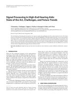
MMP induction and Bcl-2 and Bax expression in uninfected or HIV persistently-infected cell linesFigure 5
MMP induction and Bcl-2 and Bax expression in uninfected or HIV persistently-infected cell lines. H9 and H9/
HTLVIII
B
(A) and Jurkat and J1.1 (B) cells were exposed to 10 μM H
2
O
2
, 0.1 μM STS or complete medium for 24 h and har-
vested to evaluate mitochondrial membrane potential (ΔΨm) by JC-1 staining by flow cytometry. C) H9 and H9/HTLVIII
B
cells
were exposed to H
2
O
2
and STS. After 24 h, cells were washed and lysed with RIPA buffer. Equal amounts of protein (30 μg/
sample) were separated by 10% SDS-PAGE and blotted onto nitrocellulose membranes. The blots were probed with anti-Bcl-
2 and anti-Bax antibody, revealed using a peroxidase-conjugated anti-IgG and developed using a chemiluminiscence Western
blotting detection reagent. Equal loading was checked by analyzing β-actin expression. Films were analyzed with Scion image
analysis software (Scion, Frederick, MD) and the Bcl-2/Bax ratio was depicted. D) H9 and H9/HTLVIII
B
cells were exposed to
H
2
O
2
and STS and after 24 h lysates from cytosolic and mitochondrial fractions were prepared by differential centrifugation.
Equal amounts of protein (30 μg/sample) were separated by 10% SDS-PAGE and blotted onto nitrocellulose membranes. Blots
were then probed with an anti-Bax polyclonal antibody, incubated with a peroxidase-conjugated anti-rabbit secondary antibody
and developed using ECL detection reagent. Equal loading was checked by analyzing β-actin (cytosol fraction) and Complex I
(mitochondrial fraction) expression.
Retrovirology 2008, 5:19 />Page 9 of 12
(page number not for citation purposes)
mitochondrial pore is induced, apoptosis events proceed
normally. Thus, modulation of apoptosis might occur
before or during pore induction. In order to analyze the
possible mechanisms involved in this effect, expression of
Bcl-2 and Bax was analyzed in the cytosolic and mito-
chondrial compartments. Bcl-2 expression did not show
any significant difference between both cell lines, whether
they were treated or not with pro-apoptotic stimuli. How-
ever, expression of Bax was dramatically reduced in mito-
chondria of persistently-infected cells when apoptosis was
induced by exposure to H
2
O
2
or STS.
It is now widely accepted that persistent HIV-1 infection
represents a new homeostatic state of the cell, which is
likely promoted by the combination of both cellular and
viral factors. Several viral proteins have been recognized
by their ability to induce apoptosis in infected or unin-
fected cells, but some viral proteins can also protect
against cell death [5]. Decreased caspase-3 activation [26]
and p53 expression [27] were described as possible mech-
anisms implicated in apoptosis resistance in HIV-1-per-
sistently infected cells. This study provides novel evidence
showing that resistance to apoptosis in persistently-
infected cells involves direct modulation of the mitochon-
drial pathway by regulating Bax pore induction. Further
experiments are needed in order to clarify the mechanism
by which the virus decreases MMP and controls the execu-
tion of apoptosis. Viral regulation of autophagy of dam-
aged mitochondrias or Bax proteolysis might be potential
explanatory mechanisms for our observations.
The survival of viral reservoirs is a great challenge to tackle
regarding HIV eradication. Understanding the mechanis-
tic bases of the resistance to apoptosis is essential to spe-
cifically target the persistence of viral reservoirs and might
contribute to provide insights for future therapeutic strat-
egies in order to promote complete viral eradication.
Materials and Methods
Cell lines
The following uninfected cell lines of human origin were
used: lymphocytic H9, Jurkat and promonocytic U937
cell lines; and their respective HIV-1 persistently-infected
cell lines: H9/HTLVIII
B
, J1.1 and U1. All cell lines were
provided by the NIH AIDS Research and References Rea-
gent Program, except for U937. Cell lines were cultured
with RPMI 1640 medium supplemented with 2 mM L-
glutamine, 100 μg/ml streptomycin and 10% fetal calf
serum at 37°C in a humidified atmosphere (5% CO
2
in
air).
Antibodies and reagents
Annexin-V apoptosis kit, APO-BrdU apoptosis kit, active
caspase-3 antibody kit, JC-1 Mitoscreen, TNF-α, PE-conju-
gated anti-CD95 and PerCP-conjugated anti-CD4 anti-
bodies were from BD Biosciences,(CA, USA). Anti-Bcl-2
(DC21), anti-Bax (D21), anti-procaspase-3 (L-18), anti-β-
actin (I-19) polyclonal antibodies and peroxidase-conju-
gated anti-rabbit and anti-goat antibodies were from
Santa Cruz Biotechnology, (CA, USA). Anti-complex I
antibody was a generous gift from Dr. J. Poderoso (Hospi-
tal de Clínicas, University of Buenos Aires). Anti-Fas acti-
vating antibody (CH11) was from Upstate (New York,
USA). Other reagents including Hoechst, MTT, PMA, stau-
rosporine (STS), Kodak BioMax films were from Sigma
(St. Louis, MO, USA). Hydrogen peroxide (H
2
O
2
) and iso-
propanol were from Merck (New Jersey, USA). RPMI 1640
medium, fetal calf serum, L-glutamine and streptomycin
were from Gibco (New York, USA). Micro-BCA protein
assay kit was from Pierce (Rockford, USA). Chemilumi-
niscence Western blotting detection reagent and nitroce-
lulose membranes were from Amersham Biosciences, UK.
Induction of HIV-1 production
In order to induce viral production, J1.1 cells were incu-
bated for 48 h with 1000 U/ml TNF-α [28] and U1 cells
were exposed to 100 ng/ml PMA for 24 h [29]. Cells were
washed twice with PBS and fresh medium was added to
carry out experiments. Viral production was confirmed by
p24 antigen determination.
Determination of viral production
Cells were pelleted and supernatants were used to quan-
tify p24 antigen with a commercial ELISA kit (HIVAG-1
monoclonal, Abbot Laboratorios, Illinois, USA), and viral
load using a commercial assay (Quantiplex XTm HIV RNA
3.0 Assay bDNA, Chiron Corp, CA, USA). Infective virus
titration was performed by limiting dilution and syncytia
formation in MT-2 cells, and calculated by the Reed &
Müench method.
Induction of apoptosis
Cells were collected, washed with PBS, resuspended with
complete medium, and divided in a 24-well culture plate
with a final cell concentration of 150,000 cells/ml. For
apoptosis induction, H
2
O
2
, STS and the CH11 Fas activat-
ing antibody were used. Optimal concentrations for
experiments were standarized by testing different concen-
trations, which ranged from 5 to 1000 μM (H
2
O
2
), from
0.01 to 10 μM (STS) and from 20 to 40 ng/ml (CH11).
Treated cells were always compared with untreated con-
trols (Ctrl). In most experiments, cells were incubated
with the apoptosis inducers for 24 h
MTT assay
Cell viability was determined by the MTT (3-[4,4-dimeth-
ylthiazol-2-yl]-2,5-diphenyltetrazolium bromide) assay
[30]. After 24 h of exposure to pro-apoptotic stimuli,
medium was removed and cells were plated at 5 × 10
4
cells/well in 96-well plates and incubated with 0.5 mg/ml
Retrovirology 2008, 5:19 />Page 10 of 12
(page number not for citation purposes)
MTT in RPMI-1640 without Red Phenol for 1 h at 37°C in
a CO
2
incubator. Cells were pelleted and formazan crys-
tals were solubilized with 0.04 M HCl in isopropanol.
Finally, the absorbance measured at 640 nm was sub-
tracted from the absorbance at 540 nm. Each assay was
performed in triplicate. Absorbances corresponding to
treated samples were normalized to 100% of untreated
controls and expressed as percentages. In this assay, the
number of surviving cells was directly correlated with the
amount of formazan obtained.
Asssesment of apoptosis
Cells were incubated in the presence or absence of proap-
optotic stimuli for 24 h, washed twice with PBS and the
frequency of apoptotic cells was analyzed by the following
methods:
Annexin-V/PI staining
To determine the percentage of early apoptotic cells, phos-
phatidylserine (PS) cell translocation and plasma mem-
brane permeability were evaluated by dual staining with
FITC-conjugated annexin-V and propidium iodide (PI)
using the Annexin-V/PI apoptosis detection kit (BD Bio-
sciences) and analyzed by flow cytometry using a FACS-
Canto (BD Biosciences). Annexin-V
+
/PI
-
cells representing
early apoptotic cells, and annexin-V
+
/PI
+
mostly repre-
senting necrotic cells were determined.
APO-BrdU staining
Late apoptotic cells were determined with the APO-BrdU
kit by incorporation of bromodeoxyuridine triphosphate
(Br-dUTP) to 3'-hydroxyl sites in cell DNA, and analyzed
by flow cytometry in a FACSCanto (BD Bioscience).
Hoechst 33324 staining
Apoptotic cells were determined by Hoechst staining and
visualized in a fluorescence microscope (Axiophot West
Germany).
Cytofluorimetric analysis of MMP
After treatments with H
2
O
2
and STS for 24 hours, cells
were collected and resuspended in PBS, and then stained
with JC-1 (5,5',6,6'-tetrachloro-1,1',3,3'-tetraethylbenz-
imidazolcarbocyanine iodide) (JC-1 Mitoscreen, BD) for
15 min at 37°C in a CO
2
incubator. Cells were pelleted,
washed twice with buffer supplemented by the kit as indi-
cated by the manufacter and analyzed on a flow cytometer
(FACSCanto, BD Biosciences).
Cytofluorimetric analysis of caspase-3 activation
Treated and control cells were pelleted and washed twice
with PBS and the percentage of cells with active
caspase-3 was assessed using the PE-conjugated mono-
clonal active caspase-3 antibody kit (BD Pharmigen) and
analyzed on a FACSCanto flow cytometer (BD Bio-
sciences).
Flow cytometry analysis
In all cases where flow cytometry was required, 20,000
events were acquired in a FACSCanto flow cytometer (BD
Biosciences) and different parameters were analyzed
using the WinMDI 2.8 software.
Isolation and purification of mitochondria
Cells (1 × 10
7
cells) incubated in the presence or absence
of pro-apoptotic stimuli were washed and homogenized
in MSHE (0.225 M mannitol, 0.07 M sucrose, 1 mM
EGTA, and 25 mM HEPES/KOH; 1/10 w/v; pH 7.4) and
centrifuged at 5,500 × g for 10 min at 4°C. The resultant
supernatant was centrifuged at 15,000 × g for 20 min at
4°C and the pellet was resuspended in 30 μl of MSHE
(mitochondrial fraction) [31]. To remove broken mito-
chondria, contaminating organelles, and debris from the
cytosol fractions, the supernatants were further centri-
fuged at 21,000 × g for 30 min at 4°C. Protein concentra-
tion from cytosolic and mitochondrial fractions was
determined by the Micro-BCA protein assay kit (Pierce,
Rockford, USA).
Western blot analysis
After exposure to pro-apoptotic stimuli, cells were lysed in
RIPA buffer containing 20 mM Tris-HCl, 150 mM NaCl,
1% Triton X-100, 1% sodium deoxycholate, 2 mM EDTA,
0.1% SDS and protease inhibitor cocktail. Protein concen-
trations from total, cytosolic or mitochondrial lysates
were quantified using the Micro-BCA protein assay kit as
described above. Equal amounts of protein (30 μg/sam-
ple) were separated in a 10% SDS-PAGE and blotted onto
nitrocellulose membranes. Blots were then probed with
anti-pro-caspase-3, anti-Bcl-2 or anti-Bax rabbit polyclo-
nal antibodies as described [32], and incubation with per-
oxidase-conjugated anti-IgG was performed in a blocking
buffer for 1 h. Blots were then developed using a chemilu-
miniscence Western blotting detection reagent and
exposed to X-ray films. Films were analyzed using the
Scion image analysis software (Scion, Frederick, MD).
Total cell lysates were used to analyze pro-caspase-3, Bcl-
2 and Bax expression and normalized with β-actin expres-
sion. Cytosolic and mitochondrial extracts were used to
analyze the insection of Bax into mitochondria, and pro-
tein bands were compared with the expression of β-actin
(marker of cytosolic fraction) and Complex I (marker of
mitochondrial fraction).
Statistical analysis
Values represent the mean ± s.e.m. of at least three inde-
pendent experiments. Comparisons among groups were
performed by using the Student's t test and One-way
ANOVA using a SPSS 12.0 software.
Retrovirology 2008, 5:19 />Page 11 of 12
(page number not for citation purposes)
Competing interests
The author(s) declare that they have no competing inter-
ests.
Authors' contributions
PNFL was responsible for designing, performing and writ-
ing the manuscript. DAR and SEM contributed to experi-
ments of apoptosis by Hoechst and APOBrDU. DOC and
GAR were responsible of experiments using Western blot
analysis, and contributed to writing of the manuscript.
MB, RL and MS performed and interpreted the flow
cytometry experiments. LMP was responsible for the
design and writing of the manuscript. All authors read and
approved the final manuscript.
Acknowledgements
We thank the AIDS Research and Reference Program (National Institute of
Allergy and Infectious Diseases, NIH, USA) for the reagents used in this
study. We are grateful with Dr. J. Poderoso for providing essential reagents
and assisting with the procedures for mitochondria purification. This work
was supported by grants from the University of Buenos Aires (M050) and
the National Agency for Promotion of Science and Technology (PICT 05-
11734) to L.M.P. and grants from Fundación Sales, National Agency for Pro-
motion of Science and Technology (PICT 2003 05-13787) and University of
Buenos Aires (M091) to G.A.R
References
1. Benedict CA, Norris PS, Ware CF: To kill or be killed: viral eva-
sion of apoptosis. Nature immunology 2002, 3(11):1013-1018.
2. He B: Viruses, endoplasmic reticulum stress, and interferon
responses. Cell death and differentiation 2006, 13(3):393-403.
3. Brenchley JM, Price DA, Douek DC: HIV disease: fallout from a
mucosal catastrophe? Nature immunology 2006, 7(3):235-239.
4. Hel Z, McGhee JR, Mestecky J: HIV infection: first battle decides
the war. Trends in immunology 2006, 27(6):274-281.
5. Gougeon ML: To kill or be killed: how HIV exhausts the
immune system. Cell death and differentiation 2005, 12 Suppl
1:845-854.
6. Lelievre JD, Mammano F, Arnoult D, Petit F, Grodet A, Estaquier J,
Ameisen JC: A novel mechanism for HIV1-mediated
bystander CD4+ T-cell death: neighboring dying cells drive
the capacity of HIV1 to kill noncycling primary CD4+ T cells.
Cell death and differentiation 2004, 11(9):1017-1027.
7. Varbanov M, Espert L, Biard-Piechaczyk M: Mechanisms of CD4 T-
cell depletion triggered by HIV-1 viral proteins. AIDS reviews
2006, 8(4):221-236.
8. Pinti M, Biswas P, Troiano L, Nasi M, Ferraresi R, Mussini C, Vecchiet
J, Esposito R, Paganelli R, Cossarizza A: Different sensitivity to
apoptosis in cells of monocytic or lymphocytic origin chron-
ically infected with human immunodeficiency virus type-1.
Experimental Biology and Medicine 2003, 228(11):1346-1354.
9. Balestra E, Perno CF, Aquaro S, Panti S, Bertoli A, Piacentini M, For-
bici F, D'Arrigo R, Calio R, Garaci E: Macrophages: a crucial res-
ervoir for human immunodeficiency virus in the body. Journal
of biological regulators and homeostatic agents 2001, 15(3):272-276.
10. Zamborlini A, Lehmann-Che J, Clave E, Giron ML, Tobaly-Tapiero J,
Roingeard P, Emiliani S, Toubert A, de The H, Saib A: Centrosomal
pre-integration latency of HIV-1 in quiescent cells. Retrovirol-
ogy 2007, 4:63.
11. Conti L, Matarrese P, Varano B, Gauzzi MC, Sato A, Malorni W,
Belardelli F, Gessani S: Dual role of the HIV-1 vpr protein in the
modulation of the apoptotic response of T cells. Journal of
Immunology
2000, 165(6):3293-3300.
12. Gibellini D, Caputo A, Celeghini C, Bassini A, La Placa M, Capitani S,
Zauli G: Tat-expressing Jurkat cells show an increased resist-
ance to different apoptotic stimuli, including acute human
immunodeficiency virus-type 1 (HIV-1) infection. British jour-
nal of haematology 1995, 89(1):24-33.
13. Mahlknecht U, Deng C, Lu MC, Greenough TC, Sullivan JL, O'Brien
WA, Herbein G: Resistance to apoptosis in HIV-infected CD4+
T lymphocytes is mediated by macrophages: role for Nef and
immune activation in viral persistence. Journal of Immunology
2000, 165(11):6437-6446.
14. Dumont A, Hehner SP, Hofmann TG, Ueffing M, Droge W, Schmitz
ML: Hydrogen peroxide-induced apoptosis is CD95-inde-
pendent, requires the release of mitochondria-derived reac-
tive oxygen species and the activation of NF-kappaB.
Oncogene 1999, 18(3):747-757.
15. Bloom DA, Jaiswal AK: Phosphorylation of Nrf2 at Ser40 by
protein kinase C in response to antioxidants leads to the
release of Nrf2 from INrf2, but is not required for Nrf2 sta-
bilization/accumulation in the nucleus and transcriptional
activation of antioxidant response element-mediated
NAD(P)H:quinone oxidoreductase-1 gene expression. The
Journal of biological chemistry 2003, 278(45):44675-44682.
16. Arnoult D, Petit F, Lelievre JD, Estaquier J: Mitochondria in HIV-1-
induced apoptosis. Biochemical and biophysical research communica-
tions 2003, 304(3):561-574.
17. Buenz EJ, Badley AD: Impact of mitochondrial regulation of
apoptosis on the pathogenesis and treatment of HIV-1-
induced immunodeficiency. Mitochondrion 2004, 4(2-3):235-254.
18. Gieseg SP, Whybrow J, Glubb D, Rait C: Protection of U937 cells
from free radical damage by the macrophage synthesized
antioxidant 7,8-dihydroneopterin. Free radical research 2001,
35(3):311-318.
19. Pennington KN, Taylor JA, Bren GD, Paya CV: IkappaB kinase-
dependent chronic activation of NF-kappaB is necessary for
p21(WAF1/Cip1) inhibition of differentiation-induced apop-
tosis of monocytes. Molecular and cellular biology 2001,
21(6):1930-1941.
20. Marcello A: Latency: the hidden HIV-1 challenge. Retrovirology
2006, 3(1):7.
21. Petitjean G, Al Tabaa Y, Tuaillon E, Mettling C, Baillat V, Reynes J, Seg-
ondy M, Vendrell JP: Unintegrated HIV-1 provides an inducible
and functional reservoir in untreated and highly active
antiretroviral therapy-treated patients. Retrovirology 2007,
4:60.
22. Redpath S, Angulo A, Gascoigne NR, Ghazal P: Immune check-
points in viral latency. Annual review of microbiology 2001,
55:531-560.
23. Fas SC, Baumann S, Krueger A, Frey CR, Schulze-Bergkamen H, Bren-
ner D, Stumpf C, Kappes K, Krammer PH: In vitro generated
human memory-like T cells are CD95 type II cells and resist-
ant towards CD95-mediated apoptosis. European journal of
immunology 2006, 36(11):2894-2903.
24. Gougeon ML: Apoptosis as an HIV strategy to escape immune
attack. Nature Reviews Immunology 2003, 3(5):392-404.
25. Guillemard E, Jacquemot C, Aillet F, Schmitt N, Barre-Sinoussi F,
Israel N: Human immunodeficiency virus 1 favors the persist-
ence of infection by activating macrophages through TNF.
Virology 2004, 329(2):371-380.
26. Tanaka Y, Kameoka M, Ota K, Itaya A, Ikuta K, Yoshihara K: Estab-
lishment of persistent infection with HIV-1 abrogates the
caspase-3-dependent apoptotic signaling pathway in U937
cells. Experimental cell research 1999, 247(2):514-524.
27. Kim CH, Chiplunkar S, Gupta S: Chronic HIV type 1 infection
down-regulates expression of DAP kinase and p19ARF-p53
checkpoint and is associated with resistance to CD95-medi-
ated apoptosis in HUT78 T cells. AIDS research and human retro-
viruses 2004, 20(2):183-189.
28. Perez VL, Rowe T, Justement JS, Butera ST, June CH, Folks TM: An
HIV-1-infected T cell clone defective in IL-2 production and
Ca2+ mobilization after CD3 stimulation. Journal of Immunology
1991, 147(9):3145-3148.
29. Shelley CS, Teodoridis JM, Park H, Farokhzad OC, Bottinger EP,
Arnaout MA: During differentiation of the monocytic cell line
U937, Pur alpha mediates induction of the CD11c beta 2
integrin gene promoter. Journal of Immunology 2002,
168(8):
3887-3893.
30. Denizot F, Lang R: Rapid colorimetric assay for cell growth and
survival. Modifications to the tetrazolium dye procedure giv-
ing improved sensitivity and reliability. Journal of immunological
methods 1986, 89(2):271-277.
Publish with BioMed Central and every
scientist can read your work free of charge
"BioMed Central will be the most significant development for
disseminating the results of biomedical research in our lifetime."
Sir Paul Nurse, Cancer Research UK
Your research papers will be:
available free of charge to the entire biomedical community
peer reviewed and published immediately upon acceptance
cited in PubMed and archived on PubMed Central
yours — you keep the copyright
Submit your manuscript here:
/>BioMedcentral
Retrovirology 2008, 5:19 />Page 12 of 12
(page number not for citation purposes)
31. Galli S, Labato MI, Bal de Kier Joffe E, Carreras MC, Poderoso JJ:
Decreased mitochondrial nitric oxide synthase activity and
hydrogen peroxide relate persistent tumoral proliferation to
embryonic behavior. Cancer research 2003, 63(19):6370-6377.
32. Toscano MA, Bianco GA, Ilarregui JM, Croci DO, Correale J, Hern-
andez JD, Zwirner NW, Poirier F, Riley EM, Baum LG, Rabinovich
GA: Differential glycosylation of TH1, TH2 and TH-17 effec-
tor cells selectively regulates susceptibility to cell death.
Nature immunology 2007, 8(8):825-834.