Applied Radiological Anatomy for Medical Students Applied - part 9 ppsx
Bạn đang xem bản rút gọn của tài liệu. Xem và tải ngay bản đầy đủ của tài liệu tại đây (1.16 MB, 18 trang )
The adductor muscles
Gracilis arises from the body and inferior ramus of the pubis and
passes down the medial aspect of the thigh over the medial femoral
condyle to insert into the medial surface of the tibia below the
condyle. Pectineus is a flat, quadrilateral muscle arising from the
pubis; it passes posterolaterally to insert between the lesser
The lower limb a. newman sanders
133
Head
Neck
Intertrochanteric
crest
Lesser
trochanter
Greater
trochanter
Trochanteric
fossa
Quadrate
tubercle
Linea aspera
Lateral
supracondylar
line
Popliteal surface
Intercondylar
fossa
Lateral condyle
Medial condyle
Adductor
tubercle
Medial
supracondylar
line
Greater
trochanter
Fovea capitis
Head
Neck
Intertrochanteric line
Lesser trochanter
Shaft
Adductor
tubercle
Medial
epicondyle
Medial
condyle
Patellar surface
Lateral
condyle
Lateral
epicondyle
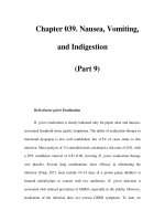
Fig. 13.8. The right femur,
(a) anterior, (b) posterior.
(a) (b)
1st year
4th year
Puberty
14th year
Fig. 13.9. (a) Ossification of femur. The secondary centres fuse with the shaft
at 18–20 years; (b) bipartite and multipartite patellae.
(a)
(b)
Vastus intermedius Rectus femoris Vastus medialis
Femur
Superficial
femoral
artery
and vein
Sartorius
Gracilis
Adductor
longus
Semimembranosus
Semitendinosus
Vastus lateralis
Long head of bicepsShort head of biceps
(a)
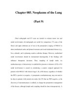
Fig. 13.10. IW MRI of the right thigh: (a) axial scan through mid-thigh and
(b) coronal image.
trochanter and the linea aspera. Adductor longus arises from the front
of the body of the pubis and is inserted by a broad aponeurosis on to
the linea aspera. Adductor brevis takes origin from the inferior ramus
and body of the pubis behind pectineus and is attached between the
lesser trochanter and the linea aspera. Adductor magnus arises from
the inferior ischio-pubic ramus and is attached along the linea aspera,
the medial supracondylar line and by a strong tendon to the adductor
tubercle of the medial femoral condyle. Its distal attachment is inter-
rupted by the adductor hiatus through which the femoral vessels pass
to reach the popliteal fossa, as the popliteal artery and vein.
The hamstrings
Semimembranosus arises by a flattened “membranous” tendon from
the ischial tuberosity. It has a complex distal attachment to the medial
tibial condyle and the medial surface of the tibia with tendinous
expansions over the popliteus muscle to the lateral femoral condyle
(the oblique popliteal ligament) and to the tibial collateral ligament
(the posterior oblique ligament).
Semitendinosus takes origin from the ischial tuberosity. Inferiorly,
its long tendon passes round the medial tibial condyle and over the
medial collateral ligament to attach to the medial surface of the tibia
posterior to the insertions of gracilis and sartorius.
Biceps femoris arises by a long head from the ischial tuberosity and
a short head from the shaft of the femur. Its tendon inserts on to the
head of the fibula.
The knee joint
The knee is a modified hinge joint and this synovial joint is the largest
in the body. Although contained within a single joint cavity, the knee
effectively comprises two condylar joints between the femoral and cor-
responding tibial condyles and a saddle joint between the patella and
the femur. The tibiofemoral compartments are each divided by a fibro-
cartilaginous meniscus (Fig. 13.11). The medial meniscus is larger and
more semicircular. It is broader and thicker posteriorly. The lateral is
smaller, thicker and forms a nearly complete ring. The anterior and
posterior horns of the menisci are attached to the intercondylar area.
The posterior horn of the lateral meniscus is also commonly attached to
the medial condyle of the femur by the meniscofemoral ligament. The
transverse ligament joins the anterior ends of the menisci.
The fibrous capsule is attached around the margins of the articular
surfaces.
The synovial membrane lines the fibrous capsule, but does not
cover the surfaces of the menisci. It lines the suprapatellar bursa,
which may be regarded as part of the knee joint and lies beneath
quadriceps femoris, extending 7–8 cm above the upper border of the
patella. Below the patella, the synovium is separated from the patellar
tendon by the infrapatellar (Hoffa’s) fat pad.
Posteriorly, the synovium is reflected anteriorly from the fibrous
capsule to cover both cruciate ligaments on their anterior and lateral
aspects. Several bursae surround the knee (Fig. 13.12).
The lower limb a. newman sanders
134
Fig. 13.10. Continued
Vastus
medialis
Vastus
lateralis
Shaft of
femur
Adductor
magnus
Gracilis
Sartorius
Transverse ligament
Anterior cruciate
ligament
Lateral meniscus
Posterior meniscofemoral ligament
Posterior cruciate ligament
Medial
meniscus
Fig. 13.11. The menisci and ligaments of the knee and their attachments
Tendon of
quadriceps
Suprapatellar
bursa
Subcutaneous
prepatellar
bursa
Infrapatellar (Hoffa’s
fat pad) extending
into infrapatellar fold
Patellar tendon
Deep infrapatellar bursa
Fibrous capsule
Anterior cruciate
ligament
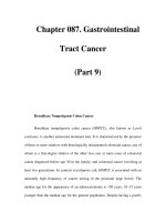
Fig. 13.12. (a) Bursae of the knee joint (sagittal section); (b) Bursae around the
knee; sagittal and axial MR arthrogram.
(a)
(b)
The knee joint is strengthened by four main ligaments, (Figs. 13.13,
13.16). The medial (tibial) collateral ligament is attached to the medial
epicondyle of the femur and to the medial tibial condyle. It is a
flattened band, which blends posteriorly with the fibrous capsule, but
anteriorly may be separated from it by a bursa. The posterolateral
complex is made up of the popliteus tendon, biceps tendon, fibular
collateral ligament, arcuate ligament, and popliteo-fibular ligament.
The lateral (fibular) collateral ligament is a cord-like structure between
the lateral epicondyle of the femur and the head of the fibula.
Popliteus muscle arises from the posterior surface of the tibia and
sweeps superolaterally behind the knee joint, where its tendon
pierces the capsule and inserts in the groove on the lateral femoral
condyle. Some of its fibers blend with the edge of the lateral
meniscus. It also gives a slip to the tip of the fibula (popliteo-fibular
ligament).
The cruciate ligaments (Figs. 13.13, 13.16), are intracapsular but
extrasynovial. The anterior cruciate ligament (ACL) passes from the
medial part of the anterior intercondylar area of the tibia upwards,
backwards and laterally to insert into the posterior part of the medial
surface of the lateral femoral condyle. It prevents the femur moving
backwards on the tibia. The posterior cruciate ligament (PCL) is
attached to the posterior intercondylar area of the tibia and passes for-
wards, upwards, and medially to insert into the anterior part of the
lateral surface of the medial femoral condyle. It is stronger and
shorter than the ACL and limits posterior sliding of the tibia on the
femur.
Movements
• Flexion: biceps, semitendinosus, semimembranosus. The extended
knee is unlocked prior to flexion by popliteus, whose action is to
rotate the femur laterally on the fixed tibia;
• Extension: quadriceps femoris;
• Medial rotation of the flexed leg: semimembranosus and semit-
endinosus;
• Lateral rotation of the flexed leg: biceps femoris.
Imaging of the knee
Plain radiography (Fig. 13.14), is able to demonstrate the bony contours
of the joint space. The normal tibio-femoral and patello-femoral joint
space is 3 mm. The fat around the joint enables visualization of the
patellar tendon, and allows an assessment of the presence or absence
of a joint effusion. If a horizontal beam lateral radiograph is taken, a
fat-fluid level (lipohaemarthrosis) in the suprapatellar bursa indicates
a fracture within the joint. Occult fractures are usually of the tibial
plateau. These may be demonstrated by coronal tomography or by
thin slice axial CT with coronal reformatting.
If an abnormality of the patella is suspected, it should be imaged by
the “skyline” view, a tangential view taken with the knee flexed. The
intercondylar fossa of the lower femur may be imaged by the “tunnel”
view, which is used to detect clinically suspected intra-articular loose
bodies (Fig. 13.15).
MRI is much the most useful imaging technique (Fig. 13.16). It
demonstrates the joint cavity, menisci, ligaments, and articular carti-
lage very well.
Dynamic scanning of the knee is also possible with modern scan-
ners, which allow assessment of patellar tracking.
The lower limb a. newman sanders
135
High signal fluid
distending the
suprapatellar
bursa
Fig. 13.12. Continued
Fig. 13.13. Ligaments of the knee Joints.
(b)
Patellar surface
Lateral condyle
Lateral meniscus
Fibular collateral
ligament
Anterior ligament
of head of fibula
Fibula
Medial condyle
Posterior cruciate
ligament
Anterior cruciate
ligament
Coronary
ligament
Medial meniscus
Transverse ligament
Tibia
Medial
condyle
Medial tibial
plateau
Medial
Lateral
Tubercles of
intercondylar
eminence
Patella
Lateral
condyle
Groove for
popliteus
tendon
Lateral
tibial
plateau
Apex
(styloid
process)
Head
Neck
Shaft
of fibula
(a)
Fig. 13.14. (a) AP, and (b) lateral radiographs of the knee.
Ultrasound scanning may be used to assess the patellar tendon, the
collateral ligaments and meniscal and popliteal cysts.
The lower leg
The tibia and fibula (Fig. 13.17)
These are joined by a tough fibrous interosseous membrane. They
give rise to the attachments of many of the muscles of the lower leg.
Ossification is shown in Fig. 13.18.
The tibiofibular joints
The superior tibiofibular joint is a plane synovial joint between the
head of the fibula and the articular surface under the lateral tibial
condyle.
The inferior tibiofibular joint is a fibrous joint (syndesmosis)
between the lower end of the fibula and the fibular notch of the tibia.
The lower limb a. newman sanders
136
(a)
Patella
Medial
femoral
condyle
Lateral
femoral
condyle
Medial
femoral
condyle
Medial
tibial
condyle
Head of
fibula
Lateral
tibial
condyle
Lateral
femoral
condyle
grooved by
popliteus
tendon
Patella
Fig. 13.15. (a). A skyline
view of the patella. Note
how the lateral femoral
condyle projects more
anteriorly, tending
to prevent lateral
patellar dislocation;
(b) intercondylar view of
the knee.
Femur Patella
Anterior cruciate
ligament
Posterior cruciate
ligament
TibiaPatellar
tendon
Hoffa’s fat
pad
Medial collateral
ligament
Anterior cruciate ligament
Fibular collateral
ligament
Posterior cruciate
ligament
Popliteus
tendon
Lateral
meniscus
Medial
meniscus
Fig. 13.16. PD MRI of the knee (a) sagittal; (b) coronal; (c) axial.
(b)
(b)
PatellaMedial retinaculum
Patellar tendon
Posterior
cruciate
ligament
Semi-membranosus
tendon
Medial and lateral
head of
gastrocnemius
(a)
(c)
(b)
Apex
Head
Shaft
of fibula
Tibial
tuberosity
Patella
Quadriceps femoris
Shaft of femur
Intercondylar fossa
Medial and lateral
femoral condyles
Tubercles of
intercondylar
eminence
Fig. 13.14. Continued
The lower limb a. newman sanders
137
Medial condyle
of tibia
Tuberosity
of tibia
Interosseous
border
Medial surface
Medial
malleolus
Tubercles of
intercondylar
eminence
Lateral condyle
of tibia
Apex
Head of fibula
Anterior border
Interosseous
border
Medial crest
Anterior surface
Medial part of
posterior surface
Lateral surface
Triangular
subcutaneous
area
Lateral
malleolus
Anterior
border
Groove for
tendon
of popliteus
Lateral condyle
of tibia
Apex of head
of fibula
Medial crest
Posterior
border
Groove for
peroneal
tendons
Lateral
malleolus
Soleal line
Nutrient
foramen
Vertical line
Medial border
Interosseous
border
Groove for tibialis
posterior tendon
Medial malleolus
Head of
fibula
Ist year
I2th year
16th–18th
year
Extensor
digitorum
longus
Tibialis
anterior
Tibialis
posterior
Posterior tibial
vessels
Flexor
digitorum
longus
Soleus
Medial
head of
gastrocnemius
Lateral
head of
gastrocnemius
Flexor hallucis
longus
Anterior
tibial
vessels
Peroneus
longus
and brevis
tendons
(a)
(b)
Fig. 13.17. The tibia and
fibula; (a) anterior,
(b) posterior.
Fig. 13.18. Ossification of the tibia and fibula. The distal and proximal epiphyses
fuse with the shaft at 16–18 years.
Fig. 13.19. T1W axial MRI of the mid-calf.
It is reinforced by the interosseous ligament of the joint and the ante-
rior and posterior inferior tibiofibular ligaments. Movements at both
joints are extremely limited.
The muscles of the lower leg (Figs. 13.19, 13.20)
Anterior compartment
Tibialis anterior takes origin from the upper part of the anterior
surface of the tibia and adjacent interosseous membrane and forms
a tendon which descends anterior to the ankle joint deep to the
extensor retinaculum to attach to the medial cuneiform and the base
of the first metatarsal.
Extensor hallucis longus (EHL) arises from the anterior surface of
the fibula. Its tendon passes under the extensor retinaculum and
inserts on to the dorsum of the base of the distal phalanx of the
hallux.
Extensor digitorum longus arises above and lateral to EHL from
the anterior surface of the fibula. Distally, it divides into four
tendons, which pass under the extensor retinaculum and insert
via a dorsal expansion onto the dorsum of the middle and distal
phalanges of the lateral four toes. Peroneus tertius arises from the
anterior surface of the fibula and inserts into the shaft of the fifth
metatarsal.
The lateral (peroneal) compartment
These muscles arise from the lateral surface of the fibula. Distally,
the tendon of peroneus longus passes behind the lateral malleolus
beneath the peroneal retinaculum, passes forwards lateral to the
calcaneus and into a groove in the inferior surface of the cuboid
before inserting on the base of the first metatarsal and the adjacent
medial cuneiform.
The tendon of peroneus brevis descends anteriorly to that of per-
oneus longus to insert on the base of the fifth metatarsal.
The posterior compartment.
Gastrocnemius, the most superficial of the muscles of the calf, arises
by two heads from the posterior surfaces of the medial and lateral
femoral condyles. A sesamoid bone, the fabella, is frequently found in
the lateral head of gastrocnemius.
Soleus arises from the upper posterior surface of the fibula and
from the posterior surface of the tibia. The tendons of gastrocnemius
and soleus unite to form the Achilles’ (or calcaneal) tendon, the thick-
est and strongest tendon in the body.
Flexor hallucis longus (FHL) takes origin from the posterior
surface of the fibula. Its tendon descends behind the lower tibia and
talus and under the sustentaculum tali, passing forward into the
fibrous sheath of the hallux and attaches to the base of its distal
phalanx.
Flexor digitorum longus (FDL) arises from the posterior aspect of the
tibia. Its tendon descends behind the medial malleolus and then
passes under the sustentaculum tali into the foot, crossing the tendon
of FHL (at the so-called Knot of Henry), and giving four slips to the
distal phalanges of the lateral four toes.
Tibialis posterior arises from the interosseous membrane and
the adjacent posterior aspects of the tibia and fibula. Its tendon
shares a groove under the medial malleolus with that of FDL and
attaches to the tuberosity of the navicular, giving slips to the
other cuneiforms and the bases of the second, third, and fourth
metatarsals.
The ankle joint
The ankle joint (Fig. 13.20), is a synovial hinge joint between the dome
of the talus and the concavity formed by the medial and lateral malle-
oli and the inferior articular surface (plafond) of the tibia. The fibrous
capsule is attached around the articular margins except anteriorly,
where its attachment extends down the anterior surface of the neck of
the talus. The synovial membrane lines the fibrous capsule.
The ankle joint is strengthened medially by the deltoid or medial
collateral ligament, which has three components attached above
to the medial malleolus and below to the tuberosity of the navicular
(tibionavicular), the sustentaculum tali of the calcaneum (tibiocal-
caneal), and the medial side of the talus and its medial tubercle
(posterior tibiotalar). The lateral ligament complex is made up of the
anterior talofibular ligament, joining the lateral malleolus to the neck
of the talus, the calcaneofibular ligament, joining the lateral malleolus
to the tubercle on the lateral side of the calcaneum (which is crossed
by the tendons of peroneus longis and brevis), and the posterior
talofibular ligament, which passes backwards from the lateral malleo-
lus to the posterior process of the talus.
The movements of the joint are dorsiflexion, produced by tibialis
anterior, extensor digitorum longus, extensor hallucis longus, and per-
oneus tertius, and plantarflexion produced in the main by gastrocne-
mius and soleus but assisted by the three other muscles of the
posterior compartment of the leg.
The foot
The tarsus consists of seven bones arranged in three rows as demon-
strated in Fig. 13.21.
The talus
This bone, which bears no muscle attachments, is made up of a body,
neck, and head (Fig. 13.22).
The calcaneum
This, the largest of the tarsal bones, is irregularly cuboidal in shape
with its long axis directed forwards upwards and slightly laterally
(Fig. 13.23).
The navicular
The proximal surface articulates with the talus. The distal surface is
divided into three facets for articulation with the three cuneiform
bones. The lateral surface may have an articular surface for the
cuboid. The medial surface bears a tuberosity, which is the principal
insertion of the tibialis posterior tendon.
The cuneiform bones
These are bones lying between the navicular and the bases of the first
three metatarsals. The medial cuneiform is the largest of the three
The lower limb a. newman sanders
138
Tibialis posterior tendon
Flexor digitorum longus
tendon
Flexor hallucis longus
tendon
Posterior tibial nerve and
vessels
Achilles tendon
Peroneus longus
and brevis tendons
Tibialis anterior tendon
(a)
Dome of talus
Talonavicular joint Middle facet of
subtalar joint
Flexor hallucis
longus tendon
Lateral malleolus
Peroneus brevis and longus tendons
(b)
Fig. 13.20. PD MRI of the ankle. (a) axial; (b) sagittal.
The lower limb a. newman sanders
139
Sesamoid
bones in
tendon of
flexor
hallucis
brevis
Medial
cuneiform
Middle
cuneiform
Lateral
cuneiform
Navicular
Talus
Distal
Middle
Proximal
1st
2nd
3rd
4th
5th
Cuboid
Calcaneum
Phalanges
Metatarsal
Distal
Proximal
phalanx
of hallux
1st-5th
metatarsals
Navicular
Talus
Medial
malleolus
Medial
Middle
Lateral
Cuboid
Lateral
malleolus
Cuneiform
(a)
(b)
Fig. 13.22. The talus: (a)
dorsal (superior), (b)
plantar (inferior), (c)
medial, (d) lateral.
Head
Neck
For anterior
ligament of
ankle joint
Trochlear
surface
Facet for lateral
malleolus
Facet for inferior
transverse ligament
Lateral tubercle
Groove for flexor
hallucis longus
Medial tubercle
For navicular bone
Anterior calcanean
articular surface
For plantar calcaneo-
navicular ligament
Middle calcanean
articular surface
Sulcus tali
Posterior
calcanean articular
surface
Groove for flexor
hallucis longus muscle
Trochlear surface
for tibia
For medial malleolus
Neck
For
navicular
bone
For plantar
calcaneonavicular
ligament
For deltoid
ligament
Medial
tubercle
Groove for
flexor
hallucis
longus
Lateral
tubercle
Neck
Trochlear surface
For lateral malleolus
Posterior
Posterior
calcaneal
facet on plantar
surface
Lateral
process
Sulcus tali
For
navicular
bone
(a)
(b)
(c)
(d)
Fig. 13.21. (a) Oblique,
and (b) dorsiplantar
radiograph of the foot.
and articulates with the base of the first metatarsal. It is wedge-
shaped, which helps to maintain the transverse arch of the foot.
The cuboid
The most lateral of the distal row of the tarsus articulates proximally
with the distal calcaneum and distally with the bases of the fourth
and fifth metatarsals. The medial surface articulates with the lateral
cuneiform and sometimes with the lateral surface of the navicular.
The lateral and plantar surfaces are grooved by the tendon of per-
oneus longus.
The metatarsal bones
The five metatarsal bones each possess a proximal base, a shaft, and a
distal head. The bases articulate with the distal row of the tarsus and
with each other. The heads articulate with the proximal phalanx of
the corresponding digit. The first metatarsal is the shortest and thick-
est. Its head bears two articular facets on its plantar surface for articu-
lation with the two sesamoid bones, which are always found in the
tendon of flexor hallucis brevis. The second metatarsal is the longest.
The base of the fifth metatarsal bears a tuberosity on its lateral aspect
to which is attached the tendon of peroneus brevis and part of the
plantar aponeurosis.
The phalanges
As in the hand, there are two phalanges in the first digit (hallux)
and three in the others. A minor degree of valgus in the great toe is
often seen. In infants, the hallux is often adducted (metatarsus
adductus) but this is physiological and usually corrects with weight
bearing.
The subtalar joint
This is functionally a single unit composed of two articulations
between the talus and the calcaneum (Fig. 13.24). The posterior talocal-
caneal joint is the articulation between the posterior of the three
facets on the inferior surface of the talus and the corresponding facet
on the upper surface of the calcaneum posterior to the sinus tarsi.
It is reinforced by medial and lateral talocalcaneal ligaments and
by the interosseous talocalcaneal ligament, which joins the sulcus
tali to the sulcus calcanei, filling in the sinus tarsi. The talocalcaneon-
avicular joint is the articulation between the head of the talus and
the concave posterior surface of the navicular anteriorly and the ante-
rior two facets on the upper surface of the calcaneum together with
the plantar calcaneonavicular (spring) ligament. This ligament con-
nects the anterior margin of the sustentaculum tali with the plantar
suface of the navicular bone.
The lower limb a. newman sanders
140
Anterior articular
surface for talus
Middle articular
surface for talus
Sulcus tali
Sulcus calcanei
Posterior articular
surface for talus
Posterior surface
Peroneal
tubercle
Middle articular
surface for talus
Anterior articular
surface for talus
Sulcus calcanei
Posterior articular
surface for talus
Peroneal
tubercle
For calcaneo-
fibular ligament
Lateral process of
calcaneal tuberosity
For cuboid bone
Anterior
tubercle
Sustentaculum
tali
Groove for flexor
hallucis longus
Medial process
Tuber calcanei
Lateral process
Posterior
surface
Posterior articular
surface for talus
Anterior articular
surface for talus
Sustentaculum tali
Middle articular
surface for talus
Medial process of
calcaneal tuberosity
Anterior tubercle
For cuboid bone
Fig. 13.23. The
calcaneum: (a) dorsal,
(b) lateral, (c) plantar,
(d) medial.
(a) (b)
(c)
(d)
Inversion of the forefoot, which is also associated with plantar
flexion, is produced by tibialis anterior and posterior and is limited
by tension in the peronei and the lateral components of the
interosseous talocalcaneal ligament. Eversion, which is associated
with dorsiflexion, is produced by peroneus longus and brevis and
limited by tibialis anterior and posterior and by the medial collateral
(deltoid) ligament.
The remainder of the joints of the foot are of less clinical interest
and will not be described.
Imaging of the foot and ankle
Plain radiography permits assessments of the bony structures and
may detect soft tissue swelling. If stress views are used, it can give
indirect information about ligamentous disruption. The ankle
joint is routinely imaged using anteroposterior and lateral radi-
ographs (Fig. 13.25). The normal joint space is 3 mm. The foot is
normally radiographed in dorsiplantar and oblique projections
(Fig. 13.21). On the dorsiplantar view, the midline of the foot,
which passes through the centre of the calcaneum and the head of
the third metatarsal, should make an angle of 15° with the long axis
of the talus.
The subtalar joint may be imaged with a series of oblique radi-
ographs with the foot internally rotated. Optimal imaging is more
normally obtained using MRI or CT.
US scanning may be used to assess the Achilles’ tendon and other
tendons of the foot and ankle. US is also employed in the evaluation
of the plantar fascia and soft tissue masses in the foot.
MRI in various planes, depending on the precise part of the ankle or
foot, can be performed to demonstrate the tendons and ligaments as
well as cartilage and bone marrow (Figs. 13.20, 13.24).
Vascular supply of the lower limb
Arterial supply
The aorta divides into the two common iliac arteries at the level
of the fourth lumbar vertebra. The internal iliac artery and its
branches are discussed in the chapter on the Pelvis. Some of the
branches of the internal iliac artery are involved in the supply of
the hip and muscles of the pelvic girdle. The blood supply to the
leg is mainly from the external iliac artery and its tributaries
(Fig. 13.26).
The lower limb a. newman sanders
141
Tibiotalar joint
Posterior facet subtalar jointInterosseous talocalcaneal
ligament
Achilles tendon
Tibiotalar component
of deltoid ligament
Calcaneus
Fig. 13.24. T1W MR arthrogram of the subtalar joint.
Inferior
tibiofibular
joint
Lateral
malleolus
Medial
malleolus
Dome of
talus
Fibula
Tibia
Lateral malleolus
Calcaneum
Sustentaculum tali
Cuboid
Base of 5th metatarsal
Medial
cuneiform
Navicular
Medial
malleolus
Dome of
talus
Head of
talus
(a)
(b)
Fig. 13.25.
(a) Anteroposterior (AP)
and (b) lateral
radiographs of the
ankle.
The lower limb a. newman sanders
142
Aorta
Lumbar
arteries
Inferior
mesenteric
artery
Common iliac artery
Median sacral artery
Internal iliac artery
Superior gluteal
artery
Deep
circumflex
iliac artery
Catheter
Lateral
circumflex
femoral
artery
Profunda
femoris
artery
Superficial
femoral
artery
Medial
circumflex
femoral
artery
Common
femoral
artery
Inferior gluteal
artery
Lateral sacral
artery
Lateral
circumflex
femoral
artery
Lateral
circumflex
femoral
artery
Common
femoral
artery
Common
femoral
artery
Medial circumflex
femoral artery
Profunda
femoris
artery
Superficial
femoral
artery
Perforating
artery
Femur
Superior
genicular
artery
Superior
femoral
artery
Perforating
arteries
Perforating
arteries
Profunda
femoral
artery
Tibia
Tibia
Tibio-
peroneal
trunk
Anterior
tibial artery
Anterior
tibial artery
Fibula
Peroneal
artery
Posterior
tibial
artery
Muscular
branch of
posterior
tibial artery
Peroneal
artery
Anterior
tibial artery
Posterior
tibial
artery
(b)
(c)
(d)
Superior gluteal
artery
Inferior gluteal
artery
Medial circumflex
femoral artery
Profunda femoris
artery
Femoral artery
Hiatus in
adductor magnus
Superior medial
genicular artery
Inferior medial
genicular artery
Posterior tibial
artery
Medial plantar
artery
Deep branch of
dorsalis pedis artery
Lateral circumflex
femoral artery
(transverse branch)
Perforating
Arteries
Superior lateral
genicualr artery
Inferior lateral
genicular artery
Anterior tibial
artery
Lateral plantar
artery
Plantar arch
Plantar metatarsal
artery
Plantar digital
arteries
Perforating
branch
Fibular (peroneal)
artery
Popliteal artery
(a)
Fig. 13.26. (a)–(d). The
lower limb arteries and
arteriography.
The external iliac artery becomes the common femoral artery at the
level of the inguinal ligament. Just before this, it gives off the inferior
epigastric artery and the deep circumflex iliac artery.
The common femoral artery gives off four superficial branches
immediately below the inguinal ligament; the superficial epigastric
artery, the superficial circumflex iliac artery, the superficial external
pudendal artery, and the deep external pudendal artery. As it passes
into the subsartorial canal, it gives off the profunda femoris artery and
continues as the superficial femoral artery. The profunda femoris has
six branches: the medial femoral circumflex artery which contributes
to the supply of the hip joint, the lateral femoral circumflex artery,
and four perforating arteries, which supply the muscles of the thigh.
The supply of the femoral head is of importance because of its rele-
vance to the management of femoral neck fractures.
As the superficial femoral artery passes through the adductor
hiatus, it gives off a descending genicular branch and enters the
popliteal fossa as the popliteal artery. This gives off seven branches to
the knee joint and adjacent muscles as it descends behind the knee
deep to the popliteal vein before dividing into the anterior and poste-
rior tibial arteries. The posterior tibial artery descends between tibialis
posterior and soleus muscles emerging on the medial side of the ankle
joint posterior to the flexor digitorum longus tendon behind the
The lower limb a. newman sanders
143
Right femoral
Right long
saphenous
Right popliteal
Right short
saphenous
Right
anterior
tibial
Right
peroneal
Right long
saphenous
Right
posterior
tibial
Right medial
plantar
Right lateral
plantar
Right plantar
arch
Inferior vena cava
Common
ilac vein
Common
femoral vein
Common
ilac vein
External
iliac vein
External
iliac vein
Common
femoral vein
(a) (b)
Fig. 13.27. (a)–(e) The lower limb veins, diagram and venography. (f) Ultrasound scans of the femoral artery, B mode ultrasound and Doppler.
medial malleolus deep to the flexor retinaculum where it is easily pal-
pable. It divides within the plantar aspect of the foot into medial and
lateral plantar arteries. The peroneal artey arises from the posterior
tibial artery at the upper end of the fibula and descends towards the
lateral aspect of the ankle. The anterior tibial artery pierces the
interosseous membrane and passes forward into the upper part of the
anterior compartment of the leg descending on the interosseous
membrane crossing the anterior aspect of the ankle joint between
the tendons of tibialis anterior and extensor hallucis longus. It contin-
ues into the foot as the dorsalis pedis artery.
Venous drainage
The deep veins of the leg correspond closely to the arterial supply
with paired veins accompanying the major arterial branches. The
superficial veins communicate with the deep veins via perforating
veins which possess valves to promote drainage of superficial veins
into the deep veins. The superficial veins also drain via the short
(small) saphenous vein, which drains the lateral side of the dorsal
venous arch and drains into the popliteal vein. The long (great) saphe-
nous vein starts at the medial side of the foot, passes anterior to the
medial malleolus, and passes up the medial side of the leg draining
into the common femoral vein in the groin (Fig. 13.27).
Nerve supply of the lower limb
The nerve supply of the lower limb is derived form the branches of
the lumbosacral plexus. The sciatic nerve is formed in the pelvis from
the L4,5 and S1 and S2 roots and passes out of the sciatic notch below
piriformis deep to the glutei into the posterior thigh. It passes within
the hamstring compartment supplying those muscles accompanied by
its own blood supply derived from the inferior gluteal artery. At the
upper end of the popliteal fossa, it divides into the common peroneal
nerve and the tibial nerve. The tibial nerve gives off some muscular
branches and the sural nerve. It then descends in the posterior com-
partment of the leg accompanying the posterior tibial artery as it
passes behind the medial malleolus deep to the flexor retinaculum. It
The lower limb a. newman sanders
144
Superficial
femoral vein
Venous valve
Femur
Popliteal vein
Patella
Femur
Popliteal
vein
Anterior
tibial
veins
Venous
valves
Perforating
veins
Common femoral artery
Common femoral artery
Superficial femoral artery
Superficial femoral artery
Profunda femoris artery
Profunda femoris artery
(d)
(e)
(f)
Fig. 13.27. Continued
External
iliac vein
Common
femoral
vein
Sapheno-
femoral
junction
Superficial
femoral
vein
Femur
(c)
divides into medial and lateral plantar branches. The common per-
oneal nerve passes laterally, winding round the neck of the fibula
where it is susceptible to compression injury. It gives off two
branches: a communicating branch to the sural nerve and the lateral
cutaneous nerve of the calf and then pierces the peroneus longus
muscle and divides into deep and superficial peroneal branches. The
superficial peroneal nerve supplies the muscles of the peroneal com-
partment and the skin of the anterior calf and dorsum of the foot. The
deep peroneal nerve supplies the muscles of the anterior compart-
ment and the cleft between the first and second toes. The sural nerve
together with the contribution from the common peroneal nerve
accompanies the short saphenous vein to supply the lateral aspect of
the foot.
The femoral nerve is derived from the L2,3 and 4 roots of the
lumbar plexus and is formed in the psoas muscle descending deep to
the inguinal ligament lateral to the femoral vessels. It supplies the
muscles of the anterior compartment and terminates in the saphe-
nous nerve, which supplies the skin on the anteromedial aspect of the
knee lower leg and foot.
The lower limb a. newman sanders
145
146
Ultrasound forms the mainstay of obstetric imaging. It may be used
throughout gestation allowing high resolution real time imaging to be
performed in any plane. MRI is occasionally used in second and third
trimester imaging for evaluation of specific abnormalities or in plan-
ning delivery. (Table 14.1).
Until 7 weeks’ gestational age (GA), the fetus is only detectable by
transvaginal scanning. Subsequently transabdominal scanning is used
with transvaginal scanning, until 11 weeks’ GA, crown rump length
being the most accurate predictor of fetal age (Fig. 14.1)
During early development, the yolk sac may be visualized soon after
5 weeks GA with a discernable embryo detected as asymmetrical
thickening of the yolk sac from 6 weeks’ GA. By 7 weeks fetal cardiac
pulsation is seen, with head, body, and limb buds being visible from
8 weeks (Fig. 14.2)
From this point, rapid development occurs with formation of limb
buds, the early brain structures, and the gut which is extra-abdominal
between weeks 8 and 13.
Once the viability of an early pregnancy is established, medical
imaging is in the first trimester used primarily for providing an accu-
rate estimation of gestational age. This is usually done at the time of
patients booking for obstetric services.
Scanning of the width of the sonolucent nuchal fold is performed
from 10–14 weeks as a screening test for chromosomal abnormality
(Fig. 14.3).
Section 6 Developmental anatomy
Chapter 14 Obstetric imaging
IAN SUCHET
and RUTH WILLIAMSON
table 14. 1. Indications for obstetric ultrasound
First trimester Second trimester Third trimester
Reserved for high-
risk pregnancies
Identification of viable Confirmation of Serial scans to assess
intrauterine pregnancy gestational age growth
Documentation of fetal Screening for Biophysical profile
number and gestational fetal structural incorporating amniotic
age abnormalities fluid volume, fetal
movement, and reactivity
Placental position Fetal lie Doppler studies of uterine
and umbilical arteries to
indicate increased fetal
risk
Screening for gross Placental position Fetal lie
fetal abnormality/
chromosome
abnormality
Fetal weight/ Placental position
amniotic fluid index
Applied Radiological Anatomy for Medical Students. Paul Butler, Adam Mitchell, and Harold Ellis (eds.) Published by Cambridge University Press. © P. Butler,
A. Mitchell, and H. Ellis 2007.
Fig. 14.1. Transvaginal scan at 5 weeks. Transvaginal scan performed at 5 weeks
GA, demonstrating an echogenic rim of tissue around the gestational sac
comprising decidua basalis (villi on the myometrial or burrowing side of the
conceptus) and the decidua capsularis or parietalis (villi covering the rest of the
developing embryo). The interface between the decidua capsularis and bright
well-vascularized endometrium is called the double decidual reaction and is
represented by two concentric rings or crescents around the gestational sac.
This implies that this is a true gestational sac associated with an intrauterine
pregnancy.
Obstetric imaging ian suchet and ruth williamson
147
Determination of gestational age
During the first trimester gestational age is determined by published
tables of the mean gestational sac diameter or by the crown rump
length (CRL) of the embryo, when it measures between 6 and 75 mm.
During the second and third trimester, gestational age is deter-
mined by measuring known body parts. Although tables have been
published for a large number of body parts, the head circumference
(HC), biparietal diameter (BPD) (Fig. 14.4), abdominal circumference
(AC) (Fig. 14.5), and femur length (FL) (Fig. 14.6) are measured routinely.
The crown rump length is no longer useful as the fetus is too large to
be measured on a single image, and it may vary considerably, depen-
dent on fetal flexion or extension.
The 20-week (Level 2) scan
This is performed as a routine anomaly screening examination in
most centers and includes the following information:
• number of fetuses
• presentation
• placental position and appearance of umbilical cord
• amount of amniotic fluid.
Fig. 14.2. Transvaginal scan at 6 weeks of gestation. By 6 weeks of gestation, the
crown rump length of the embryo reaches 5 mm and the embryo can be seen
as a separate structure from the yolk sac and cardiac pulsations should be
visible. The mean gestational sac size reaches 18–20 mm.
Fig. 14.3. Nuchal translucency scan. Nuchal translucency measurement. The
risk of chromosomal abnormality is calculated using software incorporating
maternal age, fetal gestation and, in some centers, results from serum
screening.
Fig. 14.4. Biparietal diameter/head circumference. Axial plane through the fetal
head at a level which includes the midline echo with the cavum septum
pellucidum anteriorly and the thalami more posteriorly. The skull has an ovoid
shape, the Biparietal diameter (BPD) being 80–90% of the occipitofrontal
diameter (OFD). The head circumference (HC) measurements are obtained
at the same level but involve the circumference of the cranium rather than the
diameter. The head circumference is measured from outer surface of the skull
table in the near field to the inner margin of the skull table in the far field (outer
to inner).
Obstetric imaging ian suchet and ruth williamson
148
Measurements of fetal size are plotted on normograms. These may
include any of BPD, head circumference, abdominal circumference
and femur length.
Fetal anatomy is examined with documentation of normal head,
brain, neck, spine, face, thorax, heart, abdominal wall, GIT urinary
tract, and extremities. Some centers will indicate the sex of the fetus.
This is done more routinely in multiple pregnancies.
Head and spine
Most of the intracranial structures are visualized by 20 weeks (Fig. 14.7).
Normal development of the facial structures (Fig. 14.8) is assessed along
with a coronal view of the lips and hard palate to look for midline clefts
(Fig. 14.9). The spine is examined throughout its length both in sagittal
and axial section to look for evidence of spina bifida (Fig. 14.10).
Heart and thorax
The cardiac chambers are assessed by the four-chamber view
(Fig. 14.11). The heart occupies about one-third of the chest cavity and
is situated with the apex pointing towards the left side. The left
atrium is the chamber that is most posterior (just anterior to the
spine). The left ventricle lies posterolaterally and the right ventricle
anteromedially. The foramen ovale and its flap are situated in the left
atrium. The atrioventricular valves are evident separating the atria
from their respective ventricles with the tricuspid valve situated
slightly more inferior. The interventricular septum is a thick band of
muscle separating the left from right ventricles. A band of tissue, the
Fig. 14.5. Abdominal circumference. Transverse image of fetal abdomen at a level
which demonstrates the umbilical portion of left portal vein within liver, as it
meets the “pars transversa” (horizontal portion of left portal vein) and the fluid-
filled fetal stomach on the left. This is the best parameter for assessing both
fetal size and growth as the measurement is obtained at the level of the fetal
liver, which is large in utero and constitutes 4% of total fetal weight, and which
increases in size throughout the gestation.
Fig. 14.6. Femur length. The length is measured from blunt end to blunt end
parallel to the shaft. After 32–34 menstrual weeks, the distal femoral epiphysis
should be visualized but not included in the measurement.
Obstetric imaging ian suchet and ruth williamson
149
moderator band, is usually evident in the right ventricle. The aortic
and pulmonary outflow tracts require special views for visualization.
The myocardium and pericardium are usually inseparable unless a
small amount of pericardial fluid is present.
1. Further views of the heart are obtained to demonstrate the
pulmonary and aortic outflow tracts. Two separate vessels are
present. They pulmonary artery is slightly larger than the
ascending aorta
2. The aorta arises from the posterior aspect of the left ventricle, and
sweeps to the right and cranially before turning posteriorly.
3. The pulmonary artery arises from the anterior aspect of the right
ventricle and courses posteriorly toward the descending aorta and
fetal spine.
4. The aorta and pulmonary artery cross over each other as they exit
their respective ventricles of the heart.
5. The aorta and ductus arteriosus (continuation of the pulmonary
artery) join just in front and to the left of the fetal spine.
Abdomen
The fetal liver occupies most of the upper abdominal cavity. The
left lobe is larger than the right lobe and has a uniform low
reflectivity.
Fig. 14.7. Fetal brain. Typical appearances of brain at 20 weeks of gestation.
Fig. 14.8. Face. The sagittal plane demonstrates the fetal profile and is good for
assessing the relationship between the forehead, nasal bridge, lips, and
mandible.
Obstetric imaging ian suchet and ruth williamson
150
The umbilical vein enters the liver anteriorly and runs a 45 degree
oblique course cephalad to join the posterior portal veins and enter
the inferior vena cava via the ductus venosus.
The gall bladder is an anechoic pear-shaped echo-free structure at
the right inferior border of the liver (see images of abdominal circum-
ference).
The spleen is situated posteriorly in the left upper quadrant of the
abdomen. It has a uniform reflectivity, similar to liver (Fig. 14.12).
The fetal stomach should always be visualized as a fluid-filled struc-
ture by 14–16 weeks; however, the small intestines and colon are not
usually evident until the third trimester.
The kidneys are visualized on either side of the lumbar spine on
transverse views. They have a homogeneous appearance and are con-
stantly visualized from weeks 15–16 and onwards. The renal pelvis is an
echo-free space in the central portion of the kidney, with the medullary
pyramids arranged as an echo-poor rosette around the pelvis. The renal
capsule becomes visible at about 20 weeks as a dense thin reflective
line. This line becomes brighter as perinephric fat is deposited with
advancing gestation. The outline of the kidney becomes increasingly
lobulated with advancing gestation (fetal lobulation). The ureters are
not visualized unless they are obstructed. The urinary bladder should
always be visualized as a round fluid-filled collection, while the urethra
may only be evident during fetal micturition (Fig. 14.13).
The fetal suprarenal glands are usually observed in a transverse or
sagittal plane just above the kidneys. They are usually evident by the
20th week of pregnancy and contain a dense reflective central region
(adrenal medulla) surrounded by a less dense peripheral portion
(adrenal cortex). The suprarenal glands are large in utero.
Umbilical cord and placenta
The umbilical cord contains a single umbilical vein and two umbilical
arteries. In cross-section the appearance is that of “Mickey-mouse.”
The larger vein transports oxygenated blood from the placenta to the
Fig. 14.9. Face: fetal upper lips and nose (coronal view). This view is used to
screen for cleft lip and palate.
Fig. 14.10. Spine imaged in sagittal and axial plane. In the sagittal plane the spine
appears as two parallel lines corresponding to the vertebral lamina and bodies.
These converge at the sacrum. S4 is the most caudal ossification center
sonographically visible in the second trimester, while S5 is most caudal in the
third trimester. Demonstration of the cord and dura may be possible in this plane.