Kaplan anatomy coloring book - part 2 pot
Bạn đang xem bản rút gọn của tài liệu. Xem và tải ngay bản đầy đủ của tài liệu tại đây (799.45 KB, 21 trang )
Chapter
Three:
Skeletal
System
43
FRONTAL
ASPECT
OF
THE
SKULL
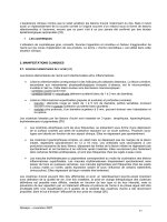
The skull is a complex structure. There are 8 cranial bones and 14 facial
bones in the skull. From the anterior view most
of
the facial bones can be
seen and some of the cranial bones are visible too. The
bone
that makes
up the forehead and extends beyond the eyebrows is the
frontal
bone.
This bone forms the upper rim
of
the
orbit,
which is a socket that
encloses the eye.In the back of the orbit is the
sphenoid
bone
and the
lateral walls of the orbit are composed of the zygomaticbones. The
bridge of the nose consists of the paired nasal
bones
and just lateral to
a. _
them are the two maxillae. These bones hold the upper teeth. The lower
teeth are held by the mandible. Inside the nasal cavity two projections
can be seen. These are the inferior nasal conchae. The wall that divides the
nasal cavity is the nasal
septum
and it consists of two bones, the ethmoid
bone and the vomer. Along the side of the skull are the temporal bones,
located posterior to the zygomatic bones. Label the major bones of the
skull and color them in. As you color in the skull try to use the same color
for the same bone on different pages. This will help you associate the
same bone with various views from which it can be seen.
d.~
e.
_
f.
g
h.
Answer Key:a.Orbit,b.
Frontal
bone, c.
Temporal
bone, d.
Sphenoid
bone, e.
Nasal
bone,
f.
Zygomatic
bone,g.
Nasal
septum, h.
Maxilla,
i.Mandible
Chapter
Three
Skeletal System
I
UPLANd'· I
me lea
45
LATERAL
VIEW
OF
THE
SKULL
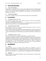
Many bones seen from the anterior view can also be seen from the lateral
view.The
frontal
bone
is joined to the
parietal
bones
by the
coronal
suture. The parietal bones span
much
of the
cranium
and articulate with
the occipital
bone
at the
lambdoid
suture.
There is a posterior
extension of the occipital
bone
known as the
external
occipital
protuberance.
The exterior aspect of the
temporal
bone
is seen from the
lateral view and many of the significant features such as the
mastoid
process, external acoustic meatus, and styloidprocess are visible. On the
side is the elongated zygomaticprocess. The temporal bone articulates
with other cranial bones by the
squamous
suture.
The bone anterior to
the temporal bone is the
sphenoid
bone.
It is a bone that is found in the
middle
of
the skull. The nasal
bone
is visible from the lateral view and its
relationship with the maxilla can be seen here. Behind the maxilla is the
a. _
lacrimal
bone
which houses the nasolacrimal canal, a duct that drains
tears from the eye into the nose. The
mandible
articulates with the rest
of
the skull at the
mandibular
condyle. A depression in front of the
condyle is the
mandibular
notch
and the anterior section of bone in
front
of
the notch is the
coronoid
process. Label the major features of
the skull seen in lateral view and color each bone a different color.
Details of the mandible can be seen in the isolated bone. In addition to
the features of the mandible listed above, find the
mandibular
foramen
and the
mental
foramen
of
the mandible. These are holes for the passage
of
nerves
and
blood vessels.The main
portion
of the mandible is the
body
and the upright part is the ramus. The angle is the posterior
junction
of
these two parts. The teeth are located in alveoli and the small
segments of bone between the teeth are the alveolar processes. Label the
features
of
the mandible.
r. _
q._
p._
0.
_
Tl, _
ffi.
_
1.
_
k. _
e. _
f. _
z.
1.
s.
Answer
Key: a. Coronal suture, b. Parietal bones, c. Zygomatic process, d. Temporal bone, e. Squamous suture, f. Lambdoid suture, g. External occipital protuberance,
h. Occipital bone, i. Mastoid process,
j. External acoustic meatus, k. Styloid process, I.Mandible,
ill.
Maxilla, n. Zygomatic bone, o. Nasal bone, p. Lacrimal bone,
q. Sphenoid bone, r. Frontal bone, s. Coronoid process,
t. Mandibular foramen, u. Mandibular notch, v. Mandibular condyle, w. Ramus, x. Angle,
y. Body, z. Mental foramen
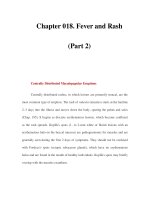
SKULL-TOP AI\ID
BOnOM
VIEWS
The superior aspect of the skull
consists of few bones and few
sutures. The
frontal
bone
is the
most anterior bone with the
parietal
bones directly posterior to it. The
coronal
suture
separates the two
and the sagittal
suture
separates the
parietal bones. The
lambdoid
suture
separates the parietal bone from the
occipital bone. Label the bones and
sutures and color the bones in the
illustrations.
The inferior aspect of the skull is
more complex than the superior
view.In the inferior view the
mandible has been removed so some
of the underlying structures can be
seen. The large opening in the
occipital bone is the
foramen
magnum.
The two bumps lateral to
the foramen magnum are the
occipital condyles and the raised
bump
at the posterior
part
of the
skull is the external occipital
protuberance. The more anterior
and lateral bone to the occipital
bone is the temporal bone. The
jugular
foramen is located between
the occipital and temporal bone.
Another opening nearby is the
carotid canal. Lateral to this is the
styloid process, an attachment point
for muscles. Lateral to this is a
depression called the
mandibular
fossa.
it
is here that the mandible
articulates with the temporal bone.
The
sphenoid
bone
spans the skull
and the major features seen from the
inferior view are the greaterwing,
and the lateral and
medial
pterygoid plates. The hard palate is
made of the palatine process of
the
maxilla and the palatine bones. The
bone that opens into the nasal cavity
is the vomer. Label and color these
features of the skull.
Answer Key: a.
Frontal
bone,
b.
Coronal
suture,c.
Parietal
bones,
d.
Sagittal
suture,e. Lambdoid suture,
f.
Occipital
bone, g.
Palatine
process
of
the maxilla, h. Palatine bone, i. Vomer,
j. Greaterwing, k.
Lateral
pterygoid
plate,
I.
Medial pterygoidplate,
m. Mandibular
fossa,
n. Styloid
process,
o.
Carotid
canal,
p. Jugularforamen,
q.
Occipital
condyles,
r.
Foramen
magnum,s.
External
occipital
protuberance
Anterior
Anterior
Posterior
g.
Chapter
Three
I
KAPLA~.
I 47
SkeletalSystem meulca
h. _
1.
Sphenoid bone:
J.
k.
1.
q._
r.
ChapterThree I UPLANd'· I 49
Skeletal System me lea
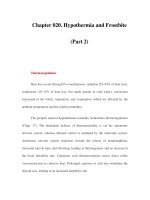
MIDSAGITIAL SECTION OF THE SKULL
Several features of the skull can be seen when it is sectioned in the
midsagittal plane. Locate the major bones of the skull and the features
seen in this section. The nasal
septum
consists of two
bony
structures, the
perpendicular
plate
of
the
ethmoid
bone
and
the
vomer.
The
crista
galli
extends superiorly from the
cribriform
plate
of the
ethmoid
bone. The
junction of the
maxilla
and the
palatine
bone
that make up the hard
palate can be seen from this view as well. The
frontal
sinus
and the
sphenoid
sinus
are two cavities seen here. Label the bones and the major
features of the midsagittal section of the skull using the terms provided.
Color the bones different colors and shade the sinuses in a darker shade
of the color used for the specific bones that hold the sinuses.
Frontal
bone
Temporal
bone
Maxilla
Styloid process
Nasal
bone
Vomer
Sphenoid
sinus
Parietal
bone
Sphenoid
bone
Mandible
Sella
turcica
Palatine
bone
Crista
galli
Occipital
bone
Ethmoid
bone
Internal
acoustic
meatus
Cribriform
plate
of
the
ethmoid
Perpendicular
plate
of
the
ethmoid
Frontal
sinus
a. _
b.
c.
m.
d.
e.
f.
n.
g.
h.
1.
J.
k.
r. _
Answer
Key:
a.
Frontal
bone, b.
Frontal
sinus, c.
Nasal
bone, d. Ethmoidbone, e.
Crista
galli,
f.
Cribriform
plate ofthe ethmoid, g. Perpendicularplate ofthe ethmoid,
h.
Vomer,
i.
Maxilla,
j. Palatinebone,
k.
Mandible,
I.
Parietal
bone, m.
Temporal
bone, n.
Sella
turcica,
o.
Occipital
bone, p.
Internal
acoustic meatus, q. Sphenoid bone,
r. Sphenoid sinus
Chapter Three I lAP
LANd'
• I 51
Skeletal System me lea
e. _
d. _
0.
_
c. _
A few bones of the skull are
frequently studied as separate bones.
The sphenoid
bone
has a superficial
resemblance to a bat or butterfly.
There are the lesser wings, the
greater
wings,
and
the
pterygoid
plates, all of which resemble wings.
The
dorsum
sellae is the posterior
part of the sella
turcica
(a
depression that holds the pituitary
gland). Locate the
foramen
rotundum
and the
foramen
ovale
on the sphenoid bone. These holes
enclose parts of the trigeminal
nerve.
The
ethmoid
bone
is located just
posterior to the nose and is best seen
isolated from the rest of the skull
bones. The cribriform plate that has
small holes called olfactory foramina
in it. Locate the
crista
galli
and
the
perpendicular
plate. The
ethmoid
has four curved structures lateral to
the perpendicular plate. These are
the two
superior
nasal
conchae
and
the two
middle
nasal
conchae. The
ethmoid
sinuses are
numerous
small holes in the bone. Locate the
structures of these skull bones. Label
the illustration and color in the
features of the bones.
The temporal
bone
has a flat
squamous
portion
and
a denser
petrous portion. The section of the
temporal
bone
that
connects to the
zygomatic
bone
is the
zygomatic
process. There are two significant
canals or meatuses for hearing.
These are the
external
acoustic
meatus
and
the internal acoustic
meatus. The
mastoid
process is a
large
bump
that can be palpated
directly posterior to the ear. The
styloid
process
anchors a
number
of
small muscles.
Answer
Key:
(Sphenoid features), a. Sellaturcica
b. Lesserwing,c. Foramen rotundum,
d. Foramen ovaIe,e. Dorsum sellae,
f.
Greaterwing
(Temporal
features), g. Squamous
portion,h.
Zygomatic
process,
i.
External
acoustic meatus,
J.
Styloid
process,
k.
Mastoidprocess
(Ethmoidfeatures),
I.
Crista
galli,
m.
Middle
nasal concha,
n. Perpendicularplate,
o. Superiornasal concha
SPHENOID, TEMPORAL,
Af\ID ETHMOID BONES
Chapter
Three I
mIAPeLA'!a cal
53
Skeletal
System
U
a. _
,
\ /""/
I~
\
d. _
c. _
b. _
I
vi
,
Answer
Key: a.
Cervical
vertebrae
(cervical
curvature), b.
Thoracic
vertebrae (thoraciccurvature),
c.Lumbarvertebrae (lumbar curvature),
d.
Sacrum
(pelvic curvature), e.
Coccyx
We are
unique
as animals because of
our
upright posture. The vertical
position
of
the spine is reflected in
the increase in size of the vertebra
from superior to inferior. The
vertebral column is divided into five
major regions. There are 7 cervical
vertebrae that occur in the neck
while the 12thoracic vertebrae have
ribs attached to them. The 5
lumbar
vertebrae are found in the lower
back and the sacrum consists
of
5
fused sacral vertebrae. The coccyx is
the terminal
portion
of the vertebral
column consisting of 4 coccygeal
vertebrae. The vertebral
column
in
the adult has curves. The
uppermost
is the cervical curvature and the
lower ones are the thoracic, lumbar,
and pelviccurvatures. Label the
illustration with the regions
and
the
curvatures and color in the regions
with different colors. Color in the
curved arrows for the curvatures.
VERTEBRAL
COLUMN
ATLAS
The atlas is the first cervical
vertebra. It is unique
among
the
vertebrae because it has no body.
Label the
vertebral
foramen,
superior
articular
facet, the
transverse
foramen,
and the lateral
masses.
AXIS
The axis is the second cervical
vertebra and it has a
body
with a
projection that arises from the body
known as the
odontoid
process or
dens. Label the axis including the
superior
articular
facets, the
transverse foramen, the
spinous
process, and the
vertebral
foramen.
Color these features in.
ATLAS
AND
AXIS
Here are the atlas
and
axis together.
Color the two bones separate colors.
HYOID
The hyoid bone is a floating bone,
which means that it has no
hard
attachments to
other
bones. The
main part of the hyoid is the
body
and the two horns that arise from
the hyoid are the
greater
cornua
and
the lessercornua. Label these parts
of the bone and color
them
in
separate colors.
Chapter
Three I
KAPLA~.
I 55
Skeletal System meulca
e. _
Answer
Key:
a.
Vertebral
foramen, b.
Lateral
masses, c.Transverse foramen, d. Superior
articular
facet,
e. Spinous process,
f.
Body,
g.Odontoid process (dens), h.
Axis,
i.
Atlas,
j. Lessercornua,
k.
Greatercornua,
I.
Body
h
1m
KAPeLA~·lcal
Chapter
Tree
u
Skeletal System
57
a.
b.
1.
bar Vertebra
Lum
Thoracic Vertebra
1.
. 1Vertebra
Cervica
b. _
/ c.
.,
.
(t
"
~!I\C'
'"
~
'~~T*P
~.:~
:tI:~
j.
d.
d.
__
e.
__
",
e.
THORACIC,
CERVICAL,
R
VERTEBRAE
AND
LUMBA
to vertebrae
mmon
Features co the spinal cord
.
gwhere
.
The operun the vertebra
IS
passes
through
t
bral
foramen.
known as the ver ertebra is
the
Th
e
body
of the ve f the vertebra
.
part
0 t
eight-bearing ss is
the
par
w .
proce
d
the
spinous
. I This process
an d
ostenor
y. al
that
exten
~
p from the
vertebr
is an extension from
the
body
arch
that
curves tebral foramen.
enclosing the ver sed
of
the two
This arch is
compo
laminae.
The
edicles and.
the
tworocess
and
the
~uperior
art~cular
facet (the flat
superior
artIcu~~cess)
are
the
parts
surface on
~he
the vertebra above.
that join with .
ular
process
and
The
infe~ior
ar1~~ar
facet are
t?e
the
infenor
art
b that join with
f the verte raparts 0
the vertebra below.
. al vertebrae
. al cervlc .
TYPIc. nd lateral
view
superior a distinct from
. I rtebrae are .
Cervica ve b having two
II
other
vertebra~
y These house
a
f
mma.
, .
transverse
ora
her characteristic
blo
od vessels.Anot
brae
iis that several
. I rte rae
of the
cervica ve
ifid
spinous
process
of them have a b
cic vertebrae
Typical thora lateral
view
superior and b e typically have
. erte ra
The thoracic v ocesses
than
l
onger
spinous
pr
d
many
of
them
t brae an Th
cervical ver
.e
rior
direction. e
int in an infe . vertebrae,
pOl in thoracic .
body
is larger I bones with
n
d they are the on y
ttachment
a that are a
costal facets h ads
of
ribs.
The
P
oints for the e ses can be seen
proces
tal
transverse
sverse cos
ith the
tran
along
WI
facets.
. lumbar verteb!ae
TYPlc~1
d lateral
view
superior an brae have larger
The
lumbar
v:r~~ey
support
~ore
bodies becaus . s process IS
The
spinou
I'
weight. re horizonta
III
.
shorter
and
mo han in thoracic
I
mbar
vertebrae t costal facets
u Thereare no bel
vertebrae. foramina. La
and no transverse brae illustrated
t
of
the verte
the
par
s .
and color
them
Ill.
, process,
. a Bifid spinous
Answer
Key
c Vertebral
' s process" , I
b Spinou , e Pedice,
. d Lamina, '
foramen, ' , lar process,
f.Superior articu ess h. Body,
g
Transverse
proc
'ssJ',
Transverse
' , lar proce ,
i Inferior artrcu , r costal facet,
' k Superio
foramen" f et
I.
Inferior costal ac
SACRUM
AND
COCCYX
Sacrum and coccyx,anterior view
The terminal
portion
of the
vertebral
column
consists of two
structures that are fused bones. The
sacrum
is 5 fused vertebrae and the
coccyx is
3-5
fused vertebrae. The
top rim of the
sacrum
is the sacral
promontory
and
the wing-like
expansion where the ilium attaches
is the ala. The area where the
vertebrae join are the
transverse
lines. The holes
running
down
each
side are the
anterior
sacral
foramina. At the top
of
the
sacrum
are the
superior
articular
processes
and they attach to the
lumbar
vertebra. Label and color the parts
of
the sacrum and the coccyx.
Sacrum
and
coccyx, posterior
view
From the posterior view the
median
sacralcrest is the fused remains
of
the spinous processes of the
vertebrae. The
posterior
sacral
foramina
are on each side of the
crest and the
lateral
sacral
crests
are
lateral to the foramina. The
superior
articular
processes can be seen from
this view and also the
auricular
surface which forms
part
of the
sacroiliac joint. Label the features of
the sacrum and the coccyx and color
them
in.
Answer Key:a. Superiorarticular
process,
b.
Ala,
c.
Sacral
promontory,
d.
Transverse
lines,e. Anterior
sacral
foramina,
f.
Coccyx,
g.
AUricular
surface,
h.
Lateral
sacral
crest,
i. Median
sacral
crest,
j.
Posterior
sacral
foramina
ChapterThree I IAPLAN
d
··
I 59
Skeletal System me lea
c. _
d. _
e. _
-f.
_
1. _
STERNUM
/
RIBS
/
HYOID
The
sternum
is commonly known as
the breastbone
and
is divided into
three areas, the upper
manubrium
with the
suprasternal
notch
and the
clavicular notches, the
body
with
the costal
notches
(where the ribs
attach),
and
the
xiphoid
process.
Between the
manubrium
and
the
body is the
sternal
angle. Label these
features on the illustration and color
the three major areas of the
sternum
different colors.
If you select a rib as a representative
bone for all of the ribs, you will find
the terminal
portion
of the rib is
expanded in a head. The constricted
region below that is the neck. The
tubercle of the rib is a
bump
that
attaches to the transverse process of
the vertebra. The
bend
in the rib is
known as the angle
and
the
depressed area of the rib where
nerves and bloodvessels are found is
the costal groove. Color in the
individual parts of a rib after you
label the figure and color the rib as it
joins with a vertebra.
a.
b. _
c. _
d. _
e
f. _
g._
1. _
ChapterThree I
KAPLAlf
d
- I
61
Skeletal System me lea
Answer Key: a.
Suprasternal
notch,
b.
Clavicular
notch,c. Manubrium,
d.
Sternal
angle,
e.
Costal
notches,
f.
Body,
g.Xiphoid
process,
h. Head,
i.
Tubercle,
j.
Neck,
k.Angleof rib,
I.
Costa
I groove
1. _
1.
_
.
.
APPEf\I
DICU
LAR
SKELETON-PECTORAL
GIRDLE
AND
UPPER
EXTREMITY
The pectoral girdle is made of the
clavicles and the scapulae. The
upper extremity consists of the
humerus
of the arm, the
radius
and
ulna
of the forearm, and the carpals,
metacarpals,
and
phalanges of the
hand. Locate these major regions of
the upper extremity and label them
on the diagram. Color these areas in
different colors on the illustration.
Answer Key:a.
Clavicle,
b. Humerus,
c.
Scapula,
d.
Radius,
e.
Ulna,
f.
Carpals,
g.
Metacarpals,
h.
Phalanges
d
_
a. _
f.
ChapterThree I UPLANd'· I 63
Skeletal System me lea
II
, ( ,
,
/
/
1
I
,
II
c
SCAPULA
The pectoral girdle consists of the
scapulae and the clavicles. Each
scapula is a triangular bone and the
three edges are known as the
superior
border,
the lateral
border,
and the
medial
border.
The
scapular
spine
is on the posterior
surface and it expands into a
terminal process known as the
acromion
process. Above the spine
isthe
supraspinous
fossa. Below the
spine is the
infraspinous
fossa and
on the anterior side of the scapula is
the
subscapular
fossa and the
coracoidprocess. The
inferior
angle
of the scapula is at the junction of
the medial and lateral borders.
Inferior to the acromion process is
the glenoidfossa. This is a
depression where the head of the
humerus articulates with the
scapula. Label the various features of
the scapula and color in the regions
of the bone with different colors.
Locate as many of the features from
the various angles presented.
Answer Key:a.Acromion
process,
b.
Superior
border,
c.
Coracoid
process,
d. Glenoid
fossa,
e.
Subscapular
fossa,
f.
Lateral
border,
g. Medial
border,
h. Inferior
angle,
i,
Supraspinous
fossa,
j.
Scapular
spine,k.Infraspinous
fossa
Chapter
Three
I UPLANd'· I 65
Skeletal System
me
lea
c. _
d. _
e.
-4~it7;:~
f. _
g._
h. _
d. _
a. _
c
d. _
f. _
h. _
CLAVICLE
The clavicle is a thin bone that stabilizes the shoulder joint in a lateral
position. It has ablunt end that articulates with the sternum (the sternal
end) and a flattened end that joins with the acromion process of the
".
' .
Chapter
Three I
lAPLA~.
I 67
Skeletal System meulCa
scapula. This is called the acromial end. A small
bump
on the inferior
part of the clavicle has a ligament that attaches to the coracoid process of
the scapula. This
bump
is called the conoid tubercle. Labelthe clavicle
and color the ends and the conoid tubercle.
Sternum
Answer
Key: a, Sternal end, b. Acromial end, c. Conoid tubercle
.; .
".
'"
".
Superior view
Inferior view
c. _
Chapter
Three
I
KAPLA~.
I 69
Skeletal System meulCa
HUMERUS
g._
~f.
__
£,.
\~
r
L
The humerus has a proximal
head
that fits into the glenoid fossa of the
scapula. Just at the edge of the head
is a rim known as the anatomical
neck. Below this neck are the greater
and lesser tubercle
and
the
depression between the two is the
intertubercular
groove. Below these
is the surgical neck of the humerus.
The deltoid muscle attaches to the
humerus at the
deltoid
tuberosity
and the two expanded wing-like
processes at the distal end of the
humerus are the
supracondylar
ridges. Inferior to these are the
medial and lateralepicondyles and
at the articulating ends of the
humerus are the lateral
capitulum
and the medial trochlea. The
depression on the anterior surface of
the humerus into which the ulna fits
is called the
coronoid
fossa and the
posterior depression where the
elbow locks into the humerus is
called the
olecranon
fossa. Label the
figure and color in the specific parts
of the illustration.
h. _
1. _
J
~ :i_=~~~L
m. _
Anterior View
Posterior View
Answer Key: a. Greatertubercle,
b. Head,
c.Anatomical neck,d.
Lesser
tubercle, e. Intertuberculargroove,
f.
Surgical
neck,g.Deltoid tuberosity,
h. Supracondylar
ridges,
i.
Lateral
epicondyle,j.Coronoid
fossa,
k.Olecranon
fossa,
I.Medial epicondyle,
m. Capitulum, n.
Trochlea
g
FOREARM
BONES
The radius has a circular
head,
a
radial
tuberosity
on the shaft
(where the biceps brachii muscle
attaches), and a distal
styloid
process. At the distal
end
of the
radius is a depression where the ulna
joins with the radius. This is
known
as the
ulnar
notch
of the radius.
The ulna has a proximal
olecranon
process, a
coronoid
process, and the
trochlear
notch
between the two.
Just distal to the coronoid process
of
the ulna is the
tuberosity
ofthe
ulna,
a projection where muscles
attach. The
head
of the ulna is distal
and it also has a
styloid
process. At
the proximal
portion
of
the ulna is a
depression where the head of the
radius articulates with the ulna. This
depression is
known
as the
radial
notch
of the ulna.
When the two bones are
joined
you
can see where each fits
into
the
other. On the edge of each
bone
is
the
interosseus
margin.
This is a
ridge where the interosseus
membrane
connects the bones.
h. _
Chapter
Three
I UPLANd'· I 71
Skeletal System me lea
Answer Key: a.
Olecranon
process,
b.
Trochlear
notch,c.Coronoid
process,
d.
Radial
notch,e.
Tuberosity
of the
ulna,
f.
Head,
g.
Radial
tuberosity,
h.
Interosseus
margin,i. Ulnarnotch,
j.
Styloid
process
1
J
ChapterThree I
ImAPelA~·ICal
n
Skeletal System U
Right Hand,
Anterior View,
Carpals
Right Hand,
Posterior View
Right Hand,
Anterior View
1.
_
g._
J
k. _
1. _
a. _
f. _
e.
~
j. _
-rS?
~YI\0f:(~
.'
A(~~}
"\J5
;:}(JJ!>
A
m.
••
~~.~~r
·l~'
1.
_
h. _
e. _
a. _
m. _
h. _
HAND
BONES
Answer Key:a.
Phalanges,
b. Head,
c.
Shaft,
d.
Base,
e. Hamate,f.
Capitate,
g.Triquetrum, h.
Lunate,
i.
Metacarpal,
j.
Trapezoid,
k.
Trapezium,
I.
Scaphoid,
m. Pisiform
The
hand
consists of 27 bones
divided into three groups: the
carpals, the metacarpals,
and
the
phalanges. The
thumb
is
known
as
the pollex
and
is listed as the first
digit of the hand. The index finger is
the second digit
and
the fingers are
listed sequentially with the little
finger being the fifth digit. The
bones of the fingers are known as
phalanges and they are named
according to what digit they belong
and as being proximal, middle or
distal. Therefore the
bone
of tip of
the little finger is the distal phalanx
of the fifth digit while the
bone
in
the place where you would normally
wear a wedding ring is the proximal
phalanx of the fourth digit. Each
phalanx has a proximal base, a shaft,
and a distal head. The
metacarpals
are the bones of the palm of the
hand. Each metacarpal also has a
proximal base, a shaft, and a distal
head. There are five metacarpals
and
they are
named
for the phalanges
that extend from them. The first
metacarpal articulates with the
thumb. The carpals are the bones of
the wrist. There are eight carpal
bones in two rows. The bone under
the
thumb
is the
trapezium.
The one
medial to it is the
trapezoid.
The
capitate is found under the third
metacarpal and the
hamate
finishes
that row. Proximal to the trapezium
is the scaphoid, which joins with the
radius. The next
bone
in line is the
lunate, followed by the
triquetrum,
and finally the little
pisiform
bone.
If you memorize the bones in this
sequence you can use a
mnemonic
device to remember them. This
mnemonic is
The Tom Cat Has
Shaken
Loose
To
Prowl.
The first
letter of the mnemonic represents
the first letter of the carpal bone.
Label the illustration and color all of
the phalanges one color. Color the
metacarpals
another
color and color
the carpal bones individual colors.
As you color the various illustrations
of the
hand
use the same color
scheme for the bones.
HIP
The hip bones are known as the os
coxae. Each os coxa is a result
of
the
fusion of three bones, the
ilium,
the
ischium, and the pubis. Label and
color in these three fused bones
using a different color for each area.
The two os coxae, when joined
together by the
pubic
symphysis,
form the pelvis and it can be divided
into an upper false pelvis
and
a
lower
true
pelvis separated by the
pelvic brim. The
anterior
superior
iliac
spine
and the
anterior
inferior
iliac spine can be seen from the
front. The top ridge of the pelvis is
the iliaccrest. The large, inferior
hole is the
obturator
foramen
and
the depression superior to it is the
acetabulum. Note the junction of
the sacrum and the ilium that forms
the sacroiliac
joint.
Label the
features of the anterior view
and
color them in.
Answer Key: a. Iliac
crest,
b.
Sacroiliac
joint, c. Greatersciaticnotch, d. Anterior
superioriliac
spine,e. Anteriorinferior
iliacspine,
f.
Acetabulum, g.Obturator m. _
foramen, h. Pubic
symphysis,
i.
False
pelvis,
j.
True
pelvis,
k.Ilium,
I.
Ischium,
m.
Pubis
Chapter
Three
I
KAPLAlf
d
- I
75
Skeletal System
me lea
a._-:
_
c. _
1. _
J
p~",-
1
_
HIP (CONTINUED)
Lateral View
When seen from a lateral view,
several features are apparent in the
os coxa. Locate the
posterior
superior
iliac spine and the
posterior
inferior
iliac
spine
along
with the greater sciaticnotch, the
spine of the ischium, and the lesser
sciatic notch. The ischial
tuberosity
is at the posterior, inferior edge of
the ischium. Just anterior to the
tuberosity is a strip of bone called
the ischial
ramus
that attaches to
the
inferior
pubic
ramus. The body
of the pubis is the most anterior part
of the pubis and the
superior
pubic
ramus
is the portion that forms part
of the acetabulum. Label and color
these features on the illustration.
MALE
AND
FEMALE
PELVIS
Differences can be seen between the
male and female pelvis. The
subpubic
angle in males is less than
90 degrees and the female angle is
greater than 90 degrees. The ilium in
males is more vertical
than
in a
pelvis of a woman who has had
children. A further distinction is
seen in the side view of a pelvis in
which the sciatic notch in the female
pelvis has a much wider angle
than
in males. Color in the upper portion
of the ilium.
Chapter Three I
KAPLAlf
d
-
I
77
Skeletal System me lea
1. _
J
k. _
1.
_
ffi.
_
ll
0.
_
Answer Key:a.Iliac
crest,
b.
Posterior
superioriliacspine, c.
Posterior
inferior
iliac
spine,
d. Greatersciaticnotch,
e. Spineof the ischium,f.
Lesser
sciatic
notch,g.
Ischial
tuberosity, h.
Ischial
ramus,
i. Anteriorsuperioriliac spine,
j. Anteriorinferioriliac spine, k.Superior
pubic
ramus,
I.
Inferior pubic
ramus,
m. Obturatorforamen, n. Acetabulum,
o. Iliacblade,p. Subpubic
angle,
q. Male (lessthan ninety degrees),
r.
Female
(more than ninety degrees)
q
r
_
Chapter
Three
I
ImAPeLA~·lcal
79
Skeletal System U
r. _
p._
n. _
h. _
The femur seen from the anterior
view shows a proximal
head
and
a
constricted neck. Two large
processes are distal to the neck.
These are the
greater
trochanter
and the lesser
trochanter.
There is a
raised section of bone between
them
called the
intertrochanteric
line.
The main part of the
bone
is the
shaft and the lateral epicondyle and
medial epicondyle are the distal
expansions of the bone. The
posterior view of the femur has
additional features such as the
intertrochanteric
ridge, the
linea
aspera, and the
lateral
condyle and
the
medial
condyle. The femur is
bowed and this can be seen from a
lateral view as well as the placement
of the patella. The base of the patella
issuperior and the
apex
is inferior.
Label the features of the femur
and
patella and color in the various
parts.
LOWER
EXTREMITY-
FEMUR/PATELLA
The lower extremity consists of the
femur
of the thigh, the
tibia
and
fibula of the leg,
and
the tarsals,
metatarsals, and
phalanges
of the
foot. Locate these
major
regions of
the lower extremity and label
them
on the diagram. Color these areas in
different colors on the illustration.
Answer Key: a.
Femur,
b.
Patella,
e.
Tibia,
d.
Fibula,
e.
Tarsals,
f.
Metatarsals,
g.
Phalanges,
h. Greater
trochanter, i. Head,
j. Neck,
k. Intertrochanteric line,
\.Intertrochantericridge,m.
Lesser
trochanter, n. Linea
aspera
, o.
Lateral
epicondyle,p.
Lateral
condyle,q. Medial
epicondyle,
r.
Medial condyle,s.
Base
of
patella,
t. Apexof patella
Anterior
s. _
Posterior
TIBIA
/
FIBULA
The
tibia
supports the weight of the
body and is the
bone
that articulates
with the femur. The fibula is more
slender and is a
bone
to
which
muscles attach. The top of the tibia is
expanded into a triangular shape
with the
medial
tibial condyle
and
lateral tibial condyle articulating
with the condyles
of
the femur. The
quadriceps femoris muscles attach to
the tibial
tuberosity
on the anterior
surface of the tibia just below the
condyles. The
anterior
tibial crest is
a large ridge that runs the length of
the bone. At the terminal portion of
the tibia is the
medial
malleolus.
This process, along with the
lateral
malleolus of the fibula, join with the
talus
of
the foot. The
head
of the
fibula is proximal.
It
is a triangular
region with a pointed apex. Label
the tibia and fibula illustrations and
color in the various regions of the
bones.
I
I I
ChapterThree I
KAPLAN
d'.
I 81
Skeletal System me lea
d. _
e. _
I
I
I
I
I
Answer Key:a.
Lateral
tibial condyle,
b. Medialtibial condyle,
c. Tibial
tuberosity, d.
Apex,
e. Headof fibula,
f.Anteriortibial
crest,
g.Shaftof tibia,
h. Shaftof fibula, i. Medialmalleolus,
j.
Lateral
malleolus Anterior Posterior
a. _
LEFT
FOOT
Color in the seven
tarsal
bones using
different colors for each bone. The
calcaneus is the heel
bone
and takes
the major weight of the body during
walking. The talus connects the foot
to the tibia and fibula forming the
ankle joint. The
cuneiforms
are so
called because they are wedge-
shaped bones and they form a
natural arch of
bone
in the foot.
Note that each
of
the
metatarsals
and each
of
the
phalanges
has a
distal head, a shaft, and a proximal
base. Color all of the five metatarsals
the same color. The first metatarsal is
under the big toe
and
the fifth is
under the smallest toe. Color all of
the fourteen phalanges
another
color. Allof the proximal phalanges
are given the same letter in the
illustration as are the middle and
distal phalanges. Write proximal,
middle, or distal in the appropriate
space next
to the toes. The big toe
(hallux) has two phalanges while the
other toes have three.
1. _
2. _
3.
_
c. _
b. _
ChapterThree I UPLANd·· I 83
Skeletal System me lea
a. _
b
c
~.lL-~'o-
d. _
Answer Key:
1.
Phalanges
2.
Metatarsals
3.
Tarsals
a.
Distal
phalanges,
b. Middle
phalanges,
c.
Proximal
phalanges,
d. Head,e.
Shaft,
f.
Base,
g.
First
(medial) cuneiform, h. Second
(intermediate) cuneiform,
i. Third(lateral) cuneiform,
j. Cuboid,
k.
Navicular,
I.
Talus,
m.
Calcaneus
1. _
h
__
g._
J
m. _
Tarsals