Kaplan anatomy coloring book - part 8 pdf
Bạn đang xem bản rút gọn của tài liệu. Xem và tải ngay bản đầy đủ của tài liệu tại đây (751.9 KB, 21 trang )
Chapter
Thirteen:
Male
Reproductive
System
295
OVERVIEW
OF THE
MALE
REPRODUCTIVE
SYSTEM
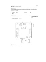
The male reproductive system consists of the two testes, the
epididymis,
the
ductus
deferens
enclosed in the spermatic cord, the
seminal
vesicles, the
prostate
gland, the bulbourethral
glands,
and
the
penis.
The testes are the glands
that
produce
testosterone
and
sperm
cells.
Sperm cells travel
from
the testes to the epididymis where they are stored
and
mature. From the epididymis
sperm
cells move into the
ductus
deferens, which enters the
body
and travels to the
posterior
bladder.
From here the
ductus
deferens
turns
into the ejaculatory duct, which
receives fluid from the sernimal vesicles. The ejaculatory duct leads to the
urethra
where secretions from the prostate
and
bulbourethral glands are
added. Finally the
sperm
cells
and
seminal fluid (together these make
semen) are ejaculated from the penis.
Label the parts of the male reproductive system
and
color the various
structures in the illustration.
<,
<,;
.~
~
h. _
g._
J
Kidneys
~
1. _
\
Answer
Key:
a. Dudus deferens, b. Seminalvesicle,c. Prostate,d. Bulbourethral gland,e.
Epididymis,
f
Testis,
g. Uncircumcised penis, h. Circumcised perns,i.
Urethra
Chapter Thirteen
Male Reproductive System
I
IAPLAN
d
·· I
me lea
297
ORGANS
OF THE
MALE
REPRODUCTIVE
SYSTEM
p
g
f. _
Answer Key: a.Ductus deferens,
b. Pampiniform
plexus,
c.
Testicular
artery,
d.
Epididymis,
e.
Testis,
f.
Cremaster
muscle and
fascia,
g.
Scrotal
skin and dartos
muscle,
h.
Ureter,
i, Urinary
bladder,
j.
Seminal
vesicle.
k.
Ejaculatory
duct,
I.
Prostate,
m. Bulbourethral gland,n.
Urethra,
o. Ductusepididymis,p.
Lobules
of
testis
The testes are enclosed in the
scrotal
sac which is lined with a
smooth
muscle layer called the
dartos
muscle. This muscle contracts when
the temperatures
drop
near the
testes, causing
them
to withdraw
closer to the body where it is
warmer. Another muscle
of
the
region is the
cremaster
muscle.
It
also contracts when it is cold
but
it is
made of skeletal muscle. The
epididymis
sits on top of the testis
like a small cap and is a place where
sperm cellsmature. The
spermatic
cord
consists of the cremaster
muscle, the
ductus
deferens, the
testicular
artery,
and
a complex
meshwork of veins called the
pampiniform
plexus. This plexus
cools arterial blood flowing to the
testes maintaining the testes at about
35 degrees C which is
important
for
proper sperm maturation.
The sperm are produced in the
seminiferous tubules of the testis.
This occurs in
lobules
of
the
testis
before they move to the epididymis.
The epididymis has a series of long
coiled tubules called the
ductus
epididymis
and the sperm cells
slowly pass through this ductwork.
After the sperm cells
mature
in the
epididymis they
then
travel to the
ductus
deferens which loops
around
the
ureters
before reaching the
seminalvesicles located on the
posterior surface of the
urinary
bladder. The seminal vesicles add a
fluid that has buffers and that
provides fructose to the sperm cells.
From the seminal vesicles the fluid
passes through the
ejaculatory
duct
to the prostate. The prostate adds
further fluid that is rich in buffers.
This fluid passes into the urethra.
The bulbourethral glands add a
protein lubricant to the fluid. Label
the organs and their features in the
illustration and color them in
different colors.
Chapter Thirteen
Male Reproductive System
I
meCtical
299
Answer Key: a.Urinary
bladder,
b.
Symphysis
pubis, c.Corpuscavernosum, d. Corpusspongiosum,e.
Glans
penis,
f.
Prepuce,
g.
Testis,
h.
Tail
of epididymis, i. Body of epididymis, j. Head of epididymis, k. Spongy
urethra,
I.
Bulbourethral
gland,m.
Prostate,
n. Seminal
vesicle,
o. Deep dorsalvein, p.
Spermatozoa,
q.
Sperrnatids,
r.
Secondary
spermatocytes,
s. Primaryspermatocytes,
1.Sertolicell, u. Spermatogonia
s. _
a.
t. _
u. _
p
k
d. _
g
f. _
e. _
d
b.
'-!'::e-~M,
c
When seen in a midsagittal section,
the relationship of the glands
that
produce seminal fluid can easily be
seen. The
prostate
is approximately
the size of a golf ball and is located
inferior to the urinary bladder. The
prostatic urethra is the
portion
of
the urethra that is enclosed in the
prostate. The
bulbourethral
glands
are located in the wall of the pelvic
floor and the
seminal
vesicles are
posterior to the urinary bladder.
Exterior to the
body
wall are the
testes and these are enclosed in the
scrotal
sac. The
epididymis
receives
sperm from the testis and has three
parts, a head, a
body,
and
a tail. The
symphysis
pubis
is an
important
reference
point
in the midsagittal
section. In males there is a flap of
tissue encircling the glans penis.
This is the
prepuce
(foreskin) and it
is sometimes removed at
birth
in a
procedure called a circumcision. The
corpus
cavernosum
can be seen in
this section along with the
corpus
spongiosum
and the
spongy
urethra.
The formation of sperm is
known
as
spermatogenesis and occurs from
spermatogonia
on the superficial
wall of the seminiferous tubules.
These produce cells called
primary
spermatocytes
which in
turn
mature into
secondary
spermatocytes.
Spermatids
derive
from secondary spermatocytes
and
they, in
turn,
become
spermatozoa
(sperm cells). Sertoli cells assist in
the process. Label the cells
and
color
each one in a separate color.
The cross section of the penis
illustrates the relative position of the
erectile tissue in the male. On the
dorsal aspect of the penis are the
paired
corpora
cavernosa
(corpus
cavernosum
singular). These
cylinders fill with blood and produce
an increase in length and diameter
of
the penis. These, along with the
corpus
spongiosum,
are involved in
making the penis erect. The corpus
spongiosum contains the
spongy
urethra.
The
deep
dorsal
vein
of
the
penis is also seen in cross section.
Label the structures seen in a cross
section of the penis and color in the
erectile tissue and the spongy
urethra.
MIDSAGITTAL
SECTION
OF
PELVIS/CROSS
SECTION
OF
PEI\IIS
AND
SEMINIFEROUS
TUBULES
Chapter
Fourteen:
Female
Reproductive
System
301
, \
\d_
~e.
c~r m
f.
~~g
.1\
f
Ir
!),
.r) h.
__
,L;'
I .
~~
1. _
The female reproductive system
consists of the two ovaries, the
uterine
tubes, a single
uterus,
vagina, and the vaginal orifice. The
uterus is held to the anterior body by
the round ligaments
and
held to the
pelvic wall by the suspensory
ligaments. Blood flows to the ovaries
by the
gonadal
arteries.
The breasts are integumentary
structures and each one has
mammary
glands, the areola, and
the nipple. Label the structures of
the female reproductive system and
color each of them in a different
color.
OVERVI
EW
OF THE
FEMALE
REPRODUCTIVE
SYSTEM
Aorta ' !-
lJreter
~
Answer Key:a.
Areola,
b. Nipple,
c.Mammary
glands,
d.
Ovary,
e. Uterine
tube,
f.
Round
ligament,
g.
Uterus,
h.
Vagina,
I.
Labium
minus,j.
Ovarian
vessels
MIDSAGITIAL
The ovaries produce the oocytes that are released into the pelvic cavity.
Locate the
suspensory
ligaments
that attach the ovaries to the pelvic
wall. The
round
ligament
attaches the uterus anteriorly. The oocytes
travel into the
uterine
tubes
and then pass into the
uterus.
The uterus
has a
domed
fundus
near the entrance of the uterine tubes and a cervix
that inserts into the vagina. The depression between the uterus and the
rectum is the
rectouterine
pouch.
The
vagina
is inferior to the uterus
and terminates with the vaginal orifice. Anterior to the vaginal orifice is
the urethral orifice, the external openingof the urethra. In this section
Sacral promonory
a. _
b. _
c. _
d. _
e. _
f. _
g
Symphysis pubis
h. _
1. _
J
Chapter Fourteen I
KAPLAlf
d
- I
303
Female Reproductive System me lea
you can see the fornix of the vagina, a pocket that surrounds the cervix
of the uterus. Youcan also see the relationship of the
labium
minus
and
the
labium
majus
in this section. The labia minora are the inner vaginal
lips and the labia majora are the outer vaginal lips. These are part of the
vulva or external genitalia. Another
part
of the vulva is the clitoris which
consists of the external glans and the body of the clitoris. The body of
the clitoris is imbedded in the body tissue. The glans is covered with a
prepuce. Anterior to the clitoris is the mons pubis, a fatty pad of tissue
overlying the symphysis pubis. Label the organs and
other
structures in
the midsagittal section of the female pelvis and color the structures in
using different colors for each structure or space.
1.
_
k. _
Answer Key: a.
Suspensory
ligaments,
b.
Ovary,
c.Uterinetube, d. Roundligament,e.
Uterus,
f.
Fundus,
g.
Cervix,
h.
Clitoris,
i. Labium majus,
J.
Labiumminus,
k
Vagina,
I.
Rectouterine pouch
Chapter Fourteen
Female Reproductive System
I
KAPLAN
d'.
I
me lea
305
OVARY
The
ovary
is the gonad of the female reproductive system. The back-
ground
tissue of the ovary is called the stroma.
It
produces oocytes in a
process known as oogenesis
and
when they are
mature
they are released
from the ovary by ovulation. The ovary has
primordial
follicles that con-
tain
primary
oocytes. When the primary oocytes get a little larger they
are located in
primary
follicles. As the ovulatory cycle progresses some of
these
primary
oocytes develop into
secondary
oocytes. These are
enclosed in
secondary
follicles. Usually only one of these oocytes
enlarges and is ovulated.
a.
e
f. _
There are two cycles
that
occur in the female reproductive system
and
they are interrelated. The
ovarian
cycle involves the
maturation
of the
oocytes, ovulation, and the development of the
corpus
luteum.
This
cycle can be divided into the
preovulatory
phase,
ovulation,
and
the
postovulatory
phase.
The
menstrual
cycle involves the changes in the
endometrium.
The
endometrium
has a
basallayer
that stays the same
thickness
during
the
menstrual
cycle
and
a
functional
layer
that
grows
larger in the early
part
of the menstrual cycle, becomes rich in glycogen
during
the middle of a woman's cycle,
and
then is shed
during
men-
struation.
b
d
1.
-,
_
~
@@
0
Endometrium
,
Stages
fenstrua~
Proliferative
Secretory
Menstrual
J-k
Answer
Key:
a.
Primary
oocytes, b. Secondary
follicles,
c.
Primary
follicle,
d. Secondary oocytes, e.
Primordial
follicles,
f. Corpus luteum, g. Preovulatory phase,
h.
Ovulation,
i.Postovulatory phase, j. Functional
iaver,
k.
Basa/layer
Chapter Fourteen
Female Reproductive System
I
IAPLAN
d
·· I
me lea
307
SECTION
OF
UTERUS
AND
VAGII\IA
The oocyte is ovulated from the
ovary
and moves into the
uterine
tube. The uterine tube is fringed by small cylindrical structures called
fimbriae. The
uterus
is a small, flask-shaped organ. The uterus has a
domed fundus, a main body, a narrowed
isthmus,
and an inferior
cervix. The uterosacral
ligament
attaches the uterus to the sacrum.
Most of the uterine wall is made of the myometrium which is a thick
layer of smooth muscle. The vagina is approximately ten centimeters in
b. _
c
__
d. _
e
f
g
h. _
length and is lined with stratified squamous epithelium and smooth
muscle. A small ring of mucous membrane called the
hymen
is present
in the vagina and is frequently
torn
during first intercourse. The hymen
can rupture prior to intercourse and is not a good indicator of
virginity. The vagina has rugae which are folds in the vaginal wall.
These stimulate the penis and also allow for expansion of the vagina
during delivery. Label the
suspensory
ligament
and ovarian ligament
as well as the structures of the uterus, ovary, and vagina. Color the
regions of the uterus, ovary, vagina, and associated structures.
1
J
Answer Key: a. Utennetube, b. Oocyte,c.
Uterus,
d.
Fundus,
e.
Body,
f.
Isthmus,
g.
Cervix,
h.
Vagina,
I.
Suspensory
ligament,j.
Fimbriae,
k.
Ovary,
I.
Ovarian
ligament,
m.
Uterosacral
ligament. n.
Rugae,
o. Hymen
Chapter
Fourteen
Female Reproductive System
I
KAPLAlfd- I
me lea
309
FEMALE
BREAST
AI\ID
EXTERNAL
GEI\IITALIA
The
mammary
glands are located in the breast. They produce milk
when a woman is lactating and lead to lactiferous ducts. These ducts
take milk to the lactiferous sinuses which drain into the nipple. Because
breast cancer is a significant cause of mortality in women, the lymph
drainage of the breast is important. Primary tumors may originate in the
breast tissue and then migrate by
lymphatic
vessels to the axillary
lymph
nodes. This is one of the main ways that breast cancer spreads.
There is a small series of
parasternal
lymph
nodes
that takes a small
portion of the lymph back to the cardiovascular system.
The floor of the pelvis is known as the perineum and can be divided into
a urogenital triangle and an
anal
triangle. The anal triangle contains
the
anus
and the urogenital triangle houses the vaginal orifice, the
urethral
orifice, and the clitoris. The
mons
pubis
isthe most anterior
part of the external genitalia and posterior to that is the prepuce. This
structure envelops the clitoris. The labia
majora
and the labia
minora
encircle the vaginal orifice. The vagina is lubricated internally by some
glands during arousal and intercourse as well as from the greater
vestibular glands located laterally and posteriorly to the vaginal orifice.
Label the structures of the female breast and the external genitalia and
color them in.
a. _
b. _
Pubic symphysis
m
n.
o.
h
~ ~~~
//
t/
i~i
~
•.
;~
• \. j
Ischial tuberosity
7~00?
f. _
g
coccyx?
p
Answer Key:a.
Axillary
lymph nodes,b.
Lymphatic
vessels,
c.
Parasternal
lymph nodes,d.
Lactiferous
sinuses,
e.
Lactiferous
ducts,
f. Urogenital
triangle,
g.Anal
triangle,
h. Mons pubis,i.
Prepuce,
j.
Clitoris,
k.
Labia
minora,I.
Labia
majora,
m.
Urethral
orifice,n.
Vaginal
orifice,
o.
Greater
vestibular gland,p.Anus
PREEMBRYONIC
STAGE
The process of development begins with the
union
of the
sperm
and
oocyte. After
ovulation,
the secondary oocyte moves
down
the uterine
tube and, if
fertilization
occurs by sperm, it usually happens in the
uterine tube. Once fertilization occurs,
the
oocyte
and
the
sperm
unite
to become a zygote. The zygote divides
during
this
preembryonic
stage
and forms a two-celled stage. These cells go
through
numerous
divisions
and
are called
blastomeres.
The two blastomeres divide
and
become four cells and this process continues until a cluster
of
cells
Chapter
Fifteen:
Development 311
(16 to 32 of them) is formed called a
morula.
As division continues this
cluster becomes a hollow ball of cells called a blastocyst. The hollow
cavity of the blastocyst is called the blastocele and most
of
the wall of
the blastocyst consists of a layer of simple squamous epithelia called the
trophoblast.
One
part
of the wall consists of an inner cell mass known
as the
embryoblast.
Some
of
these cells will develop into the embryo.
Label the structures in the preembryonic stage of development. Color
in the various stages in different colors and use one color for the
trophoblast and
another
for the embryoblast.
b.~~~~~~
f.~~~_
a. _
h. _
1
__
J.~~~~~~-
c.
~
d.~~~~~~
e
Answer
Key: a. Ovulation, b. Fertilization, c.Two-cell stage, d. Morula, e. Blastocyst, f. Zygote, g. Blastomere, h. Trophoblast, i. Embryoblast,
J.
Blastocele
Chapter Fifteen I UPLANd'· I
313
Development me lea
f. _
e. _
d
c
1. _
g._
h. _
n. _
k. _
J
EMBRYONIC
STAGE
The blastocyst is the stage
of
development in which implantation
in the uterus occurs. Implantation is
the imbedding
of
the blastocyst in
the
endometrium
of
the mother.
Once this occurs, a hollow space
develops in the embryoblast and this
is called the
amniotic
cavity. At this
time, the embryoblast isdivided into
a
bilaminar
germ
disk
with two
primitive tissues called the
epiblast
and the hypoblast. The
primitive
streak
forms along the
anterior/posterior axis of the
embryo and it becomes a region of
growth in the early stage of
development.
From the epiblast the embryoblast
begins to form three primary germ
layers.These are the
endoderm,
ectoderm, and
mesoderm.
The
structure is now referred to as a
trilaminar
germ
disk
(meaning a
developmental structure with three
layers). The development of the
notochord
begins and this structure
will make up the center part
(nucleosus pulposus) of the
intervertebral disks in the adult. The
yolk sac also forms during this
period. Once the germ layers are
formed, the preembryonic stage ends
and the developing tissue is known as
an embryo. The embryonic stage
begins about day 16 after fertilization
and lasts until about
the eighth week
of pregnancy. During the embryonic
stage, the major organs of the body
are initiated in a process called
organogenesis.
During the first part
of
the
embryonic phase, the ectoderm
begins to fold in on itself and
becomes a
neural
groove. This will
develop into the nervous system
of
the body. Other derivatives of the
ectoderm are the epidermis and
some of the facialbones and muscles.
The mesoderm givesrise to most of
the bones and muscles of the body,
the dermis, and the circulatory
system. The endodermis gives rise to
the linings of the gastrointestinal
tract and respiratory system, and
some glands. Asdevelopment
continues, the neural groove folds in
on itself and becomes a
neural
tube
and the formation of the
gut
takes
place. Label the structures in the
embryonic phase and use blue colors
for
the ectoderm and derivatives
of
the ectoderm such as the neural
tissue. Use red for the mesoderm
and
color the endoderm in yellow.
Answer
Key: a. Epiblast, b. Hypoblast, c. Ectoderm, d. Mesoderm, e. Endoderm, f.Amniotic
cavity,
g. Bilaminar germ disk, h. Primitive streak, i. Notochord,
j.Trilaminar germ disk, k. Neural
groove,
I.
Yolk sac, m. Neural tube, n. Gut
FETAL
STAGE
At the eighth week after fertilization
the organs are formed
and
the
embryo has now become a fetus.
Prior to the fetal stage the outer wall
of the
embryo
develops into a
membrane called the
chorion
and
some of this
membrane
is joined
with the maternal vasculature
and
forming the placenta. Between the
chorion and the embryo is the
chorionic
cavity. This cavity
disappears by the eighth week. A
membrane called the
amnion
folds
around
the embryo forming the
amniotic
cavity and this cavity is
filled with amnitoic fluid.
The stages
of
development can be
divided into the pre-embryo (from
fertilization to two weeks), the
embryo (up to eight weeks after
fertilization) and the final stage,
the fetus (after eight weeks). The
conceptus is the term used for the
developing cells
and
tissues from
the pre-embryo
through
the fetus.
Before delivery of the fetus, the
amniotic sac ruptures releasing
amniotic fluid, the uterus contracts
expelling the fetus from the uterus,
and the final stage occurs when the
placenta is released.
Answer
Key: a. Amniotic
cavity,
b. Embryo, c. Chorion, d. Chorionic
cavity,
e. Placenta, f. Fetus, g.
Amnion
(~o
Uterine
epithelium
Chapter Fifteen I UPLANd'· I 315
Development me lea
f
A
abdominal aorta, 207
abdominal
arteries, 217
abdominal
cavity, 19
abdominal
region, 7, 15
abdominopelvic cavity, 19
abducens nerve, 133
abduction, 101
accessory nerve, 133
accessory organs, 267
acetabular
labrum,
97
acetabulofemoral joint, 97
acetabulum, 75
acidophilic cells, 177
acinar cells, 185
acoustic meatus, 45, 49, 51
acromion
process, 65
ACTH (adrenocorticotrophic
hormone),179
adduction, 101
adenohypophysis, 177, 179
ADH (antidiuretic
hormone),
179
adipose tissue, 31
adrenal glands, 11, 175, 187
adrenocorticotrophic
hormone
(ACTH),179
afferent lymphatics, 243
agranular leukocytes, 195
air, pathway of, 265
alar cartilages, 253
alimentary canal, 267
alveolar ducts, 265
alveolar sacs, 265
alveoli,265
amnion,
315
amniotic cavity, 313
amphiarthroses, 85
amygdala, 123
anal canal, 281
anal triangle, 309
anatomical position, 1
angular
gyrus, 125
ansa cervicalis, 139
antebrachial region, 15, 17
antebrachial vein, 227
anterior
(position), 1, 15, 163
antibodies, 247
antidiuretic
hormone
(ADH), 179
antigens, 247
antrum
(pyloric region), 277
anus, 267,
281,309
aortic arch, 191,207, 217
aorta, 191, 193
aortic arteries, 209
aortic
semilunar
valve, 201,203
apical foramen, 273
apocrine
glands, 27
appendicular
skeleton, 63
arachnoid
mater, 135
areola, 301
arrector
pili muscle, 41
arterial circle (circle
of
Willis), 211
arterioles, 193,235
artery(ies), 191,
193,205,207
abdominal, 217
arcuate, 289
articulate, 215
axillary, 207, 213
basilar, 211
brain,211
carotid, 191
cerebellar, 211
cerebral,211
circumflex
humeral,
213
colic, 219
communicating,
211
coronary, 197, 199
facial,209
Index
317
femoral,
191,207,215
fibular, 207
gastric, 219
gonadal, 217
head,209
hepatic, 219
ileocolic, 219
iliac, 207
intercostal, 217
interlobular, 289
interventricular, 199
lower limb, 215
marginal, 197
maxillary, 209
mesenteric, 217, 219
metatarsal,215
obturator,
221
occipital, 209
palmar
arch, 213
pelvic, 221
pulmonary,
193, 199
pudendal,
221
radial, 207, 213
rectal, 219, 221
renal, 217, 287, 289
segmental, 289
sigmoid, 219
splenic, 219, 241
subclavian, 207, 209, 213
subscapular, 213
temporal,209
testicular, 297
thoracic, 213, 217
tibial, 207, 215
ulnar, 207,213
upper
limb, 213
uterine, 221
vaginal, 221
vertebral,211
IAPLAN°
• I
318 medical Index
articular cartilages, 89
articular disc, 95
articular facet, 55, 57
articular process, 57, 59
articulate artery, 215
articulations, 85-101
arytenoid cartilages, 259
astrocytes, 107
atlas, 55
atom,S
atrioventricular bundle, 203
atrioventricular node, 203
atrium, 193,201
auditory
association area, 125
auditory canal, 167
auditory
cortex, 125
auditory
tube
(Eustachian tube),
167,169
auricle (pinna), 167
auricular surface, 59
auricular vein, 225
autonomic
nervous system, 103,
149,151
axillary artery, 207, 213
axillary lymph nodes, 309
axillary nerve, 141
axillary vein, 223, 227
axis, 55
axon hillcock, 105
axons,37, 105, 107
azygos veins, 231
B
Bcells, 195,247
ball
and
socket joints, 91
basal layer, 305
basal nuclei, 121
basement
membrane,
23, 255
basilar artery, 211
basilar
membrane,
173
basilic vein, 223, 227
basophilic cells, 177
basophils, 195
bicuspid valve, 201, 203
bicuspids (premolars), 273
bifid spinous process, 57
bilaminar
germ disk, 313
bile canaliculi, 283
bile duct, 283, 285
bipolar layer, 165
bipolar neurons, 109
bladder, 13
gall,267,279,285
urinary, 287, 291, 297
blastocele, 311
blastocyst, 311
blastomeres, 311
blood, 35, 195
blood
vessels, 13, 165,205,273
body
cavities, 19
body
regions, 15, 17
bolus, 275
bone(s),35
forearm, 71
frontal, 43, 45, 47
hand,
73
hyoid,259
lacrimal, 45
nasal, 43, 45, 253, 255
occipital, 45, 47
palatine, 47, 49
parietal, 45, 47
pisiform, 73
sphenoid, 43, 45, 47, 51, 177
tarsal,83
temporal, 43, 45, 51
zygomatic, 43
bony
labryinth, 171
border, medial, 65
bound
ribosomes, 21
Bowman's capsule, 293
brachial artery, 191,207,213
brachial plexus, 137, 141
brachial region, 15, 17
brachial veins, 223, 227
brachiocephalic artery, 209
brachiocephalic
trunk,
207
brachiocephalic veins, 223, 225
brain,
9,103,113,
lIS,
117, 119, 121
brain
arteries, 211
breast, female, 309
Broca's area, 125
bronchi, 251, 265
bronchial tree, 261, 265
bronchioles, 265
bronchus, 261
bulbourethral
glands, 295, 299
bundle
branches, 203
bursa, 89
c
calcaneal region, 17
calcaneus, 83
calyces, 289
capillaries,
193,205,235,245,265
capitate, 73
capitulum, 69
carbohydrate chains, 21
cardia, 277
cardiac muscle, 37
cardiac notch, 263
cardiac vein, 197, 199
cardiovascular system, 13, 191-234
carina, 261
carnucle, 159
carotid artery, 191,207,209,211
carotid canal, 47
carpals, 63, 73
cartilage, 33, 87
alar, 253
articular, 89
arytenoid,259
corniculate, 259
cricoid, 257, 259
elastic, 33
hyaline, 33
nasal, 253
septal, 253, 257
thyroid,251,257,259
cartilaginous joints, 85, 87
cauda equina, 131
caudate lobe, 283
cavity(ies)
abdominal, 19
abdominopelvic, 19
amniotic, 313
anterior, 163
body, 19
chorionic, 315
cranial, 19
eye, 163
nasal, 157, 159,249,255
oral, 269, 275
pelvic, 19
pericardial, 19
pleural, 19
pulp, 273
sinus, 49
synovial, 89
thoracic, 19,263
tympanic, 169
ventral, 19
cecum, 281
celiac
trunk,
217, 219
cell membrane, 21, 23
cell(s), 5, 21-42, 37
acidophilic, 177
acinar, 185
B,195,247
basophilic, 177
chief, 277
effector (cytotoxic) T,247
ependymal, 107
follicular, 181
glial (neuroglia), 37, 107
goblet, 255
granulosa, 189
hair, 173
hepatocytes (liver), 283
interstitial, 189
olfactory, 157
oxyphilic, 183
parafollicular, 181
parietal,277
plasma, 247
principal (chiefcells), 183
Schwann, 107
sertoli, 299
spermatozoa (sperm), 299
supporting,
157
T,247
type II alveolar (septal cells), 265
cell-mediated immunity, 247
cementum,
273
central canal, 135
central nervous system, 103
central sulcus, 113, 115
central vein, 283
centrioles, 21
cephalic region, 17
cephalic vein, 223, 227
cerebellar arteries, 211
cerebellum, 113, 117, 119
cerebral aqueduct, 119, 127
I
KAPLAN·.
Index
medical
cerebral arteries, 211
cerebral cortex, 121
cerebral hemispheres, 121
cerebral peduncles, 119
cerebrospinal fluid (CSF), 127, 129
cerebrum, 125
cervical curvature, 53
cervical enlargement, 131
cervical nerve, 137
cervical plexus, 137, 139
cervical region, 15
cervical vertebrae, 53, 57
cervix, 303, 307
chiefcells, 277
choanae, 253
cholesterol molecules, 21
chondrocytes, 33
chordae tendineae, 201
chorion, 315
chorionic cavity, 315
choroid,163
chromatin,
21
cilia, 23, 255
cingulate gyrus, 123
circular folds, 279
circular layer, 277
circulation, 193,233
circumflex
branch,
197
circumflex
humeral
artery, 213
cisterna chyli, 235
clavicle, 63, 67
clavicular notches, 61
clitoris, 303, 309
coccygeal nerves, 137
coccygeal vertebrae, 53
coccyx, 53, 59
cochlea, 169, 173
cochlear nerve, 173
colic artery, 219
319
KAPLAN"
• I
320
medical Index
colic veins, 231
collagenous fibers, 29, 33
collateral ligament, 99
collecting duct, 293
colliculli, 119
colloid, 181
colon, 281
columnar
epithelium, 23
commissure, 159
communicating
arteries, 211
conduction
fibers, 203
condyle(s),45,47,
79, 81, 95
condyloid (ellipsoidal) joints, 91
cones, 165
connective tissue, 29, 31
conoid
tubercle, 67
conus elasticus, 259
conus
medullaris, 131
convoluted tubule, 293
convolutions, 115, 121
coracoid process, 65
cornea, 163
corniculate cartilages, 259
cornua, 55
coronal section, 3
coronal suture, 45, 47
coronary
artery(ies), 197, 199
coronary
sinus,
199,201
coronoid
fossa, 69
coronoid
process, 45, 71
corpora
quadrigemina, 119
corporea cavernosa, 299
corpus
callosum, 119, 121, 177
corpus
cavernosum, 299
corpusluteum,189,305
corpus
spongiosum,
299
corpuscle(s), 39,153
cortex, 115, 121, 125,
187,243,289
costal groove, 61
costal notches, 61
coxal region, 15
cranial cavity, 19
cranial nerves, 133
cranial region, 15
cranioscaral division, 149
cremaster muscle, 297
cribriform
plate,
49,157,253
cricoid cartilage, 257, 259
cricothyroid ligament, 259
crista galli, 49, 51,253
cross section, 3
crown
(tooth),
273
cruciate ligaments, 99
crural
region, 15
cubital vein, 227
cuboidal epithelium, 23
cuneiforrns, 83
cuspids, 273
cutaneous
branches, 145
cutaneous
nerve, femoral, 143
cuticle
(eponychium),
41
cystic duct, 285
cytoplasm, 21, 23
cytoskeleton, 21
cytosol,21
D
dartos
muscle, 297
deciduous
(milk) teeth, 273
deltoid, 9, 15
deltoid tuberosity, 69
dendrites,
37,105
dentin, 273
dermatomes,
147
dermis, 39
descending colon, 281
detrusor
muscle, 291
developing eye,
III
diaphragm,
249
diathroses, 85
digestive system, 11, 219, 267
digital arteries, 213, 215
digital region, 15
digital veins, 227, 229
distal (position), 1
dorsal, 1, 19
dorsal vein, 299
dorsalis pedis arteries, 215
dorsum
sellae, 51
duct(s)
bile, 283, 285
collecting, 293
cystic, 285
ejaculatory, 297
hepatic, 285
lactiferous,
309
lymphatic, 237
nasolacrimal, 159
pancreatic, 285
parotid, 271
semicircular, 169, 171
thoracic, 235, 237
ductus
arteriosus, 233
ductus
deferens, 295, 297
ductus
epididymis, 297
ductus
venosus, 233
duodenal
papilla, 285
duodenum,
277, 279
dura
mater, 135
E
ear, 167, 169, 171
ECG-conduction
pathway, 203
ectoderm, 313
effector (cytotoxic) T cells, 247
efferent lymphatics, 243
ejaculatory duct, 297
elastic cartilage, 33
elastic fibers, 29, 31, 33, 205
ellipsoidal (condyloid) joints, 91
embryoblast, 311
embryonic stage, 313
enamel,273
endocardium, 201
endocrine glands, 27
endocrine system, 11, 175
endoderm,
313
endolymph, 171
endoplasmic reticulum, 21
endothelium,
205
eosinophils, 195
ependymal cell, 107
epicondyles, 69, 79
epidermis, 39,153
epididymis, 295, 297, 299
epigastric region, 7
epiglottis, 155,259
epiphyseal plate, 87
epiploic appendages, 281
epithelium, 23,
25,157,255
eponychium (cuticle), 41
erythrocytes, 35, 195
esophagus,
11,267,269,275
esophageal sphincter, 275
ethmoid
bone, 49, 51
exocrine glands, 27
extension (joint), 101
external nares, 253, 257
external occipital protuberance,
45,47
eye,
111,159,161,163,165
eyebrow, 159
eyelids, 159
F
facial artery, 209
facial nerve, 133
facial region, 15
facial vein, 225
falciform ligament, 283
false pelvis, 75
female breast, 309
female external genitalia, 309
female pelvic arteries, 221
female pelvis, 77
female reproductive system, 301-310
femoral artery,
191,207,215
femoral cutaneous nerve, 143
femoral nerve, 143
femoral region, 15, 17
femoral vein,
191,223,229
femur, 9, 79
fertilization, 311
fetal circulation, 233
fetal stage, 315
fetus, 315
fibrious joints, 85
fibrocartilage, 33, 87, 97
fibrocytes,29
fibrous joints, 85
fibrous layer, 201
fibula, 79, 81
fibular artery, 207
fibular nerve, 145
filiform papillae, 155
filum terminale, 131
fimbriae, 307
fissure, 113, 115, 121, 135
flexion, 101
follicle
stimulating
hormone
(FSH),
179
I
KAPLAN".
Index medical
follicle(s)
hair, 41
ovarian, 189
primordial, 305
follicular cells, 181
foot, 83
foranlen,45,47,55,57,75
foramen
magnum,
47
foramen
ovale, 51, 201, 233
foramen
rotundum,
51
foramina,
59,127
forearm bones, 71
formed
elements, 195
fornix, 123
fossa
coronoid,69
glenoid, 65, 97
hypophyseal, 177
infraspinous, 65
mandibular, 47, 95
olecranon, 69
subscapular, 65
supraspinous, 65
fossa ovalis, 201
fourth
ventricle, 119, 127
fovea, 163
fovea centralis, 165
free edge, 41
free ribosomes, 21
frontal bone, 43, 45, 47
frontal lobe, 113, 115, 117, 119
frontal process
of
the maxilla, 253
frontal section, 3
frontal sinus, 49
FSH (follicle stimulating
hormone),
179
functional layer, 305
fundus, 277, 303, 307
fungiform papillae, 155
321
KAPLAN"
• I
322
medical Index
G
gall bladder, 267, 279, 285
ganglia, 151
ganglionic layer, 165
gastric artery, 219
gastric pits, 277
gastric vein, 231
gastroepiploic artery, 219
gastroepiploic vein, 231
genicular region, 15
genitalia, female external, 309
genitofemoral nerve, 143
GH (growth
hormone),
179
gingiva, 269
gland(s), 27, 39
adrenal, 11
apocrine, 27
bulbourethral, 295
endocrine, 27
holocrine, 27
lacrimal, 159
lymph,9
mammary, 301, 309
merocine,27
parathyroid,183
parotid,271
pineal, 119, 175, 177
pituitary, 117, 119, 175, 177
salivary, 267, 271
sebaceous, 41
sublingual, 271
submandibular, 271
glans penis, 299
glenoid fossa, 65, 97
glenoid
labrum,
97
glial cells (neuroglia), 37,107
gliding joints, 91
glomerular capsule, 293
glomerulus, 293
glossopharyngeal nerve, 133
glottis, 259
gluteal arteries, 221
gluteal nerve, 145
gluteal region, 17
goblet cells, 255
Golgi apparatus, 21
gomphosis, 85
gonadal arteries, 217
gonadal veins, 223, 231
gonads, 189
granular leukocytes, 195
granulosa cells, 189
gray horns, 135
gray matter, 121
growth
hormone
(GH), 179
gustatory cortex, 125
gut, 313
H
hair, 41
hair cells, 173
hair receptors, 153
hamate, 73
hand
bones, 73
hard
palate, 253, 255, 257, 269
haustra, 281
head arteries, 209
head veins, 225
heart,13,191,
197, 199,201,203
helper T cells, 247
hemiazygos veins, 231
hemoglobin, 195
hepatic artery, 219, 283
hepatic duct, 285
hepatic flexure, 281
hepatic portal system, 231
hepatic
portal
veins, 231, 283
hepatocytes (liver cells), 283
hierarchy,
of
the body,S
hilium,289
hinge joints,
91,95
hip, 75, 77
hippocampal gyrus, 123
hippocampus, 123
holocrine glands, 27
hormone(s),179
adrenocorticotrophic (ACTH),
179
antidiuretic (ADH), 179
follicle stimulating (FSH), 179
growth (GH), 179
luteinizing (LH), 179
thyroid stimulating, 179
humeroscapular joint, 97
humerus,
9, 63, 69, 97
hyaline cartilage, 33
hymen, 307
hyoid, 55, 61
hyoid bone, 259
hyperextension, 101
hypochondriac regions, 7
hypodermis, 39
hypogastric region, 7
hypoglossal nerve, 133, 139
hyponychium, 41
hypophyseal fossa, 177
hypophysis, 177
hypothalamus, 119
I
ileocolic artery, 219
ileum, 279
iliac artery, 207, 215, 217, 221, 233
iliac crest, 75
iliac region, 7
iliac spine, 75, 77
iliac vein, 223, 229
iliohypogastric nerve, 143
ilioinguinal nerve, 143
ilium, 75
immunity, 247
incisors, 273
incus, 169
inferior (position), 1
inferior lobe, 263
inferior oblique, 161
inferior vena cava, 191, 193,201, 223,
231,233
infraspinous fossa, 65
infudibulum, 177
inguinal region,
7,15
inner ear, 167, 169,171
integral proteins, 21
integument, 21-42
integumentary system, 11, 39
intercalated discs, 37
intercostal arteries, 217
intercostal veins, 231
interlobular arteries, 289
interosseus margin, 71
interstitial cells, 189
intertrochanteric line, 79
intertrochanteric ridge, 79
intertubercular groove, 69
interventricular artery, 199
interventricular branch, 197
interventricular foramina, 127
interventricular septum, 201
intestinal branches, 219
intestine
large, 267, 281
small, 245, 267, 279
iris, 159
ischial ramus, 77
ischial tuberosity, 77
ischium, 75
isthmus, 181
J
jaw joint, 95
jejunum, 279
joint capsule, 89
joint(s), 91, 95, 97, 99,101
acetabulofemoral,97
ball
and
socket, 91
condyloid, 91
extension, 101
fibrious, 85
gliding, 91
hinge, 91, 95
humeroscapular,97
jaw, 95
rotating/rotation, 91, 101
sacroiliac, 75
saddle, 91
synovial, 85, 89, 91, 93
temporomandibular, 95
tibofemoral,99
jugular foramen, 47
jugular veins, 191,223,225, 237
K
karyoplasm, 21
kidneys, 287, 289
L
labia, 269, 309
labia minora, 303
labial frenulum, 269
labrum, glenoid, 97
labryinths
of
the inner ear, 171
lacrimal apparatus, 159
I
KAPLAN".
Index medical
lacrimal bone, 45
lacrimal canals, 159
lacrimal gland, 159
lacteals, 245
lactiferous ducts, 309
lactiferous sinuses, 309
lambdoid suture, 45, 47
lameJlated corpuscles, 153
lamina elastic interna, 205
laminae, 57
large intestine, 267, 281
larynx, 249, 251,257,259
lateral (position), 1
lens, 163
leukocytes, 35, 195
levator palpebrae superioris, 161
LH (luteinizing
hormone),
179
ligament(s)
collateral, 99
cricothyroid,259
cruciate, 99
falciform, 283
medial collateral, 99
ovarian, 307
peridontal,273
round,
301, 303
suspensory, 163,303,307
uterosacral, 307
limbic system, 123
linea aspera, 79
lingual tonsils, 155,239
lipid layer, 21
liver, 267, 283, 285
liver cells (hepatocytes), 283
liver lobules, 283
LN Cries
Drum,
13
longitudinal fissure, 115, 121
longitudinal layer, 277
lower extremity, 79
323
KAPLAN·
• I
324
medical Index
lower limb veins, 229
lumbar
curvature, 53
lumbar
enlargement, 131
lumbar
nerves, 137
lumbar
plexus, 137, 143
lumbar
region, 7, 17
lumbosacral plexus, 137
lumen,
205
lunate, 73
lungs,
11,249,251,263
lunula,41
luteinizing
hormone
(LH), 179
lymph,235
lymph
capillaries, 235
lymph
glands, 9
lymph
nodes, 235, 237, 243, 309
lymphatic system, 235
lymphatic duct, 237
lymphatic system, 9
lymphatic vessels, 245, 309
lymphatics
(lymph
vessels), 9,
235,243
lymphocytes, 195
lysosomes,21
M
macula lutea, 165
male pelvic arteries, 221
male pelvis, 77
male reproductive system, 295-300,
297
malleolus, 81
malleus, 169
mammary
glands, 301, 309
mammillary
bodies, 117, 119, 123
mandible, 43, 45
mandibular
condyle, 45
mandibular
foramen, 45
mandibular
fossa, 47, 95
mandibular
notch, 45
manual
region, 15
manubrium,
61
marginal artery, 197
masses, 55
mastoid
process, 45, 51
matrix, 29, 33
maxiallary vein, 225
maxilla, 43, 45, 47, 49, 253
maxillary artery, 209
maxillary vein, 225
meatus, internal acoustic, 49
medial (position), 1
median
section, 3
mediastinum,
19
medulla,
187,243,289
medulla oblongata, 117, 119
medullary
cords, 243
Meissner corpuscles, 39
melanin, 39
melanocytes, 39
membrane(s),263
basement, 23, 255
basilar, 173
cell, 21, 23
plasma, 21
synovial, 89
tympanic
(ear
drum),
167
membranous
labyrinth, 171
memory
B cells, 247
memory
T cells, 247
meninges, 135
menisci, 89, 99
menstrual cycle, 305
mental
foramen, 45
Merkel's disks, 153
merocine
glands, 27
mesenteric artery, 217,219
mesenteric veins, 231
Mesinner's corpuscles, 153
mesoderm,
313
metacarpals, 63, 73
metatarsal arteries, 215
metatarsal veins, 229
metatarsals, 79, 83
microglia, 107
middle
ear, 167, 169
middle lobe, 263
midsagittal section, 3, 49, 303
milk (deciduous) teeth, 273
mitochondria,
21
molar
teeth, 273
molecules, 5,21
monocytes, 195
mons
pubis, 309
morula,
311
motor
association area, 125
motor
cortex, 125
motor
speech area, 125
mouth,
267, 269
mucous
sheet, 255
mucus, 255
multipolar
neuron,
109
muscle tissue, 37
muscle(s)
arrector pili, 41
cardiac, 37
cremaster, 297
dartos, 297
detrusor, 291
papillary, 201
skeletal, 37
smooth,37,205,291
trachealis, 261
muscular
branches, 145
muscular
system, 9
muscular tissue, 37
muscularis, 277, 279
musculocutaneous
nerve, 141
myelin sheath, 107
myocardium,
201
N
nails, 41
nares, 253
nasal bones, 43, 45, 253, 255
nasal cartilages, 253
nasal cavity, 157,
159,249,255
nasal conchae,
51,255,257
nasal
septum,
43, 253
nasolacrimal
duct,
159
nasopharynx, 253
neck veins, 225
nephron
loop (loop
of
Henle), 293
nerve cell
body
(soma),
37,105
nerve fibers, sensory, 155
nervets), 9, 273
abducens, 133
accessory, 133
axillary, 141
cervical, 137
coccygeal, 137
cochlear, 173
cranial, 133
facial, 133
femoral
cutaneous,
143
fibular, 145
genitofemoral, 143
glossopharyngeal, 133
iliohypogastric, 143
ilioinguinal, 143
lumbar, 137
musculocutaneous,
141
obuturator,
143
oculomotor,
133
olfactory, 133
optic, 133, 161, 163, 165
peripheral, 103
phrenic, 139
pudendal,145
radial, 141
sacral, 137
sciatic, 145
spinal, 103, 135
terminal, 137
thoracic, 137
ulnar, 141
vagus, 133
vestibulocochlear, 133
nervous
system, 9, 103-152
nervous
tissue, 37
neural development,
III
neural
groove, 313
neural
tube, 313
neuroglia (glial cells), 37, 107
neurohypophysis, 177, 179
neurolemmocyte,107
neuron
shapes, 109
neuron(s),37,
105, 109, 151
neurotransmitters,
109
neutrophils, 195
nipple, 301
Nissl bodies, 105
nose,
157,249,253
nostrils, 253, 257
n
otocho
rd, 313
nuchal
region, 17
nuclei, 23, 37
nucleolus, 21
nucleus,
21,255
o
oblique
layer, 277
obturator
artery, 221
I d I
KAPLAN"
n ex medical
obturator
foramen, 75
obturator
nerve, 143
occipital artery, 209
occipital
bone,
45, 47
occipital lobe, 113, 119
occipital
protuberance,
external
45,47
'
occiptal condyles, 47
oculomotor
nerve, 133
olecranon
fossa, 69
olecranon
process, 71
olecranon
region, 17
olfactory bulb, 157
olfactory cells, 157
olfactory
epithelium,
157
olfactory nerve, 133
oligodendrocytes, 107
oocytes, 305, 307
optic chiasma, 117, 119
optic disk, 163, 165
optic nerve, 133, 161, 163,165
oral
cavity, 269, 275
orbit
(of
skull), 43
organ
systems,S,
9-13
organelles,
5,21
organism,S
organs,S
of
the head, 177
sense,
153-174
oropharynx,
269, 275
ossicles, 167
osteocytes, 35
outer
ear, 167
oval window, 169
ovarian cycle, 305
ovarian follicles, 189
ovarian ligament, 307
ovaries, 13, 175, 189,
301,303
ovary, 305, 307
325
KAPLAN"
• I
326
medical Index
ovulation, 305, 311
oxyphilic cells, 183
oxytocin, 179
p
Pacinian corpuscles, 39,153
pain
receptors, 153
palate, soft
and
hard, 253, 255, 257,
269
palatine bones, 47, 49
palatine process of the maxilla, 47
palmar arch arteries, 213
palmar arch veins, 227
pampiniform
plexus, 297
pancreas, 175,
185,267,279,285
pancreatic islets, 185
papillary layer, 39
parasympathetic division, 149, 151
parathyroid glands, 183
parietal bones, 45, 47
parietal lobe, 113, 115, 119
parietal pericardium, 201
parietal pleura, 263
parotid
ducts, 271
parotid
glands, 271
patella, 79
patellar
tendon,
99
pathway(s)
ECG-conduction,203
of air, 265
pectoral girdle, 63
pectoral region, 15
pectoralis major, 9
pedal region, 15
pedicles,57
peduncles, cerebral, 119
pelvic arteries, 221
pelvic cavity, 19
pelvic curvature, 53
pelvis, 75, 77, 289
penis, 295, 299
pericardial cavity, 19
pericardium, 201
peridontal ligaments, 273
perilymph, 171
peripheral nerves, 103
peripheral nervous system, 103
peripheral proteins, 21
permanent
teeth, 273
peroxisomes,21
perpendicular
plate, 49, 51, 253
phagocytic vesicles, 21
phalanges, 63, 73, 79, 83
pharyngeal tonsils, 239
pharynx, 249
phosphate molecules, 21
phospholipid
bilayer, 21
photoreceptor
layer, 165
phrenic nerves, 139
pia mater, 135
pineal gland, 119, 175, 177
pituitary gland, 117, 119, 175, 177,
179
placenta, 233, 315
plantar veins, 229
plasma, 35,195
plasma cells, 247
plasma
membrane,
21
platelets, 35
platelets (thrornbocytes), 195
pleura, 249
pleural cavity(ies), 19,263
plexus
cervical, 137, 139
lumbar, 137, 143
lumbosacral, 137
pampiniform,297
sacral, 137, 145
pollex, 73
pons, 117, 119
popliteal artery, 215
popliteal region, 17
popliteal vein, 229
portal system, 231
po rtal system, hepatic, 231
portal triad, 283
portal
veins, hepatic, 231, 283
postcentral gyrus, 113, 115, 125
posterior (position), 1
postganglionic neurons, 151
postovulatory phase, 305
precentral gyrus, 113, 115, 125
preembryonic stage, 311
premolars (bicuspids), 273
preovulatory phase, 305
prepuce, 299, 309
presynaptic neuron, 109
primitive streak, 313
primordial
follicles, 305
principal cells (chiefcells), 183
process(es)
acromion, 65
articular,S7, 59
bifid spinous, 57
coracoid, 65
coronoid, 45,71
mastoid, 45, 51
of
the maxilla, 47, 253
olecranon, 71
spinous, 55, 57
styloid, 45, 47, 51, 71
transverse, 57
zygomatic, 45,51
prolactin, 179
prosencephalon,
III
prostate, 295, 297, 299
proteins, 21