Pocket Atlas of Human Anatomy 4th edition - part 1 pptx
Bạn đang xem bản rút gọn của tài liệu. Xem và tải ngay bản đầy đủ của tài liệu tại đây (1.13 MB, 51 trang )
I
Pocket Atlas of Human Anatomy
4th edition
Feneis, Pocket Atlas of Human Anatomy © 2000 Thieme
All rights reserved. Usage subject to terms and conditions of license.
II
Feneis, Pocket Atlas of Human Anatomy © 2000 Thieme
All rights reserved. Usage subject to terms and conditions of license.
III
Pocket A tlas of
Human Anatom y
Based on the International Nomenclature
Heinz Feneis
Professor
Formerly Institute of Anatomy
University of Tübingen
Tübingen, Germany
Wolfgang Dauber
Professor
Institute of Anatomy
University of Tübingen
Tübingen, Germany
Fourth edition, fully revised
800 illustrations by Gerhard Spitzer
Thieme
Stuttgart · New York 2000
Feneis, Pocket Atlas of Human Anatomy © 2000 Thieme
All rights reserved. Usage subject to terms and conditions of license.
IV
Library of Congress Cataloging-in-Publication Data
is available from the publisher.
Some of the product names, patents, and registered designs referred to in this book are in fact regis-
tered trademarks or proprietary names even though specific reference to this fact is not always
made in the text. Therefore, the appearance of a name without designation as proprietary is not to
be construed as a representation by the publisher that it is in the public domain.
This book, including all parts thereof, is legally protected by copyright. Any use, exploitation, or
commercialization outside the narrow limits set by copyright legislation, without the publisher’s
consent, is illegal and liable to prosecution. This applies in particular to photostat reproduction,
copying, mimeographing or duplication of any kind, translating, preparation of microfilms, and
electronic data processing and storage.
© 1976, 2000 Georg Thieme Verlag, Rüdigerstraße 14, D-70469 Stuttgart, Germany
Thieme New York, 333 Seventh Avenue, New York, NY 10001, USA
Typesetting by primustype R. Hurler GmbH, D-73274 Notzingen, Typeset on Textline/HerculesPro
Printed in Germany by Offizin Andersen Nexö, Leipzig
ISBN 3-13-511204-7 (GTV) ISBN 0-86577-928-7 (TNY) 123456
Important Note: Medicine is an ever-changing science undergoing continual development. Re-
search and clinical experience are continually expanding our knowledge, in particular our knowl-
edge of proper treatment and drug therapy. Insofar as this book mentions any dosage or application,
readers may rest assured that the authors, editors, and publishers have made every effort to ensure
that such references are in accordance with the state of knowledge at the time of production of
the book.
Nevertheless, this does not involve, imply, or express any guarantee or responsibility on the part of
the publishers in respect of any dosage instructions and forms of application stated in the book.
Every user is requested to examine carefully the manufacturers’ leaflets accompanying each drug
and to check, if necessary in consultation with a physician or specialist, whether the dosage sched-
ules mentioned therein or the contraindications stated by the manufacturers differ from the state-
ments made in the present book. Such examination is particularly important with drugs that are
either rarely used or have been newly released on the market. Every dosage schedule or every
form of application used is entirely at the user’s own risk and responsibility. The authors and
publishers request every user to report to the publishers any discrepancies or inaccuracies noticed.
1st German edition 1967
2nd German edition 1970
1st Italian edition 1970
3rd German edition 1972
1st Polish edition 1973
4th German edition 1974
1st Spanish edition 1974
1st Japanese edition 1974
1st Portuguese edition 1976
1st English edition 1976
1st Danish edition 1977
1st Swedish edition 1979
1st Czech edition 1981
5th German edition 1982
2nd Danish edition 1983
2nd Japanese edition 1983
1st Dutch edition 1984
2nd Swedish edition 1984
2nd English edition 1985
2nd Polish edition 1986
1st French edition 1986
2nd Polish edition 1986
6th German edition 1988
2nd Italian edition 1989
2nd Spanish edition 1989
1st Turkish edition 1990
1st Greek edition 1991
1st Chinese edition 1991
1st Icelandic edition 1992
3rd Polish edition 1992
7th German edition 1993
2nd Dutch edition 1993
2nd Greek edition 1994
3rd English edition 1994
3rd Spanish edition 1994
3rd Danish edition 1995
1st Russian edition 1996
2nd Czech edition 1996
3rd Swedish edition 1996
2nd Turkish edition 1997
8th German edition 1998
1st Indonesian edition 1998
1st Basque edition 1998
3rd Dutch edtion 1999
4th Spanish edition 2000
This book is an authorized and revised translation of the 8th German edition published and copy-
righted 1998 by Georg Thieme Verlag, Stuttgart, Germany.
Translated by David B Meyer, Detroit, Michigan, USA.
Translation revised by Suzyon O’Neal Wandrey, Berlin, Germany.
Feneis, Pocket Atlas of Human Anatomy © 2000 Thieme
All rights reserved. Usage subject to terms and conditions of license.
V
Foreword
The success of Dr. Feneis’s “Bildwörterbuch” has been phenomenal. I remember
seeing the first edition of it most vividly and wondering why no one else had
thought of producing such a useful book. And now it is in its eighth German edition,
and has also been translated into many languages. I have several such versions of it
on the shelf above my desk, and I refer to it frequently. It is, of course, much more
than a dictionary of the official “Nomina Anatomica,” for it is also a most valuable
working pocket book for anyone in the field of anatomy and medicine. It is its il-
lustrations which make it so useful and, indeed, unique; I know of no other similar
dictionary in any language in which the terms are not only defined but also shown in
clear, simple pictures. Among the large number of books on anatomy appearing year
after year, few have the originality and perennial usefulness to become of per-
manent value. This volume is undoubtedly of this elite quality. It will serve students,
academics, and clinicians throughout their working years.
Roger Warwick
Professor Emeritus
University of London
(Guy’s Hospital Medical School)
Feneis, Pocket Atlas of Human Anatomy © 2000 Thieme
All rights reserved. Usage subject to terms and conditions of license.
VI
Preface to the Fourth Edition
Professor Feneis designed the anatomic picture dictionary as a reference book that
provides illustrated short descriptions of anatomic terms in accordance with the
valid international nomenclature. The brief and clearly written text segments were
set opposite concise figures of equal educational value—a graphic task that Professor
Spitzer managed to solve brilliantly.
Since its initial publication in 1967, the Feneis work has b een published in seven edi-
tions and has been translated into numerous languages. The acceptance of the
pocket book format by our readers is proof of its successful didactic concept. Hence,
it is only logical that the eighth edition should remain dedicated to this effective
concept.
The text and figures were revised and adapted to reflect the current state of knowl-
edge. Our colleagues and students also contributed significantly with their numer-
ous suggestions. We would like to thank all of you for your efforts, especially Dr. C.
Walther, who with great commitment provided a continuous supply of expert sug-
gestions.
Proposals to add color to the illustrations of the present edition were rejected after
extensive debate, because the masterful pen-and-ink drawings by Professor Spitzer
already capture the essential elements of the structures. Furthermore, his drawings
are plastic and easy to remember. The extensive addition of color would increase
neither the informative value of the book nor the aesthetic appeal of the figures.
Instead, we selectively added color to the text when it served to make the individual
chapters and terms easier to find, also when quickly leafing through the book. The
combined use of color and different typefaces makes it easier to maintain an over-
view of the different terms. Highlighting in color the alphabetic characters of the
figures facilitates the identification of text and graphic elements that belong to-
gether.
We would like to thank Georg Thieme Verlag and its employees for their patience,
understanding, and collaboration in the production of this edition.
Tübingen, spring of 2000 Wolfgang Dauber
Feneis, Pocket Atlas of Human Anatomy © 2000 Thieme
All rights reserved. Usage subject to terms and conditions of license.
VII
1
2
3
4
5
6
7
8
9
10
11
12
13
14
15
16
17
18
19
20
21
22
23
24
25
Contents
Bones 2
Sutures, joints and ligaments 54
Muscles 74
Muscles, synovial bursae and sheaths 100
Digestive system 108
Digestive and respiratory system 134
Urogenital system 154
Peritoneum 176
Endocrine glands 182
Heart 184
Arteries 190
Veins 230
Lymphatic system 254
Spleen, meninges 268
Meninges 268
Spinal cord 272
Brain 278
Cranial nerves 320
Spinal nerves 334
Autonomic nervous system 348
Sense organs 354
Skin and its appendages 390
General terms 396
References 409
Index 412
Feneis, Pocket Atlas of Human Anatomy © 2000 Thieme
All rights reserved. Usage subject to terms and conditions of license.
IX
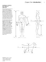
Instructions for Use
̈ The organization of the terms in
accordance with the current
Nomina Anatomica is exemplified
by the typefaces shown on the
right.
̈ Terms not organized hierarchi-
cally are printed in normal red let-
tering.
̈ The letters printed after a text seg-
ment refer to the figures on the
opposite page. The numbers in the
figures correspond to the key
word mentioned behind the
corresponding number listed in
the text.
̈ Higher-ranking terms frequently
are not represented by a number
in the figures.
̈ Fully valid alternative expressions
are listed in parentheses.
̈ The following are listed in single
square brackets:
— inconstant structures,
— terms that are unofficial but
listed in the Nomina Anatom-
ica,
— explanatory supplements.
̈ Terms not mentioned in the No-
mina Anatomica are printed in
double square brackets.
̈ Terms representing a supplement
to the older editions are marked
by lower case letters.
̈ Circled numeric marks refer to a
more extensive region.
Examples
CARDIOVASCULAR SYSTEM
ARTERIES
AORTA
ABDOMINAL AORTA
Celiac trunk
Common hepatic artery
Proper hepatic arter y
Right branch
Cystic artery
BONES OF SKULL
Neurocranium
Viscerocranium
Chondrocranium
Carpal bones (carpi)
[Sutural bones]
[Pyramidal tract]
Splenium [of corpus callosum]
[[Pouch of Douglas]]
3 aintervertebral surface of vertebra
Feneis, Pocket Atlas of Human Anatomy © 2000 Thieme
All rights reserved. Usage subject to terms and conditions of license.
1
1
2
3
4
5
6
7
8
9
10
11
12
13
14
15
16
17
18
19
20
21
22
23
24
25
A
aa
Pocket Atlas of Human Anatomy
Feneis, Pocket Atlas of Human Anatomy © 2000 Thieme
All rights reserved. Usage subject to terms and conditions of license.
2
1
2
3
4
5
6
7
8
9
10
11
12
13
14
15
16
17
18
19
20
21
22
23
24
25
SKELETON
Axial skeleton. Skeleton axiale.
1 VERTEBRAL COLUMN. Columna vertebralis. A
1a Vertebra.
2 VERTEBRAL CANAL. Canalis vertebralis. Canal
formed by the successive vertebral foramina. It
contains the spinal cord. B
3 Body of vertebra. Corpus vertebrae (verte-
brale). B C D
3a
Facies inter vertebralis. The surface of a verte-
bra facing the adjacent vertebra. B
3b
Ring apophysis (epiphysis). Apophysis anu-
laris. Ring of bone around the upper and lower
surfaces of the vertebral body. It represents a
secondary center of ossification. B
4 Vertebral arch. Arcus vertebrae (vertebralis). It
forms the posterior and lateral boundaries of
the vertebral foramen. C D
5
Pedicle. Pediculus arcus vertebrae. The portion
of the vertebral arch situated anteriorly be-
tween the body and transverse process as well
as between the superior and inferior vertebral
notches. B D
6
Lamina. Lamina arcus vertebrae (vertebralis).
The portion of the vertebral arch situated post-
eriorly between the transverse process and the
spinous process. C
6a Neurocentral junction (synchondrosis). Junc-
tio neurocentralis. Cartilaginous joint between
the left and right fetal neural arches and the
centrum. E
7 Intervertebral foramen. Foramen interverte-
brale. Opening for the passage of the spinal
nerve and small vessels. It is bordered by the
two adjacent vertebral notches, the vertebral
body and the intervertebral disc. A B
8 Superior vertebral notch. Incisura vertebralis
superior. Notch on the superior aspect of the
pedicle. B
9 Inferior vertebral notch. Incisura vertebralis
inferior. Notch on the inferior aspect of the
pedicle. B
10 Vertebral foramen. Foramen vertebrale. Space
surrounded by the vertebral arch and body. To-
gether, the series of foramina form the verte-
bral canal. C D
11 Spinous process. Processus spinosus. It is bifid
in the upper four cervical vertebrae. B C D
12 Transverse process. Processus transversus. B C
13
Costal process. Processus costalis. The trans-
verse process of a lumbar vertebra. It corre-
sponds to a rudimentary rib formed by the
embryonic costal element. D
14 Superior articular process (zygapophysis).
Processus articularis (zygapophysis) superior.
Articular process on the superior aspect of the
vertebral arch. B C D
15 Inferior articular process (zygapophysis). Pro-
cessus articularis (zygapophysis) inferior. Artic-
ular process on the inferior aspect of the verte-
bral arch. B C
16 CERVICAL VERTEBRAE. Vertebrae cervicales.
The seven uppermost vertebrae (C1−7). A
17 Uncal process or uncus. Uncus corporis. Up-
wardly projecting, hook-like process on either
side of the cervical vertebrae. It occasionally
gives rise to bony proliferations which can
exert pressure on the spinal nerve. C
18 Foramen transversarium. Hole in the trans-
verse process of cervical vertebrae for the pas-
sage of the vertebral artery and vein. C
19 Anterior tubercle. Tuberculum anterius. Ante-
rior projection on the transverse processes of
cervical vertebrae 2−7 for muscle attachment. C
20 Posterior tubercle. Tuberculum posterius.
Posterior projection on the transverse
processes of cervical vertebrae 2−7 for muscle
attachment. C
21 Carotid tubercle. Tuberculum caroticum. Well
developed anterior tubercle of C6. So named
because the common carotid artery can be
compressed against it anteriorly. A
22 Groove for spinal nerve. Sulcus n. spinalis.
Groove on the transverse processes of C3−7 for
the spinal nerves exiting from the interverte-
bral foramina. C
23 Vertebra prominens (C7). The seventh cervical
vertebra. It is so named because of its especially
well-developed spinous process (in 70% of
cases). A
24 THORACIC VERTEBRAE. Vertebrae thoracicae.
The twelve vertebrae of the thorax (T1−12). A
25 Superior costal facet. Fovea costalis superior.
Fossa for articulation with the head of a rib. It is
located near the root of the arch on the upper
edge of the body of a vertebra. B
26 Inferior costal facet. Fovea costalis inferior.
Fossa for articulation with the head of a rib. It is
located below the root of the arch on the lower
edge of the body of a vertebra. B
27 Costal facet of transverse process. Fovea
costalis processus transversi. Facet for articula-
tion with the tubercle of a rib. B
28 LUMBAR VERTEBRAE. Vertebrae lumbales (lum-
bares). The five vertebrae of the lumbar region
(L1−5). A
29 Accessory process. Processus accessorius.
Rudiment of the original lumbar transverse
process. It projects posteriorly from the base of
the costal process. D
30 Mamillary process. Processus mamillaris. A
blunt process projecting from the superior ar-
ticular process of the lumbar vertebra. D
Bones
Feneis, Pocket Atlas of Human Anatomy © 2000 Thieme
All rights reserved. Usage subject to terms and conditions of license.
3
1
2
3
4
5
6
7
8
9
10
11
12
13
14
15
16
17
18
19
20
21
22
23
24
25
A
aa
6a
3
10
5
13
14
11
30
29
4
1918 22 20 15
11
6
10
4
14
12
17
3
3b
3a
58
12
14
25
12
11
2715
9
26
3
3
2
21
23
16
24
28
4.16
4.37
7
Vertebral columnA
Thoracic vertebrae
B
Cervical vertebra
C
Lumbar vertebra,
superior view
D Infantile thoracic vertebraE
Bones
Feneis, Pocket Atlas of Human Anatomy © 2000 Thieme
All rights reserved. Usage subject to terms and conditions of license.
4
1
2
3
4
5
6
7
8
9
10
11
12
13
14
15
16
17
18
19
20
21
22
23
24
25
24 Pelvic surface. Facies pelvica. Anterior surface
of the sacrum facing the pelvis. F
25
Transverse lines. Lineae transversae. Four
anteriorly situated fusion lines of the five sacral
vertebral bodies. F
26
Intervertebral foramina. Foramina inter-
vertebralia. Openings for passage of the sacral
spinal nerves. They develop from the original
superior and inferior notches. D
27
Anterior sacral foramina. Foramina sacralia
anteriora (pelvica). Anterior openings for
nerves and vessels. D F
28 Dorsal surface of sacrum. Facies dorsalis ossis
sacri. C
29
Median sacral crest. Crista sacralis mediana.
Median ridge formed by the remnants of the
spinous processes of the sacral vertebrae. C
30
Posterior sacral foramina. Foramina sacralia
posteriora. Posterior openings for nerves and
vessels. C D
31
Intermediate sacral crests. Cristae sacralis in-
termedia. Remnants of the articular processes
located on either side the median sacral crest. C
32
Lateral sacral crest. Crista sacralis lateralis.
Posterior bilateral series of rudimentary trans-
verse processes. C
33
Sacral cornu (horn). Cornu sacrale. Hook-
shaped processes that extend downward on
either side of the sacral hiatus. C
34
Sacral canal. Canalis sacralis. Inferior end of
the vertebral canal. C D
35
Sacral hiatus. Hiatus sacralis. Opening at the in-
ferior end of the vertebral canal located usually
at the level of vertebrae S3−4. Emergence site of
filum terminale and injection site for lower
epidural anesthesia (caudal analgesia). C
36 Apex of sacrum. Apex ossis sacri. Inferior tip of
sacrum which gives attachment to the coccyx.
CF
37 COCCYGEAL VERTEBRAE I−IV. Os coccygis. Bone
that usually consists of four rudimentary verte-
brae. E
38 Coccygeal cornu (horn). Cornu coccygeus. Up-
wardly projecting process formed by the artic-
ular process. E
1 Atlas (C1). First cervical vertebra. It lacks a
body. A
2 Lateral mass of atlas. Massa lateralis atlantis.
The thickened lateral part of the atlas which
bears the skull for the lacking vertebra. A
3
Superior articular facet. Facies articularis su-
perior. Elliptical and concave facet. A
4
Inferior ar ticular facet. Facies articularis infe-
rior. Roundish and slightly concave surface
lined with cartilage.
5 Anterior arch of atlas. Arcus anterior atlantis.
A
6
Dental fovea of atlas. Fovea dentis atlantis.
Facet for articulation with the dens of the axis
on the inner surface of the anterior arch. A
7
Anterior tubercle of atlas. Tuberculum an-
terius atlantis. A
8 Posterior arch of atlas. Arcus posterior atlan-
tis. A
9
Groove for vertebral artery. Sulcus arteriae
vertebralis. Groove for the vertebral artery lo-
cated on the posterior arch of the atlas behind
the articular surfaces. A
10
Posterior tubercle. Tuberculum posterius. It is
a rudiment of the spinous process. A
11 Axis (C2) [[Epistropheus]] The second cervical
vertebra. B
12 Dens [[odontoid process]] of axis. Dens axis. B
13
Apex of dens. Apex dentis. Attachment site of
the apical ligament of the dens. B
14
Anterior articular surface of dens. Facies ar-
ticularis anterior. B
15
Posterior ar ticular sur face of dens. Facies ar-
ticularis posterior. B
16 OS SACRUM (SACRALE) / VERTEBRAE SACRALES
I−V. Sacral bone [[sacrum]] formed by five fused
vertebrae. C D F
17 Base of sacrum. Basis ossis sacri. Broad upper
end of sacrum. F
18
Promontory of sacrum. Promontorium ossis
sacri. Prominent anterior margin of the body of
the first sacral vertebra. It projects quite far into
the pelvic inlet. F
19
Ala of sacrum. Ala sacralis. Part of the base of
the sacrum situated lateral to the first sacral
vertebra.
20
Superior ar ticular process. Processus articu-
laris superior. C F
21 Lateral part or mass of sacrum. Pars lateralis
ossis sacri. The lateral part of the sacrum
derived from the transverse processes and
rudimentary ribs. C F
22
Auricular surface. Facies auricularis. Ear-
shaped articular surface for the ilium. C
23
Sacral tuberosity. Tuberositas sacralis. Rough
area behind the auricular surface for the at-
tachment of the sacroiliac ligaments. C
Bones
Feneis, Pocket Atlas of Human Anatomy © 2000 Thieme
All rights reserved. Usage subject to terms and conditions of license.
5
1
2
3
4
5
6
7
8
9
10
11
12
13
14
15
16
17
18
19
20
21
22
23
24
25
A
aa
27
1821 17
25
36
24
20
37
38
27
30
26
34
21
34
20
22
23
35
3633
32
28
29
31
30
14
13
15
12
11
1
10
9
8
4
5
742
3
6
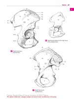
Atlas, superior viewA
Axis from leftB
Sacral bone, dorsal view
C
Sacral bone, cross-section
D
Coccyx, dorsal view
E Sacral bone, anterior viewF
Bones
Feneis, Pocket Atlas of Human Anatomy © 2000 Thieme
All rights reserved. Usage subject to terms and conditions of license.
6
1
2
3
4
5
6
7
8
9
10
11
12
13
14
15
16
17
18
19
20
21
22
23
24
25
1 [[THORAX]] Used to denote the chest and wall
consisting of ribs, cartilage and soft tissue that
encases the chest cavity.
1 THORACIC BONES. Ossa thoracis.
2 RIBS. Costae (I−XII). D
3 True ribs (1−7). Costae verae (I−VII). The first
seven ribs with individual cartilaginous con-
nections to the sternum thereby distinguishing
them from the last five ribs. D
4 False ribs (8−12). Costae spuriae (VIII−XII). The
last five ribs which have no direct cartilaginous
union with the sternum. D
5
Floating ribs (11−12). Costae fluitantes (XI−
XII). They have no connection with the costal
arch (arch of ribs). D
6 Costal cartilage. Cartilago costalis. Cartilage at
the anterior ends of the ribs. D
7 Bony rib. Os costale (costa). It is contrasted
with the cartilaginous segment of the rib. D
8 Head of rib. Caput costae. It articulates with
the vertebral column. A
9
Articular surface on head of rib. Facies artic-
ulares capitis costae. A B
10
Interarticular crest on head of rib. Crista
capitis costae. Small ridge which separates the
two articular facets. B
11 Neck of rib. Collum costae. It lies lateral to the
head of the rib. A B
12
Crest of neck of rib. Crista colli costae. Sharp
ridge on the upper border of the neck of a rib. A
13 Shaf t (body) of rib. Corpus costae. Main part of
rib adjacent to the neck. A B
14
Costal tubercle. Tuberculum costae. Posterior
elevation between the neck and the shaft of the
rib. A B
15
Articular facet of costal tubercle. Facies articu-
laris tuberculi costae. Surface for articulation
with the transverse process of the thoracic
vertebrae. A B
16
Angle of rib. Angulus costae. Posteriorly sit-
uated bend in the axis of the rib. A B
17
Costal groove. Sulcus costae. Groove for the
intercostal artery, vein and nerve on the lower
margin of the internal surface of the rib. B
17 a First rib. Costa prima. It is the only rib bent only
along the edge. A D
18
Tubercle for anterior scalene muscle. Tuber-
culum musculi scaleni anterioris. Small promi-
nence on the upper surface of the first rib for the
attachment of the anterior scalene muscle. A
19
Groove for subclavian artery. Sulcus arteriae
subclaviae. Groove on the first rib, just posterior
to the anterior scalene tubercle. A
20
Groove for subclavian vein. Sulcus venae sub-
claviae. Groove on the first rib, just anterior to
the anterior scalene tubercle. A
20 a Second rib. Costa secunda. It attaches to the
sternal angle and can easily be identified in
patients. A D
21
Tuberositas musculi serrati anterioris.
Roughened area on the outer surface of the shaft
of the second rib that gives attachment to the
serratus anterior muscle. A D
22 Cervical rib. [Costa cervicalis]. Accessory rib at
C7. It can irritate the nerves to the arm.
23 Sternum. CD
24 Manubrium sterni. The portion of the sternum
situated above the sternal angle. C D
25
Clavicular notch. Incisura clavicularis. Inden-
tation for the sternoclavicular joint. C D
26
Jugular notch. Incisura jugularis. Concavity at
the upper border of the manubrium. D
27 Sternal angle. Angulus sterni (sternalis)
[[Ludovici]]. Angle between the body and manu-
brium of the sternum. It is palpable through the
skin. C D
28 Sternal synchondroses. Synchondroses ster-
nales. The two synchondroses of the sternum
are as follows:
29 Manubriosternal synchondrosis. [Synchon-
drosis manubriosternalis]. Cartilaginous joint
between the manubrium and the body of the
sternum. C D
30 Xiphisternal synchondrosis. Synchondrosis
xiphisternalis. Cartilaginous joint between the
body of the sternum and the xiphoid process. C
D
31 Body of sternum. Corpus sterni. Situated
between the manubrium and xiphoid process. C
D
32 Xiphoid process. Processus xiphoideus. Stout
process at the lower end of the sternum. C D
33 Costal notches. Incisurae costales. Indentations
for the costal cartilages. C D
34 Suprasternal bones. [Ossa suprasternalia].
Small osseous remnants of the earlier epister-
num occurring in the ligaments of the sterno-
clavicular joint.
35 Thoracic skeleton. Compages thoracis.
35 a Thoracic cavity. Cavitas thoracis. Used to de-
note the chest and chest cavity.
36 Superior thoracic aperture (thoracic inlet).
Apertura thoracis superior. Upper thoracic
opening. D
37 Inferior thoracic aperture (thoracic outlet).
Apertura thoracis inferior. Lower opening of
thorax. D
38 Pulmonary sulcus of thorax. Sulcus pul-
monalis thoracis. Either of two large, vertical
grooves on either side of the vertebral column
that are occupied by the lungs. D
39 Costal arch. Arcus costalis. Arch of ribs formed
by the cartilages of ribs 7−10. D
40 Intercostal space. Spatium intercostale. Space
between the ribs. D
41 Infrasternal angle. Angulus infrasternalis.
Angle between the right and left costal arch. D
Bones
Feneis, Pocket Atlas of Human Anatomy © 2000 Thieme
All rights reserved. Usage subject to terms and conditions of license.
7
1
2
3
4
5
6
7
8
9
10
11
12
13
14
15
16
17
18
19
20
21
22
23
24
25
A
aa
25
24
27; 29
31
33
30
32
VII
13
17
10
9
15
14
16
11
23
3
21
25
36
2
17a
20a
58.27
4
5
41
38
39
30
31
27
24
26
6
7
40
37
29
33
32
11
15
16
21
13
II
I
20
18
19
9
8
12
14
First and second ribs, superior viewA
Seventh rib, medial view
B
Sternum
from right
C Thoracic skeleton, anterior viewD
Bones
Feneis, Pocket Atlas of Human Anatomy © 2000 Thieme
All rights reserved. Usage subject to terms and conditions of license.
8
1
2
3
4
5
6
7
8
9
10
11
12
13
14
15
16
17
18
19
20
21
22
23
24
25
20 Jugular process. Processus jugularis. Externally
and internally visible process that projects later-
ally from the jugular foramen. It corresponds to
the transverse process of a vertebra. A C
21 Intrajugular process of occipital bone. Processus
intrajugularis ossis occipitales. It occasionally
divides the jugular foramen into a lateral portion
for the internal jugular vein and a medial seg-
ment for nerves. C
22 External occipital protuberance. Protuberentia
occipitalis externa. Readily palpable bony projec-
tion in the middle of the occipital bone. B
23
Inion. Anthropometric landmark indicating the
most prominent point on the external occipital
protuberance. B
24 External occipital crest. Crista occipitalis ex-
terna. Bony ridge occasionally present between
the external occipital protuberance and the fora-
men magnum. B
25 Highest (supreme) nuchal line. Linea nuchalis
suprema. Line arching externally from the upper
margin of the external occipital protuberance. It
gives attachment to the occipital belly of the
epicranius muscle. B
26 Superior nuchal line. Linea nuchalis superior.
Transverse ridge at the level of the external occip-
ital protuberance. The trapezius muscle attaches
between it and the highest nuchal line. B
27 Inferior nuchal line. Linea nuchalis inferior.
Transverse ridge between the superior nuchal
line and the foramen magnum. The semispinalis
capitis muscle attaches between it and the super-
ior nuchal line. B
27 a Occipital plane. Planum occipitale. Outer surface
of the occipital bone located superior to the ex-
ternal occipital protuberance. B C
28 Cruciform eminence. Eminentia cruciformis.
Cross-shaped bony prominence with the internal
occipital protuberance at its center. A
29 Internal occipital protuberance. Protuberantia
occipitalis internal. Midpoint of the cruciform
eminence. A
30 Internal occipital crest. [Crista occipitalis in-
terna]. Thick bony ridge that occasionally extends
from the internal occipital protuberance to the
foramen magnum. A
31 Groove for superior sagittal sinus. Sulcus sinus
sagittalis superioris. A
32 Groove for transverse sinus. Sulcus sinus trans-
versi. A
33 Groove for the sigmoid sinus. Sulcus sinus sig-
moidei. Groove that begins before the sigmoid
sinus enters the jugular foramen. A C
33 a Groove for occipital sinus. Sulcus sinus occipi-
talis. A
34 Paramastoid process. [Processus paramas-
toideus]. Prominence that occasionally projects
from the jugular process in the direction of the
transverse process of the atlas.
34 a Cerebral fossa. Fossa cerebralis. Depression for
the occipital lobes of the cerebrum. A
34 b Cerebellar fossa. Fossa cerebellaris. Depression
for the cerebellum. A
1 Cranial bones. Ossa cranii. Bones of the skull.
1a Neurocranium. Portion of the cranium that en-
closes the brain.
Viscerocranium. Portion of the cranium that
forms the face.
Chondrocranium. Cartilaginous part of embryo-
logical skull that later forms base of skull.
2 Occipital bone. Os occipitale. It lies between the
sphenoid, temporal and parietal bones. A B C
3 Foramen magnum. Large opening in the occipital
bone for passage of the medulla oblongata, ves-
sels and nerves. A B C
4
Basion. Midpoint of the anterior border of the
foramen magnum. B
5
Opisthion. Midpoint of the posterior border of
the foramen magnum. A B
6 Basilar part of occipital bone (basioccipital
bone). Pars basilaris ossis occipitalis. Portion of
occipital bone that projects superiorly from fora-
men magnum to sphenoid bone. A C
6a
Clivus. Part of the basioccipital bone that slopes
upwardly from the foramen magnum to the dor-
sum sellae. B
7
Groove for inferior petrosal sinus of occipital
bone.
Sulcus sinus pertrosi inferioris ossis occipi-
talis. A
8
Pharyngeal tubercle. Tuberculum pharyngeum.
Prominence on the inferior surface of the
basioccipital bone, for attachment of the pharyn-
geal raphe. A C
9 Lateral (condylar) part of occipital bone. Pars
lateralis ossis occipitalis. It lies lateral to the fora-
men magnum. A B
10 Squamous part of occipital bone. Squama occip-
italis. Area extending from the posterior edge of
the foramen magnum. A B C
11
Mastoid margin. Margo mastoideus. The border
of the occipital bone united with the temporal
bone. A
12
Lambdoid margin. Margo lambdoideus. The
border of the occipital bone that articulates with
the parietal bone. A
13
Interparietal bone. [Os interparietale]. Ana-
tomic variant that forms when the upper half of
the squama occipitalis is separated by a trans-
verse suture.
14 Occipital condyle. Condylus occipitalis. Process
on the occipital bone, for articulation with the
atlas. A B C
15 Condylar canal. Canalis condylaris. Passage lo-
cated posterior to the occipital condyle, for trans-
mission of a vein from the sigmoid sinus. A B C
16 Hypoglossal canal. Canalis hypoglossalis. Pas-
sage that originates from the lateral part of the
occipital bone anterior to the foramen magnum
and ends outside, anterior to the occipital con-
dyle. It transmits the twelfth cranial nerve and
the venous plexus. A B C
17 Condylar fossa. Fossa condylaris. Depression
posterior to the occipital condyle. B
18 Jugular tubercle. Tuberculum jugulare. Small
eminence above the hypoglossal canal. A B C
19 Jugular notch. Incisura jugularis. Indentation for
the jugular foramen. A C
Bones
Feneis, Pocket Atlas of Human Anatomy © 2000 Thieme
All rights reserved. Usage subject to terms and conditions of license.
9
1
2
3
4
5
6
7
8
9
10
11
12
13
14
15
16
17
18
19
20
21
22
23
24
25
A
aa
10
27a
20 19 18
16
6
8
1421
15
20
2
33
3
10
27a
22; 23
25
26
27
17
14 4
6a
9
18
3
15
5
24
2
16
16
12
11
14 8
19
33
6
18
7
9
33a
30
28 29
32
34b
10
34a
31
5
3
2
15
20
Occipital bone,
internal surface
A
Occipital bone, inferoposterior view
B
Occipital bone, dextrolateral
and partly anterior view
C
Bones
Feneis, Pocket Atlas of Human Anatomy © 2000 Thieme
All rights reserved. Usage subject to terms and conditions of license.
10
1
2
3
4
5
6
7
8
9
10
11
12
13
14
15
16
17
18
19
20
21
22
23
24
25
1 Sphenoid bone. Os sphenoidale. Bone located
between the frontal, occipital and temporal
bones. A B C
2 Body of sphenoid bone. Corpus ossis sphe-
noidalis. Part located between the winged
processes of the sphenoid bone. A B
3
Jugum sphenoidale. Connects the lesser
wings of the sphenoid. A
4
(Pre)chiasmatic groove. Sulcus prechiasmati-
cus. Groove between the right and left optic
canals. A
5
Turkish saddle. Sella turcica. It lies above the
sphenoidal sinus and contains the hypophysis. A
6
Tuberculum sellae. Small process in front of the
hypophysial fossa. A
7
Middle clinoid process. [Processus clinoideus me-
dius]. Either of two small protuberances oc-
casionally present, one on either side of the floor
of the hypophysial fossa. A
8
Hypophysial fossa. Fossa hypophysialis. Fossa oc-
cupied by the hypophysis. A
9
Dorsum sellae. Posterior wall of the hypophysial
fossa. A C
10
Posterior clinoid process. Processus clinoideus
posterior. Either of two processes that extend
from either side of the dorsum sellae. A C
11
Carotid groove. Sulcus caroticus. Longitudinal
groove lateral to the body of the sphenoid bone
that lodges the internal carotid artery. A
12
Lingula sphenoidalis. Pointed process lateral
to the entrance of the internal carotid artery into
the cranial fossa. A
13
Sphenoidal crest. Crista sphenoidalis. Median
bony ridge on the anterior surface of the body of
the sphenoid bone that articulates with the per-
pendicular plate of the ethmoid. C
14
Sphenoidal rostrum. Rostrum sphenoidale.
Downward continuation of the sphenoidal crest
that articulates with the vomer. C
15
Sphenoidal sinus. Sinus sphenoidalis. Either of
the paired paranasal sphenoidal sinuses. C
16
Septum of sphenoidal sinus. Septum intersinuale
sphenoidale. Partition separating the sinus into
right and left parts. C
17
Aperture of sphenoidal sinus. Apertura sinus
sphenoidalis. Orifice that opens anteriorly into
the spheno-ethmoidal recess. C
18
Sphenoidal concha. Concha sphenoidalis.
Originally paired, concave bony plate which
fuses with the body of the sphenoid and forms
part of the anterior and inferior wall of the sphe-
noidal sinus and other structures. C
19 Lesser wing of sphenoid. Ala minor ossis sphe-
noidalis. A B C
20
Optic canal. Canalis opticus. Canal for the optic
nerve and the ophthalmic artery. A
21
Anterior clinoid process. Processus clinoideus
anterior. Cone-like process on either side of the
anterior part of the hypophysial fossa. A
22
Superior orbital fissure. Fissura orbitalis su-
perior. Cleft between the greater and lesser
wings of the sphenoid for the passage of nerves
and veins. A B C
23 Greater wing of sphenoid. Ala major ossis
sphenoidalis. A B C
24
Cerebral surface. Facies cerebralis. Surface of
the greater wing facing the brain. A
25
Temporal surface. Facies temporalis. Outward
surface of the greater wing. B C
26
Maxillary sur face. Facies maxillaris. Surface of
the greaterwingfacing the maxilla. The foramen
rotundum opens here. C
27
Orbital surface. Facies orbitalis. Surface of the
greater wing facing the orbit. C
28
Zygomatic border. Margo zygomaticus. Mar-
gin of the greater wing articulating with the zy-
gomatic bone. C
29
Frontal border. Margo frontalis. Margin of the
greater wing fused with the frontal bone. A
30
Parietal border.Margo parietalis. Marginofthe
greater wing fused with the parietal bone. C
31
Squamous border. Margo squamosus.
Squamous margin of the greater wing that ar-
ticulates with the temporal bone. A
32
Infratemporal crest. Crista infratemporalis.
Bony ridge between the vertical temporal sur-
face and the horizontally-oriented inferior sur-
face of the greater wing of the sphenoid. B C
33
Foramen rotundum. Round opening in the
great wing that extends anteriorly into the pter-
ygopalatine fossa. It transmits the maxillary
nerve. A B C
34
Foramen ovale. Opening for passage of the
mandibular nerve in the medial part of the great
wing, located in front of the foramen spinosum.
AB
35
[Foramen venosum]. Opening occasionally
present medial to the foramen ovale for passage
of anemissaryvein fromthecavernoussinus.AB
36
Foramen spinosum. Opening situatedlateralto
and behind the foramen ovale for passage of the
middle meningeal artery. A B
37
[Foramen petrosum]. [[Canaliculus innomina-
tus.]] Opening occasionally present between the
foramen ovale and the foramen spinosum for
transmission of the lesser petrosal nerve. A B
38
Angular spine of sphenoid. Spina ossis sphe-
noidalis. Sharp, bony spur that extends
downward from the greater wing. A B
39
Groove for the cartilaginous part of the
auditory tube.
Sulcus tubae auditoriae (audi-
tivae). Shallow groove on the underside of the
greater wing lateral to the root of the pterygoid
process. B
Bones
Feneis, Pocket Atlas of Human Anatomy © 2000 Thieme
All rights reserved. Usage subject to terms and conditions of license.
11
1
2
3
4
5
6
7
8
9
10
11
12
13
14
15
16
17
18
19
20
21
22
23
24
25
A
aa
23 27 22 26 10
18
13 9 19
30
32
16
17
181415
12.12
39
28
25
33
25
22
2
19
353937
38
36
34
33
23
32
32
64
7
20 19
29
21
31
38
3510
2
111237
36
34
24
3
33
5
8
9
23
22
Sphenoid bone, superior viewA
Sphenoid bone, anteroinferior view
B
Sphenoid bone, frontal view.
Sphenoidal sinus, fenestrated
C
Bones
Feneis, Pocket Atlas of Human Anatomy © 2000 Thieme
All rights reserved. Usage subject to terms and conditions of license.
12
1
2
3
4
5
6
7
8
9
10
11
12
13
14
15
16
17
18
19
20
21
22
23
24
25
1 Pterygoid process of the sphenoid bone.
Processus pterygoideus. A B
2
Lateral pterygoid plate. Lamina lateralis [pro-
cessus pterygoidei]. A B
3
Medial pterygoid plate. Lamina medialis [pro-
cessus pterygoidei]. A B
4
Pterygoid notch (fissure). Incisura ptery-
goidea. Fissure formed inferiorly by the diverg-
ing medial and lateral pterygoid plates. It is oc-
cupied by the pyramidal process of the palatine
bone. A
5
Pterygoid fossa. Fossa pterygoidea. Space be-
tweenthelateralandmedial pterygoidplatesfor
the medial pterygoid muscle. A B
6
Scaphoid fossa. Fossa scaphoidea. Oblong de-
pression at the root of the medial pterygoid
plate, where the end of the cartilage of the
pharyngotympanic tube is located. The tensor
velipalatinimuscle originatesatitslateralend.A
7
Vaginal process. Processus vaginalis. Small
bony ridge medial to the root of the medial pter-
ygoid plate. It borders a small furrow laterally. A
B
8
Palatovaginal groove. Sulcus palatovaginalis.
Groove whichjoinsthepalatineboneto form the
palatovaginal canal. B
9 Vomerovaginal groove. Sulcus vomerovaginalis.
Groove at the base of the pterygoid process. To-
gether with the vomer, it forms the vomerovagi-
nal canal. B
10
Pterygoid hamulus. Hamulus pterygoideus.
Hook-like process at the inferior end of the me-
dial pterygoid plate. A B
11
Sulcus of pterygoid hamulus.Sulcus hamuli pter-
ygoidei. Groove produced by a sharp bend in the
hamulus. B
12
Pterygoid (vidian) canal. Canalis ptery-
goideus [[canalis Vidii]]. Passage that extends
anteriorlyin the baseofthepterygoidprocessfor
transmission of the greater and deep petrosal
nerves to the pterygopalatine ganglion in the
pterygopalatine fossa. A see 11 C
13
Pterygospinous process. Processus ptery-
gospinosus. Sharp spine on the posterior edge of
the lateral pterygoid plate. A
14 Temporal bone.Ostemporale.Bonethatliesbe-
tween theoccipital,sphenoidandparietal bones
and consists of three parts: petrous, tympanic
and squamous. C D E
15 Petrous part (pyramid) of temporal bone.Pars
petrosa ossis temporalis. It houses the inner ear.
D
16
Occipital border. Margo occipitalis. Margin ar-
ticulating with the occipital bone. C D
17
Mastoid process. Processus mastoideus.
Process located just posterior to the external
acoustic meatus. C E
18
Mastoid notch. Incisura mastoidea. Medial
notch on the inferior surface of the mastoid
process. It gives origin to the posterior belly of
the digastric muscle. C
19
Groove for sigmoid sinus. Sulcus sinus sig-
moidei. Sulcus on the internal, posterior surface.
D
20
Groove for occipital artery. Sulcus a. occipi-
talis. It lies medial to the mastoid notch and pro-
ximal to the occipital margin. C
21
Mastoid foramen. Foramen mastoideum.
Opening behind the mastoid process for addi-
tional venous drainage from the cranial cavity. C
D
22
Facial canal. Canalis fascialis. Canal for the fa-
cial nerve. It begins at the opening of the inter-
nal acoustic meatus and ends at the stylomas-
toid foramen. C D E
23
Genu of facial canal. Geniculum canalis facialis.
Sharp bend in the facial canal just below the
anterior wall of the petrous part of the temporal
bone, near the hiatus of the canal for the greater
petrosal nerve. D
24
Canaliculus of chorda tympani nerve.
Canaliculus chordae tympani. Narrow passage-
way for the chorda tympani nerve between the
facial canal and the tympanic cavity. D E. Cf. page
381 D
25
Apex of petrous temporal bone. Apex partis
petrosae. It is directed anteromedially. C D
26
Carotid canal. Canalis caroticus. Canal for the in-
ternal carotid artery. It begins inferiorly and ex-
ternally between the jugular foramen and the
musculotubal canal. C
27
Caroticotympanic canaliculi. Canaliculi caroti-
cotypmpanici. Small channels in the wall of the
carotid canal for arterial and nerve branches to
the middle ear from the internal carotid artery
and the carotid plexus. C
28
Musculotubal canal. Canalis musculotubarius.
Double canal for the auditory tube and tensor
tympani muscle. It lies in front of the carotid
canal and leads into the tympanic cavity. C E
29
Semicanal for tensor tympani muscle. Semi-
canalis m. tensoris tympani. E
30
Semicanal for the auditory tube. Semicanalis
tubae auditoriae (auditivae). E
31
Septum of musculotubal canal. Septum canalis
musculotubarii. Bony partition between the
above-mentioned semicanals. E
Bones
Feneis, Pocket Atlas of Human Anatomy © 2000 Thieme
All rights reserved. Usage subject to terms and conditions of license.
13
1
2
3
4
5
6
7
8
9
10
11
12
13
14
15
16
17
18
19
20
21
22
23
24
25
A
aa
22
29
31
28
24
17
30
16 22
23
15
21
19
24
25
27 28
26
25
22
20
18
21
16
17
2
179810
11
3
5
12
13
4
10
7
3
1
2
5
6
Sphenoid bone, posterior viewA
Sphenoid bone, inferior view
B Right temporal bone,
inferior view
C
Right temporal bone, internal surface
D Right temporal bone, opened.
Anterolateral view
E
Bones
Feneis, Pocket Atlas of Human Anatomy © 2000 Thieme
All rights reserved. Usage subject to terms and conditions of license.
14
1
2
3
4
5
6
7
8
9
10
11
12
13
14
15
16
17
18
19
20
21
22
23
24
25
1 Anterior surface of petrous part of tem-
poral bone.
Facies anterior partis petrosae. A C
2
Roof of tympanic cavity. Tegmen tympani. Thin
bony plate anterolateral to the arcuate emi-
nence. C
3
Arcuate eminence. Eminentia arcuata. Elevation
on the anterior surface of the petrous part of the
temporal bone produced by the underlying
anterior semicircular canal. A C
4
Hiatus of canal for greater petrosal nerve. Hiatus
canalis n. petrosi majoris. Opening in the ante-
rior wall of the petrous part of the temporal
bone for passage of the greater petrosal nerve. A
C
5
Hiatus of canal for lesser petrosal nerve. Hiatus
canalis n. petrosi minoris. Opening in the ante-
rior wall of the petrous temporal below the
greater petrosal nerve. A C
6
Groove for greater petrosal nerve. Sulcus n.
petrosi majoris. It runs anteromedially from the
hiatus to the foramen lacerum. C
7
Groove for lesser petrosal nerve. Sulcus n.
petrosi minoris. Groove for the lesser petrosal
nerve, running from the respective hiatus to the
foramen lacerum. C
8
Trigeminal impression. Impressio trigeminalis.
Shallow depression in the anterior wall of the
apex of the petrous part of the temporal bone. It
lodges the trigeminal [[semilunar]] ganglion. C
9
Superior border of petrous temporal bone.
Margo superior partis petrosae. A C
10
Groove for superior petr osal sinus. Sulcus sinus
petrosi superioris. Its course is on the upper
margin of the petrous part of the temporal bone.
AC
11
Posterior surface of petrous part of tem-
poral bone.
Facies posterior partis petrosae. A
12
Porus acusticus internus. Opening of internal
acoustic meatus on the posterior wall of the
petrous part of the temporal bone. A
13
Internal acoustic (auditory) meatus. Meatus acus-
ticus internus. It transmits cranial nerves VII and
VIII and vessels. A
14
Subarcuate fossa. Fossa subarcuata. Depression
lateral and superior to the internal acoustic
meatus. In the fetus, it lodges the flocculus ofthe
cerebellum. A
15
Aqueduct of vestibule. Aqueductus vestibuli.
Narrow canal extending from the endolym-
phatic space of the inner ear to the posterior
wall of the petrous part of the temporal bone.
16
External opening of vestibular aqueduct. Aper-
tura externa aqueductus vestibuli. A
17
Posterior border of petrous par t of the tem-
poral bone.
Margo posterior partis petrosae. A
B
18
Groove for inferior petrosal sinus. Sulcus sinus
petrosi inferioris. A
19
Jugular notch. Incisura jugularis. Indentation
forming the anterior margin of the jugular fora-
men.AB
20
Intrajugular process. Processus intrajugularis. It
divides the jugular foramen into a posterolateral
part for the internal jugular vein and an an-
teromedial part for cranial nerves IX, X and XI. A
B
21
Cochlear canaliculus. Canaliculus cochleae.
Bony canal for the cochlear aqueduct.
22
External opening of cochlear canaliculus. Aper-
tura externa canaliculi cochleae. It lies medially
in front of the jugular fossa. B
23
Inferior surface of petrous temporal bone.
Facies inferior partis petrosae. B
24
Jugular fossa. Fossa jugularis. Enlargement of the
jugular foramen for the superior bulb of the in-
ternal jugular vein. B
25
Mastoid canaliculus. Canaliculus mastoideus.
Narrow canal for the auricular branch of the
vagus nerve. It begins in the jugular fossa. B
26
Styloid process. Processus styloideus. Long
process located laterally in front of the jugular
fossa. It is a vestige of the second branchial arch.
ABD
27
Stylomastoid foramen. Foramen stylomas-
toideum. External opening of the facial canal lo-
cated behind the styloid process and between
the mastoid process and the jugular fossa. B
28
Tympanic canaliculus. Canaliculus tympanicus.
Minute canal in the petrosal fossula traversed by
the tympanic nerve and inferior tympanic
artery. B
29
Petrosal fossula. Fossula petrosa. Slight depres-
sion in the bony ridge between the carotid canal
and the jugular fossa. It is occupied by the tym-
panic ganglion of the glossopharyngeal nerve. B
30
Tympanic (middle ear) cavity. Cavitas tym-
panica. Narrow, air-filled space between the os-
seous labyrinth and the tympanic membrane.
31
Petrotympanic fissure [glaserian f issure].
Fissura petrotympanica. Fissure situated dor-
somedial to the fossa ofthe temporomandibular
joint, between the tympanic part of the tem-
poral bone and thevisible petrous strip. The me-
dial part lodges the chorda tympani nerve. B D
32
Petrosquamous fissure. Fissura petrosqua-
mosa. It lies on the skull base in front of the
petrotympanic fissure between the visible
petrous strip and the squamous part of the tem-
poral bone. B C
33
Squamotympanic f issure. Fissura tympa-
nosquamosa. Lateral continuation of the two
above mentioned fissures after they unite. B D
34
Tympanomastoid fissure. Fissura tym-
panomastoidea. Suture between the tympanic
part of the temporal bone and the mastoid
process. Exit site of the auricular branch of the
vagus nerve. B D
Bones
Feneis, Pocket Atlas of Human Anatomy © 2000 Thieme
All rights reserved. Usage subject to terms and conditions of license.
15
1
2
3
4
5
6
7
8
9
10
11
12
13
14
15
16
17
18
19
20
21
22
23
24
25
A
aa
34
26
31
33
86
4
10
9
3
9
2
32
5
7
1
12.26
28
26
27
34
29
22
19
17
25
24
23
33
32
31
20
910
3
17
18
12; 13
26
19
16
1
11
12.21
20
14
5
4
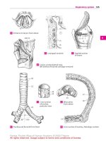
Right temporal bone,
medial view
A
Right temporal bone,
posterior view
B
Right temporal bone,
superior view
C
Right temporal bone,
lateral view
D
Bones
Feneis, Pocket Atlas of Human Anatomy © 2000 Thieme
All rights reserved. Usage subject to terms and conditions of license.
16
1
2
3
4
5
6
7
8
9
10
11
12
13
14
15
16
17
18
19
20
21
22
23
24
25
1 Tympanic part of temporal bone. Pars tym-
panica. Wall of the bony external acoustic mea-
tus with the exception of the posterior, upper
wall (tympanic notch). B
2
Tympanic ring. Anulus tympanicus. Bony ring
which is the developmental precursor of the
tympanic part of the temporal bone. The super-
ior part is still open at birth. A
3
External acoustic (auditory) meatus. Meatus
acusticus externus. B
4
Opening of external acoustic meatus. Porus
acusticus externus. B
5
Greater tympanic spine. Spina tympanica
major. Anterior end of the tympanic ring formed
by the tympanic part of the temporal bone. A
6
Lesser tympanic spine. Spina tympanica
minor. Posterior end of the ring formed by the
tympanic part of the temporal bone. A
7
Tympanic groove. Sulcus tympanicus. Groove
for attachment of the tympanic membrane. A
8
Tympanic notch. Incisura tympanica. Notch
between the greater and lesser tympanic spines.
In the newborn, it is situated superiorly in the
tympanic part of the temporal bone between
the free ends of the still open tympanic ring. A
9
Sheath of styloid process. Vagina processus
styloidei. Ridge formed by the tympanic part of
the temporal bone and partially enclosing the
root of the styloid process. A
10 Squamous part. Pars squamosa. Part of the tem-
poral bone located between the sphenoid,
parietal and occipital bones. B
11
Parietal border. Margo parietalis. Upper mar-
gin articulating with the parietal bone. B
12
Parietal notch. Incisura parietalis. Indentation
posteroinferior to the temporal line. B
13
Sphenoidal border. Margo sphenoidalis. Ante-
rior margin articulating with the sphenoid
bone. B
14
Temporal sur face. Facies temporalis. External
surface covered primarily by the temporalis
muscle. B
15
Groove for the middle temporal arter y. Sul-
cus arteriae temporalis mediae. B
16
Zygomatic process of temporal bone. Pro-
cessus zygomaticus. It contributes to the forma-
tion of the zygomatic arch. B
17
Supramastoid crest. Crista supramastoidea.
Ridge forming the posterior boundary of the
field of attachment of the temporalis muscle. B
18
Suprameatal pit. Foveola suprameatica (su-
prameatalis). Small pit superior to the su-
prameatal spine and lateral to the mastoid an-
trum. B
19
Suprameatal spine. [Spina suprameatica]. Pro-
jection for attachment of the auricular cartilage.
B
20
Mandibular fossa. Fossa mandibularis. De-
pression for the head of the mandible. B
21
Facies articularis. Surface for articulation with
the temporomandibular joint. B
22
Articular tubercle. Tuberculum articulare. Cyl-
indrical elevation in front of the mandibular
fossa. B
23
Cerebral surface. Facies cerebralis. Inner surface
of squamous part of the temporal bone facing
the brain.
24 Parietal bone. Os parietale. It is located be-
tween the frontal, sphenoid and temporal
bones. C D
25 Internal surface. Facies interna. The internal or
cerebral surface of the parietal bone. C
26
Groove for sigmoid sinus. Sulcus sinus sig-
moidei. It lies in the vicinity of the mastoid
angle. C
26 a
Groove for superior sagittal sinus. Sulcus
sinus sagittalis superioris. C
26 b
Groove for middle meningeal ar tery. Sulcus
arteriae meningeae mediae. C
27 External surface. Facies externa. The external
surface of the parietal bone facing the scalp. D
28
Superior temporal line. Linea temporalis su-
perior. Curved line for attachment of the tem-
poral fascia. It forms the upper margins of the
[[planum temporale]]. D
29
Inferior temporal line. Linea temporalis infe-
rior. Curved line for attachment of the tem-
poralis muscle. D
30
Parietal tuber. Tuber parietale. Prominence lo-
cated near the middle of the external surface of
the parietal bone. D
31 Occipital border. Margo occipitalis. Margin
facing the occiput. C D
32 Sq uamous border. Margo squamosus. Inferior
edge of the parietal bone. C D
33 Sagittal border. Margo sagittalis. Upper edge of
parietal bone that lies in the midsagittal plane. C
D
34 Frontal border. Margo frontalis. Anterior mar-
gin articulating with the frontal bone. C D
35 Frontal angle. Angulus frontalis. Anterosupe-
rior angle of the parietal bone. C D
36 Occipital angle. Angulus occipitalis. Postero-
superior angle of the parietal bone. C D
37 Sphenoidal angle. Angulus sphenoidalis. An-
teroinferior angle of the parietal bone. C D
38 Mastoid angle. Angulus mastoideus. Post-
eroinferior angle of the parietal bone. C D
39 Parietal foramen. Foramen parietale. Opening
for an emissary vein from the cranial cavity, usu-
ally located in the posterosuperior part of the
parietal bone. C D
Bones
Feneis, Pocket Atlas of Human Anatomy © 2000 Thieme
All rights reserved. Usage subject to terms and conditions of license.