Pocket Atlas of Human Anatomy 4th edition - part 4 docx
Bạn đang xem bản rút gọn của tài liệu. Xem và tải ngay bản đầy đủ của tài liệu tại đây (1.61 MB, 51 trang )
145
1
2
3
4
5
6
7
8
9
10
11
12
13
14
15
16
17
18
19
20
21
22
23
24
25
a
a
a
20
27
21
26
25
23
22
20
20
30
20° >35°
22
24
328.12
10
1
12
5
6
7
4
7a
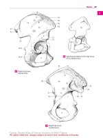
Entrance to larynx from aboveA
2
14
15
16
9
1
Laryngeal ventricleB
13
8
10
111
Sagittal section
of larynx
C
Larynx, posterolateral view,
left lamina of thyroid cartilage removed
D
Trachea and bronchi from front
E Cross secton of trachea, histologic sectionH
Cross section
of trachea,
posterior view
F Bifurcation
from above
G
Respiratory system
Feneis, Pocket Atlas of Human Anatomy © 2000 Thieme
All rights reserved. Usage subject to terms and conditions of license.
146
1
2
3
4
5
6
7
8
9
10
11
12
13
14
15
16
17
18
19
20
21
22
23
24
25
1 Lobar and segmental bronchi. Bronchi lobares
et segmentales. Bronchi for the five lobes of the
lung and their 20 segments. A B
2 Right superior lobar bronchus. Bronchus
lobaris superior dexter. Bronchus for the super-
ior lobe of the right lung. It arises just after the
tracheal bifurcation. A B
3
Apical segmental bronchus (B I). Bronchus
segmentalis apicalis. Bronchus for the apical
segment extending inferiorly asfar as the3
rd
rib.
AB
4
Posterior segmental bronchus (B II).
Bronchus segmentalis posterior. Bronchus for
the posterior segment extending forward about
as far as the midaxillary line. A B
5 Anterior segmental br onchus (B III).
Bronchus segmentalis anterior. Bronchus forthe
anterior segment extending backward about as
far as the midaxillary line. A B
6 Right middle lobar bronchus. Bronchus lobaris
medius dexter. Lobar bronchus for the middle
lobe of the right lung. A
7
Lateral segmental bronchus (B IV).
Bronchus segmentalis lateralis. Bronchus forthe
lateral segment located on the dorsal surface of
the middle lobe. A B
8
Medial segmental bronchus (B V). Bronchus
segmentalis medialis. Segmental bronchus aris-
ing anteromedially from the middle lobe. A B
9 Right inferior lobar bronchus. Bronchus
lobaris inferior dexter. Lobar bronchus for the
right inferior lobe extending posteriorly up to
the 4
th
rib. A B
10
Superior segmental bronchus (B VI).
Bronchussegmentalis superior.Bronchus for the
apical segment which borders only on the upper
lobe. B
11 Subapical segmental bronchus. [[Bronchus
segmentalis subapicalis]]. Occasionally present
accessory bronchus.
12
Medial basal segmental bronchus (B VII).
Bronchus segmentalis basalis medialis (cardia-
cus). Bronchus for the medial segment that does
not reach the external surface of the lower lobe.
AB
13
Anterior basal segmental bronchus (B VIII).
Bronchussegmentalis basalis anterior. Bronchus
for the wedge-shaped anterior end of the lower
lobe. A B
14
Lateral basal segmental bronchus (B IX).
Bronchus segmentalis basalis lateralis.
Bronchus for the small lateral segment situated
between the anterior and posterior segments. A
B
15
Posterior basal segment br onchus (B X).
Bronchus segmentalis basalis posterior.
Bronchus for the segment extending posteriorly
up to the vertebral column. A B
16 Left inferior lobar bronchus. Bronchus lobaris
superior sinister. Lobar bronchus for the left
upper lobe. A B
17
Apicoposterior segmental bronchus (B I +
II).
Bronchus segmentalis apicoposterior.
Bronchus for the left apical segment located
posterosuperiorly. A B
18
Anterior segmental bronchus (B III).
Bronchus segmentalis anterior. Bronchus for the
anterior segment of the left upper lobe situated
in front of the apical segment. A B
19
Superior lingular bronchus (B IV). Bronchus
lingularis superior. Bronchus for the second
lowest segment of the left upper lobe extending
posteriorly as far as the border of the lower lobe.
AB
20
Inferior lingular bronchus (B V). Bronchus
lingularis inferior.Bronchus for the mainly ante-
rior lowest segment of the upper lobe. A B
21 Left inferior lobar bronchus. Bronchus lobaris
inferior sinister. Lobar bronchus for the left
lower lobe extending dorsally up to the 4
th
thoracic vertebra. A B
22
Superior segmental bronchus (B VI).
Bronchussegmentalis superior.Bronchus for the
apical segment located posterosuperiorly in the
lower lobe. B
23
Subapical segmental bronchus. [[Bronchus
segmentalis subapicalis]]. Bronchus for an oc-
casionally present accessory segment.
24
Medial basal segmental bronchus (B VII).
Bronchus segmentalis basalis medialis (cardia-
cus). Bronchus for the medial basal segment
which does not reach the lateral lung surface. A
25
Anterior basal segmental bronchus (B VIII).
Bronchussegmentalis basalis anterior. Bronchus
for the anterior basal segment adjoining the
lower anterior border. A B
26
Lateral basal segmental bronchus (B IX).
Bronchus segmentalis basalis lateralis.
Bronchus for the basal middle segment located
between the anterior and posterior basal seg-
ments. A B
27
Bronchus segmentalis basalis posterior
(B X).
Bronchus for the posterior basal segment
of the lower lobe situated below the apical seg-
ment. A B
Respiratory system
Feneis, Pocket Atlas of Human Anatomy © 2000 Thieme
All rights reserved. Usage subject to terms and conditions of license.
147
1
2
3
4
5
6
7
8
9
10
11
12
13
14
15
16
17
18
19
20
21
22
23
24
25
a
a
a
144.17
16
18
27
3
5
15
144.30
4
2
7
8
13
14
12
10
9
19
20
25
26
22
21
17
144.17
144.30
3
5
15
14
27
1716
18
19
20
25
26
24
21
4
2
6
8
7
13
12
9
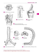
Bronchial tree, anterior viewA
Bronchial tree, posterior view
B
Respiratory system
Feneis, Pocket Atlas of Human Anatomy © 2000 Thieme
All rights reserved. Usage subject to terms and conditions of license.
148
1
2
3
4
5
6
7
8
9
10
11
12
13
14
15
16
17
18
19
20
21
22
23
24
25
1 Segmental bronchial branches. Rami
bronchiales segmentorum. Branches of in-
dividual segmental bronchi.
2 Tunica muscularis. Muscle layer in the wall of
the bronchus.
3 Tela submucosa. Connective tissue layer
beneath the bronchial mucosa.
4 Tunica mucosa. Mucous membrane of the
bronchi lined by ciliated columnar epithelium.
5
Bronchial glands. Gll. bronchiales. Mixed
glands located below the mucosa.
6 LUNGS. Pulmones. They occupy the greater por-
tion of the thoracic space. A B C D
7 RIGHT/LEFT LUNGS. Pulmo dexter/sinister. Right
lobes are larger; left lobes smaller (10%). A B C D
8 Base of lung. Basis pulmonis (pulmonalis).
Lower lung segment bordering on the dia-
phragm. A B C D
9 Apex of lung. Apexpulmonis (pulmonalis). Api-
cal portion of the lung partially occupying the
superior thoracic aperture. A B C D
10 Costal surface. [[Facies costalis]]. Lung surface
bordering the ribs. A C
11 Medial surface. [[Facies medialis]]. Medial lung
surface facing the mediastinum. B D
12
Vertebral part. Pars vertebralis. Dorsal portion
of medial surface of each lung adjacent to the
vertebral column. B D
13
Mediastinal surface. Facies mediastinalis.
Lung surface bordering the mediastinum and
lying in front of the vertebral part. B D
14
Cardiac impression of lung. Impressio cardi-
aca. Indentation on the medial surface of both
lungs produced by the heart. B D
15 Diaphragmatic surface. Facies diaphragmatica.
Concaveinferiorsurface of the lung facing the di-
aphragm. A B C D
16 Interlobar surface. Facies interlobaris. Surface
of lung tissue found in the spaces between the
lobes.
17 Anterior margin. Margo anterior. Sharp ante-
rior border at the junction of the medial and
costal surfaces of the lung. A B C D
18
Cardiac notch. Incisura cardiaca [pulmonis sin-
istri]. Notch on the anterior margin of the left
upper lobe produced by the cardiac impression.
CD
19 Margo inferior. Inferior margin of lung. Sharp
borderat the junction of the costal and diaphrag-
matic surfaces. The margin is less sharp at the
transition of the diaphragmatic surface to the
medial surface. A B C D
20 Hilum of lung. Hilum pulmonis. Site of entry of
bronchi and vessels on the medial surface. Es-
sentially, the bronchi lie posteriorly, the pulmo-
nary artery craniad and the pulmonary veins
caudad. B D
21 Root of lung. Radix [pediculus] pulmonis. It
consists of the main bronchus, blood vessels,
lymph vessels/nodes and autonomic plexuses. B
22 Lingula of left lung. Lingula pulmonis sinistri.
Portionof theupper lobe of the left lung between
the cardiac notch and the oblique fissure. C D
22 a Culmen of left lung. Culmen pulmonis sinistri.
Upper lobe without lingula.
23 Upper lobe. Lobus superior. Extends posteriorly
as far as the 4
th
rib. On the right side its lower
border runs anteriorly somewhat along the 4
th
rib. On the left side it passes as far as the car-
tilage-bone border of the 6
th
rib. A B C D
24 Middle lobe. Lobus medius (pulmonis dextri).
Presentonly in the right lung, it lies in front of the
midaxillary line between the 4
th
and 6
th
ribs. A B
25 Lower lobe. Lobus inferior. It extends mainly
dorsal. Its superior border courses obliquely
posterosuperiortoanteroinferior. It begins para-
vertebrally at the4
th
rib and ends at the intersec-
tion of the midclavicular line and the 6
th
rib. A B C
D
26 Oblique fissure. Fissura obliqua. Oblique fissure
between the lower and upper lobes of the left
lung, between the lowerand upper lobes, as well
as the middle lobe, of the right lung. Accordingly,
itpasses paravertebrallyfromthe 4
th
ribup tothe
6
th
rib in the midclavicular line. A B C D
27 Horizontal fissure of right lung. Fissura hori-
zontalis (pulmonis dextri). Fissure separating
the middle and upper lobes at the level of the 4
th
rib. A B
Respiratory system
Feneis, Pocket Atlas of Human Anatomy © 2000 Thieme
All rights reserved. Usage subject to terms and conditions of license.
149
1
2
3
4
5
6
7
8
9
10
11
12
13
14
15
16
17
18
19
20
21
22
23
24
25
a
a
a
17
25
23
11
12
22
18
14
152.23
230.2
20
144.30
190.29
9
26
26
15
8
19
13
23
10
25
17
18
22
26
8
15 19
9
23
10
24
25
24
25
12
11
23
26
19
15
8
27
17
9
190.14
230.2
21
17
144.30
26
20
14
13
152.23
19 19 26 15 8
9
Right lung, lateral viewA Right lung, medial view
B
Left lung, lateral view
C
Left lung, medial view
D
Respiratory system
Feneis, Pocket Atlas of Human Anatomy © 2000 Thieme
All rights reserved. Usage subject to terms and conditions of license.
150
1
2
3
4
5
6
7
8
9
10
11
12
13
14
15
16
17
18
19
20
21
22
23
24
25
1 BRONCHOPULMONARY SEGMENTS. Segmenta
bronchopulmonalia.Lung segments supplied by
individualbronchi and arteries and separated by
veins and connective tissue septa. A B
2 Upper lobe of right lung. (According to Dor-
land’s lobus superior pulmonis dextri). Pulmo
dexter, lobus superior. A
3
Apical segment of upper lobe of right lung.
Segmentum apicale (S I). It is inserted like a
wedge between the anterior and posterior seg-
ments. A
4
Posterior segment of upper lobe of right
lung.
Segmentum posterius (S II). It lies be-
tween the apical segment and the lower lobe. A
5
Anterior segment of upper lobe of right
lung.
Segmentum anterius (S III). It lies be-
tween the apical segmentand the middle lobe. A
6 Middle lobe of right lung. (Lobus medius pul-
monis dextri). Pulmo dexter, lobus me dius. A
7
Lateral segment of middle lobe. Segmentum
laterale (S IV). It occupies the dorsal portion
of the middle lobe and does not reach the
hilum. A
8
Medial segment of middle lobe. Segmentum
mediale (S V). It forms the medial and dia-
phragmatic surfaces of the middle lobe. A
9 Lower lobe of right lung. (Lobus inferior pul-
monis dextri). Pulmo dexter, lobus inferior. A
10
Superior segment. Segmentum superius
(S VI). Posterosuperiorly situated apical por-
tion of lower lobe. A
11 Subapical segment. [[Segmentum subapi-
cale]]. An accessory segment occasionally pre-
sent below the superior segment.
12
Medial basal (cardiac) segment (S VII). Seg-
mentum basale mediale (cardiacum). It does
not reach the lateral surface of the lung and
is only seen from the medial and inferior sur-
faces. A
13
Anterior basal segment (S VIII). Segmentum
basale anterius. It lies between the middle
lobe and diaphragm. A
14
Lateral basal segment (S IX). Segmentum
basale laterale. It lies between the posterior
and anterior basal segments. A
15
Posterior basal segment (S X). Segmentum
basale posterius. Located between the verte-
bral column and lateral basal segment. A
16 Upper lobe of left lung. (Lobus superior pul-
monis sinistri). Pulmo sinister, lobus superior.
B
17
Apicoposterior segment (S I + II). Segmen-
tum apicoposterius. It comprises two wedge-
shaped segments (apical and posterior)
which lie between the oblique fissure and the
anterior segment of the upper lobe. B
18
Anterior segment (S III) of upper lobe. Seg-
mentum anterius. It lies between the super-
ior lingular and apicoposterior segments. B
19
Superior lingular segment (S IV). Segmen-
tum lingulare superius. It lies predominantly
on the inferior lingular segment. B
20
Inferior lingular segment (S V). Segmentum
lingulare inferius. It lies between the superior
lingular segment and the oblique fissure. B
21 Lower lobe of left lung. (Lobus inferior pul-
monis sinistri). Pulmo sinister, lobus inferior
B
22
Superior segment (S VI). Segmentum su-
perius. Apical portion of lower lobe situated
posterosuperiorly near the vertebral column.
B
23
Subapical segment. [[Segmentum subapi-
cale]]. Accessory segment occasionally pre-
sent below the superior segment of the lower
lobe.
24
Medial basal (cardiac) segment (S VII). Seg-
mentum basale mediale (cardiacum). It is
often an inseparable part of the anterior
basal segment. B
25
Anterior basal segment (S VIII). Segmentum
basale anterius. It lies between the oblique
fissure and the lateral basal segment. B
26
Lateral basal segment (S IX). Segmentum
basale laterale. It lies between the anterior
and posterior basal segments. B
27
Posterior basal segment (S X). Segmentum
basale posterius. It lies beside the vertebral
column below the superior segment of the
lower lobe. B
Respiratory system
Feneis, Pocket Atlas of Human Anatomy © 2000 Thieme
All rights reserved. Usage subject to terms and conditions of license.
151
1
2
3
4
5
6
7
8
9
10
11
12
13
14
15
16
17
18
19
20
21
22
23
24
25
a
a
a
I
17
17
II
III
18
IV
19
V
20
148.22
24
VII
VIII
25
IX
26
X
27
22
VI
21
148.26
16
I
3
4
II
III
5
148.26
VI
10
IV
7
V
8
X
15
9
IX
14
VIII
13
VII
12
148.27
2
6
Segments
of the right lung
A
Segments
of the left lung
B
Respiratory system
Feneis, Pocket Atlas of Human Anatomy © 2000 Thieme
All rights reserved. Usage subject to terms and conditions of license.
152
1
2
3
4
5
6
7
8
9
10
11
12
13
14
15
16
17
18
19
20
21
22
23
24
25
1 Bronchioles. Bronchioli. Noncartilaginous
segments of the respiratory tree directly fol-
lowing the bronchi. They are lined initially by
pseudostratified, ciliated, columnar
epithelium which is subsequently replaced by
simple cuboidal epithelium. A
2 Respiratory bronchioles. Bronchioli respira-
torii. Last bronchiolar segment the wall of
which already consists partially of alveoli. A
3
Alveolar ducts. Ductuli alveolares. Terminal
branches of the respiratory bronchioles the
walls of which contain only alveoli. A
4
Alveolar sacs. Sacculi alveolares. Blind, ex-
panded ends of the alveolar ducts. A
5
Pulmonary alveoli. Alveoli pulmonis. Small-
est outpocketings, 0.1−0.9 mm. in diameter,
the thin walls of which permit the exchange
of gases. A
6 THORACIC CAVITY. Cavitas thoracis
(thoracica). Internal thoracic space enclosed
by the ribs and limited inferiorly by the dia-
phragm. B C
7 Pleuropulmonary regions. Regiones pleuro-
pulmonales. Regions connecting the pleura
and lungs.
8 Endothoracic fascia. Fascia endothoracica.
Displaceable layer of loose connective tissue
between the parietal pleura and chest wall. B
9
Suprapleural membrane. Membrana supra-
pleuralis [[Sibson]]. Thickened portion of the
endothoracic fascia in the region of the
pleural cupola. B
10
Phrenicopleural fascia. Fascia phrenico-
pleuralis. Portion of the endothoracic fascia
which connects the parietal pleura with the
diaphragm. B
11 Pleural cavity. Cavitas pleuralis. Capillary fis-
sure-like space between the parietal and
visceral pleura containing a small amount of
serous fluid. B C
12 Pleura. Serous membrane consisting of
simple squamous epithelium and underlying
connective tissue. It comprises two portions
(visceral and parietal pleura) which become
continuous at the hilum. The visceral (pulmo-
nary) pleura covers the lungs whereas the
parietal pleura lines the chest wall, dia-
phragm and mediastinum. B
13
Cupula (dome) of pleura. Cupula pleurae. It
covers the apex of the lung at the superior
thoracic aperture and forms the boundary be-
tween the neck and thorax. B
14
Visceral (pulmonary) pleura. Pleura viscer-
alis (pulmonalis). Portion of the pleura that
envelops the lung and passes into the inter-
lobar spaces. B C
15
Parietal pleura. Pleura parietalis. Serous lin-
ing of the space in which the lungs lodge. B C
16
Mediastinal part of parietal pleura (mediastinal
pleura).
Pars mediastinalis. Portion of the
parietal pleura lining the mediastinum. B C
17
Costal part of parietal pleura (costal pleura). Pars
costalis. It lines the ribs. B C
18
Diaphragmatic part of parietal pleura (diaphrag-
matic pleura).
Pars diaphragmatica. It covers
the diaphragm. B
19 Pleural recesses. Recessus pleurales. Fissure-
shaped spaces formed by the parietal pleura
for reception of the lungs during inspiration.
20
Costodiaphragmatic recess. Recessus cos-
todiaphragmaticus. Pleural recess between
the descending sides of the diaphragm and
the lateral wall of the thorax. B
21
Costomediastinal recess. Recessus costome-
diastinalis. Anterior pleural space between
the costal and mediastinal pleura; it is more
extensive on the left than on the right. C
22
Phrenicomediastinal recess. Recessus
phrenicomediastinalis. Pleural recess situated
dorsally between the diaphragm and the me-
diastinum.
23 Pulmonary ligament. Lig. pulmonale. Double
fold extending from the right and left sides of
the hilum, connecting the visceral and medi-
astinal pleura. Between both folds the lung
abuts against the mediastinal connective
tissue free of pleura. B. See also p. 149 B D
24 Mediastinum. Thoracic area between both
pleural sacs. It extends from the anterior sur-
face of the vertebral column to the posterior
surface of the sternum and from the upper
thoracic aperture to the diaphragm. B
25 Superior mediastinum. Mediastinum su-
perius. Portion of the mediastinum above the
heart. It contains the arch of the aorta to-
gether with its branches, as well as the bra-
chiocephalic veins, superior vena cava, tra-
chea, esophagus, vagus nerves, thoracic duct,
thymus, etc. B
26 Inferior mediastinum. Mediastinum inferius.
Collective term for the following three divi-
sions.
27
Anterior mediastinum. Mediastinum an-
terius. Area between the pericardium and
sternum. C
28
Middle mediastinum. Mediastinum medium.
Area occupied by the heart, pericardium and
phrenic nerves with their accompanying ves-
sels. C
29
Posterior mediastinum. Mediastinum post-
erius. Area between the pericardium and the
vertebral column. It contains the esophagus,
vagus nerves, descending aorta, thoracic duct
and the azygos and hemiazygos veins. C
Respiratory system
Feneis, Pocket Atlas of Human Anatomy © 2000 Thieme
All rights reserved. Usage subject to terms and conditions of license.
153
1
2
3
4
5
6
7
8
9
10
11
12
13
14
15
16
17
18
19
20
21
22
23
24
25
a
a
a
21
28
246.21
27
29
IX
15
14
11
16
17
21
25
9
23
14
18
10
16
24
13
15
8
11
11
17
12
20
12
4
5
3
Bronchiole and
alveolar ducts
A Frontal section
through both lungs
B
Horizontal section at level
of the ninth thoracic vertebra.
View from below
C
Respiratory system
Feneis, Pocket Atlas of Human Anatomy © 2000 Thieme
All rights reserved. Usage subject to terms and conditions of license.
154
1
2
3
4
5
6
7
8
9
10
11
12
13
14
15
16
17
18
19
20
21
22
23
24
25
1 UROGENITAL SYSTEM. Apparatus urogenitalis
(systema urogenitale). Urinary and genital or-
gans.
2 URINARY ORGANS. Organa urinaria.
3 KIDNEY. Ren (nephros). A B F
4 Lateral margin. Margo lateralis. Convex lateral
border of kidney. A
5 Medial margin. Margo medialis. Border of the
kidney which becomes concave at the hilum. A
6
Renal hilum. Hilum renale. Site of entry and
exit of renal blood vessels and ureter. A
7
Renal sinus. Sinus renalis. Very concave cavity
at the medial border for the renal hilum. B D
8 Anterior surface. Facies anterior. Curved ante-
rior surface of the kidney. A D
9 Posterior surface. Facies posterior. Nearly flat
posterior surface of the kidney. D
10 Upper pole of kidney. Extremitas superior. A B
11 Lower pole of kidney. Extremitas inferior. B
12 Renal fascia. Fascia renalis. Fibrous sheath that
separates the adipose capsule from the per-
irenal fat. D
13
Pararenal fatty body. Corpus adiposum para-
renale. Fat pad between the posterior layer of
the renal fascia and the transversalis fascia. D
14 Fatty capsule. Capsula adiposa. Fatty capsule of
the kidney, more prominent posteriorly and
medially. D
15 Fibrous capsule. Capsula fibrosa. Tough organ
capsule fused with the surface of the kidney,
but removable. D F
16 Renal segments. Segmenta renalia. Five seg-
ments of the kidney corresponding to the blood
supply regions of the branches of the renal
artery.
17 Superior segment. Segm. superius. Upper seg-
ment extending up to the posterior surface. A B
18 Upper anterior segment. Segm. anterius su-
perius. A
19 Lower anterior segment. Segm. anterius in-
ferius. A
20 Inferior segment. Segm. inferius. It reaches as
far as the posterior and anterior surfaces. A B
21 Posterior segment. Segm. posterius. B
22 Renal (uriniferous) tubule (nephron). Tubulus
renalis. Tubular system representing the struc-
tural unit of the kidney in which filtration and
selective reabsorption take place. C
23 Convoluted uriniferous tubules. [[Tubuli re-
nales contorti]]. Tortuous parts of the renal
tubules. C
24
Straight segments of renal tubules. [[Tubuli
renales recti]]. C
25 Renal lobes. Lobi renales. Still preserved in the
newborn, they correspond to renal pyramids
with cortical caps.
26 Renal cortex. Cortex renalis. About 6 mm
thick, it consists of glomeruli and predomi-
nantly convoluted uriniferous tubules. With
the renal columns, it extends up to the wall of
the renal pelvis. F
27
Convoluted part (cortical labyrinth). Pars
convoluta. Cortical region consisting of glomer-
uli and convoluted uriniferous tubules. F
28
Radiating part (medullar y rays). Pars radi-
ata. Collecting tubules coursing radially into
the cortex from the medulla. F
29
Cortical lobules. Lobuli corticales. Areas
delimited by interlobular arteries.
29 a
Medullary rays. Radii medullares. Consisting
of pale collecting tubules which project into the
cortex. F
30 Renal medulla. Medulla renalis. Medullary
tissue in the shape of renal pyramids and con-
sisting of the straight portions of the urinifer-
ous tubules and the collecting ducts. F
31
Renal pyramids. Pyramides renales. Six to 20
pyramidal areas separated by renal columns.
They form the medullary substance. F
32
Base of pyramid. Basis pyramidis. It lies at the
corticomedullary border. F
33
Renal papillae. Papillae renales. Rounded apical
portion of the renal pyramid projecting into the
renal calyx. F
34
Area cribrosa. Surface of renal papillae with
sieve-like perforations created by the openings
of the uriniferous tubules. F
35
Papillary foramina. Foramina papillaria. Holes
in the area cribrosa produced by the openings
of the uriniferous tubules.
36
Renal columns. Columnae renales. Cortical
substance which extends toward the hilum be-
tween the renal pyramids. F
37 Renal corpuscle. Corpusculum renale. Com-
posed of a glomerulus and its capsule; it lies in
the convoluted part of the cortex. E
38
Glomerulus. Capillary tuft within a renal cor-
puscle. E
39
Glomerular [[Bowman’s]] capsule. Capsula
glomerularis [[Bowman’s]]. The capsule around
a capillary tuft (glomerulus) of a renal cor-
puscle. It is continuous with a convoluted
tubule. E
Urogenital system
Feneis, Pocket Atlas of Human Anatomy © 2000 Thieme
All rights reserved. Usage subject to terms and conditions of license.
155
1
2
3
4
5
6
7
8
9
10
11
12
13
14
15
16
17
18
19
20
21
22
23
24
25
a
a
a
Urogenital system
28
156.15
26
36
15
31
30
33
29a
32
34
27
37
38
39
15
14
13
12
15
9
14
7812
23
22
22
24
23
17
21
7
20
11
10
4
18
19
20
6
5
10
17
8
4
Kidney segments,
anterior view
A Kidney segments
posterior view
B Schema
of a nephron
C
Cross-section through
kidney in situ
D
Glomerulus, semischematic
E Section of left kidneyF
Feneis, Pocket Atlas of Human Anatomy © 2000 Thieme
All rights reserved. Usage subject to terms and conditions of license.
156
1
2
3
4
5
6
7
8
9
10
11
12
13
14
15
16
17
18
19
20
21
22
23
24
25
1 Renal arteries. Arteriae renales.
2 Interlobar arteries. Arteriae interlobares. Ar-
teries that run between the pyramids. B
3 Arcuate arteries. Arteriae arcuatae. Arising
from the interlobar arteries, they take an
arched course along the medullo-cortical
border. B
4
Interlobular arteries. Arteriae interlobulares.
Radially oriented branches of the arcuate arter-
ies lying between two medullary rays. B
5
Afferent glomerular arteriole. Arteriola glomeru-
laris afferens (vas afferens). Arteriole arising
from an interlobular artery and entering a renal
corpuscle where it subdivides to form the capil-
lary tuft (glomerulus). B
6
Efferent glomerular arteriole. Arteriola glomeru-
laris efferens (vas efferens). Arteriole leaving
the glomerulus and forming a capillary net-
work between the convoluted tubules. B
7
Capsular branches. Rami capsulares. Small ar-
teries passing from the cortex to the capsule. B
8
Straight ar terioles. Arteriolae rectae (vasa
recta). Straight vessels coursing from the effer-
ent glomerular arterioles to the capillary net-
work of the tubules or coming from the arcuate
arteries into the medulla. B
9 Renal veins. Venae renales.
10 Interlobar veins. Venae interlobares. Veins
coming from the periphery between the renal
pyramids. B
11 Arcuate veins. Venae arcuatae. Veins that run
an arched course along the corticomedullary
border. B
12
Interlobular veins. Venae interlobulares.
Lobular veins corresponding to the interlobular
arteries. B
13
Straight venules. Venulae rectae. Fine veins in
the medullary substance which open into the
arcuate veins. B
14
Stellate venules. Venulae stellatae. Veins form-
ing a stellate network beneath the capsule and
emptying into the interlobular veins. B
15 Renal pelvis. Pelvis renalis. Funnel-shaped
beginning of the ureter occupying the renal
hilum. A
16 Renal calices. Calices renales. More or less long
tubular processes of the renal pelvis that drain
the renal papillae. A
17
Major calices of kidney. Calices renales ma-
jores. Two to three primary tubular diverticula
of the renal pelvis. A
18
Minor calices of kidney. Calices renales minores.
Seven to 13 calices forme d by further division
of the major calices. Each receives a renal
papilla. A
19 Ureter. Excretory duct of the kidney situated
retroperitoneally. It connects the renal pelvis
with the urinary bladder. A C
20 Abdominal part of ureter. Pars abdominalis.
Part of ureter that extends from the renal pel-
vis to the terminal line of the pelvis.
21 Pelvic part of ureter. Pars pelvica. Part of ure-
ter that extends from the terminal line to the
urinary bladder.
22 Tunica adventitia. Superficial connective
tissue which unites the ureter with the sur-
rounding tissues and permits ureteral mobil-
ity. C
23 Tunica muscularis. Muscular layer in the wall
of the ureter. C
24 Tunica mucosa. Mucous membrane lined by
transitional epithelium with underlying con-
nective tissue. C
25 Urinary bladder. Vesica urinaria. Receptacle
that holds about 350−500 ml or more of
urine. D
26 Apex of urinary bladder. Apex vesicae (vesi-
calis). Anterosuperiorly directed apical portion
of urinary bladder. D
27 Body of urinary bladder. Corpus vesicae. Por-
tion of the urinary bladder situated between
the fundus and apex. D
28 Fundus of urinary bladder. Fundus vesicae.
Posterior wall of the urinary bladder lying op-
posite to the apex, specifically in its lower
segment between the ureters. D
29 Cervix (neck) or urinary bladder. Cervix ves-
icae. The urethra arises from it. D
30 Median umbilical ligament. Lig. umbilicale
medianum. Fibrous cord derived from the ura-
chus; it extends from the apex of the bladder
to the umbilicus. D
31 Urachus. Connecting passage between the
cloaca and allantois present only during
embryonic development.
Urogenital system
Feneis, Pocket Atlas of Human Anatomy © 2000 Thieme
All rights reserved. Usage subject to terms and conditions of license.
157
1
2
3
4
5
6
7
8
9
10
11
12
13
14
15
16
17
18
19
20
21
22
23
24
25
a
a
a
Urogenital system
27
30
28
25
26
29
22
23
24
7
14
12
3
3
2
10
11
5
4
6
8
13
8
15
19
17
17
16
18
16
18
17
Cross section of ureterC
Left renal pelvis, frontal view
A
Renal vessels, schematicB
Urinary bladder, sagittal sectionD
Feneis, Pocket Atlas of Human Anatomy © 2000 Thieme
All rights reserved. Usage subject to terms and conditions of license.
158
1
2
3
4
5
6
7
8
9
10
11
12
13
14
15
16
17
18
19
20
21
22
23
24
25
1 Tunica serosa. Peritoneal covering of the uri-
nary bladder. C
2 Tela subserosa. Connective tissue layer
beneath the serosa of the urinary bladder. C
3 Tunica muscularis. Entire musculature of the
urinary bladder with the following four parts.
4
Detrusor muscle of bladder. Musculus detru-
sor vesicae. True musculature of the wall of
the bladder. It consists of an inner and outer
longitudinal layer as well as a middle circular
layer. B C
5
M. pubovesicalis. Smooth muscle extending
from the lower portion of the pubic symphysis
to the neck of the bladder. A
6
M. rectovesicalis.Smooth muscle passing from
the longitudinal musculature of the rectum to
the lateral base (fundus) of the bladder. A
7
M. rectourethralis. Smooth muscle extending
from the longitudinal muscules of the rectum to
the urethra in males. See p. 126.22 A
8 Tela submucosa. Connective tissue layer
beneath the mucosa of the bladder.It is absent in
the trigone. C
9 Tunica mucosa. Mucous membrane of the uri-
nary bladder. It is lined by transitional
epithelium. C
10 Trigone of bladder. Trigonum vesicae. Triangu-
lar region between the openings of the ureters
and the exit site of the urethra. Here the mucosa
is firmly united with the muscularis and con-
sequently there are no folds. B
11
Interureteric ridge. Plica interureterica. Trans-
verse mucosal fold between the two ureteric
openings. B
12
Ostium ureteris. Slit-like opening of the ureter.
B
13
Ostium urethrae internum. Initial portion of
the urethra at the anterior apex of the trigone. B
14 Uvula of bladder. Uvula vesicae. Sagittal ridge
located behind the urethral opening and above
the middle lobe of the prostate. B
15 INTERNAL MALE GENITALIA: Organa genitalia
masculina interna.
16 Testis (Orchis). It measures about 5 cm in
length. D E
17 Superior end of testis. Extremitas superior. D
18 Inferior end of testis. Extremitas inferior. D
19 Lateral, flattened surface of testis. Facies later-
alis. D
20 Medial, flattened surface of testis. Facies me-
dialis. D
21 Anterior, free margin of testis. Margo anterior.
D
22 Posterior margin of testis. Margo posterior, at-
tached to a serous reflected fold. D
23 Tunica vaginalis testis. Serous covering of the
testis forme d developmentally by the vaginal
process of the peritoneum. See also p. 162.1−6
24 Tunica albuginea. Tough connective tissue cap-
sule of the testis. D
25 Mediastinum testis. Connective tissue mass
projecting into the interior of the testis from the
posterior margin of the tunica albuginea. D
26 Septa of testis. Septula testis. Connective tissue
partitions radiating out from the mediastinum
to the tunica albuginea. D E
27 Lobules of testis. Lobuli testis. Compartmental-
ized lobules of testicular parenchyma formed by
the septa. D E
28 Parenchyma testis. Specific testicular tissue
made up of seminiferous tubules. D
29 Convoluted seminiferous tubules. Tubuli sem-
iniferi contorti. Tortuous testicular tubules
which occupy the lobules of the testis. E
30 Straight seminiferous tubules. Tubuli semi-
niferi recti. Short straight tubules extending
from the convoluted seminiferous tubules to the
rete testis. E
31 Rete testis. Network of canals within the medi-
astinum testis. Lined by simple cuboidal
epithelium, they connect the straight seminifer-
ous tubules with the efferent ductules. E
32 Efferent ductules of testis. Ductuli efferentes
testis. 10−12 ductulus between the rete testis
and the duct of the epididymis. D E
Urogenital system
Feneis, Pocket Atlas of Human Anatomy © 2000 Thieme
All rights reserved. Usage subject to terms and conditions of license.
159
1
2
3
4
5
6
7
8
9
10
11
12
13
14
15
16
17
18
19
20
21
22
23
24
25
a
a
a
Urogenital system
26
30
30
26
29
27
31
160.1
25
22
28
27
32
17
24
21
19
18
26
1
2
9
8
4
4
10
14
13
12
11
5
6
7
Muscles of neck of urinary bladderA
Urinary bladder and prostate,
opened, frontal view
B
Wall of urinary bladder
C
Testicle and epididymis
D Testicle, schematicE
Feneis, Pocket Atlas of Human Anatomy © 2000 Thieme
All rights reserved. Usage subject to terms and conditions of license.
160
1
2
3
4
5
6
7
8
9
10
11
12
13
14
15
16
17
18
19
20
21
22
23
24
25
1 Epididymis. Lying on the posteromedial surface
of the testis, it serves as a storage receptacle for
sperm. A
2 Head of epididymis. Caput epididymidis. It is
occupied by the efferent ductules. A
3 Body of epididymis. Corpus epididymidis.
Middle segment of the epididymis consisting of
the convolutions of theduct of the epididymis. A
4 Tail of epididymis. Cauda epididymidis. Infe-
rior, terminal portion of the epididymis con-
sisting of the convolutions of the duct of the
epididymis. A
5 Lobules (cones) of epididymis. Lobuli coni
epididymidis. Wedge-shaped lobules in the
head of the epididymis separated by connective
tissue and formed by one or two efferent duc-
tules. A
6 Duct of epididymis. Ductus epididymidis.
Coiled duct, 5−6 meters long, beginning at the
end of the head of the epididymis where it re-
ceives the efferent ductules. It terminates at the
end of the tail where it is continuous with the
ductus deferens. A
7 Aberrant ductules. Ductuli aberrantes. Blind
branches of the efferent ductules and duct of the
epididymis representing vestiges of the caudal
mesonephric tubules.
8
[Ductulus aberrans superior]. Superior aber-
rant ductule in the head of the epididymis.
9
[Ductulus aberrans inferior]. Inferior aber-
rant ductule in the tail of the epididymis. A
10
Appendix testis. Vesicular appendage superior
to the testis (vestige of the paramesonephric
duct). A
11
[Appendix epididymidis]. Appendix of epid-
idymis. Pedunculate appendage at the head of
the epididymis (vestige of the mesonephros). A
12 Paradidymis. Bilateral blind ductules superior
to the head of the epididymis and in front of the
spermatic cord (remnant of mesonephric
tubules). A
13 Ductus deferens. Spermatic duct, about 60 cm
long, between the epididymis and the seminal
vesicle. It is initially coiled and then becomes
straight. A B D E
14 Ampulla of ductus deferens. Ampulla ductus
deferentis. Oval enlargement of ductus deferens
just prior to joining the duct of the seminal ves-
icle. B
15
Diverticula of ampulla. Diverticula ampullae.
Lateral sacculations in the wall of the ampulla of
the ductus deferens. B
16 Tunica adventitia. Connective tissue covering
of the ductus deferens. E
17 Tunica muscularis. Relatively very thick muscle
layer of the ductus deferens. E
18 Tunica mucosa. Mucous membrane of ductus
deferens lined by pseudostratified, stereocil-
iated, columnar epithelium. E
19 Ejaculatory duct. Ductus ejaculatorius. Sper-
matic duct formed by the union of the ductus
deferens and the duct of the seminal vesicle. It
traverses the prostate and empties into the
prostatic urethra. B
20 Seminal vesicle. Vesicula seminalis. Er-
roneously designated as a receptacle for sperm,
this organ is a vesicular glandwhich consists of a
coiled tube, about 12 cm in length. B C
21 Tunica adventitia. Connective tissue covering
of the seminal vesicle. C
22 Tunica muscularis.Muscular layer in the wall of
the seminal vesicle. C
23 Tunica mucosa. Multilocular mucous mem-
brane of the seminal vesicle lined by a simple
secretory epithelium. C
24 Excretory duct. Ductus excretorius. Efferent
duct of the seminal vesicle. It unites with the
ductus deferens to form the ejaculatory duct. B
25 Spermatic cord. [[Funiculus spermaticus]]. It
consists of the ductus deferens, accompanying
vessels, nerves and connective tissue, together
with its coverings. D
26 Tunicae funiculi spermatici. Coverings of the
spermatic cord and the testis, described below.
D
27 External spermatic fascia. Fascie spermatica
externa. Outer covering of the spermatic cord,
which is continuous with the fascia of the exter-
nal oblique m. of the abdomen. It also envelops
the testis together with its remaining coverings.
D
28 M. cremaster. Elevator of the testis. It is derived
mainly from internal abdominal oblique
muscle. D
29 Cremasteric fascia. Fascia cremasterica. Con-
nective tissue on and between the cremaster
muscle fibers. D
30 Internal spermatic fascia. Fascia spermatica in-
terna [tunica vaginalis communis]]. The finger-
like inner covering of the spermatic cord, which
is derived from the transverse fascia. It lies
beneath the cremaster muscle and surrounds
the testis, epididymis and ductus deferens to-
gether with blood vessels and nerves. D
31 Vestige of vaginal process. [Vestigium proces-
sus vaginalis]. Remnant of the not completely
obliterated embryological vaginal process of the
peritoneum. D
Urogenital system
Feneis, Pocket Atlas of Human Anatomy © 2000 Thieme
All rights reserved. Usage subject to terms and conditions of license.
161
1
2
3
4
5
6
7
8
9
10
11
12
13
14
15
16
17
18
19
20
21
22
23
24
25
a
a
a
Urogenital system
16
18
17
176.23
86.28
13
26
28
29
30
31
27
25
21
22
23
13 13
15
14
20
20
24
19
5
5
2
3
12
11
10
4
6
13
9
1
Testicle and epididymisA Postate and seminal vesicle,
opened, frontal view
B
Seminal vesicle, histological section
C
Sperm duct (ductus deferens),
cross section
E Coverings of the spermatic
cord and the testis
D
Feneis, Pocket Atlas of Human Anatomy © 2000 Thieme
All rights reserved. Usage subject to terms and conditions of license.
162
1
2
3
4
5
6
7
8
9
10
11
12
13
14
15
16
17
18
19
20
21
22
23
24
25
1 Tunica vaginalis testis. Double-layered serous
covering of the testis, a remnant of the vaginal
process of the peritoneum. A
2
Parie tal l ayer. Lamina parietalis [[peri-
orchium]]. External layer of the serous tunica
vaginalis testis. A
3
Visceral layer. Lamina visceralis [[eporchium]].
Layer of the tunica vaginalis testis that attaches
superiorly to the testis. A
4
Superior ligament of epididymis. Lig. epid-
idymidis superius. Reflected fold of the tunica
vaginalis testis located superiorly at the head of
the epididymis. A
5 Inferior ligament of epididymis. Lig. epididy-
midis inferius. Reflected fold of the tunica vagi-
nalis testis situated inferiorly at the tail of the
epididymis. A
6 Sinus of epididymis. Sinus epididymidis. Ser-
ous cleft between the testis and epididymis. It is
accessible laterally and is bordered above and
below by the superior and inferior ligaments of
the epididymis. A
7 Descent of testis. [[Descensus testis]]. Down-
ward migration of the fetal testis during the last
weeks of pregnancy. It descends from the peri-
toneal cavity into the scrotal sac via the inguinal
canal.
8 [[Gubernaculum testis]]. Fetalconnectivetissue
band which arises from the caudal gonadal fold
and guides the testis during its descent.
9 Genitoinguinal ligament. [[Lig. genitoingui-
nale]]. Embryonic precursor of the guber-
naculum testis.
10 Prostate. Prostata (glandula prostatica). Chest-
nut-sizedorganconsistingof30−50tubulo-alve-
olarglands.Situatedb elowtheurinarybladder,it
is penetrated by the urethra. B C D
11 Base of prostate. Basis prostatae. Part of the
prostate fused with the urinary bladder. B
12 Apex of prostate. Apex prostatae. Portion of
prostate directed downward and forward and
containing the urethra. B
13 Anterior surface. Facies anterior. Surface of the
prostate facing the symphysis. B D
14 Posterior surface. Facies posterior. Surface of
the prostate facing the rectum. B
15 Inferolateral surface. Faciesinferolateralis.Sur-
face of the prostate directed downward and
lateral. D
16 Right/left lobe. Lobus (dexter/sinister). Part of
the prostate arising from the caudal anlage. B D
17 Isthmus of prostate. Isthmus prostatae.Median
part of the prostatelocated in front of the urethra
and connecting the right and left lobes. It is
devoid of glands and possesses a fibromuscular
stroma. D
18 Middle lobe. [Lobus medius]. Prostatic lobe sit-
uated between the ejaculatory duct and the
urethra. It tends to undergo hormone-induced
hypertrophy in the elderly, thus closing the
urethral canal like a valve. B D
19 Capsule of prostate. Capsula prostatica. Pro-
vided with smooth muscle fibers, it is firmly
fused to the prostate. D
20 Parenchyma. Glandular component of the pros-
tate.
21 Prostatic ductules. Ductuli prostatici. 15−30
glandular excretory ductules which open into
the prostatic urethra. C
22 Substantia muscularis. Smooth musclesituated
between the glandular alveoli. C
23 M. puboprostaticus. Tracts of smooth muscle
contained within the puboprostatic (pubovesi-
cal) ligament extending from the pubic symphy-
sis to the prostate.
24 Bulbourethral [[Cowper’s]] gland. Glandula
bulbourethralis [[Cowper’s]]. Pea-sized mucous
gland located in the urogenital diaphragm. E
25 Duct of bulbourethral gland. Ductus gl. bul-
bourethralis.Excretoryductof the bulbourethral
gland, 3−4 cm long. E
26 EXTERNAL MALE GENITALIA . Organa genitalia
masculina externa. E
27 Penis. Male copulatory organ consisting of
cavernous bodies and the urethra. E
28 Root of penis. Radix penis. Portion of the penis
attached to the pubis. E
29 Body (shaft) of penis. Corpus penis. Portion of
the penis situated between the root and the
glans. E
30 Crus penis. Cavernousbody attached to the infe-
rior ramus of the pubis. E
31 Dorsum penis. Flattened upper surface of penis.
32 Urethral surface. Faciesurethralis.Undersideof
penis. It bears the urethra within the corpus
spongiosum. E
Urogenital system
Feneis, Pocket Atlas of Human Anatomy © 2000 Thieme
All rights reserved. Usage subject to terms and conditions of license.
163
1
2
3
4
5
6
7
8
9
10
11
12
13
14
15
16
17
18
19
20
21
22
23
24
25
a
a
a
Urogenital system
27
32
30
2828
30
25
29
24
13
19
15
17
18
16 16
21
21
13
12
16
18
14
11
3
6
5
4
2
3
1
Right testicle with epididymis and
investing layers, lateral view
A Prostate, sagittal section
B
Prostate, histologic viewC
Prostate, horizontal section
D Penis from belowE
Feneis, Pocket Atlas of Human Anatomy © 2000 Thieme
All rights reserved. Usage subject to terms and conditions of license.
164
1
2
3
4
5
6
7
8
9
10
11
12
13
14
15
16
17
18
19
20
21
22
23
24
25
1 Glans penis. Expanded end of the corpus spon-
giosum penis. A D
2
Corona glandis. Raised posterior margin of the
glans. A D
3
Septum glandis. Median partition in the glans.
C
4
Collum glandis. Neck of glans. Constricted por-
tion behind the corona. A
5 Prepuce (foreskin) of penis. Preputium penis.
Double layer of skin over the glans. A
6
Frenulum of prepuce. Frenulum preputii. Re-
flected fold passing from the prepuce to the un-
derside of the glans. A
7 Raphe penis. Seam on the skin on the underside
of the penis that forms during development. B
8 Corpus cavernosum penis. Cavernous body
divided into two halves by the septum of the
penis. A B D
9 Corpus spongiosum penis.Cavernousbodysur-
rounding the urethra. A B D
10 Bulb of penis. Bulbus penis.Posterior thickened
end of the corpus spongiosum. D
11 Tunica albuginea of corpora cavernosa.Tunica
albuginea corporum cavernosorum. Tough con-
nectivetissue covering of the corpora cavernosa.
B
12 Tunica albuginea of corpus spongiosum.
Tunica albuginea corporis spongiosi. Less firm
connective tissue covering of the corpus spon-
giosum. B
13 Septum of penis. Septum penis [[septum pec-
tiniforme]]. Pectinate partition between the
right and left corpus cavernosum. Gaps are pre-
sent. B
14 Trabeculae corporum cavernosorum. Connec-
tivetissuetractswithinthecorporacavernosain-
terspersed with smooth muscle. A B D
15 Trabeculae corporis spongiosi. Connective
tissue tracts within the corpus spongiosum in-
terspersed with smooth muscle. B
16 Cavernae corporum cavernosorum. Wide-
meshed, blood-filled spaces within the corpora
cavernosa. B D
17 Cavernae corporis spongiosi Blood-filled,
finely meshed spongy network within the cor-
pus spongiosum. A B
18 Helicine arteries. Arteriae helicinae. Coiled
branches of the deep artery of the penis. D
19 Cavernous veins. Vena cavernosae. Dilated
veins in the cavernous bodies.
20 Superficial fascia of penis.Fascia penis superfi-
cialis. Delicate subcutaneous fascia with in-
dividual smooth muscle fibers, continuous with
the tunica dartos of the scrotum. B
21 Deep fascia of penis. Fascia penis profunda.
Deeper, somewhat tougher fascia surrounding
the three cavernous bodies. B
22 Preputial glands. Gll. preputiales. Sebaceous
glands, mainly on the corona of the glans.
23 Male urethra. Urethra masculina. D
24 Prostatic part of urethra. Pars prostatica. Por-
tion of male urethra passing through the pros-
tate. D
25
Urethral crest. Crista urethralis.Mucosalfoldin
the dorsal wall of the prostatic urethra continu-
ous with the uvula of the urinary bladder. D
26
Colliculus seminalis. Elevated portion (veru-
montanum) of the urethral crest containing the
openings of the ejaculatory duct. D
27
Prostatic utricle. Utriculus prostaticus. Blind
sac in the colliculus seminalis measuring up to
1 cm in length and representing a rudiment of
the paramesonephric duct. D
28
Prostatic sinus. Sinus prostaticus. Furrow on
both sides of the colliculus seminalis containing
the openings of the prostatic ductules. D
29 Membranous part of urethra. Pars mem-
branacea. Portion of the male urethra passing
through the urogenital diaphragm. D
30 Spongy part of urethra.Parsspongiosa. Portion
of male urethra surrounded by the corpus spon-
giosum. D
31
Navicular fossa of urethra. Fossa navicularis
urethrae. Oval dilatation of the male urethra
before its external opening. A D
32
Valveofnavicularfossa.[Valvulafossaenavic-
ularis]. Mucosal fold on the upper wall of the
navicular fossa.
33 External urethral orifice. Ostium urethrae ex-
ternum. D
34 Urethral lacunae. Lacunae urethrales. Numer-
ous outpocketings in the urethral mucosa with
the openings of the urethral glands. D
35 Urethral glands. Gll. urethrales. Small mucous
glands opening into the urethral lacunae.
36
Ductus (canales) paraurethrales. Inconstant
paraurethralductsthatdraintheurethralglands.
They open in the vicinity of the external urethral
orifice.
Urogenital system
Feneis, Pocket Atlas of Human Anatomy © 2000 Thieme
All rights reserved. Usage subject to terms and conditions of license.
165
1
2
3
4
5
6
7
8
9
10
11
12
13
14
15
16
17
18
19
20
21
22
23
24
25
a
a
a
Urogenital system
8
9
1
23
14
34
2
31
33
27
10
28
26
25
29
18
16
24
30
3
15
8
9
252.5
250.11
346.22
224.17
20
21
11
11
13
14
20
21
16
712 17
8
9
14
1
6
17 31
42 5
End of penis, longitudinal sectionA
Penis, cross section
B
Glans, cross-section
C
Penis with prostate and base of bladder
opened from dorsal up to the urethra
D
Feneis, Pocket Atlas of Human Anatomy © 2000 Thieme
All rights reserved. Usage subject to terms and conditions of license.
166
1
2
3
4
5
6
7
8
9
10
11
12
13
14
15
16
17
18
19
20
21
22
23
24
25
1 Scrotum. The scrotal sac containing the two
testes. A
2 Raphe of scrotum. Raphe scroti (scrotalis).
Developmental median skin suture on the
scrotum. A
3 Septum of scrotum. Septum scroti (scrotale).
Median connective tissue partition in the
scrotum. A
4 Dartos muscle. Tunica [musculus] dartos. Layer
of subcutaneous smooth muscle fibers that
wrinkle the skin of the scrotum. A
5 INTERNAL FEMALE GENITALIA. Organa geni-
talia feminina interna. B C D
6 Ovary. Ovarium. Intraperitoneal, almond-
shaped gonad, about 2.5−4.5 cm long and 0.5−
1 cm thick. C D
7 Hilum of ovary. Hilum ovarii. Place of entry and
exitof the ovarian vessels and attachment site of
the mesovarium. C
8 Medial surface. Facies medialis. Surface of the
ovary directed medial to the intrapelvic space. D
9 Lateral surface. Facies lateralis. Surface of the
ovary adjoining the wall of the pelvis. D
10 Free margin. Margo liber. Free margin of the
ovary lying opposite the hilum of the ovary. C D
11 Mesovarian border. Margo mesovaricus. Mar-
gin of attachment of the mesovarium lying op-
posite the free margin. D
12 Tubal extremity. Extremitas tubaria (tubalis).
Upper pole of the ovary that faces the uterine
tube. D
13 Uterine extremity. Extremitas uterina. Lower
pole of the ovary facing the uterus. D
14 Tunica albuginea. Thin organ capsule beneath
the epithelial covering of the ovary, sometimes
called the ”germinal epithelium.” C
15 Stroma of ovar y. Stroma ovarii. Highly nu-
cleated connective tissue framework of the
ovary. C
16 Cortex of ovary.Cortexovarii.Corticalregion of
the ovary with follicles of variable maturity. C
17 Medulla of ovary. Medulla ovarii. Tissue and
massofbloodvesselsthatformthecentralareaof
the ovary. C
18 Primary ovarian follicles. Folliculi ovarici pri-
marii. Immature ovarian follicles, each con-
sistingofanovumsurroundedbyasingle layerof
follicular epithelial cells without a lumen. C
19 Vesicular ovarian [[Graafian]] follicles.Folliculi
ovarici vesiculosi. Maturing ovarian follicles,
each with a fluid-filled cavity (antrum). B C
20
[[Thecae folliculi]]. Specific connective tissue
investments of the follicles. B
21
Theca externa. External, fibrous layer of the
theca folliculi. B
22
Theca interna. Internal cell and vessel-rich
layer of the theca folliculi. It produces estradiol
when the follicle is mature. B
23
Follicular epithelium. Epithelium folliculare
(stratum granulosum). Granular, stratified layer
of follicular epithelial cells. B
24 Cumulus oophorus (ovifer). Mass of follicular
epithelial cells projecting into the antrum of the
follicle. It surrounds the ovum. B C
25
Ovum (oocyte). Ovocytus. B
26 Corpus luteum. Endocrine gland arising from
the follicular and thecal cells of the ruptured fol-
licle. C
27 Corpus albicans. Connectivetissuereplacement
of the degenerated corpus luteum. C
28 Ovarian ligament. Lig. ovarii proprium [[chorda
utero-ovarica]]. Ligament between the uterine
extremity of the ovary and the tubal angle. It
arises from the caudal gonadal fold. D
29 Uterine (Fallopian) tube, oviduct. Tuba uterina
(salpinx). Thin connection tube, about 10 cm in
length, extending from the region of the ovary to
the uterus. D
30 Abdominal opening of uterine tube. Ostium
abdominale tubae uterinae. Opening at the base
of the infundibulum which communicates with
the peritoneal cavity. D
31 Infundibulum of uterine tube. Infundibulum
tubae uterinae. Funnel-shaped beginning of the
uterine tube at the ovary. D
32 Fimbriae of uterine tube. Fimbriae tubae.
Fringe-like processes of the infundibulum. D
33
Ovarian fimbria. Fimbria ovarica.An especially
long fimbria projecting from the base of the in-
fundibulum to the ovary where it is attached. D
34 Ampulla of uterine tube. Ampulla tubae uter-
inae. Lateral 2/3 of the tube. Its lumen tapers
toward the isthmus. D
35 Isthmus of uterine tube. Isthmus tubae uter-
inae. Narrow medial 1/3 of the tube. D
36 Uterine part of uterine tube. Pars uterina. Por-
tion of the tube within the wall of the uterus. D
37 Uterine opening of uterine tube. Ostium uter-
inum tubae. Openingof thetube into the uterine
cavity. D
Urogenital system
Feneis, Pocket Atlas of Human Anatomy © 2000 Thieme
All rights reserved. Usage subject to terms and conditions of license.
167
1
2
3
4
5
6
7
8
9
10
11
12
13
14
15
16
17
18
19
20
21
22
23
24
25
a
a
a
Urogenital system
6
8
31
32
12
30 33
10 9
29 11 13 28 37 168.9
35 3634
10
26
19
17
2414
15
18
16
27 7
22
21
20
24
23
25
1
3
4
2
Scrotum, frontal viewA
Ripening follicle
B
Ovary
C
Uterine tube, ovary and uterus, posterior view
D
Feneis, Pocket Atlas of Human Anatomy © 2000 Thieme
All rights reserved. Usage subject to terms and conditions of license.
168
1
2
3
4
5
6
7
8
9
10
11
12
13
14
15
16
17
18
19
20
21
22
23
24
25
1 Tunica serosa.Peritonealcoveringof the uterine
tube. B
2 Tela subserosa. Connective tissue layerbeneath
the peritoneal covering of the uterine tube. B
3 Tunica muscularis.Layerofmusclein the wallof
the uterine tub e. B
4 Tunica mucosa. Mucous membrane lined by cil-
iated columnar epithelium with glandular cells
forming abundantly branched folds. B
5 Tubal folds. Plicae tubariae (tubales). Exten-
sively branched mucosal folds which in some
areasfilluptheentirelumenoftheuterinetube. B
6 Uterus [[Metra]]. It measures about 7.5 cm in
length. A C
7 Body of uterus. Corpus uteri. Portion of the
uterus situated between the cervix and fundus.
Itslumenisflattenedfromanteriortoposterior.C
8 Fundus of uterus. Fundus uteri. Dome of the
uterus lying above the entrance of the uterine
tube. C
9 Right/left horn of uterus.Cornu uteri dextrum/
sinistrum. Pointed extension of the uterus at the
entrance of the uterine tube owing to the in-
complete union of both paramesonephric ducts.
See p. 167 D
10 Right/left margin of uterus. Margo uteri dex-
ter/sinister. Blunt lateralmargins of the uterus to
which the broad ligament of the uterus is at-
tached. A
11 Intestinal surface. Facies intestinalis. Surface of
uterus facing posterosuperiorly and bordering
on the intestine. C
12 Cavity of uterus. Cavitas uteri. It is lined by mu-
cosa. A C
13 Vesical surface. Facies vesicalis. Uterine surface
directed anteroinferiorly and facing the urinary
bladder. C
14 Isthmus uteri. Portion of the uterus betweenthe
body and cervix. It is about 1 cm long. C
15 Cer vix uteri. More tubular lower third of the
uterusadjacent to the isthmus and about 2.5 cm
long. C
16
Supravaginal part of cervix.Portiosupravagi-
nalis cervicis. Portion of the cervix surrounded
on all sides by connective tissue. C
17
Vaginal part of cervix. Portio vaginalis cervi-
cis. Cone-shaped portion of the cervix that pro-
jects into the vaginaand is covered on all sides by
vaginal epithelium. C
18 External os of uterus. Ostium uteri. Opening
of the uterine lumen into the vagina. It is pit-like
in nullipara and becomes slit-like after child-
birth. C
19
Anterior lip. Labium anterius. Anterior border
of the uterine ostium. C
20
Posterior lip. Labium posterius. Posterior
border of uterine ostium. C
21 Cervical canal. Canalis cervicis uteri. Tube-
shaped canal of the cervix. C
22
Palmate folds. Plicae palmatae. Mucosal folds
in the cervix organized like leaves of a palm tree.
C
23
Cervical glands Gll. cervicalis (uteri).Branched,
tubular mucous glands in the mucosa of the cer-
vix.
24 Parametrium. Connective tissue between the
two layers of the broad ligament. A
25 Paracervix. Continuation of the parametrium
into the cervical region.
26 Tunica serosa (perimetrium). Peritoneal cover-
ing of the uterus. A
27 Tela subserosa.Connective tissue layer beneath
the peritoneal covering of the uterus. A
28 Tunica muscularis (myometrium). Very thick
muscular layer of thewall of the uterus. Its fibers
are arranged in a spiral system. A
29 Tunica mucosa (endometrium). Mucous mem-
brane of uterus lined by simple columnar
epithelium and glands. It undergoes changes
corresponding to the menstrual cycle. A
30
Uterine glands.Gll. uterinae. Simple, branched,
tubular glands within the endometrium. A
31 M. recto-uterinus. Smooth muscle within the
rectouterine fold. C
32 Round ligament of uterus. Lig. teres uteri.
Derived from the caudal gonadal fold, it extends
fromthetubeangletothelabiummajusbywayof
the broad ligament and the inguinal canal. C
33 [[ Processus vaginalis peritonei]]. Transient
developmental diverticulum of the peritoneum
extending through the inguinal canal. In rare
cases it is the site of a congenital inguinal hernia
in the female.
Urogenital system
Feneis, Pocket Atlas of Human Anatomy © 2000 Thieme
All rights reserved. Usage subject to terms and conditions of license.
169
1
2
3
4
5
6
7
8
9
10
11
12
13
14
15
16
17
18
19
20
21
22
23
24
25
a
a
a
Urogenital system
6
7
8
12 11
31
32
13
22
20
21
18
19
171614
15
3
5
2
1
4
12
28
28
27 26
24
10
30 29
10
24
Cross section of uterusA
Cross section of uterine tube
B
Sagittal section of female pelvisC
Feneis, Pocket Atlas of Human Anatomy © 2000 Thieme
All rights reserved. Usage subject to terms and conditions of license.