Pocket Atlas of Human Anatomy 4th edition - part 8 ppt
Bạn đang xem bản rút gọn của tài liệu. Xem và tải ngay bản đầy đủ của tài liệu tại đây (1.4 MB, 51 trang )
349
1
2
3
4
5
6
7
8
9
10
11
12
13
14
15
16
17
18
19
20
21
22
23
24
25
a
a
a
Autonomic nervous system
8
9
4
6
6
5
5
5
13
14
22
18
12
17
24
23
17
19
12
11
14
13
28
15
16
25
26; 27
20
19
11
21
21
18
Lower part of
sympathetic system
A
Cardiac plexus
B
Celiac plexus
C
Feneis, Pocket Atlas of Human Anatomy © 2000 Thieme
All rights reserved. Usage subject to terms and conditions of license.
350
1
2
3
4
5
6
7
8
9
10
11
12
13
14
15
16
17
18
19
20
21
22
23
24
25
1 Inferior mesenteric plexus. Plexus mesentericus
inferior. Continuation of the abdominal aortic
plexus along the inferior mesenteric artery in-
cluding its branches. D
2 Superior rectal plexus. Plexus rectalis superior.
Continuation of the inferior mesenteric plexus on
the superior rectal artery and rectum. It also con-
tains parasympathetic fibers from the inferior hy-
pograstric plexus. D
3 Enteric plexus. Plexus entericus. Collective term
for the autonomic plexuses in the wall of the in-
testinal tract.
4
Subserosal plexus. Plexus subserosus. Fine au-
tonomic plexus located directly beneath the
serosa. C
5
Myenteric (Auerbach’s) plexus. Plexus my-
entericus [[Auerbach]]. Prominent plexus situated
between the longitudinal and circular muscle
layers. It contains ganglion cells and regulates the
peristaltic action of the intestine. C
6
Submucosal (Meissner’s) plexus. Plexus sub-
mucosus [[Meissner]]. Prominent plexus occupy-
ing the submucosa. It contains ganglion cells and
regulates the activity of the muscularis mucosae
and villi. C
7 Iliac plexus. Plexus iliaci. Continuation of the
abdominal aortic plexus onto both iliac arteries.
DE
8 Femoral plexus. Plexus femoralis. Continuation
of the iliac plexus onto the femoral artery. E
8a PARS PELVICA AUTONOMICA. Pelvic part of the
autonomic nervous system.
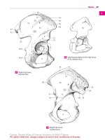
9 Superior hypogastric plexus (presacral nerve).
Plexus hypogastricus superior (n. praesacralis).
Plexus-like connection between the abdominal
aortic and inferior hypogastric plexuses located
predominantly in front of the 5
th
lumbar verte-
bral body and receiving branches from the lum-
bar sympathetic ganglia. D E
10 Right/left hypogastric nerve. N. hypogastricus
dexter/sinister. Right and left branches of the su-
perior hypogastric plexus to the pelvic viscera.
They communicate with the inferior hypogastric
plexus. D E
11 Inferior hypogastric plexus (pelvic plexus).
Plexus hypogastricus inferior (plexus pelvicus).
Network of sympathetic and parasympathetic
fibers located to the right and left of the rectum
and in front of it. D
12 Middle rectal plexus. Plexus rectalis medius.
Continuation of the inferior hypogastric plexus
onto the wall of the rectum. E
13 Inferior rectal plexus. Plexus rectalis inferior.
Autonomic nerve plexus located around the
branches of the internal iliac artery and passing
to both sides of the rectum. E
14 Prostatic plexus. Plexus prostaticus. Nerve
plexus mainly located at the posterior and infe-
rior surfaces of the prostate and extending as far
as the membranous part of the urethra. E
15 Deferential plexus. Plexus deferentialis. Nerve
plexus around the ductus deferens. E
16 Uterovaginal plexus. Plexus uterovaginalis.
Nerve plexus occupying the parametrium and in-
filtrated with many ganglia. It sends branches to
the uterus, vagina, uterine tube and ovary and
communicates with the inferior hypogastric
plexus in the rectouterine fold. D
17
Vaginal nerves. Nervi vaginales. Branches of the
uterovaginal plexus passing to the vagina. D
18 Vesical plexus. Plexusvesicalis. Plexussituated on
both sides of the urinary bladder. It contains para-
sympatheticfibers andisinvolvedinregulatingthe
voiding mechanism of the urinary bladder. E
19 Cavernous nerves of penis. Nn. cavernosi penis.
Rami from the prostatic plexus to the cavernous
bodies of the penis. E
20 Cavernous nerves of clitoris. Nn. cavernosi cli-
toridis. Nerves corresponding to the cavernous
nerves of the penis. E
21 SYMPATHETIC PART (SYSTEM). Pars sympathet-
ica. Thoracolumbar part of the autonomic
nervous system represented in the sympathetic
trunk. Excitable by adrenalin (adrenergic), it has a
stimulatory effect on the circulation and an in-
hibitory effect on the intestinal tract.
22 Sympathetic trunk. Truncus sympatheticus.
Chain of ganglia connected by nerve fibers. It lies
to the right and left of the vertebral column and
extends from the base of the skull to the coccyx. B
23 Ganglia of sympathetic trunk. Ganglia trunci
sympathetici. Groups of small, mostly multipolar
ganglion cells producing macroscopic thicken-
ings and forming synaptic sites between myeli-
nated preganglionic and nonmyelinated postgan-
glionic fibers. B
24 Interganglionic branches. Rami interganglion-
ares. Bundles of white and gray fibers linking the
sympathetic ganglia. B
25 Rami communicates. Communicating branches
(afferent and efferent) between the spinal nerves
and sympathetic trunk. B
26 Intermediate ganglia. Ganglia intermedia. Addi-
tional accumulations of sympathetic ganglion
cells mainly in the rami communicantes of the
cervical and lumbar regions. B
27 Superior cervical ganglion. Ganglion cervicale
superius. Uppermost sympathetic trunk gan-
glion, about 2.5 cm long and lying 2 cm below the
base of the skull between the longus capitis and
posterior belly of the digastric. A
28 Jugular nerve. N. jugularis. Branch to the inferior
ganglion of the glossopharyngeal nerve and to
the superior ganglion of the vagus. A
29 Internal carotid nerve. N. caroticus internus. It
contains postganglionic fibers and forms the in-
ternal carotid plexus in the carotid canal. A
30 Internal carotid plexus. Plexus caroticus inter-
nus. Nerve plexus in the carotid canal giving rise to
the deep petrosal nerve and branches to the inner
ear. It supplies the eye with sympathetic fibers. A
31 External carotid nerves. Nn. carotici externi.
Nerves for the external carotid plexus descending
along the external carotid artery. A
Autonomic nervous system
Feneis, Pocket Atlas of Human Anatomy © 2000 Thieme
All rights reserved. Usage subject to terms and conditions of license.
351
1
2
3
4
5
6
7
8
9
10
11
12
13
14
15
16
17
18
19
20
21
22
23
24
25
a
a
a
Autonomic nervous system
C II
C III
C IV
C V
30
29
27
28
31
24
23
2625
22
4
5
6
7
9
11
16
10
2
1
17
7
9
10
12
15
18
14
19; 20
8
13
352.35
12
Superior cervical
ganglion with branches
A
Sympathetic trunk
from in front
B
Plexuses in intestinal wall
C
Abdominal and
pelvic plexuses
D
Pelvic plexus
E
Feneis, Pocket Atlas of Human Anatomy © 2000 Thieme
All rights reserved. Usage subject to terms and conditions of license.
352
1
2
3
4
5
6
7
8
9
10
11
12
13
14
15
16
17
18
19
20
21
22
23
24
25
1 External carotid plexus. Plexus caroticus exter-
nus. Autonomic nerve plexus around the external
carotid artery. A
2 Common carotid plexus. Plexus caroticus com-
munis. It surrounds the common carotid artery. A
3 Laryngopharyngeal branches. Rami laryn-
gopharyngeales. Postganglionic fibers to the
pharyngeal plexus. A
4 Superior cervical cardiac nerve. N. cardiacus
cervicalis superior. Fibers to the cardiac plexus. A
5 Middle cervical ganglion. Ganglion cervicale me-
dium. Often very small ganglion of the sympa-
thetic trunk that lies at the level of C6 in front of or
behind the inferior thyroid artery. A E
6 Vertebral ganglion. Ganglion vertebrale. Small
accessory ganglion usually on the vertebral artery
in front of its entrance into the foramen transver-
sarium. A
7 Middle cervical cardiac nerve. N. cardiacus cervi-
calis medius. It passes from the middle cervical
ganglion to the deep part of the cardiac plexus. A
8 Cervicothoracic (stellate) ganglion. Ganglion
cervicothoracicum (stellatum). Fusion of the infe-
rior cervical ganglion with the 1
st
or, in many cases
(ca. 75%) 2
nd
thoracic ganglion. A E
9 Ansa subclavia. Cord of nerve fibers forming a
loop around the subclavian artery. A
10 Inferior cervical cardiac nerve. N. cardiacus
cervicalis inferior. It passes to the deep part of the
cardiac plexus. A
11 Subclavian plexus. Plexus subclavius. Autonomic
nerve plexus around the subclavian artery. A
12 Vertebral nerve. N. vertebralis. Located behind
the vertebral artery; it forms the vertebral plexus.
A
13 Vertebral plexus. Plexus vertebralis. Network of
nerves around the vertebral artery. A
14 Thoracic ganglia. Ganglia thoracica. 11−12 thick-
enings in the thoracic sympathetic trunk. A E
15 Thoracic cardiac branches. Rami cardiaci
thoracici. Rami with efferent and afferent (pain)
fibers passingfrom T2−4(5) thoracic ganglia to the
cardiac plexus. A E
15 a Thoracic plumonary branches. Rami pulmonales
thoracici. Efferent fibers from T2−4 ganglia to the
pulmonary plexus at the lung hilum.
15 b Esophageal branches. Rami oesophageales. Effer-
ent fibers from T2−5 ganglia.
16 Greater splanchnic nerve. N. splanchnicus major.
It passes from sympathetic trunk ganglia 5−9(10)
to the celiac ganglion and contains pre- and post-
ganglionic fibers which conduct pain and other
sensations from the upper abdominal organs. E
17 Thoracic splanchnic ganglion. Ganglion thoraci-
cum splanchnicum. Accessory ganglion at the
level of T9. It is incorporated into the greater
splanchnic nerve. E
18 Lesser splanchnic nerve. N. splanchnicus minor.
Arises from sympathetic trunk ganglia 9−11 and is
similar to the greater splanchnic nerve. E
19 Renal branch. Ramus renalis. Occasional branch
from the lesser splanchnic nerve to the renal
plexus. E
20 Lowest splanchnic nerve.N.splanchnicus imus. It
often arises independently from the T12 ganglion
and passes to the renal plexus. E
21 Lumbar ganglia. Ganglia lumbalia (lumbaria).
The sympathetic ganglia of the lumber vertebral
column, usually four on either side. B
22 Lumber splanchnic nerves. Nervi splanchnici
lumbales (lumbares). Usually four nerves from the
lumbar sympathetic trunk forming a plexus on L5.
B
23 Sacral ganglia. Ganglia sacralia. Four smaller gan-
glia lying medial to the pelvis sacral foramina. B
24 Sacral splanchnic nerves. Nervi splanchnici
sacrales. Two to three fine nerves from sacral gan-
glia 2−4. B
25 Ganglion im par. Last unpaired sympathetic trunk
ganglion lying in front of the coccyx. B
26 PARASYMPATHETIC PART (SYSTEM). Pars para-
sympathetica. Craniosacral component of the au-
tonomic nervous system involving cranial nerves
III, VII, IX, and X and sacral spinal nerves 2−4. An-
tagonistic to the sympathetic division, it slows
down the heart beat and stimulates intestinal and
sexual functions.
26 a
Cranial part. Pars cranialis.
27 Terminal nerve.N. terminalis.Aslender, partly in-
terwoven nerve of unknown function, but prob-
ably autonomic. It unites the olfactory region with
the anterior perforated substance. C
28 Terminal ganglion. Ganglion terminale. It com-
prises all of the ganglion cells dispersed in the ter-
minal nerves.
29 Ciliary ganglion. Ganglion ciliare. Located lateral
to the optic nerve. It receives preganglionic fibers
from the oculomotor nerve and gives off postgan-
glionic fibers which constrict the pupil and con-
tract the ciliary muscles during accomodation. D
30 Pterygopalatine ganglion. Ganglion pterygo-
palatinum. Nerve that lies lateral to the spheno-
palatine foramen, receives motor fibers from the
facial nerve via the nerve of the pterygoid canal
and supplies the lacrimal and nasal glands. D
31 Otic ganglion. Ganglion oticum. Situated below
the foramen ovale and medial to the mandibular
nerve. It receives motor fibers from the glos-
sopharyngeal nerve via the lesser petrosal nerve
and innvervates the parotid gland. D
32 Submandibular ganglion. Ganglion subman-
dibulare. Located along the lingual nerve below
the mandible. It receives motor fibers from the fa-
cial nerve via the chorda tympani and sends effer-
ent fibers to the sublingual and submandibular
glands. D
33 Sublingual ganglion. Ganglion sublinguale.Small
accumulations of cells occasionally present on the
glandular branches.
33 a Pelvic part (Pars pelvica). Sacral part of parasym-
pathetic division.
34 Pelvic splanchnic nerves. Nn. pelvici splanchnici
(nn. erigentes). Parasympathetic fibers from S2−4
spinal nerves to the pelvic ganglia for the pelvic
and genital organs. They also contain afferent
fibers. B
35 Pelvic ganglia. Ganglia pelvica. Groups of auton-
omic cells in the inferior hypogastric plexus. They
give rise to the postganglionic axons. See p. 351 E
Autonomic nervous system
Feneis, Pocket Atlas of Human Anatomy © 2000 Thieme
All rights reserved. Usage subject to terms and conditions of license.
353
1
2
3
4
5
6
7
8
9
10
11
12
13
14
15
16
17
18
19
20
21
22
23
24
25
a
a
a
Autonomic nervous system
21
23
22
34
24
25
1
2
3
13
9
4
15
14
10
8
11
7
6
12
5
27
32
31
30
29
19
20
18
17
16
14
15
8
5
Cervical sympathetic trunkA
Lumbosacral sympathetic trunk
B
Terminal nervesC
Autonomic ganglia of the head
D Splanchnic nervesE
Feneis, Pocket Atlas of Human Anatomy © 2000 Thieme
All rights reserved. Usage subject to terms and conditions of license.
354
1
2
3
4
5
6
7
8
9
10
11
12
13
14
15
16
17
18
19
20
21
22
23
24
25
1 SENSE ORGANS. Organa sensoria (sensuum).
In the narrow sense, the organs of vision, hear-
ing, smell and taste.
2 ORGAN OF VISION. Organum visus (visuale).
3 EYE. Oculus.
4 Optic nerve. N. opticus. Fiber bundle beginning
in the retina and extending as far as the optic
chiasm. Histologically and embryologically
speaking, it is the tract of the brain that is ac-
cordingly enclosed by meninges up to the pos-
terior aspect of the eyeball. Its axons have no
neurilemma (sheath of Schwann) but are myeli-
nated. The myelin sheath is formed by the oligo-
dendroglia. A C E
5 Intracranial part. Pars intracranialis. Segment
of the optic nerve betweeen the optic canal and
the chiasm. E
6 Intracanalicular part. Pars intracanicularis.
Segment of the optic nerve located in the optic
canal. It is partially connected with the canal
wall. E
7 Orbital part. Pars orbitalis. Slightly tortuous
segment of the optic nerve measuring about
3 cm in length and occupying the orbit. E
8 Intraocular part. Pars intraocularis. Segment of
optic nerve located in the wall of the eyeball.
9
Postlaminar part. Pars postlaminaris. In-
traocular segment located behind the lamina
cribrosa and thus at the site where the external
sheath of the optic nerve (dura) blends into the
sclera. A
10
Intralaminar part. Pars intralaminaris. In-
traocular segment lying within the lamina cri-
brosa. A
11
Prelaminar part. Pars preliminaris. Intraocular
segment extending between the lamina cri-
brosa and the nerve fiber layer of the retina. A
12 External sheath. Vagina externa. Dural cover-
ing of the optic nerve extending up to the eye-
ball. A
13 Internal sheath. Vagina interna. Pia and
arachnoid coverings acoompanying the optic
nerve to the eyeball. A
14
Intervaginal spaces. Spatia intervaginalia.
Subarachnoid space accompanying the optic
nerve and the capillary space between the
arachnoid and dura. A
15 Eyeball. Bulbus oculis. Globe of the eye. It con-
sists of the cornea and sclera together with all of
the structures they enclose. D
16 Anterior pole. Polus anterior (center of anterior
curvature) of the eyeball, which is determined
by the corneal vertex. D
17 Posterior pole. Polus posterior (center of poste-
rior curvature) of the eyeball, which lies lateral
to the exit of the optic nerve and opposite to the
anterior pole. D
18 Equator. Aequator. Greatest circumference of
the eyeball located equidistant from the ante-
rior and posterior poles. D
19 Meridians. Meridiani. Semicircles oriented at
right angles to the equator between the anterior
and posterior poles. D.
20 External axis of eyeball. Axis bulbi externus.
Line connecting anterior and posterior poles. C
21 Internal axis of eyeball. Axis bulbi internus.
Distance from posterior surface of cornea to the
inner surface of retina measured along an im-
aginary line (external axis of eyeball) through
the anterior and posterior poles. C
22 Optic axis. Axis opticus. Line passing through
the midline of the cornea and lens and bisecting
the retina between the fovea centralis and optic
disc. C
23 FIBROUS TUNIC OF EYEBALL. Tunica fibrosa
bulbi. External wall of eyeball comprising the
cornea and sclera. C
24 Sclera. The bluish-white outer coat of the eye-
ball, which consists of irregulatory arranged
collagenous fibers visible through the conjunc-
tiva.ABC
25 Scleral sulcus. Sulcus sclerae. Shallow groove
between the cornea and sclera caused by the
greater curvature of the cornea. B C D
26 Corneoscleral junction. Limbus. The concave
border of the sclera adjacent to the cornea. B
27 Trabecular meshwork (pectinate ligament).
Reticulum trabeculare (lig. pectinatum)
[[spongium iridocorneale]]. Connective tissue
framework at the iridocorneal (filtration) angle.
28
Corneoscleral par t. Pars corneoscleralis. Part
of the meshwork attached to the sclera. B
29
Uveal part. Pars uvealis. Part of the trabecular
meshwork attached to the iris. B
30 Canal of Schlemm. Sinus venosus sclerae.
Circular vessel occupying the interior aspect of
the trabecular meshwork. It can be interrupted
or doubled and is involved in the discharge of
aqueous humor from the anterior chamber. B
31 Episclera. Lamina episcleralis. Delicate dis-
placeable connective tissue between the outer
surface of the sclera and [[Tenon’s capsule]]
(bulbar fascia).
32 Substantia propria sclerae corneal stroma.
The proper substance, i. e., main part of the
sclera. It consists of irregularly arranged col-
lagenous fibers with sparse elastic fibers. A B
33 Lamina fusca sclerae. Layer of loose connective
tissue connecting the sclera and the choroid
lying below it. It appears yellowish owing to the
pigment cells dispersed within it. A
34 Lamina cribrosa. Fine, perforated layer of the
slcera for the passage of optic nerve fibers from
the retina. A
Sense organs
Feneis, Pocket Atlas of Human Anatomy © 2000 Thieme
All rights reserved. Usage subject to terms and conditions of license.
355
1
2
3
4
5
6
7
8
9
10
11
12
13
14
15
16
17
18
19
20
21
22
23
24
25
a
a
a
34
4
14
24
32
11
1213
33
10
9
22 2021
24
23
4
28
29
26
2530
32
24
7
5
6
16
19 25
18
17
25
Optic nerve with coverings
at point of exit
A
Iridocorneal angle
B
Eye, schematic
C
Eye, lines of orientationD
Segments of optic nerve
E
Sense organs
Feneis, Pocket Atlas of Human Anatomy © 2000 Thieme
All rights reserved. Usage subject to terms and conditions of license.
356
1
2
3
4
5
6
7
8
9
10
11
12
13
14
15
16
17
18
19
20
21
22
23
24
25
1 Cornea. The transparent anterior part (1/6) of
the eyeball with an anterior convex curvature
and a posterior concave curvature. It is 0.9 mm
thick in the middle, 1.2 mm thick at its margins.
BD
2 Conjunctival ring. Anulus conjunctivae. Junc-
tion between bulbar conjunctival epithelium
and the anterior epithelium of the cornea. D
3 Corneoscleral junction. Limbus corneae. D
4 Vertex corneae. The most prominent point on
the anterior surface of the cornea.
5 Anterior surface. Facies anterior. Corneal sur-
face facing the outside air. D
6 Posterior surface. Facies posterior. Corneal sur-
face facing the anterior chamber. D
7 Anterior epithelium. Epithelium anterius.
Stratified (about 5 layers) squamous
epithelium covering the anterior surface of the
cornea with a very smooth surface. B D
8 Anterior limiting (Bowman’s) membrane.
Lamina limitans anterior [[Bowman]]. Basal
membrane of the anterior epithelium, about
10−20 mm thick. It is continuous posteriorly
with the substantia propria. B
9 Substantia propria. Predominant part of the
avascular cornea consisting of highly organized
lamellar connective tissue embedded within a
mucopolysaccharide substance. The state of
turgescence of its fibers and the distribution of
its colloidal matrix affect the transparency of
the cornea. B
10 Posterior limiting (Descemet’s) membrane.
Lamina limitans posterior [[Descemet]]. Basal
membrane of the corneal (posterior) en-
dothelium. At its lateral margin it divides into
fibers which radiate into the trabecular mesh-
work of the sclera and iris. Aqueous humor
passes through its interstices to drain into the
sinus venosus sclerae. B D
11 Posterior epithelium (endothelium). Epithe-
lium posterius. Simple squamous epithelium
lining the posterior surface of the cornea. B D
12 VASCULAR TUNIC OF EYEBALL (UVEAL TRACT).
Tunica vasculosa bulbi (tractus uvealis). It rep-
resents the middle layer of the wall of the eye-
ball and consists of the choroid, ciliary body and
iris.
13 Choroid. Choroidea. The vascular coat lying be-
tween the retina and sclera. A
14 Suprachoroid lamina (lamina fusca). Lamina
suprachoroidea. Displaceable layer directly
beneath the sclera. It contains only a few vessels
and pigment; its fibers are partly covered by en-
dothelium. A
15 Perichoroidal space. Spatium perichoroideale.
Spatial system in the suprachoroid lamina, part
of which forms lymph pathways. It houses the
ciliary nerves, long and short posterior ciliary
arteries and the vorticose veins. A
16 Vascular lamina. Lamina vasculosa. It contains
the branchings of the short posterior ciliary ar-
teries. A
17 Choriocapillaris. Lamina choroidocapillaris.
Pigment-free layer of connective tissue with a
dense network of capillaries extending as far as
the ora serrata. It is often delimited from the
vascular lamina by a special connective tissue
layer. A
18 Basal lamina [[Bruch’s membrane]]. Com-
plexus (lamina) basalis. Homogeneous zone
about 2−4 mm thick between the choriocapil-
laris and the pigment epithelium of the retina. A
19 Ciliary body. Corpus ciliare. Enlarged uveal seg-
ment situated between the ora serrata and root
of the iris. It contains ciliary muscles and
processes. C
20 Pars plicata (Corona ciliaris). Circular zone oc-
cupied by ciliary processes. C
21 Ciliary processes. Processus ciliares. 70−80
radially oriented, capillary-rich folds, 0.1−
0.2 mm wide, 1 mm high and 2−3 mm long.
Their epithelium produces aqueous humor. C
22 Ciliary folds. Plicate ciliares. Low folds in the re-
gion of the corona ciliaris and between the cili-
ary processes. C
23 Pars plana. Orbiculus ciliaris. Circular zone
lying between the corona and ora serrata. It is
occupied by ciliary folds. C
24 Ciliary muscle. M. ciliaris. Smooth muscle oc-
cupying the ciliary body. It pulls the choroid for-
ward and, in so doing, relaxes the zonule fibers
so that the lens can become more strongly
curved for accomodation of near objects. D
25
Meridional (longitudinal) fibers. Fibrae mer-
idionales [fibrae longitudinales]. Larger muscle
fibers oriented meridionally (longitudinally).
Anteriorly they are attached to the posterior
limiting lamina above the trabecular mesh-
work; posteriorly, they insert into the choroid.
D
26
Circular fibers. Fibrae circulares. Circular
muscle lying internal to the meridional fibers. D
27
Radial fibers. Fibrae radiales. Muscle fibers
crossing perpendicular to the two other muscle
systems and coursing outwardly.
28 Basal lamina. Lamina basalis. Continuation of
the basal membrane of the choroid. It supports
the epithelium. D
Sense organs
Feneis, Pocket Atlas of Human Anatomy © 2000 Thieme
All rights reserved. Usage subject to terms and conditions of license.
357
1
2
3
4
5
6
7
8
9
10
11
12
13
14
15
16
17
18
19
20
21
22
23
24
25
a
a
a
24
354.24 13
14 16 17 1815
11
10
9
7
8
9
23
20
21
22
2
5
7
1
3
10
11
6
26
25
28
ChoroidA
Cornea
B
Ciliary body from behind
C
Iridocorneal angle, schematic
D
Sense organs
Feneis, Pocket Atlas of Human Anatomy © 2000 Thieme
All rights reserved. Usage subject to terms and conditions of license.
358
1
2
3
4
5
6
7
8
9
10
11
12
13
14
15
16
17
18
19
20
21
22
23
24
25
1 Iris. Frontally-located, round, variably colored
disk about 10−12 mm in diameter, with a cen-
tral aperture (pupil). The iris forms the poste-
rior border of the anterior chamber of the eye.
Its lateral margins become continuous with the
ciliary body. A
2 Pupillary margin. Margo pupillaris. Medial (in-
ternal) margin of the iris bordering the pupil. A
B
3 Ciliary margin. Margo ciliaris. Lateral (external)
margin of iris attached to ciliary body at the ir-
idocorneal angle. B
4 Anterior surface. Facies anterior. It faces the
anterior chamber. B
5 Posterior surface. Facies posterior. Surface
facing the posterior chamber. A B
6 Greater ring (circle) of iris. Anulus iridis major.
Ciliary segment of the iris, and outer cirucular
zone on the anterior surface of the iris. It is
coarser and broader than the lesser ring. A
7 Lesser ring (circle) of iris. Anulus iridis minor.
Pupillary segment of iris. Narrow, circular inner
zone on the anterior surface of iris. Its structure
is finer than that of the greater ring. A
8 Iridial folds. Plicae iridis. Folds passing around
the pupillary margin on the anterior side of the
iris. They make the pupillary margin appear
slightly serrated. A
9 Pupil. Pupilla. Aperture in the iris surrounded
by the pupillary margin of the iris. Its diameter
varies depending upon the intensity of light and
the focal distance of the observed object. A
10
M. sphincter pupillae. Network of spirally
coursing muscle fibers the longitudinal axes of
which run approximately parallel to the pupil-
lary margin when the pupil is dilated. It is in-
nervated by parasympathetic fibers from the
oculomotor nerve. B
11
M. dilator pupillae. Thin layer of smooth
muscle mainly comprised of radially oriented
fibers. It is innervated by sympathetic fibers
from the carotid plexus.
12 Stroma iridis. Vascular framework of the iris in-
filtrated by pigmented connective tissue cells.
Its anterior and posterior portions are thicker
than the rest and are divided by a fine fibrous
network. A B
13 Pigmented (posterior) epithelium. Epithelium
pigmentosum. Bilayered epithelium on the
posterior surface of the iris. It is so heavily pig-
mented that no nuclei are visible on the surface
facing the posterior chamber. A
14 Spaces of iridocorneal angle [spaces of Fon-
tana]. Spatia anguli iridocornealis. Interstices
between the fibers of the trabecular meshwork.
They form passageways that convey aqueous
fluid to the sinus venosus sclerae. A
15 Greater arterial circle of iris. Circulus arterio-
sus iridis major. Ringlike vascular system with
radiating branches. It is formed by anastomoses
between the long and short posterior ciliary ar-
teries. A
16 Lesser arterial circle of iris. Circulus arteriosus
iridis minor. Ringlike vascular system in the vi-
cinity of the pupillary margin formed by anas-
tomoses between the radial branches of the
greater arterial circle. A
17 Pupillary membrane. [Membrana pupillaris].
Anterior part of embryonical vascular mem-
brane around the lens that is situated behind
the pupil. It is fused to the pupillary margin and
receives blood vessels from there.
18 INTERNAL (SENSORY) TUNIC OF EYEBALL.
Tunica interna bulbi. It comprises the retina
with its pigment epithelium.
19 Retina. Inner lining of eyeball developed from
the two layers of the optic cup. Most of it is
light-sensitive (pars optica). B
20 Pars optica retinae. Retinal segment capable of
transforming light stimuli into nerve impulses.
It lines the posterior aspect of the eyeball and
extends as far anteriorly as the ora serrata. B
21
Pigmented part. Pars pigmentosa. Pigment
epithelium arising from the external layer of the
optic cup. B
22
Nervous par t. Pars nervosa. Retina proper con-
sisting essentially of three nuclear layers lying
internal to the pigment epithelium. B
23
Neuroepithelial (photosensitive) layer. Stratum
neuroepitheliale (photosensorium). Outer layer
of the cerebral stratum. It consists of rods and
cones, the outer segments of which affect the
transformation of light stimuli into nerve im-
pulses. Cell bodies of rods and cones form the
outermost layer of the retinal nuclei (external
nuclear layer). D
24
Internal nuclear layer. [[Stratum ganglionare reti-
nae]]. Middle layer of cell nuclei mainly con-
sisting of the cell bodies of bipolar and amacrine
cells. D
25
Ganglion cell layer. [[Stratum ganglionare n. op-
tici]]. Internal layer of nuclei consisting of multi-
polar cell bodies of initially non-myelinated
ganglion cells the axons of which form the optic
nerve. D
26 Ora serrata. Serrated margin between the
light-sensitive and light-insensitive parts of the
neural retina. B C
27 Pars ciliaris retinae. Light-insensitive retinal
segment consisting of a bilayered cuboidal
epithelium (ciliary epithelium) forming the
posterior surface of the ciliary body. Its outer
layer of epithelium is continuous with the pig-
ment epithelium of the retina and is pigmented,
whereas the innermost epithelium is continu-
ous with the pars nervosa of the retina and is
devoid of pigment. B
28 Pars iridica retinae. Light-insensitive retinal
segment on the posterior surface of the iris. It is
continuous with the pars ciliaris retinae and
forms the bilayered posterior epithelium of the
iris. Both layers are heavily pigmented. B
Sense organs
Feneis, Pocket Atlas of Human Anatomy © 2000 Thieme
All rights reserved. Usage subject to terms and conditions of license.
359
1
2
3
4
5
6
7
8
9
10
11
12
13
14
15
16
17
18
19
20
21
22
23
24
25
a
a
a
1
2
8
16
9
6
12
14
15
1313
5
10
28
12
2
5
43
27
20
26
2221
19
26
25
24
23
Iris, schematicA
Sections of retina
B
Ora serrata retinae
C
Retinal layers
D
Sense organs
Feneis, Pocket Atlas of Human Anatomy © 2000 Thieme
All rights reserved. Usage subject to terms and conditions of license.
360
1
2
3
4
5
6
7
8
9
10
11
12
13
14
15
16
17
18
19
20
21
22
23
24
25
1 Optic disc (papilla). Discus nervi optici [papilla
nervi optici]. Beginning of the optic nerve as
visualized in the fundus about 3−4 mm medial
to the macula. It is about 1.6 mm in diameter. C
2
Physiological cup. Excavatio disci. Depression
in the middle of the optic disc with the stems of
the central retinal artery and vein. C
3 Macula [[lutea]]. Transversely oval, yellowish
area, 2−4 mm in diameter, at the posterior pole
of the retina. C
4
Fovea centralis. Central fovea, a small depres-
sion in the macula caused by thinning of the
upper retinal layers. Its diameter, measured
from the beginning of the decrease in retinal
thickness from one side to the other, is approxi-
mately 1−2 mm. B C
5
Foveol a. Thinnest area of fovea centralis with a
diameter of about 0.2−0.4 mm. Here, the retina
is comprised entirely of approx. 2500 closely
packed cones. B
6 Retinal blood vessels. Vasa sanguinae retinae.
Branches of the central retinal artery and vein
located on the internal aspect of the retina.
7 Circle of arteries around the optic nerve. Cir-
culus vasculosus nervi optici. Small vascular
ring penetrating the sclera around the optic
nerve.
8 Superior temporal arteriole/venule or retina.
Arteriola/venula temporalis retinae superior.
Lateral upper branch of the central retinal
artery and vein. C
9 Inferior temporal arteriole/venule of retina.
Arteriola/venula temporalis retinae inferior.
Lateral lower branch of the central retinal artery
and vein. C
10 Superior nasal arteriole/venule of retina.
Arteriola/venula nasalis retinae superior. Upper
medial branch of the central retinal artery and
vein. C
11 Inferior nasal arteriole/venule of retina. Arte-
riola/venula nasalis retinae inferior. Lower me-
dial branch of the central retinal artery and vein.
C
12 Superior macular arteriole/venule. Arteriola/
venula macularis superior. They supply and
drain the upper part of the macula. C
13 Inferior macular arteriole/venule. Arteriola/
venula macularis inferior. They supply and
drain the lower part of the macula. C
14 Medial arteriole/venule of retina. Arteriola/
venula medialis retinae. Small branches that
supply and drain the medial part of retina proxi-
mal to the optic disc. C
14 a CHAMBERS OF THE EYE. Camerae bulbi.
15 Anterior chamber. Camera anterior. Space that
extends from the anterior surface of the iris to
the posterior surface of the cornea and com-
municates with the posterior chamber via the
pupil. A
16
Iridocorneal angle. Angulus iridocornealis.
Angle between the iris and cornea. It houses the
trabecular meshwork, the interstices of which
serve as passageways that drain aqueous humor
into the sinus venosus sclerae. A
17
Aqueous humor. Humor aquosus. Fluid pro-
duced by the epithelium of the ciliary processes
(total quantity: 0.2−0.3 cm
3
). The clear fluid
consists of 98% water, 1.4% NaCl and traces of
protein and sugar. It has a refractive index of
1.336.
Sense organs
Feneis, Pocket Atlas of Human Anatomy © 2000 Thieme
All rights reserved. Usage subject to terms and conditions of license.
361
1
2
3
4
5
6
7
8
9
10
11
12
13
14
15
16
17
18
19
20
21
22
23
24
25
a
a
a
15
16
5
8
10
12
4
14
13
9
11
1
2
4
3
Ciliary margin of irisA
Fovea centralis
B
Fundus
C
Sense organs
Feneis, Pocket Atlas of Human Anatomy © 2000 Thieme
All rights reserved. Usage subject to terms and conditions of license.
362
1
2
3
4
5
6
7
8
9
10
11
12
13
14
15
16
17
18
19
20
21
22
23
24
25
1 Posterior chamber. Camera posterior. It ex-
tends from the iris and ciliary body to the ante-
rior surface of the vitreous. A
2
Aqueous humor. Humor aquosus. Produced by
the ciliary processes. It flows between the in-
terstices of the suspensory ligaments of the lens
to the anterior surface of the lens and then be-
tween the iris and lens to the pupil, through
which it enters the anterior chamber.
3 Vitreous chamber. Camera vitrea. Space filled
up by the vitreous body. B
4
Vitreous body. Corpus vitreum. It consists of
about 9 8% water and primarily contains traces
of protein and NaCl and a mixture of fine fibrils
which thicken near the surface to form a lim-
iting membrane. It has a gelatinous consistency
due to its high content of hyaluronic acid. A
5
Hyaloid ar tery. [A. hyaloidea]. Branch of the
ophthalmic artery supplying the vascular mem-
brane of the lens. Present only during embry-
onic development. The proximal portion per-
sists in the optic nerve as the central retinal
artery. B
6
Hyaloid canal. Canalis hyaloideus. Canal
within the vitreous body formerly occupied by
the embryonic hyaloid artery which degener-
ates in this region. The canal assumes a
downward sagging corkscrew shape; it extends
from the optic disc to the posterior surface of
the lens. Its wall is formed by condensed fibers.
A
7
Hyaloid (lenticular, patellar) fossa. Fossa hy-
aloidea. Fossa on the anterior surface of the vit-
reous body adjacent to the lens. A
8
Vitreous (hyaloid) membrane. Membrana
vitrea. Condensation of fibers on the surface of
the vitreous body. See (4), vitreous body. A
9
Stroma of vitreous body. Stroma vitreum.
Fine network of fibers in the virtreous body. Its
surface thickens to form the vitreous mem-
brane.
9a
Vitreous humor. Humor vitreus. Fluid part of
vitreous body. Primarily consists of mucupoly-
saccharides and is situated between the fibers
of the stroma.
10 LENS. Structure of the eye situated between the
pupil and vitreous body. It is suspended by the
ciliary zonule (suspensory ligaments), has a di-
ameter of 9−10 mm and is about 4 mm thick. B C
D
11 Substantia lentis. Lens substance situated
beneath the lens epithelium and comprising
the lens nucleus and lens cortex with a refrac-
tive index of 1.44−1.55. C
12 Lens cortex. Cortex lentis. External zone of the
lens. It is softer owing to its high water content
and blends into the lens nucleus without a
sharp boundary. C
13 Nucleus of lens. Nucleus lentis. Harder core of
the lens with a low water content, as is espe-
cially evident in the elderly. C
14 Lens fibers. Fibrae lentis. Fibers corresponding
to the lens epithelium from which they develop.
They form the lens substance measuring 2.5−
12 µm thick and up to 10 mm long. C
15 Epithelium of lens. Epithelium lentis. Part of
the lens confined to the anterior surface and ex-
tending as far as the equator. It is derived
embryologically from the anterior epithelium
of the lens vesicle. C
16 Lens capsule. Capsula lentis. Transparent mem-
brane, up to 15 µm thick, covering the lens in-
cluding its epithelium. Its anterior pole is
thicker than the posterior pole. It gives attach-
ment to the suspensory ligaments. C
17 Anterior pole. Polus anterior. D
18 Posterior pole. Polus posterior. D
19 Anterior surface. Facies anterior. Less curved
lens surface with a radius of 8.3−10 mm. C
20 Posterior surface. Facies posterior. More
curved lens surface with a radius of about
6.5 mm. C
21 Axis. Line connecting anterior and posterior
poles. D
22 Equator. Margin of lens. D
23 Radii of lens. Suture line of the individual lens
fibers. In the young it resembles a triradiate
seam. D
24 Ciliary zonule. Zonula ciliaris. Suspensory ap-
paratus together with its interstices. It encircles
the lens equator and consists of a radially
oriented system of f ibers of variable length and
the folds situated between them. C
25
Zonular f ibers (suspensory ligaments). Fi-
brae zonulares. Suspensory fibers attached to
the equator and the adjacent anterior and post-
erior surfaces of the lens. They arise distally
from the basal lamina of the ciliary body and the
pars ciliaris retinae. C
26
Zonular spaces. Spatia zonularia. Spaces be-
tween the zonule fibers filled with percolating
aqueous humor. C
Sense organs
Feneis, Pocket Atlas of Human Anatomy © 2000 Thieme
All rights reserved. Usage subject to terms and conditions of license.
363
1
2
3
4
5
6
7
8
9
10
11
12
13
14
15
16
17
18
19
20
21
22
23
24
25
a
a
a
1
4
8
6
8
7
3
5
10
18
22
3
21
17
23
24
12
19
13
12
20
11
26
14
15 16
25
Posterior chamber of eyeA
Hyaloid artery
B
Lens and zonula ciliaris
C
Lens of eye
D
Sense organs
Feneis, Pocket Atlas of Human Anatomy © 2000 Thieme
All rights reserved. Usage subject to terms and conditions of license.
364
1
2
3
4
5
6
7
8
9
10
11
12
13
14
15
16
17
18
19
20
21
22
23
24
25
1 ACCESSORY ORGANS OF EYE. Organa oculi ac-
cessoria.
2 Muscles of eye. Musculi bulbi. Extrinsic ocular
muscles.
3 Orbital muscle. M. orbitalis. Thin layer of
smooth muscle which bridges the inferior orbi-
tal fissure. C
4 Superior rectus. M. rectus superior. o: Common
tendinous ring. i: Along an oblique line in front
of the equator, 7−8 mm posterior to the corneal
margin. A: Elevation and medial rotation of su-
perior pole of eyeball. I: Oculomotor nerve. B C
D
5 Inferior rectus. M. rectus inferior. o: Common
tendinous ring. i: Along an oblique line about
6 mm behind the corneal margin. A: Depression
and lateral rotation of superior pole of eyeball. I:
Oculomotor nerve. B C D
6 Medial rectus. M. rectus medialis. o: Common
tendinous ring. i: About 5.5 mm from the cor-
neal margin. A: Adduction of corneal pole. I:
Oculomotor nerve. B C
7 Lateral rectus. M. rectus lateralis. o: Common
tendinous ring and lesser wing. i: 5.5 mm be-
hind corneal margin. A: Abduction of corneal
pole. I: Abducent nerve. B C D
8
Tendon of lateral rectus at greater wing.
Lacertus musculi recti lateralis. C
9 Common tendinous ring (common annular
tendon). Anulus tendineus communis. Ten-
dinous ring for attachment of the recti ocular
muscles. It surrounds the optic canal and me-
dial part of the superior orbital fissure. C
10 Superior oblique. M. obliquus superior. o: Body
of sphenoid medial to common tendinous ring.
i: Posterolateral aspect of sclera behind the
equator after its tendon passes through the
trochlea and approaches sclera obliquely from
the medial margin of orbit. A: Abduction, me-
dial rotation and depression. I: Trochlear nerve.
B
11
Trochlea. Cartilaginous sling attached to the
medial wall of the orbit [[trochlear spine]] and
serving as a pulley for the tendon of the super-
ior oblique muscle. B
12
Tendon sheath of superior oblique muscle
(synovial bursa of trochlea).
Vagina tendinis
m. obliqui superioris [[bursa synovialis
trochlearis]]. Synovial sheath (bursa) for the
tendon of the superior oblique muscle separat-
ing the tendon from the trochlea. B
13 Inferior oblique. M. obliquus inferior. o: Lateral
to the nasolacrimal canal. i: Posterior to equa-
tor. A: Elevation, abduction and lateral rotation.
I: Oculomotor nerve. D
14 M. levator palpebrae superioris. o: Bone above
optic canal and dura of optic nerve. Its tendon
broadens anteriorly and splits to form an upper
and lower layer. I: Oculomotor nerve. A C D
15
Superf icial lamina of levator tendon.
Lamina superficialis. It passes between the tar-
sus and orbicularis oculi to insert into the sub-
cutaneous connective tissue of the upper eyelid.
It is so broad that it extends mainly laterally to
the wall of the orbit. A
16
Deep lamina of levator tendon. Lamina pro-
funda. It inserts into the upper margin and the
anterior surface of the tarsus. A
17 Orbital fasciae. Fasciae orbitales.
18 Periosteum of orbit. Periorbita. It is delicate
and fused solidly to the bone at the inlet and
outlet of the orbit. Anteriorly, it is continuous
with the adjacent periosteum, posteriorly with
the dura. A
19 Orbital septum. Septum orbitale. Connective
tissue septum partly reinforced by tendon. It
passes from the orbital margin below the orbic-
ularis oculi to the external margins of the tarsi
and forms the anterior end of the orbit. A
20 Muscular fasciae. Fasciae musculares. Sheaths
of Tenon’s capsule enveloping the tendons and
muscular bellies of the 6 extrinsic ocular
muscles. A
21 Tenon’s capsule (fascia bulbi). Vagina bulbi.
Connective tissue gliding membrane between
the eyeball and orbital fat. It is fused to the
sclera posteriorly at the optic nerve. Anteriorly
it ends beneath the conjunctiva. It is separated
from the sclera primarily by the episcleral
space. A
22 Episcleral space. Spatium episclerale [[inter-
vaginale]]. Gliding space between the eyeball
and Tenon’s capsule. It is traversed by long, deli-
cate connective tissue fibers. A
23 Orbital fat body. Corpus adiposum orbitae.
Adipose tissue fills the spaces around the ocular
muscles, the eyeball and the optic nerve and is
bordered anteriorly by the orbital septum. A D
Sense organs
Feneis, Pocket Atlas of Human Anatomy © 2000 Thieme
All rights reserved. Usage subject to terms and conditions of license.
365
1
2
3
4
5
6
7
8
9
10
11
12
13
14
15
16
17
18
19
20
21
22
23
24
25
a
a
a
15
19
16
21
22
22
21
20
1423
18
12
11
10
6
5
4
14
7
14 4
7
9
8
6
5
3
14
4
23
23
57
Orbit,
sagittal section
A
Eye muscles from above
B
Orbit, anterior view
C
Eye muscles, lateral viewD
Sense organs
Feneis, Pocket Atlas of Human Anatomy © 2000 Thieme
All rights reserved. Usage subject to terms and conditions of license.
366
1
2
3
4
5
6
7
8
9
10
11
12
13
14
15
16
17
18
19
20
21
22
23
24
25
1 Eyebrow. Supercilium. The transverse eleva-
tion above the eyes, covered by thick, bristle-
like hairs. A
2 Eyelids. Palpebrae.
3 Upper eyelid. Palpebra superior. A
4 Lower eyelid. Palpebra inferior. A
5 Anterior palpebral surface. Facies anterior pal-
pebralis. The anterior external (skin-covered)
surface of the eyelid. E
6 Epicanthus (mongolian fold). [Plica palpe-
bronasalis] [[epicanthus]]. Vertical fold covering
the medial angle of the eye. It is a continuation
of the upper eyelid at the lateral nasal wall. C
7 Posterior palpebral surface. Facies posterior
palpebralis. Surface lined by conjunctival
epithelium and containing dispersed goblet
cells. E
8 Palpebral fissure. Rima palpebrarum. Space
between the margins of the upper and lower
eyelids. A E
9 Lateral palpebral commissure. Commissura
palpebralis lateralis. Lateral junction of the
upper and lower eyelids. A
10 Medial palpebral commissure. Commissura
palpebralis medialis. Medial junction of the
upper and lower eyelids. A
11 Lateral angle (cant hus) of eye. Angulus oculi
lateralis. Acute lateral angle of the eye; it is also
the lateral end of the palpebral fissure. A
12 Medial angle (canthus) of eye. Angulus oculi
medialis. More rounded medial end of the
palpebral fissure which delimits a triangular
space, the lacrimal lake. A
13 Limbi palpebrales anteriores. Anterior edges
of the free margins of the eyelids adjacent to the
external skin. E
14 Limbi palpebrales posteriores. Posterior edges
of the free margins of the eyelids adjacent to the
conjunctiva. E
15 Eyelashes. Cilia. The 3−4 rows of hair growing
near the anterior edge of the free margin of the
eyelids. E F
16 Superior tarsal plate. Tarsus superior. Curved
plate about 10 mm high occupying the upper
eyelid and consisting of compact, interwoven
collagenous connective tissue with tarsal
glands. B E
17 Inferior tarsal plate. Tarsus inferior. Plate about
5 mm high within the lower eyelid. It likewise
consists of firm, interwoven collagenous con-
nective tissue with tarsal glands. B E
18 Medial palpebral ligament. [[Lig. palpebrale
mediale]]. Band ofconnective tissuebetweenthe
medial palpebral commissure and the medial
walloftheorbit.Itliesinfrontofthelacrimalsac.
BD
19 Lateral palpebral raphe. [[Raphe palpebralis
lateralis]]. Delicate band on the lateral palpebral
ligament. It is reinforced by the orbicularis oculi
muscle. D
20 Lateral palpebral ligament. Lig. palpebrale
laterale. Fibrous band that attaches the lateral
palpebral commissure to the lateral wall of the
orbit in front of the orbital septum. B
21 Tarsal [[Meibomian]] glands. Glandulae tar-
sales. Elongated holocrine glands located in the
superior and inferiortarsal plates with openings
near the posterior edge of the free margin of the
eyelids. They produce a sebaceous secretion for
lubrication of the lid margins. E
22 Superior tarsal muscle. M. tarsalis superior.
Smooth muscle fibers between the muscle-ten-
don border of the levator palpebrae muscle and
the superior tarsal plate. E
23 Inferior t arsal muscle. M. tarsalis inferior.
Smooth muscle fibers between the inferior for-
nix of the conjunctiva and the inferior tarsal
plate. E
24 Tunica conjunctiva. The lining of the inner sur-
face of the eyelids, which consists of two or more
layers of columnar epithelium with goblet cells
and a loose, cell-rich lamina propria containing
multiple blood vessels. The tunica extends
around the fornix of the conjunctiva to the eye-
ball, which it covers with a layer of stratified
squamousepitheliumthatextends up tothecor-
neal margin. E
25 Semilunar fold of conjunctive. Plica semi-
lunarisconjunctivae.Itliesinthe medial angle of
the eye between the fornix of the upper and
lower eyelid. F
26 Lacrimal caruncle. Caruncula lacrimalis. Mu-
cosal mass in the medial angle of the eye covered
by stratified squamous or columnar epithelium.
F
Sense organs
Feneis, Pocket Atlas of Human Anatomy © 2000 Thieme
All rights reserved. Usage subject to terms and conditions of license.
367
1
2
3
4
5
6
7
8
9
10
11
12
13
14
15
16
17
18
19
20
21
22
23
24
25
a
a
a
25
26
15
16
5
13
13
15
8
17
5
21
7
14
21
7
24
23
22
1819
6
20
16
17
18
9
3
1
12
8
11
4
10
Palpebral fissureA Tarsal plates and ligamentsB
Epicanthus (epicanthic fold)
C
Orbicular muscle of eye
from behind
D
Eyelids, sagittal section
E
Inner (nasal) canthus of eye
F
Sense organs
Feneis, Pocket Atlas of Human Anatomy © 2000 Thieme
All rights reserved. Usage subject to terms and conditions of license.
368
1
2
3
4
5
6
7
8
9
10
11
12
13
14
15
16
17
18
19
20
21
22
23
24
25
1 Bulbar conjunctiva. Tunica conjunctiva bul-
baris. Part of conjunctiva covering the eyeball. It
consists of stratified, nonkeratinized squamous
epithelium with only a few goblet cells and a
loose, cell-poor lamina propria permeated with
elastic fibers. A
2 Palpebral conjunctiva. Tunica conjunctiva pal-
pebralis. The portion of theconjunctiva covering
the posterior surface of the eyelid. It consists of
two or more layers of columnar epithelium with
goblet cells and a loose, vascularized lamina
propria. A
3 Superior fornix of cunjunctiva. Fornix con-
junctivae superior. Reflected fold of conjunctiva
extendingfrom the eyeball (bulbar) to the upper
eyelid (palpebral). A
4 Inferior fornix of conjunctiva. Fornix conjunc-
tivae inferior. Reflected fold of conjunctiva from
the eyeball (bulbar) on to the lower eyelid
(palpebral). A
5 Conjunctival sac. Saccus conjunctivalis. Space
between palpebral and bulbar conjunctivae. Its
upper and lower ends form the superior and in-
ferior fornices of the conjunctiva. A
6 Ciliary glands (of Moll). Glandulae ciliares
[[Molli]]. Apocrine glands on the lid margin.
They open either into the hair follicles of the
eyelashes or at the lid margin. A
7 Sebaceous glands (of Zeiss). Glandulae se-
baceae [[Zeiss]]. Small sebaceous glands with
openings into the hair follicles of the eyelashes.
A
8 Conjunctival glands. Glanduale conjunctivales.
Follicular aggregations of lymphocytes at the
medial angle of the eye.
9 Lacrimal apparatus. Apparatus lacrimalis. The
system of structures that lubricate the cornea
and conjunctiva. B
10 Lacrimal gland. Glandula lacrimalis. Gland lo-
cated above the lateral angle of the eyelids; it is
separated into an upper and lower portion by
the tendon of levator palpebrae muscle. Its ex-
cretory ducts open laterally into the superior
fornix of the conjunctiva. B
11
Orbital par t. Pars orbitalis. Larger portion of
lacrimal gland located above the tendon of the
levator palpebrae muscle. B
12
Palpebral part. Pars palpebralis. Smaller por-
tion of lacrimal gland located below the tendon
of the levator palpebrae muscle. B
13
Excretory ducts of lacrimal gland. Ductuli
exretorii [[glandulae lacrimalis]]. 6−14 ducts
opening into the superior fornix of the conjunc-
tiva. B
14 Accessory lacrimal glands. [Gll. lacrimales ac-
cessoriae]. Additional smaller lacrimal glands
found scattered especially in the vicinity of the
superior conjunctival fornix. A
15 Rivus lacrimalis. Pathway that conducts tears
from the excretory ducts to the lacrimal lake. It
lies within the conjunctival sac between the
closed eyelids and the eyeball.
16 Lacrimal lake. Lacus lacrimalis. Spaceinthe me-
dial angle of the eye around the lacrimal
caruncle. B C
17 Papilla lacrimalis. Small cone-shaped elevation
medial to the inner edge of both the upper and
lower eyelids. Each apex houses an opening or
lacrimal punctum. C
18 Lacrimal punctum. Punctum lacrimale. Small
opening marking the beginning of the lacrimal
fluid drainage system. C
19 Lacrimal canaliculus. Canaliculus lacrimalis.
Small canal, up to 1 cm long, from each lacrimal
punctum to the lacrimal sac. C
20 Ampulla of lacrimal canaliculus. Ampulla
canaliculi lacrimalis. Slight enlargement at the
bend of the lacrimal canaliculus. C
21 Lacrimal sac. Saccus lacrimalis. It is located in
the lacrimal fossa and is about 1.5 cm long and
about 0.5 cm wide. It descends directly into the
nasolacrimal duct. C
22
Fornix of lacrimal sac. Fornix sacci lacrimalis.
Dome-shaped upper margin of the lacrimal sac.
C
23 Nasolacrimal duct. Ductus nasolacrimalis. Duct
that is directly continuous withthe larcrimal sac
and about 1.2−2.4 cm in length. It passes
through the nasolacrimal canal and opens into
the inferior nasal meatus. Its flattened lumen is
lined by a mucosa containing two or more layers
of columnar epithelium bearing cilia at some
sites. C
24
Lacrimal fold. Plica lacrimalis. Mucosal fold at
the opening of the nasolacrimal duct. It is lo-
cated in the inferior nasal meatus about 3−
3.5 cm posterior to the external naris. C
Sense organs
Feneis, Pocket Atlas of Human Anatomy © 2000 Thieme
All rights reserved. Usage subject to terms and conditions of license.
369
1
2
3
4
5
6
7
8
9
10
11
12
13
14
15
16
17
18
19
20
21
22
23
24
25
a
a
a
7
6
5
2
5
2
1
14
4
13
11
12
16
3
19
20
21
22
23
24
18
18
16
17
17
20
19
Eyelids, sagittal sectionA
Lacrimal gland
B
Lacrimal systemC
Sense organs
Feneis, Pocket Atlas of Human Anatomy © 2000 Thieme
All rights reserved. Usage subject to terms and conditions of license.
370
1
2
3
4
5
6
7
8
9
10
11
12
13
14
15
16
17
18
19
20
21
22
23
24
25
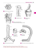
1 VESTIBULOCOCHLEAR ORGAN. Organum vesti-
bulocochleare. Sensory apparatus housed in the
temporal bone for the perception of sound,
equilibrium and positional changes.
2 INTERNAL EAR. Auris interna. Part of the vesti-
bulocochlear organ residing in the petrous tem-
poral bone.
3 MEMBRANOUS LABYRINTH. Labyrinthus mem-
branaceus. Complicated system of ducts and di-
latations within the bony labyrinth, which con-
tains sensory epithelium and is suspended by
connective tissue. A
4 Endolymph. Fluid contained within the mem-
branous labyrinth.
5 Perilymph. Fluid occupying the osseous laby-
rinth and surrounding the membranous laby-
rinth.
6 Vestibular labyrinth. Labyrinthus vestibularis.
Portion of membranous labyrinth constituting
the organ of equilibrium. It includes the semi-
circular ducts.
7 Endolymphatic duct. Ductus endolymphaticus
[[aquaeductus vestibuli]]. Slender duct arising
from the utriculosaccular duct and passing
through the osseous aqueduct of the vestibule to
terminate as the endolymphatic sac. A
8 Endolymphatic sac. Saccus endolymphaticus.
Blind sac of endolymphatic duct located be-
tween two dural layers at the posterior wall of
the petrous temporal. A
9 Utriculosaccular duct. Ductus utriculosaccu-
laris. Slender duct between the saccule and
utricle. It gives rise to the endolymphatic duct. A
10 Utricle. Utriculus. Sac 2.5−3.5 mm in diameter,
serving as the base for the three semicircular
ducts. A
11 Semicircular duct. Ductus semicirculares.
Three membranous ducts that resemble two-
thirds of a circular arch, each occupying its own
osseous semicircular canal oriented perpendic-
ular to the others.
12
Anterior (superior) semicircular duct. Duc-
tus semicircularis anterior. It is oriented verti-
cally and somewhat perpendicular to the
petrous part of the temporal bone. A
13
Posterior semicircular duct. Ductus semi-
circularis posterior. It is oriented somewhat ver-
tically in a plane which runs parallel to the longi-
tudinal axis of the petrous part of the temporal
bone. A
14
Lateral semicircular duct. Ductus semicircu-
laris lateralis. The most lateral, horizontally
oriented semicircular duct. It may create a bulge
in the medial wall of the tympanic cavity. A
15 Proper membrane of semicircular duct. Mem-
brana propria ductus semicircularis. Layer
below the basal membrane consisting primarily
of densely packed fibers which extends into the
looser network of the perilymphatic space. C
16 Basal membrane of semicircular duct. Mem-
brana basalis ductus semicircularis. Appears
upon light microscopy as a homogeneous basal
membrane situated directly below the
epithelium. C
17 [[Epithelium of semicircular duct]].
[[Epithelium ductus semicircularis]]. Simple
epithelium lining the inner aspect of the mem-
branous semicircular duct. The cells are flat and
become cuboidal on their concave side. C
18 Membranous ampullae. Ampullae mem-
branaceae. Dilatations of the semicircular ducts
in the vicinity of the utricle.
19
Anterior membranous ampulla. Ampulla
membranacea anterior. Dilatation at the end of
anterior (superior) semicircular duct located
near the lateral membranous ampulla. A
20
Posterior membranous ampulla. Ampulla
membranacea posterior. Dilatation at the end of
the posterior semicircular duct located distal to
the other two membranous ampullae. A
21
Lateral membranous ampulla. Ampulla
membranacea lateralis. Ampulla of the lateral
semicircular duct located proximal to the ante-
rior membranous ampulla. A
22 Sulcus ampullaris. Indentation below the am-
pullary crest bearing branches from the ampul-
lar nerve for innervation of the ampullary crest.
B
23 Ampullary crest. Crista ampullaris. Crescent-
shaped ridge projecting into the ampullary
space. It is covered by sensory epithelium and
has a base of nerve fibers and connective tissue.
B
24
[[Neuroepithelium]]. Sensory epithelium of
ampullae consisting of supporting cells and
sensory cells with hairs (microvilli) projecting
from the surface into an overlying cupula. B
25
Cupula. Gelatinous body suspended above the
ampullary crest as far as the roof of the ampulla
and penetrated by hairs of the sensory cells. B
26 Membranous crura. Crura membranacea.
Limbs of semicircular ducts opening into the
utricle.
27
Simple membranous crus. Crus mem-
branaceum simplex. Posterior limb of lateral
semicircular duct opening independently into
the utricle. A
28
Ampullary membranous crura. Crura mem-
branacea ampullaria. Semicircular duct seg-
ments situated between the ampullae and the
utricle. A
29
Common membranous crus. Crus mem-
branaceum commune. Common limb formed by
the anterior and posterior semicircular ducts
and opening into the utricle. A
Sense organs
Feneis, Pocket Atlas of Human Anatomy © 2000 Thieme
All rights reserved. Usage subject to terms and conditions of license.
371
1
2
3
4
5
6
7
8
9
10
11
12
13
14
15
16
17
18
19
20
21
22
23
24
25
a
a
a
12
19
21
29
27
8
20
25
10
28
7
14
13
25
23
24
16
15
17
9
22
Membranous labyrinthA
Ampulla of semicircular duct
B
Semicircular duct, cross section
C
Sense organs
Feneis, Pocket Atlas of Human Anatomy © 2000 Thieme
All rights reserved. Usage subject to terms and conditions of license.
372
1
2
3
4
5
6
7
8
9
10
11
12
13
14
15
16
17
18
19
20
21
22
23
24
25
1 Ductus reuniens. Fine tube connecting the sac-
cule with the cochlear duct. B
2 Saccule. Sacculus. Round vesicle, 2−3 mm in
size, equipped with a sensory field. B
3 Maculae [[staticae]]. Sensory fields for the per-
ception of the position of the head in space. A B
4
Utricular macula. Macula utriculi. Horizon-
tally oriented sensory field, 2.3−3 mm in size,
occupying the floor of the utricle. B
5
Saccular macula. Macula sacculi. Vertically
oriented, arched sensory field, about 1.5 mm
wide; in the medial wall of the saccule. B
6
Statoconia. Calcium concretions, up to 15 µm
in size, embedded in a gelatinous substance to-
gether with the sensory hairs. A
7
Statoconial membrane. Membrana stato-
coniorum. Membrane covering the maculae and
consisting of a gelatinous ground substance
with statoconia on itssurface. It is penetrated by
bristle-like processes from underlying macular
sensory cells. A
8
[[Neuroepithelium]]. Pseudostratified, pris-
matic, sensory epithelium of the macula con-
sisting of supporting and sensory cells. The
sensory cells bear 20−25 µm long bristle-like
processes which project into the statoconial
membrane. A
9 Cochlear labyrinth. Labyrinthus cochlearis.
Complex contents of the osseous cochlea. C
10 Perilymphatic space. Spatium perilymphati-
cum. Space occupied by perilymph and partially
permeated by connective tissue fibers. It in-
cludes the scala vestibuli and tympani. A B
11
Scala vestibuli. Perilymphatic canal located
above the osseous spiral lamina and cochlear
duct. It ascends as far as the apex of the cochlea
(helicotrema). C
12 Scala tympani. Perilymphatic canal below the
osseous spiral lamina and basilar membrane. C
13 Cochlear aqueduct (perilymphatic duct).
Aquaeductus cochleae. Pathway connecting the
perilymphatic space with the subarachoid
space. B
14
External aperture of perilymphatic duct.
Apertura externa aquaeductus cochleae. Open-
ing for the tympanic nerve in the vicinity of the
canaliculus. See p. 14.22
15 Cochlear duct. Ductus cochlearis. A spiral en-
dolymphatic tube taking 2
1
/2−2
3
/4 turns around
a bony axis (modiolus) before ending blindly at
the apex of the cochlea. It houses the sensory
epithelium for the perception of sound. B C E
16 Cupular cecum. Caecum cupulare. Blind end of
cochlear duct located at the apex of the cochlea.
B
17 Vestibular cecum. Caecum vestibulare. Blind
end of the cochlear duct facing the vestibule. B
18 Tym panic wall of cochlear duct (spiral mem-
brane). Paries tympanicus ductus cochlearis
(membrana spiralis). Inferior wall of cochlear
duct situated above the scala tympani. E
19 Spiral organ (of Corti). Organum spirale
[[Corti]]. Sensory field on the basilar membrane
that transforms sound waves into nerve im-
pulses. D
20 Basilar membrane. Lamina basilaris. Plate of
connective tissue between the cochlear duct
and scala tympani. It extends between the tym-
panic lip of the osseous spiral lamina and the
spiral crest. E
21 Spiral crest (ligament). Crista spiralis (lig. spi-
rale). Spirally arranged system of fibers arising
from the periosteum of the cochlear canal and
radiating into the basilar lamina. E
22 Nerve foramina. Foramina nervosa. Holes in the
basilar lamina for transmission of cochlear
nerve fibers from the hair cells to the spiral gan-
glion. D
23 Limbus of osseous spiral lamina. Limbus
laminae spiralis osseae. Thickening and trans-
formation of the endosteum on the upper layer
of the osseous spiral lamina indented externally
by the internal spiral sulcus. E
24 Vestibular lip of limb of osseous spiral lamina.
Labium limbi vestibulare. Upper, shorter
process of the limbus. Site of attachment of the
tectorial membrane. E
25 Typmpanic lip of limb of osseous spiral
lamina. Labium limbi tympanicum. Lower,
longer process of the limbus lying on the basilar
membrane. D E
26 Tectorial membrane. Membrana tectoria.
Fibrous membrane resting on the organ of Corti.
It is narrow at the site where it attaches to the
vestibular lip and ends freely beyond the row of
outer hair cells. D E
27 Auditory teeth. Dentes acustici. The ridge-like
rows of cells on the surface of the vestibular lip.
The tectorial membrane attaches near here. D
28 Internal spiral sulcus. Sulcus spiralis internus.
Groove between the vestibular and tympanic
lips. D E
29 External spiral sulcus. Sulcus spiralis externus.
Groove on theouter wallof the cochlear duct be-
tween the spiral prominence and the spiral
organ. E
Sense organs
Feneis, Pocket Atlas of Human Anatomy © 2000 Thieme
All rights reserved. Usage subject to terms and conditions of license.
373
1
2
3
4
5
6
7
8
9
10
11
12
13
14
15
16
17
18
19
20
21
22
23
24
25
a
a
a
7
8
6
10
10
13
17
15
16
4
2
5
1
11
12
15
27
26
28
25 22
2524 20
21
29
28
26
23
15
18
Macula staticaA
Membranous labyrinth
B
Cochlea, opened
C
Organ of Corti
D Cochlear ductE
Sense organs
Feneis, Pocket Atlas of Human Anatomy © 2000 Thieme
All rights reserved. Usage subject to terms and conditions of license.