Báo cáo y học: "Alveolar instability caused by mechanical ventilation initially damages the nondependent normal lung" ppt
Bạn đang xem bản rút gọn của tài liệu. Xem và tải ngay bản đầy đủ của tài liệu tại đây (425.66 KB, 10 trang )
Open Access
Available online />Page 1 of 10
(page number not for citation purposes)
Vol 11 No 5
Research
Alveolar instability caused by mechanical ventilation initially
damages the nondependent normal lung
Lucio Pavone
1
, Scott Albert
1
, Joseph DiRocco
1
, Louis Gatto
2
and Gary Nieman
1
1
Upstate Medical University, Department of Surgery, 750 E Adams Street, Syracuse, NY 13210 USA
2
Department of Biology, Cortland College, P.O. Box 2000 Cortland, NY 13045 USA
Corresponding author: Scott Albert,
Received: 26 Jun 2007 Revisions requested: 27 Jul 2007 Revisions received: 6 Sep 2007 Accepted: 18 Sep 2007 Published: 18 Sep 2007
Critical Care 2007, 11:R104 (doi:10.1186/cc6122)
This article is online at: />© 2007 Pavone et al., licensee BioMed Central Ltd.
This is an Open Access article distributed under the terms of the Creative Commons Attribution License ( />2.0), which permits unrestricted use, distribution, and reproduction in any medium, provided the original work is properly cited.
Abstract
Background Septic shock is often associated with acute
respiratory distress syndrome, a serious clinical problem
exacerbated by improper mechanical ventilation. Ventilator-
induced lung injury (VILI) can exacerbate the lung injury caused
by acute respiratory distress syndrome, significantly increasing
the morbidity and mortality. In this study, we asked the following
questions: what is the effect of the lung position (dependent
lung versus nondependent lung) on the rate at which VILI occurs
in the normal lung? Will positive end-expiratory pressure (PEEP)
slow the progression of lung injury in either the dependent lung
or the nondependent lung?
Materials and methods Sprague–Dawley rats (n = 19) were
placed on mechanical ventilation, and the subpleural alveolar
mechanics were measured with an in vivo microscope. Animals
were placed in the lateral decubitus position, left lung up to
measure nondependent alveolar mechanics and left lung down
to film dependent alveolar mechanics. Animals were ventilated
with a high peak inspiratory pressure of 45 cmH
2
O and either a
low PEEP of 3 cmH
2
O or a high PEEP of 10 cmH
2
O for 90
minutes. Animals were separated into four groups based on the
lung position and the amount of PEEP: Group I, dependent +
low PEEP (n = 5); Group II, nondependent + low PEEP (n =
4);Group III, dependent + high PEEP (n = 5); and Group IV,
nondependent + high PEEP (n = 5). Hemodynamic and lung
function parameters were recorded concomitant with the filming
of alveolar mechanics. Histological assessment was performed
at necropsy to determine the presence of lung edema.
Results VILI occurred earliest (60 min) in Group II. Alveolar
instability eventually developed in Groups I and II at 75 minutes.
Alveoli in both the high PEEP groups were stable for the entire
experiment. There were no significant differences in arterial PO
2
or in the degree of edema measured histologically among
experimental groups.
Conclusion This open-chest animal model demonstrates that
the position of the normal lung (dependent or nondependent)
plays a role on the rate of VILI.
Introduction
Mechanical ventilation (MV) is essential in the treatment of the
acute respiratory distress syndrome (ARDS), but casual MV
can lead to a secondary ventilator-induced lung injury (VILI)
significantly increasing the morbidity and mortality [1-3]. High
tidal volume MV has been shown to significantly worsen the
outcome of the critically ill patient, and reducing or eliminating
VILI would greatly improve the prognosis of these patients
[1,4]. One of the primary mechanisms of VILI is alveolar recruit-
ment/derecruitment, which causes a shear stress-induced
mechanical injury to the pulmonary parenchyma [5]. Alveolar
instability (recruitment/derecruitment) causes a cascade of
pathologic events, including a direct mechanical injury to pul-
monary tissue that causes a release of cytokines that can exac-
erbate the systemic inflammatory response syndrome typical
of ARDS [6].
ARDS is a heterogeneous injury with both normal and dis-
eased tissue throughout the lung. A study by Schreiber and
colleagues showed that large tidal volumes (20 ml/kg) can
rapidly injure normal rat lungs as compared with low tidal vol-
ume ventilation (4 ml/kg) [7]. Although recent experiments
ARDS = acute respiratory distress syndrome; H & E = hematoxylin and eosin; %I - EΔ = percentage change in alveolar area; MV = mechanical
ventilation; PCO
2
= partial pressure of carbon dioxide; P
control
= control pressure; PEEP = positive end expiratory pressure; PIP = peak inspiratory
pressure; PO
2
= partial pressure of oxygen; VILI = ventilator-induced lung injury.
Critical Care Vol 11 No 5 Pavone et al.
Page 2 of 10
(page number not for citation purposes)
have shown that improper MV can injure both diseased and
normal lung tissue [3,7,8], several questions concerning the
pathophysiology of VILI in the normal lung remain unanswered:
are different lung regions (dependent versus nondependent)
more susceptible to VILI during high-volume, high-pressure
ventilation? If VILI is dependent upon the lung position, will a
positive end-expiratory pressure (PEEP) be protective in all
lung areas?
In the present study we addressed these questions by meas-
uring alveolar mechanics (that is, the dynamic change in alve-
olar size and shape with tidal ventilation) utilizing in vivo
microscopy in both the dependent lung and the nondependent
lung. Lung injury (VILI) was determined by a change from nor-
mal, stable alveolar mechanics to highly unstable alveoli that
collapse and expand with each breath [5,8-13].
Our experimental model investigated the time it took, following
initiation of injurious MV, to reach a predetermined level of lung
injury. This model shifted the main endpoint to the time neces-
sary to cause lung injury with injurious MV, rather than to a pre-
determined endpoint of time. In our study we defined lung
injury to be a 20% increase in alveolar instability. We also
assessed whether the 'time to alveolar instability' could be
modified with the lung position (that is, nondependent versus
dependent lung regions) and with increased PEEP.
To our knowledge this is the first study to directly visualize the
influence of lung position on alveolar instability caused by inju-
rious MV. We postulated that alveolar instability would
develop first in the nondependent lung, since this lung region
is more compliant and should receive a larger percentage of
the tidal volume as compared with the dependent lung. We
postulated that instability would develop in the dependent
lung, but that it would take a longer time on injurious MV for
injury to develop. We postulated that PEEP would prevent the
development of alveolar instability in both regions, by increas-
ing the functional residual capacity and therefore changing the
location of ventilation on the pressure volume curve.
Methods
Surgical preparation and ventilator settings
Adult male Sprague–Dawley rats weighing 330–600 g were
anesthetized with intraperitoneal ketamine (90 mg/kg) and
xylazine (10 mg/kg) at the onset of the procedure, and as
needed throughout the procedure to maintain surgical
anesthesia. A tracheostomy was established with a 2.5 mm
pediatric endotracheal tube. Paralysis was then achieved with
intravenous pancuronium (0.8 mg/kg) and the rats were
placed on pressure control ventilation with 50% oxygen deliv-
ered via a Galileo ventilator (Hamilton Medical Inc., Reno, NV,
USA). Baseline ventilator settings included a control pressure
(P
control
, the pressure applied above that of PEEP during the
inspiratory phase) of 14 cmH
2
O and a PEEP of 3 cmH
2
O. The
respiratory rate was initially titrated to maintain a PCO
2
of 35–
45 mmHg.
Rats were then placed on zero PEEP and a midline sternotomy
was performed with removal of the right third through sixth
ribs. Lung volume history was standardized by generating a
single inflation from zero PEEP to a peak pressure of 25
cmH
2
O at a constant rate of 3 cmH
2
O/sec (PV Tool™; Hamil-
ton Medical Inc.).
Blood pressure measurement and fluid resuscitation
A carotid arterial catheter was placed for blood gas analysis
(model ABL5; Radiometer Inc., Copenhagen, Denmark) and
for inline measurement of the mean arterial pressure (Tru-
Wave™; Baxter Healthcare Corp., Irvine, CA, USA). The inter-
nal jugular vein was cannulated for fluid and drug infusion.
Fluid resuscitation was performed with a 1 cm
3
bolus of
warmed lactated Ringer's solution when the mean arterial
pressure fell below 60 mmHg.
The protocol was as follows. After surgical instrumentation,
the rats remained on MV and were randomly assigned to one
of four groups: Group I, dependent + low PEEP (n = 5), P
control
= 45 cmH
2
O, PEEP = 3 cmH
2
O; Group II, nondependent +
low PEEP (n = 4), P
control
= 45 cmH
2
O, PEEP = 3 cmH
2
O;
Group III, dependent + high PEEP (n = 5), P
control
= 45
cmH
2
O, PEEP = 10 cmH
2
O; and Group IV, nondependent +
high PEEP (n = 5), P
control
= 45 cmH
2
O, PEEP = 10 cmH
2
O.
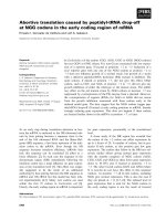
The only difference between the dependent and nondepend-
ent groups with similar PEEP was the position of the animal
(Figure 1). Animals were placed in the lateral decubitus posi-
tion, left lung up to measure the nondependent lung alveolar
mechanics (Groups II and IV) and left lung down to film the
dependent lung alveolar mechanics (Groups I and III) (Figure
1). In vivo microscopy was accomplished in the dependent
lung by rotating the microscope 180° so that the objective was
pointing up, and the microscope was positioned under the rat
and attached to the pleural surface (Figure 1).
Concomitant with the initiation of the injurious ventilator strat-
egy, the respiratory rate was set to 20 breaths/min in all
groups. Time zero was designated as the time immediately fol-
lowing initiation of the experimental ventilatory strategy. Hemo-
dynamic data, lung function data, and in vivo microscopic data
were recorded at baseline and every 15 minutes after initiation
of the experimental protocol. The protocol was terminated
after 90 minutes.
In vivo microscopy
A microscopic coverslip mounted on a ring was lowered onto
the pleural surface, and the lung was held in place by gentle
suction (≤5 cmH
2
O) at end inspiration for placement of an in
vivo videomicroscope (epi-objective microscope with epi-illu-
mination; Olympus America Inc. Melville, NY USA). At each
Available online />Page 3 of 10
(page number not for citation purposes)
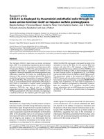
timepoint, the apparatus was reattached to the lung so that a
different cohort of alveoli was sampled every 15 minutes. The
microscope objective was moved from the top to the bottom
of the coverslip field by field, and each new field was photo-
graphed for the measurement of alveolar mechanics (Figure
2). Microscopic images of alveoli were viewed at a final mag-
nification of 130× with a color video camera (model CCD
SSC-S20; Sony, Tokyo, Japan) and recorded on Pinnacle Stu-
dio Plus software (Pagasus Imaging Corporation Tampa, FL)
Each field measured 1.22 × 10
6
μm
2
and was filmed through-
out five complete tidal ventilations for subsequent analysis of
alveolar mechanics.
Image analysis of alveoli
Frame-by-frame analysis was performed by capturing still
images of alveoli at peak inspiration and at end expiration. For
each visual field, the subset of alveoli analyzed consisted of
those that contacted a vertical line bisecting the visual field
and represented approximately 10 alveoli per field, the length
of that line measuring approximately 1 mm. Five microscopic
fields were analyzed for each animal at each timepoint (Figure
2). The outer walls of individual alveoli were manually traced at
both end inspiration and end expiration. The areas of these
tracings were computed with image analysis software (Empire
Imaging Systems; Image Pro, Syracuse, NY, USA) and are
referred to as the area at peak inspiration (I) and the area at
end expiration (E). The degree of alveolar stability (%I - EΔ)
was defined as the percentage decrease in alveolar size dur-
ing tidal ventilation:
%I - E
Δ
= 100 × [(I - E)/I]
For each animal at each timepoint, the mean I and the mean E
values were calculated, yielding a single value. These aggre-
gate values were then used in the statistical analysis. Similarly,
%I - EΔ was calculated for each individual alveolus, and the
mean %I - EΔ value for each animal at each timepoint was then
compared using standard statistics (see Statistical analysis).
Lung function measurements
Arterial blood gases, systemic arterial pressures, and pulmo-
nary parameters (tidal volume) were recorded at baseline and
then at 15-minute intervals. Pulmonary parameters were meas-
ured inline by the Galileo ventilator (Hamilton Medical Inc.).
Necropsy
The trachea was cannulated and the lung was inflated with
10% formalin by gravity to a pressure of 25 cmH
2
O. Each lung
was identified as a dependent lung or a nondependent lung
and was grouped for histological assessment. After 24 hours,
the tissue was blocked in paraffin and serial sections were
made for staining with H & E. The slides were reviewed at high
magnification for edema (400×).
Mechanism of alveolar collapse
Alveolar instability was caused in two additional rats by 30
minutes of injurious MV (peak inspiratory pressure (PIP) = 45
cmH
2
O, PEEP = 3 cmH
2
O), similar to injury in Group I and
Group II of this study. This injurious ventilation caused the alve-
olar mechanics of subpleural alveoli to change from stable
(that is, little to no change in size with ventilation) to unstable
(that is, very large change is size with tidal ventilation), deter-
mined by in vivo microscopy within 60 minutes of application.
Once unstable alveoli developed, the animals were sacrificed
and the lungs were removed en bloc and perfused through the
Figure 1
Schematic demonstrating in vivo videomicroscopy procedure for the nondependent and dependent lungSchematic demonstrating in vivo videomicroscopy procedure for the
nondependent and dependent lung. The right lung was filmed in all
groups (that is, dependent and nondependent lung and high and low
positive end-expiratory pressure). (a) To film the nondependent lung,
the rat was placed in the left lateral decubitus position and the micro-
scope was lowered from the top. (b) To film the dependent lung, the rat
was in the lateral decubitus position with an open chest and the micro-
scope was elevated from the bottom.
Figure 2
Alveolar sampling techniqueAlveolar sampling technique. The microscope objective was moved to
the top of the coverslip and the first field was filmed (F1). The objective
was than moved down one field, viewing all new alveoli. This was
sequentially repeated to the bottom of the coverslip, filming five entirely
different microscopic fields of alveoli.
Critical Care Vol 11 No 5 Pavone et al.
Page 4 of 10
(page number not for citation purposes)
pulmonary artery with 10% formalin at an intravascular pres-
sure of 20 cmH
2
O for 24 hours.
The lungs of one rat were inflated and held constant at an air-
way pressure of 45 cmH
2
O (when subpleural alveoli were
observed to be fully inflated with the in vivo microscope), and
the lungs of the second rat were fixed at an airway pressure of
3 cmH
2
O (when subpleural were observed to be mostly col-
lapsed with the in vivo microscope). Following 24 hours of fix-
ation at constant perfusion and airway pressure, the lungs
were blocked, sliced, and stained with H & E. These data were
used to identify the potential mechanism of alveolar collapse.
Vertebrate animals
Experiments described in this study were performed in
accordance with the National Institutes of Health guidelines
for the use of experimental animals in research. The protocol
was approved by the Committee for the Humane Use of Ani-
mals at our institution.
Statistical analysis
All results are presented as the mean ± standard error of the
mean. An all-pairs, Tukey HSD (honestly significantly different)
test was used to compare more than two groups. Student's t
test was applied for all pair-wise comparisons. We accepted
P < 0.05 as significant. Data were analyzed using JPM soft-
ware (version 5; SAS Institute Cary, NC, USA).
Results
Alveolar mechanics
Normal alveoli before injurious ventilation are very stable, and
they did not change size appreciably during tidal ventilation
(Additional file 1). Injurious MV caused alveolar instability
faster (60 minutes) in the nondependent + low PEEP group
(Figure 3 and Additional file 2) as compared with the depend-
ent + low PEEP group (Figure 3 and Additional file 3). By 75
minutes, however, the %I - EΔ was no longer different
between these groups although it trended higher in the non-
dependent + low PEEP group. The addition of 10 cmH
2
O
PEEP prevented the development of alveolar instability for the
entire experiment in both the nondependent and dependent
lungs (Figures 3 and 4, and Additional file 4)
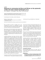
Mechanism of alveolar collapse
At 45 cmH
2
O airway pressure (PIP) most alveoli in the in vivo
microscopic field are inflated (Figure 5a,c), and at 3 cmH
2
O
(PEEP) most alveoli collapsed (Figure 5b,d). Alveoli at the PIP
are inflated and fill the in vivo microscopic field (Figure 5a, dot-
ted line surrounds representative alveoli), and the alveolar
walls are very thin (Figure 5c, arrows). At the PEEP the subp-
leural alveoli collapse (Figure 5b, dark atelectatic areas identi-
fied by arrows), and the alveolar walls are thickened (Figure
5d, arrows). The thickened alveolar walls suggest that alveolar
collapse is by folding of the alveolar walls [14].
Blood gases
The arterial PO
2
and PCO
2
were not significantly different in
the low PEEP versus the high PEEP groups (Table 1) even
though alveoli were unstable only in the low PEEP groups (Fig-
ures 3 and 4). There were no significance changes in intra-
Figure 3
Change in alveolar stability over timeChange in alveolar stability over time. The change in alveolar stability
(inspiration–expiration percentage change, %I - E) was monitored over
time in four groups: Group I, dependent + low positive end-expiratory
pressure (PEEP) (n = 5); Group II, nondependent + low PEEP (n = 4);
Group III, dependent + high PEEP (n = 5); and Group IV, nondepend-
ent + high PEEP (n = 5). Data are the mean ± standard error.
#
P <
0.05, Group IIversus Groups III and IV; *P < 0.05, Group II versus
Group I.
Figure 4
Alveolar stability at 60 minutesAlveolar stability at 60 minutes. The degree of alveolar stability (inspira-
tion–expiration percentage change, %I - E) was monitored at 60 min-
utes in four groups: Group I, dependent + low positive end-expiratory
pressure (PEEP) (n = 5); Group II, nondependent + low PEEP (n = 4);
Group III, dependent + high PEEP (n = 5); and Group IV, nondepend-
ent + high PEEP (n = 5). Data are the mean ± standard error There is
greatest instability in Group II, nondependent + minimal PEEP. Group
III and Group IV have a PEEP of 10 cmH
2
O applied.
Available online />Page 5 of 10
(page number not for citation purposes)
alveolar edema or in interstitial edema between groups (Table
2).
Lung function
There was a significantly smaller tidal volume in the PEEP 10
cmH
2
O groups compared with the PEEP 3 cmH
2
O groups.
There was no significant difference in lung compliance or
mean arterial pressure at 90 minutes between groups. There
were no differences in intravenous fluid resuscitation between
groups.
Discussion
The four most important findings from this study are the follow-
ing: 1) the development of alveolar injury, assessed by alveolar
stability, occurred earlier following initiation of injurious ventila-
tion in the nondependent lung with low PEEP as compared
with the dependent lung with low PEEP. 2) increasing the
PEEP to 10 cmH
2
O prevented alveolar instability in both the
nondependent and dependent lung areas. 3) alveolar instabil-
ity was not correlated with a decrease in PO
2
. 4) preventing
alveolar instability with PEEP did not decrease the pulmonary
edema. To our knowledge, the present study is the first to
show that the position of the normal lung can influence the
development of abnormal alveolar mechanics secondary to
injurious MV. It is tempting to use these results and to hypoth-
esize on the impact of the body position and VILI in humans,
but extreme caution must be taken when extrapolating data
from a rodent experiment into a human scenario.
Although it is beyond the scope of this paper to discuss in
detail normal and abnormal alveolar mechanics (that is, the
dynamic change in alveolar size and shape with tidal ventila-
tion), it is important to understand that normal alveoli do not
change size during tidal ventilation by expanding and contract-
ing like a balloon in order to appreciate the significance of our
experimental results. There are several excellent reviews on
this subject [15,16] but a brief overview is as follows. The
laboratory of Gil and colleagues produced the first high-quality
experiments demonstrating the possibility that there may be
many mechanisms by which the alveolar volume changed
during ventilation [17,18]. Their experiments lead them to
hypothesize that the lung volume change could be due to
expansion and contraction of the alveolar ducts with little
change in alveolar volume, could be due to successive alveolar
recruitment/derecruitment, could be due to alveolar crumpling
and uncrumpling (like a paper bag), and could be due to pleat-
ing and unpleating of alveolar corners.
More recent experiments have all demonstrated that alveoli do
not expand and contract like balloons. Carney and colleagues
studied lung inflation from the residual volume to 80% of the
total lung capacity and found that alveoli do not change size
appreciably even during large changes in lung volume; they
concluded that the lung volume change is by alveolar recruit-
ment and derecruitment [15]. These data were confirmed by
Escolar and colleagues, using a sophisticated morphometric
analysis, who demonstrated that there is little change in alveo-
Figure 5
Comparison of abnormal alveoli at peak inspiration and end expirationComparison of abnormal alveoli at peak inspiration and end expiration. Abnormal alveoli at peak inspiration and end expiration as seen with an in vivo
microscope (a, b) and as a histologic comparison (c, d). (a) Normal alveoli fill the microscopic field at peak inspiration, and (b) collapse during expi-
ration. (c) Alveolar walls are very thin at peak inspiration, and (d) become thickened at end expiration.
Critical Care Vol 11 No 5 Pavone et al.
Page 6 of 10
(page number not for citation purposes)
lar size during ventilation but there is a significant change in
alveolar number [19,20].
It is also possible that the lung volume change is due to
changes in the size of the alveolar mouth and duct. Kitaoka and
colleagues have designed a working four-dimensional model
of an alveolus and alveolar duct in which the major change in
volume is due to opening and closing of the alveolar mouth
[16]. The example movie (Additional file 5) demonstrates that
the vast majority of the size change that occurs in a single
alveolus during ventilation could be due to changes in the size
of the alveolar mouth. As the size of the mouth of all alveoli
comprising an air sac concomitantly open and close, the size
of the alveolar duct changes size greatly; it is the expansion
and contraction of the alveolar duct, not of the alveolus, that
occurs during ventilation in the normal lung [16].
There is a potential artifact in our experimental technique. It is
possible that the suction prevents normal pleural expansion
and contraction, and thus prevents healthy alveoli from chang-
Table 1
Lung and hemodynamic parameters
Baseline 15 minutes 30 minutes 45 minutes 60 minutes 75 minutes 90 minutes
Ventilation positive end-expiratory pressure 10 cmH
2
O (n = 10)
PCO
2
32.5 ± 4.40 35.7 ± 4.38 32.6 ± 4.73 31.2 ± 4.26* 28.2 ± 4.67 26.5 ± 4.21 24 ± 4.39
PO
2
239.6 ± 15.44 293.1 ± 17.18 300.6 ± 10.66 294.5 ± 17.62 292.5 ± 21.46 331.7 ± 1.79 333.8 ± 14.23
Tidal volume (ml) 6.2 ± 0.55 3.8 ± 1.06* 3.1 ± 1.08* 3.1 ± 1.08* 2.4 ± 1.02* 2.5 ± 1.05* 2.5 ± 1.07*
Lung static
compliance
(ml/cmH
2
O)
0.5 ± 0.03 0.19 ± 0.03* 0.53 ± 0.29 0.47 ± 0.23 0.35 ± 0.09 0.34 ± 0.09 0.61 ± 0.34
Mean arterial
pressure (mmHg)
88.5 ± 6.86 93.6 ± 14.90 87.1 ± 11.48 77.2 ± 10.75 77.8 ± 10.76 77.9 ± 10.36 58.1 ± 8.91
Fluid total
a
9.9 ± 2.88
Ventilation positive end-expiratory pressure 3 cmH
2
O (n = 9)
PCO
2
31 ± 3.67 26.4 ± 4.27 22.5 ± 2.17 17.8 ± 1.82 17.4 ± 2.34 18 ± 2.40 17.25 ± 3.26
PO
2
228.5 ± 24.91 293.4 ± 18.33 302.2 ± 17.62 289 ± 18.33 296.4 ± 20.85 290.78 ± 26.85 308.4 ± 32.11
Tidal volume (ml) 6.9 ± 1.53 11.5 ± 1.01 11.9 ± 1.37 12.3 ± 1.16 12.1 ± 1.18 12.9 ± 1.25 11.7 ± 1.33
Lung static
compliance
(ml/cmH
2
O)
0.47 ± 0.04 0.34 ± 0.02 0.32 ± 0.01 0.6 ± 0.27 0.74 ± 0.42 0.57 ± 0.25 0.53 ± 0.23
Mean arterial
pressure (mmHg)
88.2 ± 8.42 78.5 ± 6.81 83.1 ± 6.81 76.4 ± 4.36 83.1 ± 9.15 82.9 ± 9.21 76.4 ± 8.12
Fluid total
a
9.8 ± 2.74
a
Total amount of normal saline infused over the entire experiment (ml). *P < 0.05 between groups.
Table 2
Pulmonary edema assessed by histological measurement of intra-alveolar edema and interstitial (alveolar wall thickness) edema
Nondependent lung Dependent lung
Positive end-expiratory pressure 10 cmH
2
O
Intra-alveolar edema 3.22 ± 0.27 3.28 ± 0.25
Alveolar wall thickness 2.9 ± 0.42 2.72 ± 0.38
Positive end-expiratory pressure 3 cmH
2
O
Intra-alveolar edema 3.5 ± 0.30 3.7 ± 0.09
Alveolar wall thickness 2.51 ± 0.45 2.62 ± 0.34
A score for both intra-alveolar and interstitial edema was used to measure edema in both nondependent and dependent lung sections: 0, no
edema; 1, mild scattered edema; 2, moderate scattered edema; 3, severe scattered edema; and 4, severe universal edema. Data presented as the
mean ± standard error of the mean. No significant difference was seen among groups.
Available online />Page 7 of 10
(page number not for citation purposes)
ing size normally with ventilation. There is evidence for this
occurring since the pleural surface changes size to the one-
third power of lung volume, and thus there must be either a
change in size of or in the number of alveoli to account for this
change. If this is true, than normal alveoli would be artificially
stabilized and this may account for the minimal alveolar size
change during tidal ventilation.
We believe, however, our microscopic technique was ade-
quate to answer the questions we asked in this paper. We
intended to demonstrate a change in alveolar mechanics from
normal to abnormal, understanding that there was a potential
alveolar-stabilizing artifact with our microscopic technique.
Our results clearly show a dramatic change in alveolar stability
from the normal to the injured, even if the microscopic prepa-
ration was preventing the full degree of alveolar volume
change. The absolute changes in alveolar size may therefore
not be totally accurate but the qualitative changes are very dra-
matic, allowing us to adequately answer our experimental
question and to test our hypothesis.
In summary, normal alveoli are very stable, with changes in
lung volume accommodated by normal alveolar recruitment
and derecruitment and/or changes in the size of the alveolar
mouth and duct. The unstable alveoli that develop 60 minutes
following injurious MV are pathologic and will exacerbate the
development of VILI [21]. The mechanism of this pathologic
alveolar collapse and re-expansion appears to be alveolar fold-
ing and unfolding (Figure 5).
VILI and body position
Our data are contrary to the findings of Nishimura and col-
leagues, who showed that lung injury was not gravity depend-
ent [22]. Using a closed-chest rabbit VILI model they found
that lung injury was not uniformly greatest in the dependent
portions of the lung. Nishimura and colleagues demonstrated
that lung injury was very regional but that the most severe
injury always occurred in the dorsal portion of the lung regard-
less of whether the dorsal lung was in the dependent or non-
dependent position. In contrast, our study showed that the
nondependent lung was the first to develop alveolar instability.
Nishimura and colleagues, however, did show that prone posi-
tion slowed the onset of atelectasis (VILI) [22], which supports
our finding that body position affects the rate at which VILI
develops.
Both of these studies suggest that VILI is not uniform through-
out the lung, but rather occurs preferentially in specific areas;
however, there is no consensus whether this specificity of
injury is due to the gravitational or anatomical position of the
lung. The reason for the discrepancy may involve the species
being studied (rat versus rabbit), or the tools used to measure
the injury (in vivo microscopy versus computed tomography
scan). It is possible that there was more injury in the dorsal por-
tions of the lung in our study, which could be not identified with
in vivo microscopy. Likewise, there may have been a gravity-
dependent increase in alveolar instability in Nishimura and col-
leagues' study that was not identified with the computed tom-
ography scan. Finally, our study looked at open-chest rats
whereas the Nishimura and colleagues study used closed-
chest rabbits. Perhaps the influence of the chest wall resist-
ance to inflation changed the location of injury in the two
models.
In addition, the interpretation of the computed tomography
scan has recently been called into question. Hubmayr sug-
gests that the increased density seen by computed tomogra-
phy scan in ARDS patients is caused by open alveoli flooded
with edema rather than by atelectasis [23]. Perhaps the dorsal
injury seen on the computed tomography scan occurs
regardless of whether the animal is in the prone or the supine
position because the anatomical shape of the rabbit lung
causes increased edema in that dorsal portion of the lung.
Alveolar instability and lung position
The lung can be described as an elastic sponge that is com-
pressed by its own weight, especially when edematous (that
is, nondependent lung compresses dependent lung), and by
the weight of other organs (that is, the heart). Albert and Hub-
mayr [24] confirmed by computed tomography scan in
humans that the heart compresses a significant amount of lung
tissue and that the prone position relieves much of this com-
pression. The weight of the nondependent lung and the heart
would cause the dependent lung to become less compliant
and would divert a larger percentage of the tidal volume into
the more compliant nondependent lung. Veldhuizen and col-
leagues have previously shown that large tidal volumes cause
pulmonary surfactant dysfunction [25,26]. The development of
alveolar instability in our VILI model was therefore probably
due to a large tidal volume-induced surfactant deactivation. In
addition, if a larger tidal volume was being delivered to the
more compliant nondependent lung, surfactant deactivation
would be exacerbated – which may explain why alveolar insta-
bility occurred more rapidly in the nondependent lung.
These findings have clinical significance since the amount of
healthy lung tissue is drastically reduced in ARDS [27], and
thus a 'normal' tidal volume might direct excessively large vol-
umes into the healthy tissue and cause VILI similar to that in
the present study. Indeed, it has been shown that smaller tidal
volumes significantly reduce mortality in ARDS patients [1].
Alveolar instability and PEEP
In this study, the addition of PEEP prevented repetitive recruit-
ment and derecruitment in both the nondependent and
dependent lung regions. Our study used a PEEP of 10
cmH
2
O, since it was previously shown in our laboratory by
Halter and colleagues that 10 cmH
2
O PEEP stabilized alveoli
following a recruitment maneuver [13]. These data support
those of Dreyfuss and colleagues that PEEP will reduce injury
Critical Care Vol 11 No 5 Pavone et al.
Page 8 of 10
(page number not for citation purposes)
to the normal lung ventilated with high volumes and peak pres-
sures [2,28]. Therefore it appears that it is not the high PIP that
causes VILI, but rather the large change in pressure from PIP
to the end-expiratory pressure that causes injury that ultimately
results in altered alveolar mechanics.
The mechanisms by which PEEP reduces VILI and stabilizes
alveoli are twofold: the increase in end-expiratory pressure
could prevent alveolar collapse, or the decreased tidal volume
when 10 cmH
2
O PEEP was applied could prevent alveolar
overdistension. Although either mechanism could be respon-
sible for the results in this paper, the literature supports the
concept of a large tidal volume-induced deactivation of pulmo-
nary surfactant causing alveolar instability [29]. We therefore
conclude that the most probable mechanism of PEEP-induced
alveolar stabilization is by prevention of alveolar collapse.
Our results are complex, however, since high PEEP prevented
alveolar instability but did not reduce pulmonary edema meas-
ured histologically. This suggests that PEEP prevents the
onset of mechanical VILI (that is, unstable alveoli) but not
inflammatory VILI (that is, injury secondary to sequestered neu-
trophils). Neutrophil-released proteases and reactive oxygen
species could cause an increase in vascular permeability with
resultant edema formation without alveolar instability. It is pos-
sible that if we had allowed the study to continue past 90 min-
utes, the combination of mechanical and inflammatory injury in
the low PEEP group would have caused more edema than that
in the lung with high PEEP and stable alveoli. Another explana-
tion for the increase in edema with high PEEP possibility is that
barotrauma occurred in the absence of alveolar instability due
to the high peak inflation pressure.
Mechanism of alveolar collapse
Lung histology was studied at the PIP and at the PEEP to
determine a potential mechanism of abnormal alveolar col-
lapse and re-expansion. We used the histological configura-
tion of the collapsed alveoli to speculate on the mechanism of
this collapse. Tschumperlin and colleagues found that the
alveolar walls were thickened at low airway pressure [14], very
similar to those in the present study fixed at 3 cmH
2
O (Figure
5c, arrows). Using electron microscopy they demonstrated
that the thickened alveolar walls were due to alveolar wall fold-
ing, and concluded that alveoli do not change size by balloon-
like expansion and contraction but rather by folding and
unfolding like a paper bag [14]. We conclude that the proba-
ble mechanism by which unstable alveoli collapse and expand
in the injured lung is not by balloon-like isotropic expansion,
but rather due to the folding of the alveolar walls.
Alveolar instability and arterial PO
2
Another interesting finding was that the arterial PO
2
was not
significantly reduced (actually it was slightly higher) in the low
PEEP group with abnormal, unstable alveolar as compared
with that in the high PEEP ventilation group with normal, stable
alveoli.
The present study clearly demonstrated that alveoli in the low
PEEP group were unstable, and we know from previous stud-
ies that alveolar instability leads to VILI if alveoli are unstable
for 3–4 hours [5,12]. A normal arterial PO
2
does not therefore
necessarily identify a healthy lung with normal alveolar
mechanics, and nor does it identify a lung that is not being sub-
jected to mechanical VILI.
We postulate that the arterial PO
2
remained elevated in our
study even with unstable alveoli because oxygen was
exchanged during the portion of the ventilatory cycle in which
the unstable alveoli are inflated. This hypothesis was
supported by Pfeiffer and colleagues, who demonstrated a
cyclic change in arterial PO
2
utilizing an ultrafast inline PO
2
sensor [11]. The arterial PO
2
in these studies fluctuated with
each breath in an animal ARDS model with unstable alveoli.
The arterial PO
2
can therefore be maintained if the PIP is high
enough to open most of the alveoli during inflation. Forcing
collapsed alveoli open to improve the PO
2
, however, will
greatly increase lung injury since alveolar recruitment/dere-
cruitment is one of the primary mechanisms of VILI. These data
can loosely be extrapolated to the bedside, and would suggest
that it might be possible to normalize PO
2
by increasing the air-
way pressure, but at the expense of causing a significant VILI.
Critique of methodology
Our microscope has a limited depth of field (70 μm), and
therefore only allows for alveolar analysis in two dimensions.
Also, the subpleural alveolar mechanics might still differ from
those within the lung parenchyma. Subpleural alveoli have less
structural support since these alveoli are not surrounded on all
sides by adjacent alveoli (that is, one wall of a subpleural alve-
olus is attached to the visceral pleura rather than to another
alveolus). This anatomic arrangement may lessen the struc-
tural support provided by alveolar interdependence, causing
subpleural alveoli to become unstable sooner than those
within the lung. A classic paper by Mead and colleagues
showed that even if not surrounded by alveoli on all sides,
there is still a significant structural interdependence between
alveoli [30].
The suction that stabilizes the lung tissue on the cover slip
might prevent normal pleural expansion and contraction, and
thus may prevent healthy alveoli from changing size normally
with ventilation. Although we have not totally eliminated this
possibility, we have shown in a previous study that suction
slightly but significantly increased both the alveolar size and
stability. These changes were very subtle, with an alveolar size
change from expiration to inspiration being 1.1% in the suction
group increasing to 8.3% in the nonsuction group [21]. This
slight change in alveolar size with ventilation even without suc-
tion was in stark contrast to the dramatic change in alveolar
Available online />Page 9 of 10
(page number not for citation purposes)
size (for example, total collapse at end expiration or 100%
change in size) that occurred following prolonged exposure to
injurious MV. Suction therefore does not seem to artificially
stabilize normal alveoli nor does it prevent alveoli from becom-
ing unstable following injury.
Finally, the fact that we must open the chest to obtain our in
vivo microscopy may alter the way that normal and injured
alveoli behave mechanically.
Conclusion
Injurious MV, over time, will cause damage to pulmonary alve-
oli, significantly altering their mechanics of ventilation. The
mechanism of injury is probably a combination of tissue dam-
age leading to alveolar flooding and deactivation of pulmonary
surfactant by both direct mechanisms (large tidal volumes
have been shown to deactivate surfactant) and indirect mech-
anisms (surfactant being washed off of the alveolar surface by
edema fluid and deactivated by plasma proteins). Surfactant
loss results in alveolar instability during ventilation. In the
present study we demonstrated that the body position affects
the timing of injurious MV-induced alveolar instability. We pos-
tulate that the normal dependent lung was less compliant than
the nondependent lung, and thus received a smaller percent-
age of the total tidal volume; the larger tidal volume delivered
to the nondependent lung was the cause of a more rapid injury
(that is, alveolar instability). These data support the concept of
volutrauma occurring in normal areas of the heterogeneously
injured lung of ARDS patients. The arterial PO
2
is not a good
indicator of alveolar stability, and thus the PO
2
alone would not
be appropriate to identify protective MV strategies.
Competing interests
The authors declare that they have no competing interests.
Authors' contributions
LP conducted the experiments, and analyzed and graphed the
data. SA contributed to manuscript writing and editing, and to
data analysis. JD assisted LP in conducting the experiments
and analyzing the data. LG contributed to the experimental
design, data analysis and interpretation, and performed the
histologic analysis. GN contributed to the design and
development of the protocol, to data analysis and interpreta-
tion, and to writing of the manuscript.
Additional files
Key messages
• Nondependent regions of the normal lung are the first
to develop alveolar instability when ventilated with high
PIP and low PEEP.
• Alveolar instability occurs without significant differences
in lung edema.
• The addition of PEEP prevents high peak-pressure-
induced alveolar instability but not the increase in pul-
monary edema.
• Oxygenation is not an effective indicator of alveolar
instability or of VILI.
The following Additional files are available online:
Additional file 1
A Windows media player file containing a movie showing
normal alveoli ventilated at a P
control
of 14 cmH
2
O and a
PEEP of 3 cmH
2
O. Individual alveoli fill the microscopic
field and do not change size appreciably with ventilation.
Note the blood flowing around and over the alveoli.
See />supplementary/cc6122-S1.mpg
Additional file 2
A Windows media player file containing a movie showing
alveolar instability in the nondependent low PEEP group
60 minutes following injurious ventilation. At end
expiration there is a great deal of atelectasis, which
appears as dark-red areas without the presence of
alveolar structures. During inspiration, the collapsed
alveoli reach the critical opening pressure and 'pop'
open. When the critical closing pressure is reached
during exhalation, the alveoli collapse. The mechanism of
this collapse and re-expansion appears to be by alveolar
folding and unfolding (Figure 5).
See />supplementary/cc6122-S2.mpg
Additional file 3
A Windows media player file containing a movie showing
that alveoli are stable and appear normal (Additional file
1) in the dependent lung with low PEEP 60 minutes
following injurious ventilation.
See />supplementary/cc6122-S3.mpg
Additional file 4
A Windows media player file containing a movie showing
that alveoli are stable and appear normal (Additional file
1) with a high PEEP 90 minutes following injurious
ventilation.
See />supplementary/cc6122-S4.mpg
Critical Care Vol 11 No 5 Pavone et al.
Page 10 of 10
(page number not for citation purposes)
Acknowledgements
The authors would like to thank Kathy Snyder for her expert technical
assistance.
References
1. Acute Respiratory Distress Syndrome Network: Ventilation with
lower tidal volumes as compared with traditional tidal volumes
for acute lung injury and the acute respiratory distress
syndrome. N Engl J Med 2000, 342:1301-1308.
2. Dreyfuss D, Saumon G: Ventilator-induced lung injury: lessons
from experimental studies. Am J Respir Crit Care Med 1998,
157:294-323.
3. Gajic O, Dara SI, Mendez JL, Adesanya AO, Festic E, Caples SM,
Rana R, St Sauver JL, Lymp JF, Afessa B, Hubmayr RD: Ventilator-
associated lung injury in patients without acute lung injury at
the onset of mechanical ventilation. Crit Care Med 2004,
32:1817-1824.
4. Rubenfeld GD, Caldwell E, Peabody E, Weaver J, Martin DP, Neff
M, Stern EJ, Hudson LD: Incidence and outcomes of acute lung
injury. N Engl J Med 2005, 353:1685-1693.
5. Steinberg JM, Schiller HJ, Halter JM, Gatto LA, Lee HM, Pavone
LA, Nieman GF: Alveolar instability causes early ventilator-
induced lung injury independent of neutrophils. Am J Respir
Crit Care Med 2004, 169:57-63.
6. Gattinoni L, Carlesso E, Cadringher P, Valenza F, Vagginelli F,
Chiumello D: Physical and biological triggers of ventilator-
induced lung injury and its prevention. Eur Respir J 2003,
47(Suppl):15s-25s.
7. Schreiber T, Niemann C, Schmidt B, Karzai W: A novel model of
selective lung ventilation to investigate the long-term effects
of ventilation-induced lung injury. Shock 2006, 26:50-54.
8. Su F, Nguyen ND, Creteur J, Cai Y, Nagy N, Anh-Dung H, Amaral
A, Bruzzi de Carvalho F, Chochrad D, Vincent JL: Use of low tidal
volume in septic shock may decrease severity of subsequent
acute lung injury. Shock 2004, 22:145-150.
9. Carney D, DiRocco J, Nieman G: Dynamic alveolar mechanics
and ventilator-induced lung injury. Crit Care Med 2005,
33:S122-S128.
10. DiRocco JD, Pavone LA, Carney DE, Lutz CJ, Gatto LA, Landas
SK, Nieman GF: Dynamic alveolar mechanics in four models of
lung injury. Intensive Care Med 2006, 32:140-148.
11. Pfeiffer B, Syring RS, Markstaller K, Otto CM, Baumgardner JE:
The implications of arterial PO
2
oscillations for conventional
arterial blood gas analysis. Anesth Analg 2006,
102:1758-1764.
12. Halter JM, Steinberg JM, Gatto LA, Dirocco JD, Pavone LA, Schiller
HJ, Albert S, Lee HM, Carney DE, Nieman GF: Effect of positive
end-expiratory pressure and tidal volume on alveolar instabil-
ity-induced lung injury. Crit Care 2007, 11:R20.
13. Halter JM, Steinberg JM, Schiller HJ, DaSilva M, Gatto LA, Landas
S, Nieman GF: Positive end-expiratory pressure after a recruit-
ment maneuver prevents both alveolar collapse and recruit-
ment/derecruitment. Am J Respir Crit Care Med 2003,
167:1620-1626.
14. Tschumperlin DJ, Margulies SS: Alveolar epithelial surface
area–volume relationship in isolated rat lungs. J Appl Physiol
1999, 86:2026-2033.
15. Carney DE, Bredenberg CE, Schiller HJ, Picone AL, McCann UG,
Gatto LA, Bailey G, Fillinger M, Nieman GF: The mechanism of
lung volume change during mechanical ventilation. Am J
Respir Crit Care Med 1999, 160:1697-1702.
16. Kitaoka H, Nieman GF, Fujino Y, Carney D, Dirocco J, Kawase I: A
4-dimensional model of the alveolar structure. J Physiol Sci
2007, 57:175-185.
17. Gil J, Bachofen H, Gehr P, Weibel ER: Alveolar volume–surface
area relation in air- and saline-filled lungs fixed by vascular
perfusion. J Appl Physiol 1979, 47:990-1001.
18. Gil J, Weibel ER: Morphological study of pressure-volume hys-
teresis in rat lungs fixed by vascular perfusion. Respir Physiol
1972, 15:190-213.
19. Escolar JD, Escolar A: Lung hysteresis: a morphological view.
Histol Histopathol 2004, 19:159-166.
20. Escolar JD, Escolar MA, Guzman J, Roques M: Morphological
hysteresis of the small airways. Histol Histopathol 2003,
18:19-26.
21. Pavone LA, Albert S, Carney D, Gatto LA, Halter JM, Nieman GF:
Injurious mechanical ventilation in the normal lung causes a
progressive pathologic change in dynamic alveolar
mechanics. Crit Care 2007, 11:R64.
22. Nishimura M, Honda O, Tomiyama N, Johkoh T, Kagawa K, Nishida
T:
Body position does not influence the location of ventilator-
induced lung injury. Intensive Care Med 2000, 26:1664-1669.
23. Hubmayr RD: Perspective on lung injury and recruitment: a
skeptical look at the opening and collapse story. Am J Respir
Crit Care Med 2002, 165:1647-1653.
24. Albert RK, Hubmayr RD: The prone position eliminates com-
pression of the lungs by the heart. Am J Respir Crit Care Med
2000, 161:1660-1665.
25. Veldhuizen RA, Tremblay LN, Govindarajan A, van Rozendaal BA,
Haagsman HP, Slutsky AS: Pulmonary surfactant is altered dur-
ing mechanical ventilation of isolated rat lung. Crit Care Med
2000, 28:2545-2551.
26. Veldhuizen RA, Welk B, Harbottle R, Hearn S, Nag K, Petersen N,
Possmayer F: Mechanical ventilation of isolated rat lungs
changes the structure and biophysical properties of
surfactant. J Appl Physiol 2002, 92:1169-1175.
27. Gattinoni L, Pesenti A: The concept of 'baby lung'. Intensive
Care Med 2005, 31:776-784.
28. Dreyfuss D, Soler P, Basset G, Saumon G: High inflation pres-
sure pulmonary edema. Respective effects of high airway
pressure, high tidal volume, and positive end-expiratory
pressure. Am Rev Respir Dis 1988, 137:1159-1164.
29. Verbrugge SJ, Bohm SH, Gommers D, Zimmerman LJ, Lachmann
B: Surfactant impairment after mechanical ventilation with
large alveolar surface area changes and effects of positive
end-expiratory pressure. Br J Anaesth 1998, 80:360-364.
30. Mead J, Takishima T, Leith D: Stress distribution in lungs: a
model of pulmonary elasticity. J Appl Physiol 1970,
28:596-608.
Additional file 5
A Windows media player file containing a movie showing
a computer-assisted design rendition of the three-
dimensional changes in alveolar volume over time
(addition of the time element creates a four-dimensional
representation). The alveolar mouth is highlighted in red.
Note the large change in the size of the mouth and the
minimal changes in the size of the other portions of the
alveolus. When functioning together in an air sac, the
change in alveolar mouth size results in a large change in
the size of the alveolar duct [16].
See />supplementary/cc6122-S5.avi