Báo cáo y học: " Cellular turnover and expression of hypoxic-inducible factor in acute acalculous and calculous cholecystitis" ppt
Bạn đang xem bản rút gọn của tài liệu. Xem và tải ngay bản đầy đủ của tài liệu tại đây (3.55 MB, 6 trang )
Open Access
Available online />Page 1 of 6
(page number not for citation purposes)
Vol 11 No 5
Research
Cellular turnover and expression of hypoxic-inducible factor in
acute acalculous and calculous cholecystitis
Merja Vakkala
1
, Jouko J Laurila
1
, Juha Saarnio
2
, Vesa Koivukangas
2
, Hannu Syrjälä
3
,
Tuomo Karttunen
4
, Ylermi Soini
4
and Tero I Ala-Kokko
1
1
Department of Anesthesiology, Division of Intensive Care, Oulu University Hospital, Kajaanintie 52, Oulu, Finland, FIN-90029
2
Department of Surgery, Oulu University Hospital, Kajaanintie 52, Oulu, Finland, FIN-90029
3
Department of Infection Control, Oulu University Hospital, Kajaanintie 52, Oulu, Finland, FIN-90029
4
Department of Pathology, Oulu University, Kajaanintie 50, Oulu, Finland, FIN-90029
Corresponding author: Tero I Ala-Kokko,
Received: 24 Aug 2007 Revisions requested: 24 Oct 2007 Revisions received: 31 Oct 2007 Accepted: 31 Oct 2007 Published: 31 Oct 2007
Critical Care 2007, 11:R116 (doi:10.1186/cc6170)
This article is online at: />© 2007 Vakkala et al.; licensee BioMed Central Ltd.
This is an open access article distributed under the terms of the Creative Commons Attribution License ( />),
which permits unrestricted use, distribution, and reproduction in any medium, provided the original work is properly cited.
Abstract
Introduction Epithelial corrective and destructive mechanisms
have not been studied in inflammatory gallbladder disease.
Methods Epithelial apoptosis, cell proliferation and expression
of hypoxia-inducible factor (HIF)-1α were compared in
gallbladders from patients with acute acalculous cholecystitis
(AAC; n = 30) and acute calculous cholecystitis (ACC; n = 21),
and from patients undergoing surgery for other reasons (normal
gallbladders; n = 9), which were removed during open
cholecystectomy. The immunohistochemical stains included
antibodies to Ki-67 (proliferation), M30 (apoptosis) and HIF-1α.
Proliferation and apoptosis were expressed as percentages of
positive cells. HIF-1α expression was expressed as absent,
weak, or strong.
Results Apoptosis (median [25th to 75th percentile]) was
significantly increased in AAC (1.31% [0.75% to 1.8%], P <
0.001) and ACC (1.10% [0.63% to 1.64%], P = 0.001),
compared with control samples (0.20% [0.07% to 0.45%]. The
proliferation rate was significantly increased in AAC (8.0%
[4.0% to 17.0%], P < 0.001) and ACC (14% [7.5% to 26.5%],
P = 0.001) compared with control samples (1.0% [1.0% to
3.0%]). Strong HIF-1α staining was observed in 57% of AAC,
in 100% of ACC and in 44% of control specimens (P < 0.001).
Intense HIF-1α expression was associated with increased cell
proliferation (P = 0.002).
Conclusion Cell proliferation and apoptosis were increased in
AAC and ACC, as compared with normal gallbladders.
Expression of HIF-1α was lower in AAC than in ACC.
Introduction
Acute acalculous cholecystitis (AAC) is an acute inflammation
of the gallbladder in the absence of gallstones. It has been
diagnosed with increasing frequency in critically ill patients [1-
5]. Systemic inflammatory response and disturbances in
splanchnic circulation combined with visceral hypoperfusion,
and ischaemia-reperfusion injury are assumed to play impor-
tant roles in the pathogenesis of AAC [6,7]. AAC has also
been shown to be associated with multiple organ dysfunction
syndrome [6,8]. In contrast, the more common form of acute
cholecystitis, namely acute calculous cholecystitis (ACC), is
caused by gallstones, which lead to occlusion, distension,
oedema, bile stasis and often bacterial infection of the gall-
bladder [9,10].
Epithelial integrity depends on cell proliferation and cell
destruction. The mucosal cell proliferation rate has previously
been studied in normal gallbladder mucosa [11]. It has been
reported to be low and comparable to that in normal colorectal
mucosa [12]. There are no data on the epithelial proliferation
rate or apoptosis in inflammatory conditions involving the gall-
bladder. Apoptosis is (at least in rat intestinal epithelium) the
major mode of cell death in ischaemia and ischaemia-reper-
fusion [13]. Apoptosis is a regulated process, and it is medi-
ated by a sequential cascade of intracellular enzymes [14].
Hypoxia-inducible factor (HIF)-1 is a key factor in the regula-
tion of epithelial integrity [15]. It is a transcription factor that
regulates the pathophysiological response to hypoxia and
AAC = acute acalculous cholecystitis; ACC = acute calculous cholecystitis; HIF = hypoxia-inducible factor; ICU = intensive care unit; SD = standard
deviation.
Critical Care Vol 11 No 5 Vakkala et al.
Page 2 of 6
(page number not for citation purposes)
ischaemia by increasing the transcription of various proteins
that are involved in angiogenesis, glycolysis, erythropoiesis
and cell survival, ensuring cellular function in low-oxygen con-
ditions [16-18]. HIF-1 consists of a constitutively expressed
subunit (HIF-1β) and an oxygen-regulated subunit (HIF-1α; or
its paralogs HIF-2α and HIF-3α) [15]. No studies of HIF-1
expression in normal or inflammatory gallbladder mucosa have
yet been reported. HIF-1 is involved in ischaemia-reperfusion
and tumour growth, and its expression is regulated by hypoxia
and cytokines or nitric oxide [19,20].
Because the epithelial corrective and destructive mechanisms
have not been studied in inflammatory gallbladder disease, we
were interested in determining whether these phenomena dif-
fer in the main patterns of acute cholecystitis, namely AAC and
ACC. Thus, expression of HIF-1α and markers of apoptosis
and cell proliferation were compared in these entities and nor-
mal gallbladders to elucidate the pathogenesis of these
conditions.
Materials and methods
Patients
This study was approved by the Ethics Committee of Oulu Uni-
versity Hospital, and informed consent was not required
because the data had been collected for clinical purposes and
no additional interventions were done. During the years 2000
to 2001, 39 of the 3,984 intensive care unit (ICU) patients
treated in this hospital underwent cholecystectomy because
of AAC during their ICU stay. The operative finding was necro-
sis and gangrene in the gallbladder wall in 17 patients (44%)
and a thickened gallbladder wall in 22 patients (56%). A
detailed report of the clinical and diagnostic features and the
outcomes of these 39 patients was published previously [3].
The basic histopathology, including assessment of epithelial
necrosis, and epithelial detachment of these gallbladders
were also previously described [10].
The AAC group in the present study included 30 randomly
chosen patients out of this series of 39. The age (mean ±
standard deviation [SD]) of these patients was 60 ± 12.5
years and 19 out of 30 were men. Severity of illness scores on
admission (mean ± SD) were as follows: Acute Physiology
and Chronic Health Evaluation score II 23.6 ± 6.1, Simplified
Acute Physiology Score II 47.2 ± 12.3 and Sequential Organ
Failure Assessment score 9.6 ± 3.5. Sepsis (10/30), cardio-
vascular surgery (8/30) and pneumonia (5/30) were the most
common admission diagnoses. The median (25th to 75th per-
centile) length of ICU stay before cholecystectomy was 7.5
days (2.8 to 15.3 days). Sixteen patients (53.3%) had three or
more failing organs at the time of cholecystectomy. Two of the
30 bile samples (6.7%) taken during cholecystectomy were
positive for bacterial growth. Hospital mortality was 36.6%
(11/30).
The ACC group included 21 consecutive patients undergoing
surgery at our hospital during the years 2000 and 2001. An
operative finding of an inflamed gallbladder with gallstones
was used as the inclusion criterion. All of these patients were
admitted to the hospital because of ACC, and none was
treated in the ICU. The median (25th to 75th percentile) time
from onset of symptoms to surgery was 3 days (2 to 4.5 days),
and the median (25th to 75th percentile) time from admission
to the hospital to surgery was 2 days (1 to 2 days). The age of
these patients (mean ± SD) was 57.9 ± 10.3 years, and six out
of 21 were men. Nine out of 16 bile cultures were positive for
bacterial growth (56%).
The control group included nine samples taken from normal-
looking gallbladders removed during pancreatic tumour resec-
tion. These patients had local disease remote from the gall-
bladder, and they did not have a history of biliary obstruction.
The age (mean ± SD) of the patients in the control group was
59.1 ± 17.8 years, and three out of nine were men.
Immunohistochemical staining
The gallbladder samples were fixed in neutral buffered formalin
and embedded in paraffin. Sections (5 μm) were cut from the
specimens and placed on glass slides. The sections were
stained in haematoxylin and eosin for conventional histopatho-
logical diagnosis [10]. The immunohistochemical analyses
were conducted in accordance with the manufacturer's rec-
ommendations, and they consisted of Ki-67 (proliferation), M-
30 (apoptosis) and HIF-1α antibodies. For Ki-67 and M30
stainings, the slides were pretreated with Tris/EDTA (pH 9; 15
minutes). The primary antibodies consisted of a mouse mono-
clonal Ki-67 antibody (NCL-Ki67-MMl, dilution 1:100, incuba-
tion time 30 minutes; Novocastra Laboratories Ltd, Newcastle
upon Tyne, UK) and a mouse monoclonal M30 CytoDEATH™
antibody (dilution 1:1,000, incubation time 30 minutes;
Roche, Mannheim, Germany). With these two antibodies, the
EnVisio kit (Dako, Glostrup, Denmark) was used and the col-
our was developed with diaminobenzidine. Pretreatment for
HIF-1α consisted of 10 mmol/l sodium citrate (pH 6; 10 min-
utes). The primary antibody was mouse monoclonal HIF-1α
antibody (dilution 1:50, at +4°C overnight; Neomarkers, Lab-
Vision Corporation, Fremont, CA, USA). The Power Vision kit
(Immunovision Technologies, Brisbane, CA, USA) was used
for detection, and the colour was developed with
diaminobenzidine.
Evaluation of immunohistochemical staining reactions
Ki-67 positivity was expressed as a nuclear staining pattern.
Positive cells were counted in approximately 450 epithelial
cells in each sample. The Ki-67 index was expressed as the
percentage of Ki-67-positive cells.
M30 detects apoptosis in epithelial cells and is expressed as
a cytoplasmic staining pattern. Apoptotic activity, expressed
as a M30 index, was determined as the percentage of M30-
Available online />Page 3 of 6
(page number not for citation purposes)
positive epithelial cells in 10 high-power fields (HPFs) with
40× objective.
HIF-1α expression was assessed in both the cytoplasm and
the nucleus by calculating the percentage of positively stained
cells. The HIF-1α staining reaction was expressed as absent,
weak, or strong, based on the intensity and extent of cytoplas-
mic and nuclear staining as follows: complete absence of HIF
reactivity (score = 0); cytoplasmic reactivity in fewer than 50%
of epithelial cells and/or nuclear expression in sporadic cells
(<10% of epithelial cells) (score = 1); and cytoplasmic expres-
sion in more than 50% of epithelial cells and/or nuclear
expression in more than 10% of epithelial cells (score = 2).
All assessments were made by two investigators experienced
in immunohistochemistry (MV and either YS or TK).
Statistical analysis
SPSS 13.0 for Windows (SPSS Inc., Chicago, IL, USA) was
used for statistical analysis. The normality of distribution was
assessed using the Kolmogorov-Smirnov test. The data are
expressed as percentage, as mean ± SD in the case of nor-
mally distributed data, and as median (25th to 75th percentile)
in the case of non-normally distributed data. Categorical data
were analyzed using Fisher's exact test. The Kruskal-Wallis
test was used to describe the differences in the percentage of
positively stained epithelial cells between the AAC, ACC and
control groups. The Mann-Whitney U-test was applied to ana-
lyze the differences between two groups (AAC versus control,
ACC versus control and AAC versus ACC). Differences were
considered significant at P < 0.05.
Results
Immunoreactivity in AAC, ACC and control samples is shown
in Figure 1. Apoptosis (M30 index) was significantly increased
in both AAC (1.31% [0.75% to 1.80%], P < 0.001) and ACC
patients (1.10% [0.63% to 1.64%], P = 0.001), as compared
with control individuals (0.20% [0.07% to 0.45%]. However,
the difference in extent of apoptosis between the AAC and
ACC groups was not statistically significant (P = 0.50).
Proliferation rate (Ki67 index) was significantly increased in
AAC (8.0% [4.0% to 17.0%], P < 0.001) and ACC (14.00%
[7.5% to 26.50%], P = 0.001) compared with control speci-
mens (1.0% [1.0% to 3.0%]. Although higher in ACC than in
AAC, the increase in cell proliferation did not reach statistical
significance (P = 0.196).
Nuclear HIF-1α positivity was higher in the ACC group (80%)
than in the control group (20%), and this difference was
almost statistically significant (P = 0.05). Differences in
nuclear positivity in AAC patients (50%) compared with con-
trol individuals or in AAC patients compared with ACC
patients were not statistically significant (P = 0.33 and P =
0.34, respectively). There were no significant differences in
distribution of cytoplasmic staining reactions between the dif-
ferent groups. Slides were scored in a two-scale system
according to the intensity and extent of cytoplasmic and
nuclear staining. According to this grading system, intense
HIF-1α staining was observed in 57% (16/28) of AAC, in
100% (20/20) of ACC and in 44% (4/9) of control specimens
(P < 0.001).
There was a statistically significant positive correlation
between apoptosis and cell proliferation in ACC (r = 0.56, P
= 0.016). The correlations between apoptosis and cell prolif-
eration in the AAC (r = -0.16, P = 0.41) and control groups (r
= 0.017, P = 0.97) were not statistically significant.
In cases in which HIF-1α expression was low (score = 1), the
cell proliferation rate was markedly lower (4.0% [1.0% to
8.0%]) than in cases with more intense HIF-1α staining (score
= 2; 12% [5.50% to 26.50%], P = 0.002; Figure 2). There
were no significant differences in apoptotic indices between
the two HIF-1α groups; for cases with HIF-1α score of 1, the
apoptotic index was 1.03% (0.47% to 2.16%) and for cases
with HIF-1α score of 2, the apoptotic index was 0.93%
(0.45% to 1.60%; P = 0.67).
The proliferation to apoptosis ratio tended to be higher in ACC
(13.3 [6.3 to 27.0]) than in AAC patients (5.6 [3.2 to 19.9], P
= 0.149) and control individuals (6.7 [0.7 to 15.8], P = 0.362).
Furthermore, the proliferation to apoptosis ratio was signifi-
cantly greater in the group with intense HIF-1α expression
(13.7 [6.1 to 27.8]) than in the group with weak HIF-1α
expression (3.7 [0.8 to 7.8], P = 0.001).
Discussion
The present study shows that cellular proliferation and apop-
tosis were increased in AAC and ACC compared with normal
gallbladders, indicating a higher cellular turnover rate, whereas
the expression of HIF-1α was stronger in ACC than in AAC.
On the other hand, the apoptosis rate in normal gallbladder
epithelium was low.
To date, this study is the first to compare the gallbladder epi-
thelial cell response to systemic inflammatory (AAC) or local
inflammatory disease processes (ACC). Our previous histo-
logical and immunohistochemical studies have shown distinc-
tive pathophysiological backgrounds in these two disease
states [10,21]. Those studies and the present one provide evi-
dence that both necrosis and apoptosis are involved in the
destruction of gallbladder epithelium in both ACC and AAC.
However, the extent of apoptosis is greater than that of necro-
sis in both types of cholecystitis. Although cell proliferation
and apoptosis were increased compared with normal biliary
mucosa, there were no differences between AAC and ACC.
In our series, apoptosis was significantly increased in AAC
compared with control specimens. Apoptosis is a controlled
Critical Care Vol 11 No 5 Vakkala et al.
Page 4 of 6
(page number not for citation purposes)
process that is initiated through the expression of an endog-
enous cell death programme that requires sequential and co-
ordinated action of various intracellular enzymes, which is
modulated by HIF-1 and apoptosis-related genes [22]. Ischae-
mia-reperfusion injury has been suggested to be central to the
pathogenesis of AAC, which has been shown to be associ-
ated with systemic sepsis, visceral arterial hypoperfusion and
multiple organ dysfunction syndrome [3,4]. In rat models
apoptosis has been found to be a major mode of cell death in
ischaemia and ischaemia-reperfusion injury in the intestinal
epithelium [13]. On the other hand, purulent inflammation and,
frequently, bacterial infection are typical of ACC [3,9,10].
Apoptosis was also increased in ACC, which increase may
have been triggered by a local neutrophil reaction, as has been
demonstrated in intestinal epithelial cells [23].
The proliferation rate and the proliferation to apoptosis ratio
appeared to be higher in the ACC than in the AAC group. HIF-
1α expression was significantly higher in ACC samples. HIF-
1α promotes cellular proliferation by inducing several growth
factors [18,24]. Accordingly, the proliferation to apoptosis
ratio was significantly higher in the samples with intense HIF-
1α expression. It may well be that the stronger HIF expression
accounts for the better preserved epithelial regeneration of the
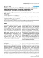
Figure 1
Immunohistochemical staining in AAC, ACC and normal gallbladder mucosaImmunohistochemical staining in AAC, ACC and normal gallbladder mucosa. (a) Ki67 expression in acute acalculous cholecystitis (AAC). (b) One
M30-positive apoptotic cell in AAC. (c) Hypoxia-inducible factor (HIF)-1α expression in AAC; nuclear expression and moderate cytoplasmic expres-
sion can be seen in this sample. (d) Abundant Ki67 expression in acute calculous cholecystitis (ACC). (e) two M30-positive cells in ACC. (f) strong
nuclear expression of HIF-1α in ACC. (g) Ki67 in control sample. (h) M30-positive cell in control sample. (i) Abundant nuclear and weak cytoplasmic
expression of HIF-1α in control sample.
Available online />Page 5 of 6
(page number not for citation purposes)
ACC mucosa. In murine experimental colitis, decreased HIF-1
expression correlated with more severe clinical symptoms,
whereas increased levels were protective of mucosal epithelial
barrier integrity [25]. Our previous studies also suggest signif-
icant differences in the integrity of gallbladder epithelia
between AAC and ACC. Bile infiltration in the gallbladder
mucosa is more abundant and extends deeper in AAC than in
ACC [10], and there are differences in tight junction protein
expression between ACC and AAC, which are important for
epithelial barrier function [21].
The explanations for decreased HIF-1α expression in AAC are
not clear. In a murine model, HIF expression is only sustained
for a few hours in a hypoxic environment, implying that factors
other than hypoxia are necessary for sustained elevation of HIF
levels [26]. It has been suggested that over-expression is due
to induction by cytokines released from inflammatory cells
rather than hypoxia [27]. In addition, in human ischaemic coli-
tis, HIF-1α is over-expressed in acutely ischaemic lesions
compared with normal epithelia, and the expression is normal-
ized after tissue recovery [28]. It could thus be speculated
that, being an ischaemia-reperfusion phenomenon, HIF
release is also time dependent and short-lived in AAC. Further-
more, during ischaemia and ischaemia-reperfusion, cell
destruction and repair are affected in the various phases of the
injury, and repeated ischaemia may also reduce epithelial
apoptosis [29]. It should be noted that our patients were in dif-
ferent phases of their disease, which certainly could have
affected the correlation of the different modulators.
In a rat model of gut ischaemia-reperfusion, HIF-1α activation
rapidly disappeared on subsequent re-oxygenation, and this
response was potentiated and preserved by the presence of
bacteria or lipopolysaccharide [30]. This also agrees well with
our results, which demonstrated stronger HIF-1α expression
in ACC, which is associated with the intense mucosal neu-
trophil reaction. More than half of the gallbladders in the ACC
group had bacterial growth, in contrast to only 7% in the AAC
group. The histopathology of ACC is clearly a purulent local
inflammation of the gallbladder. This further supports the
notion that, in addition to inflammatory cells, the presence of
bacteria potentiates the HIF response in ACC, keeping up the
corrective cellular proliferation of the biliary mucosa. In addi-
tion, proliferation activity has been shown to be increased by
gallbladder distension, which is also typical for ACC [31].
Conclusion
In conclusion, cellular proliferation and apoptosis were
increased in AAC and ACC, indicating higher cellular turnover
compared with normal gallbladders. However, the expression
of HIF was stronger in ACC than in AAC. These differences
are probably accounted for by a local purulent inflammation of
the gallbladder in ACC and systemic inflammation with vis-
ceral hypoperfusion in AAC.
Competing interests
The authors declare that they have no competing interests.
Figure 2
Epithelial cell proliferation and apoptosisEpithelial cell proliferation and apoptosis. Provided are scatter plots showing epithelial cell (a) proliferation and (b) apoptosis in acute acalculous
cholecystitis (AAX), acute calculous cholecystitis (AXX) and normal gallbladder. In each group, cases with a high hypoxia-inducible factor (HIF)-1α
expression are shown with solid circles and those with low HIF-1α expression with open circles. Line indicates the median for each group.
Key messages
• Cellular proliferation and apoptosis are increased in
AAC and ACC compared with normal gallbladder.
• Expression of HIF-1α is lower in AAC than in ACC.
• Intense expression of HIF-1α is associated with
increased cell proliferation.
Critical Care Vol 11 No 5 Vakkala et al.
Page 6 of 6
(page number not for citation purposes)
Authors' contributions
VM conducted the immunohistochemical assessments and
participated in drafting of the manuscript. LJ participated in
designing of the study, acquisition of data, analysis and inter-
pretation of the results, and drafting of the manuscript. SJ con-
tributed to designing of the study, acquisition of data, and
analysis and interpretation of the results. KV contributed to
designing and acquisition of data, and analysis and interpreta-
tion of the results. SH participated in designing and analysis,
and interpretation of the data, as well as in critical revision of
the manuscript. KT participated in the immunohistochemical
analysis and in drafting of the manuscript. SY participated in
the immunohistochemical analysis and in the critical revision of
the manuscript. AKT participated in designing and coordina-
tion and in drafting of the manuscript, as well as in critical revi-
sion of the manuscript. All authors read and approved the final
manuscript.
Acknowledgements
We thank Ms. Riitta Vuento for skilful technical assistance. The study
complies with the current Finnish laws.
References
1. Glenn F, Becker CG: Acute acalculous cholecystitis. An
increasing entity. Ann Surg 1982, 195:131-136.
2. Kalliafas S, Ziegler D, Flancbaum L, Choban PS: Acute acalcu-
lous cholecystitis. Incidence, risk factors, diagnosis, and
outcome. Am Surg 1998, 64:471-475.
3. Laurila J, Syrjälä H, Laurila PA, Saarnio J, Ala-Kokko TI: Acute acal-
culous cholecystitis in critically ill patients. Acta Anesthesiol
Scand 2004, 48:986-991.
4. McChesney JA, Northup PG, Bickston SJ: Acute acalculous
cholecystitis associated with systemic sepsis and visceral
arterial hypoperfusion. A case series and review of
pathophysiology. Dig Dis Sci 2003, 48:1960-1967.
5. Rady MY, Kodavatiganti R, Ryan T: Perioperative predictors of
acute cholecystitis after cardiovascular surgery. Chest 1998,
114:76-84.
6. Laurila J, Laurila PA, Saarnio J, Koivukangas V, Syrjälä H, Ala-Kokko
TI: Organ system dysfunction following open cholecystectomy
for acute acalculcous cholecystitis in critically ill patients. Acta
Anaesthesiol Scand 2006, 50:173-179.
7. Savoca PE, Longo WE, Pasternak B, Gusberg RJ: Does visceral
ischemia play a role in the pathogenesis of acute acalculous
cholecystitis? J Clin Gastroenterol 1990, 12:33-36.
8. Taoka H: Experimental study on pathogenesis of acute acalc-
ulous cholecystitis, with special reference to the roles of
microcirculatory disturbances, free radicals and membrane-
bound phospholipase A2. Gastroenterol Jpn 1991, 26:633-644.
9. Csendes A, Becerra M, Burdiles P, Demian I, Bancalari K,
Csendes P: Bacteriological studies of bile from the gallbladder
in patients with carcinoma of the gallbladder, cholelithiasis,
common bile duct stones and no gallstones disease. Eur J
Surg 1994, 160:363-367.
10. Laurila JJ, Ala-Kokko TI, Laurila PA, Saarnio J, Koivukangangas V,
Syrjälä H, Karttunen TJ: Histopathology of acute acalculous
cholecystitis in critically ill patients. Histopathology 2005,
47:
485-495.
11. Saarnio J, Parkkila S, Parkkila AK, Pastorekova S, Haukipuro K,
Pastorek J, Juvonen T, Karttunen TJ: Transmembrane carbonic
anhydrase, MN/CA IX, is a potential biomarker for biliary
tumours. J Hepatol 2001, 35:643-649.
12. Saarnio J, Parkkila S, Parkkila AK, Haukipuro K, Pastoreková S,
Pastorek J, kairaluoma MI, Karttunen T: Immunohistochemical
study of colorectal tumors for experession of novel transmem-
brane carbonic anhydrase, MN/CA IX, with potentila value as a
marker of cell proliferation. Am J Pathol 1998, 153:279-285.
13. Ikeda H, Suzuki Y, Suzuki M, Koike M, Tamura J, Tong J, Nomura
M, Itoh G: Apoptosis is a major mode of cell death caused by
ischemia and ischemia/reperfusion injury to the rat intestinal
epithelium. GUT 1998, 42:530-537.
14. Majno G, Joris J: Apoptosis, oncosis and necrosis. An overview
of cell death. Am J Pathol 1995, 146:3-15.
15. Ke Q, Costa M: Hypoxia-inducible factor-1 (HIF-1). Mol
Pharmacol 2006, 70:1469-1480.
16. Carroll VA, Ashcroft M: Role of hypoxia-inducible factor (HIF)-
1alpha versus HIF-2alpha in the regulation of HIF target genes
in response to hypoxia, insulin-like growth factor-1, or loss of
von Hippel-Lindau function: implications for targeting the HIF
pathway. Cancer Res 2006, 66:6264-6270.
17. Iyer NV, Kotch LE, Agani F, Leung SW, Laughner E, Wenger RH,
Gassmann M, Gearhart JD, Lawler AM, Yu AY, et al.: Cellular and
developmental control of O2 homeostasis by hypoxia-induci-
ble factor 1 alpha. Genes Dev 1998, 12:149-162.
18. Nakamura M, Yamabe H, Osawa H, Nakamura N, Shiemada M,
Kumasaka R, Murakami R, Fujita T, Osanai T, Okumura K: Hypoxic
conditions stimulate the production of angiogenin and vascu-
lar endothelial growth factor by human renal proximal tubular
epithelial cells in culture. Nephrol Dial Transplant 2006,
21:1489-1495.
19. Sandau KB, Fandrey J, Brune B: Accumulation of HIF-1alpha
under the influence of nitric oxide. Blood 2001, 97:1009-1015.
20. Sandau KB, Zhou J, Kietzmann T, Brune B: Regulation of the
hypoxia-inducible factor 1alpha by the inflammatory media-
tors nitric oxide and tumor necrosis factor-alpha in contrast to
desferroxamine and phenylarsine oxide. J Biol Chem 2001,
276:39805-39811.
21. Laurila JJ, Karttunen TJ, Koivukangas V, Laurila PA, Syrjälä H,
Saarnio J, Soini Y, Ala-Kokko TI: Expression of tight junction pro-
teins in acute acalculous and calculous cholecystitis. J Histo-
chem Cytochem 2007, 55:567-573.
22. Katada K, Naito Y, Mizushima K, Takagi T, Handa O, Kokura S,
Ichikawa H, Yoshida N, Matsui H, Yoshikawa T: Gene expression
profiles on hypoxia and reoxygenation in rat gastric epithelial
cells: a high-density DNA microarray analysis. Digestion 2006,
73:89-100.
23. Le'Negrate G, Selva E, Auberger P, Rossi B, Hofman P: Sus-
tained polymorphonuclear leukocyte transmigration induces
apoptosis in T84 intestinal epithelial cells. J Cell Biol 2006,
6:1479-1488.
24. Bos R, van Diest PJ, de Jong JS, van der Groep P, van der Valk P,
van der Wall E: Hypoxia-inducible factor-1alpha is associated
with angiogenesis, and expression of bFGF, PDGF-BB, and
EGFR in invasive breast cancer. Histopathology 2005,
46:31-36.
25. Karhausen J, Furuta GT, Tomaszewski JE, Johnson RS, Colgan SP,
Haase VH: Epithelial hypoxia-inducible factor-1 is protective in
murine experimental colitis. J Clin Invest 2004,
114:1098-1106.
26. Stroka DH, Burkhardt T, Desbaillets I, Wenger RH, Neil DAH,
Bauer C, Gassmann M, Candinas D: HIF-1 is expressed in nor-
moxic tissue and displays an organ-spesific regulation under
systemic hypoxia. FASEB J 2001, 15:2445-2453.
27. Giatromanolaki A, Sivridis E, Maltezos E, Papazoglou D, Simopou-
los C, Gatter KC, Harris AL, Koukourakis MI: Hypoxia inducible
factor 1alpha and 2 alpha overexpression in inflammatory
bowel disease. J Clin Pathol 2003, 56:209-213.
28. Okuda T, Azuma T, Ito Y, Kuriyma M: Hypoxia-inducible factor 1
aplha and vascular endothelial growth factor overexpression
in ischemic colitis. World J Gastroenterol 2005, 11:1535-1539.
29. Cinel I, Avlan D, Cinel L, Polat G, Atici S, Mavioglu I, Serinol H,
Aksoyek S, Oral U: Ischemic preconditioning reduces intestinal
epithelial apoptosis in rats. Shock 2003, 19:588-592.
30. Koury J, Deitch EA, Homma H, Abungu B, Gangurde P, Condon
MR, Lu Q, Xu DZ, Feinman R: Persistent HIF-1alpha activation
in gut ischemia/reperfusion injury: potential role of bacteria
and lipopolysaccharide. Shock 2004, 22:270-277.
31. Putz P, Willems G: Cell proliferation in the human gallbladder
epithelium: effect of distension. GUT 1979, 20:246-248.