Báo cáo y học: " The conserved dileucine- and tyrosine-based motifs in MLV and MPMV envelope glycoproteins are both important to regulate a common Env intracellular trafficking" doc
Bạn đang xem bản rút gọn của tài liệu. Xem và tải ngay bản đầy đủ của tài liệu tại đây (1.65 MB, 19 trang )
BioMed Central
Page 1 of 19
(page number not for citation purposes)
Retrovirology
Open Access
Research
The conserved dileucine- and tyrosine-based motifs in MLV and
MPMV envelope glycoproteins are both important to regulate a
common Env intracellular trafficking
Vincent Blot*
†1,3,4,5,6
, Sandra Lopez-Vergès
†2,3,4,5
, Marie Breton
1,3,4,5
,
Claudine Pique
1,3,4,5
, Clarisse Berlioz-Torrent
2,3,4,5
and Marie-
Pierre Grange
1,3,4,5
Address:
1
Institut Cochin, DépartementBiologie Cellulaire, Paris, F-75014 France,
2
Institut Cochin, DépartementMaladies Infectieuses, Paris, F-
75014 France,
3
Inserm, U567, Paris, F-75014 France,
4
CNRS, UMR 8104, Paris, F-75014 France,
5
Université Paris 5, Faculté de Médecine René
Descartes, UMR3, Paris, F-75014 France and
6
Weill Medical College of Cornell, Biochemistry Dept, New York, NY10021 USA
Email: Vincent Blot* - ; Sandra Lopez-Vergès - ; Marie Breton - ;
Claudine Pique - ; Clarisse Berlioz-Torrent - ; Marie-Pierre Grange - marie-
* Corresponding author †Equal contributors
Abstract
Background: Retrovirus particles emerge from the assembly of two structural protein
components, Gag that is translated as a soluble protein in the cytoplasm of the host cells, and Env,
a type I transmembrane protein. Because both components are translated in different intracellular
compartments, elucidating the mechanisms of retrovirus assembly thus requires the study of their
intracellular trafficking.
Results: We used a CD25 (Tac) chimera-based approach to study the trafficking of Moloney
murine leukemia virus and Mason-Pfizer monkey virus Env proteins. We found that the cytoplasmic
tails (CTs) of both Env conserved two major signals that control a complex intracellular trafficking.
A dileucine-based motif controls the sorting of the chimeras from the trans-Golgi network (TGN)
toward endosomal compartments. Env proteins then follow a retrograde transport to the TGN
due to the action of a tyrosine-based motif. Mutation of either motif induces the mis-localization
of the chimeric proteins and both motifs are found to mediate interactions of the viral CTs with
clathrin adaptors.
Conclusion: This data reveals the unexpected complexity of the intracellular trafficking of
retrovirus Env proteins that cycle between the TGN and endosomes. Given that Gag proteins
hijack endosomal host proteins, our work suggests that the endosomal pathway may be used by
retroviruses to ensure proper encountering of viral structural Gag and Env proteins in cells, an
essential step of virus assembly.
Published: 15 September 2006
Retrovirology 2006, 3:62 doi:10.1186/1742-4690-3-62
Received: 20 July 2006
Accepted: 15 September 2006
This article is available from: />© 2006 Blot et al; licensee BioMed Central Ltd.
This is an Open Access article distributed under the terms of the Creative Commons Attribution License ( />),
which permits unrestricted use, distribution, and reproduction in any medium, provided the original work is properly cited.
Retrovirology 2006, 3:62 />Page 2 of 19
(page number not for citation purposes)
Background
Retroviruses are surrounded by a lipid envelope acquired
by the virus from cellular membranes through a budding
process. Anchored in this lipid envelope are the viral enve-
lope glycoproteins (Env), which are heterodimers
between a transmembrane subunit (TM) and a covalently
or non-covalently attached extracellular subunit (named
SU for surface). Both subunits emerge from the cleavage
of a single type-1 transmembrane envelope glycoprotein
precursor (for review on retrovirus structural protein syn-
thesis, see [1].
The Gag proteins precursor, simply referred to here as
Gag, is the only viral structural protein that is both neces-
sary and sufficient to produce virus-like particles (VLPs)
by budding into the extracellular medium, even in the
absence of Env [2,3]. However, VLPs devoid of Env are
non infectious since Env glycoproteins are necessary for
the attachment of the virions to their receptor(s) and sub-
sequent fusion of viral and target cell membranes leading
to virus entry. The Env precursor is co-translationally
anchored in the membrane of the endoplasmic reticulum
and then follows the trafficking of transmembrane and
soluble proteins along the secretory pathway. By contrast,
Gag is synthesized by free ribosomes in the cytosol, before
being able to bind to internal membranes through signals
in its amino-terminus. Given that both structural compo-
nents are being translated in different subcellular com-
partments, some specific mechanisms must account for
their encounter at the site of virus assembly and budding.
Studying the precise steps of the intracellular trafficking of
envelope glycoproteins should then bring some under-
standing as to how they encounter Gag in cells. In the case
of human immunodeficiency virus (HIV) Env, it has been
shown that the cytoplasmic tail (CT) of the TM subunit
contains several motifs that regulate Env trafficking. A
tyrosine-based motif (YxxΦ where Φ is a bulky hydropho-
bic amino-acid) has been implicated in Env endocytosis
after its arrival at the cell surface by mediating interaction
with the AP-2 clathrin adaptor complexes [4-7]. A dileu-
cine-based motif (consensus sequence LL or LΦ) has also
been shown to control some post-Golgi trafficking step by
recruiting the AP-1 adaptor complexes [5,8]. Finally, HIV
Env is also able to undergo a retrograde endosome to
trans-Golgi network (TGN) route through the interaction
of a diaromatic YW motif, located in the cytoplasmic
domain of Env, with the TIP47 protein [9].
The intracellular transport of HIV Env glycoproteins has
been extensively examined, however little is known about
the trafficking of envelope glycoproteins of retroviruses
that do not belong to the lentivirus genus. The cytoplas-
mic tails of human T-cell leukemia virus (HTLV) and
Moloney murine leukemia virus (MLV) Env possess a
tyrosine-based motif that is able to target them to the
basolateral membrane of polarized MDCK cells [10].
Dileucine- and tyrosine-based motifs in the CT of bovine
leukemia virus (BLV) Env are responsible for low surface
expression of Env, although the details of Env intracellular
trafficking were not elucidated [11]. We have shown in a
previous study that engrafting the CTs of different retrovi-
rus Env to the carboxy-terminus of the CD25 reporter
molecule leads to specific intracellular trafficking path-
ways of the resulting chimeras [12]. Indeed, HTLV, BLV
and Rous sarcoma virus (RSV) CD25 chimeras are endo-
cytosed after reaching the cell surface, whereas chimeras
containing either MLV or Mason-Pfizer monkey virus
(MPMV) CT appeared mainly retained inside the cells in a
Rab6-positive Golgi or post-Golgi compartment.
In this study, we aimed to precisely define the intracellular
routes followed by MLV and MPMV envelope glycopro-
teins. Using the same CD25 chimera-based approach, we
found that these proteins accumulated in the TGN as a
result of a dynamic transport involving a retrograde route
from endosomes to the TGN. A membrane proximal
dileucine-based motif and a more distal tyrosine-based
motif conserved between both CTs governed this peculiar
trafficking. The dileucine-based motif is implicated in the
sorting of the chimeras at the level of the TGN, whereas
the tyrosine-based motif is required in the retrograde
transport step. We also documented that both motif
mediate in vitro interaction with clathrin adaptors, linking
their functional role in Env trafficking with their capacity
to physically interact with cellular trafficking machineries.
Results
CD25-MuLV and CD25-MPMV chimera accumulated in
the TGN
We have previously shown that engrafting the cytoplasmic
tail of either MLV or MPMV envelope glycoprotein to the
carboxyl-terminus of the CD25 protein induced the intra-
cellular retention of the resulting chimeras [12]. Both chi-
meras colocalized at steady state with the small GTPase
Rab6, a protein distributed between the Golgi apparatus
and the TGN [13,14].
To define more precisely the intracellular site of accumu-
lation of the chimeras, we treated transiently transfected
HeLa cells with cycloheximide, which acted by preventing
new synthesis of proteins. CD25-MuLV and CD25-MPMV
chimeras appeared then mainly concentrated in a tubular-
shaped perinuclear compartment as well as in dots dis-
persed throughout the cytoplasm (figure 1, CD25 panels)
whereas the control CD25 protein accumulated at the cell
surface (data not shown and [12]).
We then compared the distribution of the chimeras with
those of different intracellular markers: the Mannose-6-
Retrovirology 2006, 3:62 />Page 3 of 19
(page number not for citation purposes)
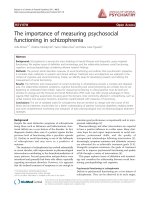
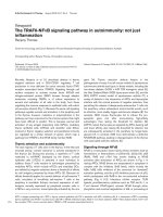
CD25-MLV and CD25-MPMV accumulate in the TGNFigure 1
CD25-MLV and CD25-MPMV accumulate in the TGN. Forty-eight hours after transfection with the appropriate chi-
mera cDNA, cells were treated with cycloheximide for 3 hours prior to fixation and staining. A. Co-staining of CD25 chimeras
and Mannose 6-phosphate receptor of 46 kDa (MPR46), a protein that accumulates in the TGN at steady state. B. Co-staining
of CD25 chimeras and internalized Cy3-conjugated tranferrin revealing the early/recycling endosomes. C. Co-staining of CD25
chimeras and Lamp1, a protein resident of the lysosomes.
A.
CD25 Transferrin Merge
CD25-MPMV WT
B.
CD25 Lamp-1 Merge
CD25-MuLV WT
CD25-MPMV WT
C.
CD25 MPR46 Merge
CD25-MuLV WT
CD25-MPMV WT
CD25-MuLV WT
19 µ
19 µ
19 µ
19 µ
19 µ
19 µ
Retrovirology 2006, 3:62 />Page 4 of 19
(page number not for citation purposes)
phosphate receptor of 46kDa (MPR46) that cycles
between the TGN and late endosomes and is mainly local-
ized in the TGN at steady state [15], internalized cyanin3-
conjugated transferrin that reveals the general early and
recycling endosomal pathway and Lamp1, a marker of lys-
osomes [16]. CD25-MLV and CD25-MPMV did not colo-
calize with either endocytosed transferrin or Lamp1,
indicating that they do not accumulate in the endocytic
pathway (figure 1B and 1C). By contrast, both proteins
showed extensive colocalization with MPR46 revealing
that their intracellular compartment of retention is the
TGN (figure 1A).
A dileucine- and a tyrosine-based motifs are both required
for the TGN localization of CD25-MuLV and CD25-MPMV
chimeras
To define the motifs in MLV and MPMV cytoplasmic tails
important for this peculiar localization, we compared
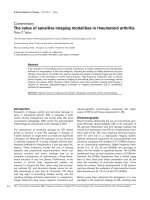
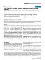
their primary sequences (figure 2A). The two sequences
shared 10 amino acids conserved in position, amongst
which two clusters fit potential conventional sorting sig-
nals: the dileucine-based motifs
3
LV
4
/
3
LM
4
and the tyro-
sine-based motif
23
YHQL
26
/
23
YHRL
26
in MLV and MPMV
sequences respectively (where 1 is the position of the first
amino-acid in each viral cytoplasmic tail). MPMV cyto-
plasmic tail possesses a second tyrosine-based motif
(
35
YLTL
38
) that is not conserved in the MLV cytoplasmic
domain.
To investigate the implication of these putative sorting
motifs in the trafficking of the chimeras, we produced a
diversity of point mutations in the cytoplasmic tails by
site-directed mutagenesis (figure 2B). We then analyzed
the effects of these mutations on the intracellular localiza-
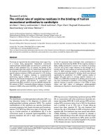
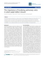
tion of the resulting mutated chimeras. Mutation of the
tyrosine 23 to serine in either MLV and MPMV CT pro-
voked a relocalization of the chimeras to peripheral dots
dispersed throughout the cytoplasm that do not colocal-
ize with MPR46 (figure 3A). By contrast, mutation of the
distal
35
YLTL
38
tyrosine-based motif in MPMV cytoplas-
mic tail had no effects (figure 3A lower panels). Changing
the leucine 3 into a serine resulted in a partial shift of the
localization of the chimeras from the TGN to peripheral
dots and the mutated chimeras still colocalized to some
Sequences of wild type and mutant MLV and MPMV cytoplasmic tailsFigure 2
Sequences of wild type and mutant MLV and MPMV cytoplasmic tails. A. The 10 amino acids conserved between
MLV and MPMV cytoplasmic tails (CT) are noted ● Bold letters indicate the position of the conserved dileucine- and tyrosine-
based motifs, whereas underlined letters indicate the position of the extra tyrosine-based motif in MPMV CT. B. Sequences of
the mutated CD25 chimeras that we used in this study. The mutants are named CD25-retrovirus X amino acid position Z,
where X and Z are the wild-type and mutant amino-acids, respectively. The amino-acid position 1 corresponds to the first res-
idue of the corresponding viral CT.
NRLVQFVKDRISVVQALVLTQQYHQLKPIEYEP
NKLMTFIKHQIESIQAKPIQVHYHRLEQEDSGGSYLTL
T
NRS
VQFVKDRISVVQALVLTQQYHQLKPIEYEP
NRLVQFVKDRISVVQALVLTQQS
HQLKPIEYEP
NRS
VQFVKDRISVVQALVLTQQSHQLKPIEYEP
NKS
MTFIKHQIESIQAKPIQVHYHRLEQEDSGGSYLTLT
NKLMTFIKHQIESIQAKPIQVHS
HRLEQEDSGGSYLTLT
NKLMTFIKHQIESIQAKPIQVHYHRLEQEDSGGSSLTL
T
NKS
MTFIKHQIESIQAKPIQVHSHRLEQEDSGGSYLTLT
A.
Wild type CD25 chimeric proteins
CD25-MuLV WT
CD25-MPMV WT
B.
Mutant CD25 chimeric proteins
CD25-MuLV L3S
CD25-MuLV Y23S
CD25-MuLV L3S/Y23S
CD25-MPMV L3S
CD25-MPMV Y23S
CD25-MPMV Y35S
CD25-MPMV L3S/Y23S
1 10 20 30
Retrovirology 2006, 3:62 />Page 5 of 19
(page number not for citation purposes)
Mutation of either the dileucine- or the tyrosine-based motifs affect the TGN localization of CD25 chimerasFigure 3
Mutation of either the dileucine- or the tyrosine-based motifs affect the TGN localization of CD25 chimeras.
Forty-eight hours after transfection with the appropriate chimera cDNA, cells were treated with cycloheximide for 3 hours
prior to fixation and permeabilization. Co-stainings of MPR46 and chimeras bearing either (A) the Y23S or the Y35S mutation,
(B) the L3S mutation, or (C) both L3S and Y23S mutations.
CD25 MPR46 Merge
CD25-MuLV Y23S
B.
A.
CD25-MuLV L3S/Y23S
CD25-MPMV L3S/Y23S
CD25-MPMV Y23S
CD25-MPMV Y35S
C.
CD25-MuLV L3S
CD25-MPMV L3S
19 µ
Retrovirology 2006, 3:62 />Page 6 of 19
(page number not for citation purposes)
extent with MPR46 (figure 3B). Finally, MLV and MPMV
chimeras mutated on both leucine 3 and tyrosine 23
mainly accumulated at the plasma membrane (figure 3C),
thus behaving as the control CD25.
Thus, extensive localization of the CD25-MLV and the
CD25-MPMV chimeras in the TGN required both the
dileucine-based motif in position 3 and the tyrosine-
based motif in position 23. By contrast, the tyrosine-based
motif in position 35 of the MPMV cytoplasmic tail does
not play a significant role in the TGN localization of the
protein.
CD25-MLV and CD25-MPMV with mutated dileucine- or
tyrosine-based motifs accumulate in different endocytic
compartments
We then assess whether the changes in localization of the
CD25-MLV and CD25-MPMV chimeras that we observed
after mutating either the dileucine- or the tyrosine-based
motif revealed a relocalization of the protein in endocytic
compartments. We used internalized transferrin as a
marker of early/recycling endosomes, Lamp1 as a marker
of lysosomes and dextran internalized for 30 minutes and
chased for an equivalent amount of time to reveal late
endosomal compartments.
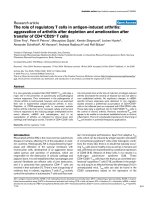
Chimeras with mutations in the dileucine-based motif
showed partial colocalization with the three markers of
the endosomal pathway (figure 4A, 4B and 4C, arrows).
Colocalization of chimeras with Lamp1, however, is
weaker than with endocytosed transferrin or dextran.
Thus, the fraction of L3S mutated chimeras that is delocal-
ized from the TGN is redistributed throughout the endo-
somal pathway. By contrast, chimeras bearing the Y23S
mutation did not colocalize with either transferrin or
Lamp1 (figure 5A and 5C), indicating that they are absent
from early/recycling endosomes or lysosomes. However,
these mutant proteins did colocalize to some extent with
internalized and chased dextran (figure 5B, arrows). Thus,
mutation of the tyrosine-based motif in position 23
induced the relocalization of both CD25-MLV and CD25-
MPMV chimeras in non well-defined late endosomal
compartments.
Internalization of chimeras from the plasma membrane is
mainly driven by the tyrosine-based motif in position 23
That the chimeras are mainly detected in intracellular sites
at steady state could either reflect an active retention of the
proteins within the cells or their slow recycling to the
plasma membrane followed by their rapid internaliza-
tion. We thus wanted to determine whether the chimeras
could be endocytosed from the plasma membrane. To
that extent, we compared the abilities of the different WT
and mutant chimeras to allow uptake of monoclonal anti-
CD25 antibody. Transiently-transfected HeLa cells were
then incubated for 30 min at 4°C with anti-CD25 anti-
body and shifted or not at 37°C for 30 additional min-
utes. For each chimera, we then compared the amount of
anti-CD25 antibody remaining at the cell surface after 30
minutes at 37°C relative to the amount of anti-CD25 at
the cell surface at time 0.
After 30 minutes, approximately 50% of bound anti-
CD25 antibody was internalized in cells expressing either
CD25-MLV or CD25-MPMV chimeras. This is similar to
the amount of CD25 internalized in cells expressing
CD25-TFR, a control chimera containing the well defined
YRTF endocytic signal of the transferrin receptor (figure
6A and 6B). By contrast, the CD25 control protein that
lacks specific internalization signals or viral cytoplasmic
tail does not allow measurable uptake of anti-CD25 anti-
body. This indicates that viral cytoplasmic tails in CD25-
MLV and CD25-MPMV chimeras contain specific internal-
ization signals.
Mutation of the dileucine-based motifs in MLV or MPMV
chimera did not impair the capacity of the proteins to
mediate specific uptake anti-CD25 antibody (fig 6A and
6B; L3S). By contrast, chimeras bearing the Y23S mutation
had a decreased ability to allow anti-CD25 antibody
retrieval from the cell surface (figure 6A and 6B). Chime-
ras bearing both L3S and Y23S mutations behave like the
single Y23S mutant indicating that the lack of detectable
effects of the single L3S mutation was not due to redun-
dancy with the Y23 tyrosine-based motif.
Altogether, these results indicate that CD25-MLV and
CD25-MPMV chimeras are internalized from the plasma
membrane, and that the tyrosine-based motif in position
23 acts as their main endocytosis signal.
The tyrosine-based motif in position 23 drives a retrograde
transport step toward the TGN
The steady state TGN localization of proteins like MPRs,
furin or TGN38 is the results of a complex trafficking
involving a retrograde transport from endosomes to the
TGN [15,17]. We thus assessed the capacity of MLV and
MPMV cytoplasmic tails to target the chimeras to the TGN
following their internalization in endosomes.
One hour after their internalization from the cell surface,
anti-CD25 antibodies taken up by either the CD25-MLV
or CD25-MPMV chimera were found concentrated in a
perinuclear region of the cells (figure 7A). Both chimeras
then extensively colocalized with MPR46, indicating that
they reached the TGN (figure 7A). By contrast, anti-CD25
taken up by the control CD25-TFR construct that follows
the recycling pathway of the transferrin receptor did not
colocalize with MPR46 (figure 7A), indicating that both
MLV and MPMV cytoplasmic tails contain specific infor-
Retrovirology 2006, 3:62 />Page 7 of 19
(page number not for citation purposes)
Chimeras bearing the L3S mutation are relocated throughout the endosomal pathwayFigure 4
Chimeras bearing the L3S mutation are relocated throughout the endosomal pathway. Forty-eight hours after
transfection with the L3S mutant chimeras cDNA, HeLa cells were treated with cycloheximide for 3 hour prior to fixation and
permeabilization. A. Co-staining of L3S mutant chimeras and internalized Cy3-conjugated transferrin revealing the early/recy-
cling endosomes.B. Cells were allowed to take up FITC-conjugated dextran for 30 min. Cells were then extensively washed,
and dextran was chased for another 30 min prior to fixation and CD25 staining. FITC-dextran thus revealed some late endo-
somal compartment. C. Co-staining of CD25 chimeras and Lamp1, a protein resident of the lysosomes.
CD25 transferrin Merge
CD25-MuLV L3S
CD25-MPMV L3S
Dextran-FITC Merge
CD25-MuLV L3S
CD25
CD25-MPMV L3S
CD25 Lamp-1 Merge
CD25-MuLV L3S
CD25-MPMV L3S
B.
C.
A.
19 µ
19 µ
19 µ
19 µ
19 µ
19 µ
Retrovirology 2006, 3:62 />Page 8 of 19
(page number not for citation purposes)
Chimeras bearing the Y23S mutation are mainly relocated to a late endosomal compartmentFigure 5
Chimeras bearing the Y23S mutation are mainly relocated to a late endosomal compartment. Forty-eight hours
after transfection with the Y23S mutant chimeras cDNA, HeLa cells were treated with cycloheximide for 3 hour prior to fixa-
tion and permeabilization. A. Co-staining of Y23S mutant chimeras and internalized Cy3-conjugated transferrin revealing the
early/recycling endosomes.B. Before fixation, cells were allowed to take up FITC-conjugated dextran for 30 min. The cells
were then extensively washed, and dextran was chased for another 30 min prior to fixation thus accumulating in late endo-
somal compartments. C. Co-staining of CD25 chimeras and Lamp1, a protein resident of the lysosomes.
A.
CD25 Transferrin Merge
CD25-MuLV Y23S
CD25-MPMV Y23S
CD25 Lamp-1 Merge
CD25-MuLV Y23S
CD25-MPMV Y23S
Dextran-FITC MergeCD25
CD25-MuLV Y23S
CD25-MPMV Y23S
B.
C.
19 µ
19 µ
19 µ
19 µ
19 µ
19 µ
Retrovirology 2006, 3:62 />Page 9 of 19
(page number not for citation purposes)
mation capable of driving their retrograde transport to the
TGN.
Mutation of the dileucine motif in position 3 did not dras-
tically affect the capacity of the chimeras to be targeted to
the TGN following internalization (Figure 7B). By con-
trast, chimeras mutated in the tyrosine-based motif in
position 23 appeared localized in dispersed dots through-
out the cytoplasm after their internalization. No colocali-
zation was then apparent with MPR46 (Figure 7C).
Altogether, these data indicate that the TGN localization
of the MLV and MPMV chimeras is the result of a complex
trafficking involving retrieval of these proteins from endo-
somal compartments towards the TGN. This last step is
driven by the tyrosine-based motif in position 23 that is
conserved between both retroviruses.
MLV cytoplasmic tail interacts with adaptor protein
complexes (AP) 1, 2 and 3
To better understand the molecular basis of the intracellu-
lar sorting of the viral chimeras, we assessed the ability of
the viral CT to physically interact with components of the
adaptor protein complexes AP-1, AP-2 and AP-3 in a yeast
two-hybrid assay. Because we have shown that both MLV
and MPMV Env share the same trafficking, we decided to
restrict our biochemical analysis to one virus. Thus, MLV
CT was fused to the N-terminus of the LexA binding
domain (BD), whereas the µ1, γ and β1 chains of AP1, the
µ2, α and β2 chains of AP2 and the µ3, δ and β3 chains of
AP3 were fused to the Gal4 activation domain (AD). MLV
CT did not interact with γ or β1 subunits of AP1, α or β2
subunits of AP2, or δ and β3 subunits of AP3 in yeast two-
hybrid system (data not shown). By contrast, MLV CT
bound to µ1, µ2 and µ3 medium chains as indicated by
the expression of the HIS3 reporter gene, which allows cell
growth in the absence of histidine (figure 8A, 8B and 8C).
However, interaction with µ2 only appeared after 72
hours growth (figure 8B), whereas interaction with µ1 and
µ3 were present after 30 hours growth (figure 8A and 8C),
indicating that binding to µ2 was weaker than the other
interactions.
Mutation of the tyrosine in position 23 completely abol-
ished interaction of the MLV CT with all three µ1, µ2 and
µ3 chains of AP complexes (figure 8A, 8B and 8C). On the
contrary, mutation of the leucine in position 3 did not
affect interaction with any of the µ chains (figure 8A, 8B
and 8C). These results therefore indicate the tyrosine 23 is
critical for binding of the MLV cytoplasmic tail to the iso-
lated µ subunits, and further demonstrate the specificity
of these interactions.
We then examined whether a GST fusion of the MLV CT
was able to recruit the whole preformed AP complexes
Effects of the L3S and/or Y23S mutations on the chimeras ability to be retrieved from the plasma membraneFigure 6
Effects of the L3S and/or Y23S mutations on the chi-
meras ability to be retrieved from the plasma mem-
brane. HeLa cells were cotransfected with GFP vector and
with the appropriate A MLV or B MPMV chimera cDNA.
Cells were then incubated for 1 hour at 4C with anti-CD25
antibody before being either shifted for 30 min at 37°C or
not. Anti-CD25 stainings were revealed using phycoeryth-
rine-conjugated secondary antibodies. Stained cells were ana-
lyzed using flow cytometry excluding the none transfected
GFP-negative cells. We then plotted the percentage of inter-
nalization as the ratio between the CD25-associated fluores-
cence that disappeared during the 30 min uptake at 37°C and
the CD25-associated fluorescence at time 0. CD25 is the ref-
erence protein without any viral cytoplasmic tail (negative
control) and CD25-TFR is the CD25 reference protein in the
cytoplasmic tail of which the well described YTRF endocyto-
sis motif of the transferrin receptor has been inserted (posi-
tive control).
A.
20
45
70
CD25
CD25-TFR
CD25-MuL
V
WT
CD25-
MuL
V
L3S
CD25-MuL
V
Y23S
CD25-
MuL
V
L3S/Y23
S
% internalisation
20
45
70
CD25
CD25
-TFR
CD25-
MPM
V
WT
CD25-
MPM
V
L3S
CD25-MPM
V
Y23S
CD25-
MPM
V
V L3S/Y2
3
S
% internalisation
B.
Retrovirology 2006, 3:62 />Page 10 of 19
(page number not for citation purposes)
The tyrosine-based motif in position 23 allows the chimera to follow a retrograde route from endosomes to TGNFigure 7
The tyrosine-based motif in position 23 allows the chimera to follow a retrograde route from endosomes to
TGN. HeLa cells were transfected with A wild type, B L3S mutated or C Y23S mutated chimeras. Forty-eight hours after
transfection, cells were treated with cycloheximide for 2 h. Chimeras present on cell surface were stained with the anti-CD25
antibody at 4°C for 1 hour and cells were then shifted at 37°C for another hour. After fixation, internalized anti-CD25 was
revealed using FITC-conjugated secondary antibodies, and MPR46 was revealed as in figure 1.
CD25 MPR46 Merge
CD25-MuLV WT
CD25-MPMV WT
CD25-MuLV L3S
CD25-MPMV L3S
CD25-MuLV Y23S
CD25-MPMV Y23S
CD25-TFR
A.
B.
C.
19 µ
19 µ
19 µ
19 µ
19 µ
19 µ
19 µ
Retrovirology 2006, 3:62 />Page 11 of 19
(page number not for citation purposes)
Interaction of the MLV TM-CD with µ1, µ2 and µ3 subunits in the yeast two-hybrid systemFigure 8
Interaction of the MLV TM-CD with µ1, µ2 and µ3 subunits in the yeast two-hybrid system. The yeast reporter
strain L40 was co-transformed with plasmids encoding Gal4 AD-µ1, µ2 and µ3-adaptin (A, B and C, respectively), and plas-
mids encoding lexA BD fused to either the wild type (WT) or mutated (L3S, Y23S) cytoplasmic tail of MLV. Cotransformants
were analyzed for histidine auxotrophy. They were patched on medium with histidine and then replica-plated on medium with-
out histidine (- His medium). Growth in the absence of histidine indicates interaction between hybrid proteins. The positive
control was the interaction between Ras and Raf proteins which bind to each other efficiently (lane 4, lower patch in panels A,
B, C). Binding specificity was verified by the absence of interaction between the retroviral cytoplasmic tails (WT, L3S and Y23S)
and the Gal4AD alone (none).
P1-adaptin
none
Gal4 AD
WT L
3
S Y
23
S none
Ras/Raf
LexA BD MLV ENV CD
A
P2-adaptin
none
Gal4 AD
WT L3S Y23S none
Ras/Raf
LexA BD MLV ENV CD
B
30H growth
(-His medium)
72H growth
(-His medium)
1 2 3 4
1 2 3 4
P3-adaptin
none
Gal4 AD
WT L3S Y23S none
Ras/Raf
LexA BD
MLV ENV CD
C
30H growth
(-His medium)
1 2 3 4
Retrovirology 2006, 3:62 />Page 12 of 19
(page number not for citation purposes)
from HeLa cells lysates. AP1, AP2 and AP3 complexes
were revealed using antibodies to γ-adaptin, α-adaptin
and δ-adaptin, respectively. Immunoblot analysis of the
cellular proteins retained on GST-MLV beads indicated
that AP1, AP2 and AP3 bound specifically to the viral cyto-
plasmic tail (figure 9A, 9B and 9C). Mutation of either the
tyrosine 23 or the leucine 3 affected the binding of the
resulting GST-MLV to the AP2 complex (figure 9B). Inter-
estingly, mutating the leucine 3 strongly affected the bind-
ing to AP1 and AP3, whereas mutation of the tyrosine 23
had no effect (figure 9A and 9C).
We thus showed that MLV cytoplasmic tail interacts not
only with the µ chains of clathrin adaptors type 1, 2 and
3, but also associates with the AP complexes from cell
lysates. Optimal interaction with AP2 requires both the
dileucine- and the tyrosine-based motif in position 3 and
23, respectively. On the other hand, interactions with AP1
and AP3 complexes depend only on the dileucine-based
motif.
Discussion
In this study, we analyzed the intracellular trafficking of
two oncoretroviral envelope proteins, these of MLV and
MPMV retroviruses. Their peculiar trafficking resulted in
the dynamic intracellular retention of the proteins in the
TGN and was driven by the association of two conven-
tional sorting signals conserved in position between the
two envelope glycoprotein cytoplasmic tails: a membrane
proximal dileucine-based motif (
3
LV
4
/
3
LM
4
in MLV and
MPMV sequences respectively) and a more distal tyrosine-
based motif (
23
YHQL
26
/
23
YHRL
26
).
To evaluate the roles of the CT of MLV and MPMV enve-
lope glycoproteins in regulating their trafficking, we used
an approach based on the study of chimeras between the
whole CD25 chain and the cytoplasmic tail of retroviral
Env proteins. Study of CD25 chimeras is a broadly used
approach to assess the role of the cytoplasmic tail of dif-
ferent cellular proteins in their trafficking [17-19]. We and
others have used this approach to study the trafficking of
different retroviral glycoproteins [5,9,11,12]. Using CD25
chimeras permitted us to conduct a comparative work and
to avoid complications inherent to the use of native viral
envelope glycoproteins such, as shedding of the SU subu-
nit in the extracellular medium or cytopathogenic effects
due to envelope induced cell-cell fusion. Importantly, we
previously demonstrated that native viral glycoproteins
displayed the same intracellular trafficking as their chime-
ras counterparts, thus legitimazing the use of this
approach [8,9,12].
We previously showed that CD25-MLV and CD25-MPMV
chimera appeared retained in an intracellular tubular-
shaped perinuclear compartment [12]. We now show that
this compartment is distinct from the early/recycling
endosomes or lysosomes and is enriched with the MPR46
protein. MPR46 mediates the transport of lysosomal
enzymes from the TGN to endosomal prelysosomal com-
partments. After delivery of its cargo in acidic compart-
ments, MPR returns to the TGN [20]. At steady state,
MPR46 is found in the TGN, although a fraction may be
found in endosomes [21,22]. We therefore concluded that
the majority of CD25-MLV and CD25-MPMV localized in
the TGN.
The TGN localization of both chimeras was dependent on
the integrity of two sorting motifs. Mutating the dileucine-
based motif in position 3 resulted in a partial delocaliza-
tion of the chimeras throughout the endosomal pathway,
without affecting their capacity to follow the retrograde
route to the TGN when internalized from the plasma
membrane. By contrast, mutation of the tyrosine-based
motif in position 23 totally abolished the TGN localiza-
tion of the chimeras, as well as their ability to be retrieved
from endosomes to the TGN. The Y23S mutated chimeras
then appeared accumulated in an endosomal compart-
ment. Because this compartment was stained after inter-
nalization of dextran followed by a chase and was distinct
from the early/recycling endosomes and lysosomes, we
conclude that it must represent some late endosomal
compartment. This compartment, however, did not con-
tain CD63, a typical marker for late endosmes/multivesic-
ular bodies (data not shown) and its exact nature remains
to be identified. Regardless, these data allow us to propose
the following model for the complex intracellular traffick-
ing of the CD25-MLV and CD25-MPMV chimeras:
While exiting the biosynthetic pathway at the TGN level,
the chimeric proteins are sorted to a specific late endo-
somal compartment. This sorting step involves the dileu-
cine-based motif and mutation of this motif results in
misrouting the proteins throughout the endosomal path-
way (Table 1). Alternatively, Env proteins can also reach
Table 1: Summary of the roles of the motifs in MLV and MPMV Env CT in subcellular Env trafficking.
MOTIF (MLV/MPMV) ROLE IN TRAFFICKING EFFECT OF MUTATION
23
YHQL
26
/
23
YHRL
26
Endocytosis Increase plasma membrane localization
23
YHQL
26
/
23
YHRL
26
TGN retrieval Delocalization in unidentified late-endocytic compartments
3
LV
4
/
3
LM
4
Sorting from TGN Delocalization in the endosomal pathway
Retrovirology 2006, 3:62 />Page 13 of 19
(page number not for citation purposes)
Interaction between GST fusions of WT or mutated MLV cytoplasmic tail and AP-1, AP-2 and AP-3 complexesFigure 9
Interaction between GST fusions of WT or mutated MLV cytoplasmic tail and AP-1, AP-2 and AP-3 com-
plexes. Identical quantities of GST (5 µg) (lanes 2, panels A-C), GST-MLV (lanes 3), GST-MLV-L3S (lanes 4,), GST-MLV-Y23S
(lanes 5) were incubated with HeLa cell lysates (2.5 × 10
6
cells). The binding of AP-1, AP-2 and AP-3 complexes to GST fusion
proteins was revealed by Western blotting with anti-γ adaptin mAb (panel A), anti-α adaptin mAb (panel B) and anti-δ adaptin
mAb (panel C). The positions of the α-adaptin (Mr~100,000), γ-adaptin (Mr~104,000) and δ-adaptin (Mr~90,000) are indicated
in the crude cell lysate from 10
6
cells (lanes 1).
AP-1
(J-adaptin)
AP-2
(D-adaptin)
B
HeLa Lystae
MLV WT
GST
MLV L3S
MLV Y23S
A
HeLa Lystae
MLV WT
GST
MLV L3S
MLV Y23S
1 2 3 4 5
C
HeLa Lystae
MLV WT
GST
MLV L3S
MLV Y23S
AP-3
(G-adaptin)
1 2 3 4 5
1 2 3 4 5
Retrovirology 2006, 3:62 />Page 14 of 19
(page number not for citation purposes)
the late endosomal compartment after internalization
from the plasma membrane. Once the chimeras reach the
specific late endosomal compartment, their tyrosine-
based motif in position 23 mediates their trafficking in a
retrograde pathway up to the TGN. If the tyrosine-based
motif is mutated, the chimeras accumulate in the late
endosomal compartment (Table 1). On the contrary, the
wild type chimeras continuously cycle between endo-
somes and the TGN. The fact that chimeric proteins
appear accumulated in the TGN at steady state indicates
that the limiting step of their trafficking is the sorting
event at the TGN level. Lastly, chimeras that make it to the
plasma membrane are being internalized and delivered
back to the TGN, both steps implicating the tyrosine-
based motif in position 23 (Table 1).
This model is reinforced by the biochemical study we con-
ducted on the MLV tail. Indeed, GST pull down assays
confirmed that the dileucine-based motif is important for
interaction with AP1 and AP3 complexes that are impli-
cated in sorting cargos at the TGN level (review in [23]),
whereas the tyrosine-based motif is somehow dispensable
for this process. On the contrary both dileucine- and tyro-
sine-based motifs are important for the optimal recruit-
ment of the AP2 complexes that function in endocytosis
[24]. However, the direct interaction we detected in yeast
two-hybrid between MLV cytoplasmic tail and the µ2
chain of AP2 appeared weaker than interaction with µ1 or
µ3. This is consistent with our findings that trafficking of
CD25-MLV chimeras is mainly restricted between intrac-
ellular compartments and that the MLV tyrosine-based
signal is not optimized for the binding to the AP2 com-
plexes. These findings nevertheless indicate that when the
chimeras eventually reach the plasma membrane, they
can be cleared from cell surface following endocytosis.
Our biochemical data does not elucidate how the tyro-
sine-based motif in position 23 mediates the retrograde
route of the CD25-MLV and CD25-MPMV chimeras from
late endosomal compartment to the TGN. One retrograde
transport pathway to TGN involves the AP1 clathrin adap-
tor [25,26]. In our yeast two-hybrid experiments, we elu-
cidated an interaction between the µ1 chain of AP1 and
MLV cytoplasmic tail. This interaction depended on the
integrity of the tyrosine-based motif in position 23. In our
GST pull down assay, however, the tyrosine 23 appears
dispensable for the interaction with AP1 complexes. These
contrasting results might indicate that, although the tyro-
sine-based motif is capable to bind the isolated µ chains
of AP complexes in the yeast two-hybrid system, it might
not be able to mediate the interaction with the whole AP1
complex. Alternatively, it is also possible that the interac-
tion of the MLV and MPMV Env CTs with AP1 complexes
during the retrograde transport might involve additional
informations, like other determinants located in the Env
CTs. Another possibility is that the tyrosine-based motif
functions in the retrograde transport by recruiting other
adaptors than AP1. Indeed, different retrograde pathways
have been described for the furin and MPRs proteins. Ret-
rograde transport of the furin protein involves an interac-
tion of an acid cluster of amino-acids with the adaptor
protein PACS1 [27,28] while retrograde transport of the
MPRs requires either interaction of a motif constituted by
two successive aromatic amino-acids (MPR46) or by pro-
lines (MPR300) with the TIP47 adaptor [29,30]. None of
these kinds of motifs can be found in either MLV or
MPMV cytoplasmic tails suggesting that these viral pro-
teins follow a different retrograde route using an
unknown mechanism or use new motifs to interact with
these adaptors. Formal demonstration of this would how-
ever require more biochemical analysis to directly test the
interaction of MLV and MPMV cytoplasmic tails with
PACS1 and TIP47.
Interestingly, it has been demonstrated that Epsin R can
recruit clathrin either directly or through AP1 [31-34].
This recruitment is important for an AP1-independent ret-
rograde pathway followed by TGN38 and MPR300 [35].
Because it has proposed that Espin R may function as a
cargo adaptor [36], further studies should also assess the
putative relationship between Epsin R and the retrograde
transport mediated by MLV and MPMV cytoplasmic tails.
The dileucine 3- and tyrosine 23-based motifs in MLV and
MPMV cytoplasmic tails are very similar to each other in
term of sequence and are conserved in position (Fig 2).
Nevertheless, MLV and MPMV infect different hosts,
belong to two different retrovirus genuses and appear
highly divergent on a phylogenetic tree. We can thus pos-
tulate that these two motifs and the intracellular traffick-
ing they regulate must be essential for efficient replication
and propagation of these viruses. We and others have
shown that, regardless of whether they are from the
oncoretrovirus or the lentivirus family, all retrovirus enve-
lope glycoproteins follow complex intracellular routes
[5,8-10,12]. It has been proposed that these different
intracellular trafficking routes were part of mechanisms
allowing the virus to escape the host immune response by
limiting the amount of antigenic envelope glycoproteins
at the cell surface of infected cells. Thus, a simian immun-
odeficiency virus bearing a mutation in the tyrosine-based
endocytosis signal of its envelope glycoproteins is attenu-
ated in vivo [37], although envelope incorporation into
virions and virions infectivity are both normal in vitro
[38]. Moreover, mutation of the same tyrosine-based
endocytosis signal in HIV enhances the immunogenicity
of a vaccine preparation, in correlation with enhanced
surface expression of the protein [39]. However, the fact
that the intracellular trafficking pathways we describe are
much more complex than just endocytosis suggests that
Retrovirology 2006, 3:62 />Page 15 of 19
(page number not for citation purposes)
they play other roles than just limiting the amount of
envelope glycoproteins present at the cell surface.
In the last few years, it was demonstrated that the Gag
polyproteins of retroviruses hijack various host proteins
and use them for assembly and budding of particles
(reviewed in [2,3]). All the cellular factors described so far
to participate in this phenomenon are proteins that func-
tion in different stages of the endosomal trafficking:
Nedd4 [40-42], Tsg101 and other ESCRT-1 components
[40,42-44], AIP1/ALIX [45], AP2 [46] and AP3 [47].
Growing number of evidence indicate that Gag proteins
are transported along the endosomal pathway prior to
assembly and budding [42,47-56]. Thus, targeting enve-
lope glycoproteins in the endosomal pathway might help
Env encountering Gag and being subsequently incorpo-
rated in the nascent virion. That Env glycoproteins con-
stantly traffic in a cycling pathway between TGN and
endosomes as we described here would furthermore allow
them to wait inside the cell until they encounter the Gag
proteins in endosomes, and are subsequently rerouted to
be incorporated into budding particles.
Supporting this hypothesis, it has been shown that HIV-1
envelope glycoproteins also follow an anterograde/retro-
grade pathway and that this trafficking step is required for
optimal incorporation of Env into virions and subsequent
infectivity of the virus [9]. We also found that bovine
leukemia virus envelope cytoplasmic tail possesses dileu-
cine- and tyrosine-based motifs that drive its trafficking in
a TGN-endosome cycling pathway (Blot et al, unpub-
lished data). Finally, the tyrosine-based motifs in position
23 in the cytoplasmic tail of MLV and MPMV envelope
glycoproteins, which we found are necessary to maintain
the protein in the endosomes to the TGN retrograde route,
are also necessary for efficient incorporation of Env into
virus particles [57,58].
Finally, regulated Env intracellular trafficking might also
be important for intracellular Gag sorting and subsequent
efficient virus release. Indeed, it has been shown that Env
can influence Gag intracellular localization for both MLV
[53] and MPMV [48]. MLV can be released in a polarized
manner and this process depends on the integrity of the
tyrosine-based motif in Env CT [10,59]. MPMV follows
the Type-D assembly pathway in which Gag pre-assem-
bled in the cytoplasm. It has been recently shown that pre-
assembled MPMV Gag are localized on pericentriolar
microdomains and Env is required to promote Gag trans-
port out of this perinuclear site [48]. It will thus be of great
interests to test the importance of Env tyrosine-based and
dileucine-based motifs in this process. It was long thought
that the whole process of retrovirus assembly occurs at the
plasma membrane of infected cells. Accumulating evi-
dence now complicates this simple scheme and suggests
that retroviruses developed strategies ensuring the specific
sorting of their structural proteins into intracellular com-
partments. These complex routes may be viewed as a fun-
nel, concentrating the different structural components of
the viruses from their synthesis sites dispersed throughout
the cell towards a unique platform of assembly. The dis-
covery of such mechanisms may provide new targets to
develop antiretroviral drugs. Understanding the precise
mechanisms that underlie the transport of viral proteins
inside the cells and their interactions with host cell factors
during assembly and budding appears then as an impor-
tant future challenge for retrovirology.
Conclusion
We found here that two unrelated retroviruses, MLV and
MPMV, share the capacity to acutely regulate the traffick-
ing of their envelope glycoprotein inside the cells. Env
intracellular trafficking involves a cycling loop between
the TGN and endosomes. Due to the presence of dileu-
cine- and tyrosine-based motifs conserved in sequence
and position in MLV and MPMV Env cytoplasmic tails,
Env interact with clathrin adaptors. Thus, both structural
Gag and Env proteins hijack the host cell machinery
involved in trafficking in the endosomal pathway, which
could be used as an assembly platform.
Methods
Plasmids and cells
The CD25, CD25-TFR, CD25-MLV and CD25-MPMV chi-
meras were previously described [12]. Mutagenesis was
performed by PCR using the Quickchange™ Site-directed
mutagenesis kit (Stratagene) according to the manufac-
turer's instructions and plasmids were then sequenced by
automatic sequencing (sequencing core facility, Institut
Cochin, Paris, France). The amino acid sequences of the
resulting chimeric proteins are shown in Fig. 2B.
HeLa cells were grown in Dulbecco's Modified Eagle
Medium (DMEM) supplemented with 10% fetal calf
serum (FCS), gentamycine and 2 mM L-glutamine. Tran-
sient transfections were performed using the calcium
phosphate procedure. For indirect immunofluorescence
assays, 2.10
4
cells plated per well of 24-well plates were
transfected with 300 ng (steady state analysis) or 500 ng
(antibody uptake assays) plasmid. The total quantity of
DNA was normalized to 1 µg by adding empty vector
(pcDNA3). For flow cytometry analysis, 7.10
5
HeLa cells
plated in 100 mm dishes were co-transfected with 4 µg of
chimera encoding plasmid and 2 µg of pEGFP1 vector
(Clontech), which allowed the detection of transfected
cells by the expression of green-fluorescent protein (GFP).
The total amount of DNA was normalized to 10 µg by
adding empty vector.
Retrovirology 2006, 3:62 />Page 16 of 19
(page number not for citation purposes)
Antibodies and Fluorescent Reagents
The 7G7B6 and 2A3A1H monoclonal antibodies (MAb)
directed to CD25 were obtained from ascites fluids (gift of
A. Dautry-Varsat, Institut Pasteur, Paris, France). The anti-
MPR46 is an affinity purified rabbit serum provided by S.
Höning (University of Göttingen, Germany)[22]. The
anti-Lamp 1 MAb coupled to FITC was purchased from
Pharmingen (San Diego CA, USA). The transferrin recep-
tor was revealed using cyanine 3-conjugated human trans-
ferrin (gift of A. Dautry-Varsat, Institut Pasteur, Paris,
France). The FITC-conjugated dextran was purchased
from Molecular Probes.
Intracellular staining and confocal microscopy
Forty-eight hours after transfection, cells grown on glass
coverslips (Polylabo, France) were treated with cyclohex-
imide (500 µM) (Sigma) for 3 h before fixation for 15 min
in PBS-4% paraformaldehyde at room temperature, and
quenching for 15 min in PBS-0.1 M glycine. Cells were
then permeabilized for 40 min with PBS containing
0.05% saponin and 0.2% bovine serum albumin (BSA)
(permeabilizing buffer). CD25 chimeras and human
MPR46 marker were co-stained using the anti-CD25
7G7B6 MAb (ascites fluid, 1/500 dilution in permeabiliz-
ing buffer) and anti-MPR46 affinity purified rabbit antise-
rum (1/500) for 1 h. After washes, the staining was
revealed using FITC-coupled goat anti-mouse Ig and cya-
nine 3-coupled goat anti-rabbit Ig (Jackson Immunore-
search Laboratories, Inc., 1/300). Cells were mounted in
mowiol (Calbiochem, California, USA) and examined
under a confocal microscope (MRC-1024, Bio Rad). All
images presented are single slices from median sections of
cells.
For colocalization with Lamp-1, cells were saturated using
non immune murine sera (1/100, in permeabilizing
buffer) following the CD25 staining. Lamp-1 was then
revealed by a 1 h incubation with 1/50 dilution of FITC-
conjugated anti-lamp-1 MAb (Pharmingen, CA, USA) in
permeabilizing buffer complemented with a 1/50 dilu-
tion of non-immune murine sera. Late endosomal com-
partments were revealed by incubating living transfected
cells with FITC-conjugated dextran (2 mg/ml in complete
medium) for 30 min at 37°C followed by a 30 min chase
using complete medium, whereas early/recycling com-
partments were stained after a 30 min internalization of
cyanin3-conjugated transferrin (100 nM in serum free
medium). Cells were then fixed and CD25 chimeras were
revealed as described above.
Flow cytometry
Cells were collected 48 hours after transfection by incuba-
tion with PBS containing 5 mM EDTA for 10 min, pelleted
and suspended in ice-cold PBS. They were then incubated
for 1 h with the anti-CD25 2A3A1H MAb (ascites fluid, 1/
2000) in 100 µl of PBS at 4°C, washed 2× with chilled
PBS, and stained with phycoerythrine conjugated goat
anti-mouse Ig (Caltag, California, USA) for 1 h at 4°C.
The cells were washed and fixed in PBS containing 2% for-
maldehyde (FAD), and analyzed by flow cytometry after
gating on the GFP-positive population.
Internalization assay
Transfected cells were collected as described above, incu-
bated with the anti-CD25 2A3A1H MAb (ascites fluid, 1/
2000) for 1 h on ice, and washed in chilled PBS. Cells
were then either kept at 4°C (t = 0) or shifted to 37°C for
30 min, rapidly cooled to 4°C and washed once (t = 30).
MAbs bound to the cell surface were then revealed by
incubation with phycoerythrine-coupled goat anti-mouse
Ig for 1 h at 4°C. After two washes with chilled PBS, cells
were fixed in PBS-2% FAD and analyzed by flow cytome-
try. GFP-negative cells were excluded from the analysis.
The internalization of chimeras was estimated as follow:
[(mfi
t = 0
)-(mfi
t = 30
)]/[(mfi
t = 0
)-(mfi
neg
)] × 100, where mfi
t
is the mean fluorescence intensity of cells harvested after
incubation for either 0 min or 30 min at 37°C and mfi
neg
is the background staining without primary antibody.
Analysis of the retrograde transport
For analysis of the endosome-to-TGN retrograde trans-
port, HeLa cells were transiently transfected with 500 ng
chimera expressing vectors and analyzed 48 h after trans-
fection. Cells were treated 2 h with cycloheximide (500
µM) and incubated with the 7G7B6 MAb (1/500 in
chilled PBS) for 1 h on ice. Then cells were shifted at 37°C
in complete medium containing cycloheximide for 1 h,
fixed and permeabilized as described above. The TGN
compartment was revealed by anti-MPR46 affinity puri-
fied rabbit serum (1/500), followed by a co-incubation of
FITC conjugate anti-mouse Ig (1/300) and cyanine 3 con-
jugate anti-rabbit Ig (1/300).
Yeast two-hybrid assays
DNA fragment encoding MLV cytoplasmic tail (amino
acid residues 1 to 33; Figure 2A) was generated by PCR
and cloned in frame with the LexA binding domain (BD)
into the pFBL2-3 vector, a gift of J. Camonis (Institut
Curie, Paris), (pFBL-MLV). Point mutations of the tyro-
sine 23 and leucine 3 residues were introduced by PCR-
based site-directed mutagenesis using the appropriate
primers to generate the following constructs: pFBL-MLV-
L3S, pFBL-MLV-Y23S. Mutations were verified by DNA
sequencing. Plasmids for expressing the µ1, γ and β1
chains of AP1 complex, the µ2, α and β2 chains of AP2
and the µ3, δ and β 3 chains of AP3 fused to the Gal4 acti-
vation domain (AD) in the pACTII vector were kindly pro-
vided by J. S. Bonifacino (NIH, Bethesda, MD) [7] and M.
Robinson (University of Cambridge, Cambridge) [60].
The yeast reporter strain L40 containing the HIS3 LexA
Retrovirology 2006, 3:62 />Page 17 of 19
(page number not for citation purposes)
were co-transformed with the indicated LexA BD and Gal4
AD expression vectors, and plated on selective medium
lacking tryptophan and leucine. Double transformants
were patched on the same medium and then analyzed for
histidine auxotrophy by replica-plating on selective
medium lacking tryptophan, leucine and histidine [5].
GST-pull down assays
DNA fragment containing the cytoplasmic tail of MLV was
obtained by PCR and cloned in-frame with GST (glutath-
ione S-transferase) into the pGex-2TH vector to generate
pGex-MLV. Point mutations of the essential L3 and Y23
residues were introduced by PCR and the following con-
structs were obtained: pGex-MLV-L3S, pGex-MLV-Y23S.
Bacterially-expressed GST chimeric proteins and unfused
GST (control) were purified and immobilized on GSH-
agarose beads as previously described [5]. Coomassie blue
staining of polyacrylamide gel was used to control that the
beads were coated with the same amount of GST recom-
binant proteins. GST-fusion proteins (5 µg) immobilized
on GSH-agarose beads were incubated 1h at 4°C in PBS
containing 2 mg/ml BSA and 0.05% Tween. HeLa cells
were lysed in lysis buffer (50 mM Tris pH 8, 150 mM
NaCl, 5 mM EDTA, 1% Triton X-100). HeLa cell lysates
corresponding to 2.5.10
7
cells were incubated overnight at
4°C with 5 µg GST fusion proteins or GST control immo-
bilized on GSH-agarose beads. The beads were then
washed five times with lysis buffer. Bound proteins were
eluted, separated by SDS-PAGE and revealed by Western
blotting with anti-γ adaptin mAb (Transduction laborato-
ries), anti-α adaptin mAb (clone 100/2, Sigma) and anti-
δ adaptin mAb (Transduction laboratories).
Abbreviations
MLV: Moloney murine leukemia virus; MPMV: Mason-
Pfizer monkey virus; HIV: human immunodeficiency
virus; Env: envelope glycoprotein; CT: cytoplasmic tail;
AP: adaptor protein; TGN: Trans Golgi network
Competing interests
The author(s) declare that they have no competing inter-
ests.
Authors' contributions
VB and MPG designed and conducted the study, per-
formed the experiments and wrote the manuscript. MB
helped setting up the assays. CBT contributed to draft the
manuscript and conducted the yeast two-hybrid and GST
pull down studies. SLV performed the yeast two-hybrid
and GST pull down experiments, and contributed to draft
the manuscript. CP contributed to the data interpretation
and to draft the manuscript.
Acknowledgements
Thanks are due to J. Bonifacino and to J. Camonis for the kind gift of rea-
gents and to L. Tortorella for her critical reading of the manuscript. SLV
received a fellowship from MENRT (Université Paris 7 – Denis Diderot).
This work was supported by grants from the ANRS, the FRM and SIDAC-
TION.
References
1. Hunter E, Swanstrom R: Retrovirus envelope glycoproteins.
Curr Top Microbiol Immunol 1990, 157:187-253.
2. Demirov DG, Freed EO: Retrovirus budding. Virus Res 2004,
106:87-102.
3. Rowell JF, Stanhope PE, Siliciano RF: Endocytosis of endogenously
synthesized HIV-1 envelope protein. Mechanism and role in
processing for association with class II MHC. J Immunol 1995,
155:473-488.
4. Berlioz-Torrent C, Shacklett BL, Erdtmann L, Delamarre L, Bouchaert
I, Sonigo P, Dokhelar MC, Benarous R: Interactions of the cyto-
plasmic domains of human and simian retroviral transmem-
brane proteins with components of the clathrin adaptor
complexes modulate intracellular and cell surface expres-
sion of envelope glycoproteins. J Virol 1999, 73:1350-1361.
5. Boge M, Wyss S, Bonifacino JS, Thali M: A membrane-proximal
tyrosine-based signal mediates internalization of the HIV-1
envelope glycoprotein via interaction with the AP-2 clathrin
adaptor. J Biol Chem 1998, 273:15773-15778.
6. Ohno H, Aguilar RC, Fournier MC, Hennecke S, Cosson P, Bonifacino
JS: Interaction of endocytic signals from the HIV-1 envelope
glycoprotein complex with members of the adaptor medium
chain family. Virology 1997, 238:305-315.
7. Wyss S, Berlioz-Torrent C, Boge M, Blot G, Honing S, Benarous R,
Thali M: The highly conserved C-terminal dileucine motif in
the cytosolic domain of the human immunodeficiency virus
type 1 envelope glycoprotein is critical for its association
with the AP-1 clathrin adaptor [correction of adapter]. J Virol
2001, 75:2982-2992.
8. Blot G, Janvier K, Le Panse S, Benarous R, Berlioz-Torrent C: Tar-
geting of the human immunodeficiency virus type 1 envelope
to the trans-Golgi network through binding to TIP47 is
required for env incorporation into virions and infectivity. J
Virol 2003, 77:6931-6945.
9. Lodge R, Delamarre L, Lalonde JP, Alvarado J, Sanders DA, Dokhelar
MC, Cohen EA, Lemay G: Two distinct oncornaviruses harbor
an intracytoplasmic tyrosine-based basolateral targeting sig-
nal in their viral envelope glycoprotein. J Virol 1997,
71:5696-5702.
10. Novakovic S, Sawai ET, Radke K: Dileucine and YXXL motifs in
the cytoplasmic tail of the bovine leukemia virus transmem-
brane envelope protein affect protein expression on the cell
surface. J Virol 2004, 78:8301-8311.
11. Grange MP, Blot V, Delamarre L, Bouchaert I, Rocca A, Dautry-Varsat
A, Dokhelar MC: Identification of two intracellular mecha-
nisms leading to reduced expression of oncoretrovirus enve-
lope glycoproteins at the cell surface. J Virol 2000,
74:11734-11743.
12. Goud B, Zahraoui A, Tavitian A, Saraste J: Small GTP-binding pro-
tein associated with Golgi cisternae. Nature 1990, 345:553-556.
13. Antony C, Cibert C, Geraud G, Santa Maria A, Maro B, Mayau V,
Goud B: The small GTP-binding protein rab6p is distributed
from medial Golgi to the trans-Golgi network as determined
by a confocal microscopic approach. J Cell Sci 1992, 103 ( Pt
3):785-796.
14. Dell'Angelica EC, Payne GS: Intracellular cycling of lysosomal
enzyme receptors: cytoplasmic tails' tales. Cell 2001,
106:395-398.
15. Chen JW, Murphy TL, Willingham MC, Pastan I, August JT: Identifi-
cation of two lysosomal membrane glycoproteins. J Cell Biol
1985, 101:85-95.
16. Mallet WG, Maxfield FR: Chimeric forms of furin and TGN38
are transported with the plasma membrane in the trans-
Golgi network via distinct endosomal pathways. J Cell Biol
1999, 146:345-359.
17. Aguilar RC, Boehm M, Gorshkova I, Crouch RJ, Tomita K, Saito T,
Ohno H, Bonifacino JS: Signal-binding Specificity of the {mu}4
Retrovirology 2006, 3:62 />Page 18 of 19
(page number not for citation purposes)
Subunit of the Adaptor Protein Complex, AP-4. J Biol Chem
2001.
18. Subtil A, Delepierre M, Dautry-Varsat A: An alpha-helical signal in
the cytosolic domain of the interleukin 2 receptor beta chain
mediates sorting towards degradation after endocytosis. J
Cell Biol 1997, 136:583-595.
19. Korner C, Braulke T: Inhibition of IGF II-induced redistribution
of mannose 6-phosphate receptors by the phosphatidylinosi-
tol 3-kinase inhibitor, wortmannin. Mol Cell Endocrinol 1996,
118:201-205.
20. Geuze HJ, Stoorvogel W, Strous GJ, Slot JW, Bleekemolen JE, Mell-
man I: Sorting of mannose 6-phosphate receptors and lyso-
somal membrane proteins in endocytic vesicles. J Cell Biol
1988, 107:2491-2501.
21. Klumperman J, Hille A, Veenendaal T, Oorschot V, Stoorvogel W,
von Figura K, Geuze HJ: Differences in the endosomal distribu-
tions of the two mannose 6-phosphate receptors. J Cell Biol
1993, 121:997-1010.
22. Bonifacino JS, Traub LM: Signals for Sorting of Transmembrane
Proteins to Endosomes and Lysosomes. Annu Rev Biochem
2003.
23. Pearse BM, Smith CJ, Owen DJ: Clathrin coat construction in
endocytosis. Curr Opin Struct Biol 2000, 10:220-228.
24. Meyer C, Zizioli D, Lausmann S, Eskelinen EL, Hamann J, Saftig P, von
Figura K, Schu P: mu1A-adaptin-deficient mice: lethality, loss
of AP-1 binding and rerouting of mannose 6-phosphate
receptors. Embo J 2000, 19:2193-2203.
25. Meyer C, Eskelinen EL, Guruprasad MR, von Figura K, Schu P: Mu 1A
deficiency induces a profound increase in MPR300/IGF-II
receptor internalization rate. J Cell Sci 2001, 114:4469-4476.
26. Crump CM, Xiang Y, Thomas L, Gu F, Austin C, Tooze SA, Thomas
G: PACS-1 binding to adaptors is required for acidic cluster
motif-mediated protein traffic. Embo J 2001, 20:2191-2201.
27. Wan L, Molloy SS, Thomas L, Liu G, Xiang Y, Rybak SL, Thomas G:
PACS-1 defines a novel gene family of cytosolic sorting pro-
teins required for trans-Golgi network localization. Cell 1998,
94:205-216.
28. Carroll KS, Hanna J, Simon I, Krise J, Barbero P, Pfeffer SR: Role of
Rab9 GTPase in facilitating receptor recruitment by TIP47.
Science 2001, 292:1373-1376.
29. Diaz E, Pfeffer SR: TIP47: a cargo selection device for mannose
6-phosphate receptor trafficking. Cell 1998, 93:433-443.
30. Kalthoff C, Groos S, Kohl R, Mahrhold S, Ungewickell EJ: Clint: a
novel clathrin-binding ENTH-domain protein at the Golgi.
Mol Biol Cell 2002, 13:4060-4073.
31. Wasiak S, Legendre-Guillemin V, Puertollano R, Blondeau F, Girard
M, de Heuvel E, Boismenu D, Bell AW, Bonifacino JS, McPherson PS:
Enthoprotin: a novel clathrin-associated protein identified
through subcellular proteomics. J Cell Biol 2002, 158:855-862.
32. Mills IG, Praefcke GJ, Vallis Y, Peter BJ, Olesen LE, Gallop JL, Butler
PJ, Evans PR, McMahon HT: EpsinR: an AP1/clathrin interacting
protein involved in vesicle trafficking. J Cell Biol 2003,
160:213-222.
33. Hirst J, Motley A, Harasaki K, Peak Chew SY, Robinson MS: EpsinR:
an ENTH domain-containing protein that interacts with AP-
1. Mol Biol Cell 2003, 14:625-641.
34. Saint-Pol A, Yelamos B, Amessou M, Mills IG, Dugast M, Tenza D,
Schu P, Antony C, McMahon HT, Lamaze C, Johannes L: Clathrin
adaptor epsinR is required for retrograde sorting on early
endosomal membranes. Dev Cell 2004, 6:525-538.
35. Chidambaram S, Mullers N, Wiederhold K, Haucke V, von Mollard
GF: Specific interaction between SNAREs and epsin N-termi-
nal homology (ENTH) domains of epsin-related proteins in
trans-Golgi network to endosome transport. J Biol Chem 2004,
279:4175-4179.
36. Fultz PN, Vance PJ, Endres MJ, Tao B, Dvorin JD, Davis IC, Lifson JD,
Montefiori DC, Marsh M, Malim MH, Hoxie JA: In vivo attenuation
of simian immunodeficiency virus by disruption of a tyrosine-
dependent sorting signal in the envelope glycoprotein cyto-
plasmic tail. J Virol 2001, 75:278-291.
37. Sauter MM, Pelchen-Matthews A, Bron R, Marsh M, LaBranche CC,
Vance PJ, Romano J, Haggarty BS, Hart TK, Lee WM, Hoxie JA: An
internalization signal in the simian immunodeficiency virus
transmembrane protein cytoplasmic domain modulates
expression of envelope glycoproteins on the cell surface. J
Cell Biol 1996, 132:795-811.
38. Bu Z, Ye L, Vzorov A, Taylor D, Compans RW, Yang C: Enhance-
ment of immunogenicity of an HIV Env DNA vaccine by
mutation of the Tyr-based endocytosis motif in the cytoplas-
mic domain. Virology 2004, 328:62-73.
39. Gottwein E, Bodem J, Muller B, Schmechel A, Zentgraf H, Krausslich
HG: The Mason-Pfizer monkey virus PPPY and PSAP motifs
both contribute to virus release. J Virol 2003, 77:9474-9485.
40. Kikonyogo A, Bouamr F, Vana ML, Xiang Y, Aiyar A, Carter C, Leis J:
Proteins related to the Nedd4 family of ubiquitin protein
ligases interact with the L domain of Rous sarcoma virus and
are required for gag budding from cells. Proc Natl Acad Sci U S
A 2001, 98:11199-11204.
41. Blot V, Perugi F, Gay B, Prevost MC, Briant L, Tangy F, Abriel H, Staub
O, Dokhelar MC, Pique C: Nedd4.1-mediated ubiquitination
and subsequent recruitment of Tsg101 ensure HTLV-1 Gag
trafficking towards the multivesicular body pathway prior to
virus budding. J Cell Sci 2004, 117:2357-2367.
42. Garrus JE, von Schwedler UK, Pornillos OW, Morham SG, Zavitz KH,
Wang HE, Wettstein DA, Stray KM, Cote M, Rich RL, Myszka DG,
Sundquist WI: Tsg101 and the vacuolar protein sorting path-
way are essential for HIV-1 budding. Cell 2001, 107:55-65.
43. Martin-Serrano J, Zang T, Bieniasz PD: Role of ESCRT-I in retro-
viral budding. J Virol 2003, 77:4794-4804.
44. Strack B, Calistri A, Craig S, Popova E, Gottlinger HG: AIP1/ALIX
is a binding partner for HIV-1 p6 and EIAV p9 functioning in
virus budding. Cell 2003, 114:689-699.
45. Puffer BA, Watkins SC, Montelaro RC: Equine infectious anemia
virus Gag polyprotein late domain specifically recruits cellu-
lar AP-2 adapter protein complexes during virion assembly.
J Virol 1998, 72:10218-10221.
46. Dong X, Li H, Derdowski A, Ding L, Burnett A, Chen X, Peters TR,
Dermody TS, Woodruff E, Wang JJ, Spearman P: AP-3 directs the
intracellular trafficking of HIV-1 Gag and plays a key role in
particle assembly. Cell 2005, 120:663-674.
47. Sfakianos JN, Hunter E: M-PMV capsid transport is mediated by
Env/Gag interactions at the pericentriolar recycling endo-
some. Traffic 2003, 4:671-680.
48. Sfakianos JN, LaCasse RA, Hunter E: The M-PMV cytoplasmic tar-
geting-retention signal directs nascent Gag polypeptides to
a pericentriolar region of the cell. Traffic 2003, 4:660-670.
49. Bouamr F, Melillo JA, Wang MQ, Nagashima K, de Los Santos M, Rein
A, Goff SP: PPPYVEPTAP motif is the late domain of human
T-cell leukemia virus type 1 Gag and mediates its functional
interaction with cellular proteins Nedd4 and Tsg101 [cor-
rected]. J Virol 2003, 77:11882-11895.
50. Sherer NM, Lehmann MJ, Jimenez-Soto LF, Ingmundson A, Horner
SM, Cicchetti G, Allen PG, Pypaert M, Cunningham JM, Mothes W:
Visualization of retroviral replication in living cells reveals
budding into multivesicular bodies. Traffic 2003, 4:785-801.
51. Nydegger S, Foti M, Derdowski A, Spearman P, Thali M: HIV-1
egress is gated through late endosomal membranes. Traffic
2003, 4:902-910.
52. Basyuk E, Galli T, Mougel M, Blanchard JM, Sitbon M, Bertrand E: Ret-
roviral genomic RNAs are transported to the plasma mem-
brane by endosomal vesicles. Dev Cell 2003, 5:161-174.
53. Segura-Morales C, Pescia C, Chatellard-Causse C, Sadoul R, Bertrand
E, Basyuk E: Tsg101 and Alix interact with murine leukemia
virus Gag and cooperate with Nedd4 ubiquitin ligases during
budding. J Biol Chem 2005, 280:27004-27012.
54. Sandrin V, Muriaux D, Darlix JL, Cosset FL: Intracellular traffick-
ing of Gag and Env proteins and their interactions modulate
pseudotyping of retroviruses. J Virol 2004, 78:7153-7164.
55. Grigorov B, Arcanger F, Roingeard P, Darlix JL, Muriaux D: Assem-
bly of Infectious HIV-1 in Human Epithelial and T-Lymphob-
lastic Cell Lines. J Mol Biol 2006, 359:848-862.
56. Taylor GM, Sanders DA: Structural criteria for regulation of
membrane fusion and virion incorporation by the murine
leukemia virus TM cytoplasmic domain. Virology 2003,
312:295-305.
57. Song C, Dubay SR, Hunter E: A tyrosine motif in the cytoplasmic
domain of mason-pfizer monkey virus is essential for the
incorporation of glycoprotein into virions. J Virol 2003,
77:5192-5200.
58. Weclewicz K, Ekstrom M, Kristensson K, Garoff H: Specific inter-
actions between retrovirus Env and Gag proteins in rat neu-
rons. J Virol 1998, 72:2832-2845.
Publish with BioMed Central and every
scientist can read your work free of charge
"BioMed Central will be the most significant development for
disseminating the results of biomedical research in our lifetime."
Sir Paul Nurse, Cancer Research UK
Your research papers will be:
available free of charge to the entire biomedical community
peer reviewed and published immediately upon acceptance
cited in PubMed and archived on PubMed Central
yours — you keep the copyright
Submit your manuscript here:
/>BioMedcentral
Retrovirology 2006, 3:62 />Page 19 of 19
(page number not for citation purposes)
59. Page LJ, Robinson MS: Targeting signals and subunit interac-
tions in coated vesicle adaptor complexes. J Cell Biol 1995,
131:619-630.