Báo cáo y học: " HIV-1 integrase inhibitors are substrates for the multidrug transporter MDR1-P-glycoprotein" ppt
Bạn đang xem bản rút gọn của tài liệu. Xem và tải ngay bản đầy đủ của tài liệu tại đây (1.43 MB, 10 trang )
BioMed Central
Page 1 of 10
(page number not for citation purposes)
Retrovirology
Open Access
Research
HIV-1 integrase inhibitors are substrates for the multidrug
transporter MDR1-P-glycoprotein
Maurizio Cianfriglia*
†1
, Maria Luisa Dupuis
†1
, Agnese Molinari
1
,
Antonio Verdoliva
2
, Roberta Costi
3
, Clementina Maria Galluzzo
1
,
Mauro Andreotti
1
, Andrea Cara
1
, Roberto Di Santo
3
and Lucia Palmisano
1
Address:
1
Department of Drug Research and Evaluation, Istituto Superiore di Sanità, Viale Regina Elena 299, 00161 Rome, Italy,
2
Tecnogen SCpA,
Piana di Monte Verna, Caserta, Italy and
3
Istituto Pasteur-Fondazione Cenci Bolognetti, Dipartimento Di Studi Farmaceutici, P.le Aldo Moro 5,
00185 Rome, Italy
Email: Maurizio Cianfriglia* - ; Maria Luisa Dupuis - ;
Agnese Molinari - ; Antonio Verdoliva - ; Roberta Costi - ;
Clementina Maria Galluzzo - ; Mauro Andreotti - ; Andrea Cara - ; Roberto Di
Santo - ; Lucia Palmisano -
* Corresponding author †Equal contributors
Abstract
Background: The discovery of diketoacid-containing derivatives as inhibitors of HIV-1 Integrase
(IN) (IN inhibitors, IINs) has played a major role in validating this enzyme as an important target
for antiretroviral therapy. Since the in vivo efficacy depends on access of these drugs to intracellular
sites where HIV-1 replicates, we determined whether the IINs are recognized by the multidrug
transporter MDR1-P-glycoprotein (P-gp) thereby reducing their intracellular accumulation. To
address the effect of IINs on drug transport, nine quinolonyl diketo acid (DKA) derivatives active
on the HIV-1 IN strand transfer (ST) step and with EC50 ranging from 1.83 to >50 µm in cell-based
assays were tested for their in vitro interaction with P-gp in the CEM-MDR cell system. IINs were
investigated for the inhibition and induction of the P-gp function and expression as well as for
multidrug resistance (MDR) reversing ability.
Results: The HIV-1 IINs act as genuine P-gp substrates by inhibiting doxorubicin efflux and inducing
P-gp functional conformation changes as evaluated by the modulation of UIC2 mAb epitope.
Further, IINs chemosensitize MDR cells to vinblastine and induce P-gp expression in drug sensitive
revertants of CEM-MDR cells.
Conclusion: To our knowledge, this is the first demonstration that HIV-1 IINs are P-gp substrates.
This biological property may influence the absorption, distribution and elimination of these novels
anti HIV-1 compounds.
Published: 7 March 2007
Retrovirology 2007, 4:17 doi:10.1186/1742-4690-4-17
Received: 27 November 2006
Accepted: 7 March 2007
This article is available from: />© 2007 Cianfriglia et al; licensee BioMed Central Ltd.
This is an Open Access article distributed under the terms of the Creative Commons Attribution License ( />),
which permits unrestricted use, distribution, and reproduction in any medium, provided the original work is properly cited.
Retrovirology 2007, 4:17 />Page 2 of 10
(page number not for citation purposes)
Background
The emergence of HIV-1 strains resistant to reverse tran-
scriptase and protease inhibitors and the toxicity associ-
ated to the chronic use of antiretroviral agents highlights
the need to develop antiviral compounds with novel
mechanisms of action [1].
The virally encoded integrase (IN) protein is an essential
enzyme in the life cycle of the HIV-1 virus and represents
an attractive and validated target for the development of
antiretroviral agents [2]. Drugs that selectively inhibit this
enzyme (IN inhibitors, IINs), when used alone and in
combination regimens, have shown potent anti-HIV
activity and a good safety profile in phase II clinical trials
conducted in treatment-naïve and treatment-experienced
HIV+ patients [3-5]
Drug disposition and interaction are important compo-
nents of the activity and response to antiretroviral drugs.
Determinants of drug disposition include the ATP bind-
ing cassette (ABC) drug transporter proteins [6]. In partic-
ular, considerable attention is now given to
understanding the role of the multidrug transporter
MDR1-P-glycoprotein (P-gp) in modulating drug bioa-
vailability in cells and tissues [7].
P-gp, which is encoded in humans by the multidrug resist-
ance (MDR) gene 1 (mdr1), is a membrane phosphoglyc-
oprotein that functions as an ATP-dependent drug efflux
system for structurally different compounds [8,9]. P-gp
was initially studied in the setting of anticancer treatment
and was identified as the agent removing a number of
drugs from the cells, resulting in what has been termed
MDR in tumor cells [10-13]. Concerning HIV-1 infection,
it has been recently shown that MDR1-P-gp binds and
removes from the drug-treated cells several HIV-1 protease
inhibitors (PIs), including the recently approved Atazana-
vir [8,14-18].
P-gp is naturally present in CD4+ lymphocytes [19-21],
one of the main cell targets of HIV-1, and in the endothe-
lial cells lining the small blood capillaries of blood-brain,
blood-testis and blood-nerve barriers, preventing the
entry of toxic compounds under physiological conditions
in potential HIV-1 sanctuary sites in the body [22-24]. The
oral bioavailability of drugs and their penetration into the
foetus also appear to be hindered by P-gp activity [25].
These findings indicate that P-gp plays an important role
in the pharmacokinetic of anti-HIV-1 compounds; how-
ever, the inhibition of P-gp induced by different agents or
by the combination of anti-HIV-1 drugs themselves may
affect the efficacy and penetration of other anti-HIV-1
compounds [8].
On the basis of these considerations, it appears that the
effect on MDR1-P-gp expression is an important compo-
nent of the preclinical evaluation of new antiretroviral
compounds, especially IINs, which are among the most
promising new anti-HIV-1 agents [26], currently in phase
III of clinical development. This study was designed to
investigate, by a variety of assays, interactions between
IINs and P-gp, potentially influencing their pharmacolog-
ical activity.
Results and Discussion
Antiviral activity of IINs
Nine in house synthesized IINs [27], selected for their
inhibitory activity on the stand transfer (ST) step of HIV-
1 integration, were assessed for anti-HIV-1 activity and
cytotoxicity on HIV-infected H9 target cells. The results are
summarized in Table 1, and show that all tested IINs act
as efficient enzyme inhibitors. Three of them (RDS 1974,
RDS 1981 and RDS 2022) possessed a relatively low cyto-
toxicity but exerted a weak antiviral activity (EC50 > 50
µM) in the cell based assay, whereas the RDS 1983, RDS
1984, RDS 1992, RDS 1997 and RDS 2012 exerted a good
antiviral activity associated to a relatively low cytotoxicity.
In contrast, the good antiviral activity of the RDS 1996
was associated with a relatively high cytoxicity that dis-
couraged its further development as an anti HIV-1 com-
pound.
IINs induce a functional P-gp conformation
Previous studies demonstrated that the reactivity of the
mAb (monoclonal antibody) UIC2 with cells expressing
P-gp on their surface is increased at 37°C in the presence
of P-gp transport substrates or agents inducing ATP deple-
tion [28-30]. We therefore analyzed the effect of the IINs
on UIC2 epitope modulation (Fig 1 and Table 2). To bet-
ter appreciate this phenomenon the CEM-VBL10 cell line
was used as MDR1 cell substrate because of the relatively
low number of P-gp binding sites/cell (< 1 × 10
4
) [31-33].
In general, cell lines with a higher level of P-gp require
higher concentrations of P-gp substrates for maximal
stimulation. As shown in Fig. 1, mAb UIC2 reactivity was
increased in the presence of 50 µg/ml each of RDS 1974,
RDS 1981, RDS 1983, RDS 1984 and 10 µg/ml vinblast-
ine (VBL), a conventional P-gp substrate, while the bind-
ing level of the anti-P-gp mAb, MM4.17 was not modified
(Fig. 1). This latter mAb recognizes an extracellular
epitope of the human P-gp [30], but does not show the
same P-gp ability of mAb UIC2 in intercepting the P-gp
modulation during drug transport activity [34]. Impor-
tantly, the IINs did not modulate other cell surface anti-
gens such as CD4 (data not shown).
Retrovirology 2007, 4:17 />Page 3 of 10
(page number not for citation purposes)
Induction of P-gp expression by the IINs in CD4+ CEM
cells
Similarly to some of the isozymes involved in drug metab-
olism, the expression of P-gp is inducible [35]. It is there-
fore important to assess whether P-gp induction occurs
upon exposure to IINs, with potential implications for
drug metabolism. To this aim, we investigated whether
the treatment with the IINs RDS 1974, RDS 1983, RDS
1984 and RDS 1996, all having different biological and
physicochemical properties (Table 1), modulate P-gp
expression in the human CD4+ CEM cell system. The
parental drug sensitive CEM cell line and the revertant of
its derivative CEM-VBL10 MDR cell variant (CEMrev),
expressing very low or undetectable amount of P-gp [33],
were cultured in the presence of the indicated IINs (10 µg/
ml for RDS 1974 and RDS 1996; 25 µg/ml for RDS 1983
and RDS 1984) for 28 days (RDS 1983, RDS 1984 and
RDS 1996) or 104 days (RDS 1974). As a control for P-gp
modulation, CEMrev cells were exposed to VBL, shown to
increase P-gp expression.
Flow cytometry studies (Table 3) showed that, compared
with the drug diluent control, exposure to IINs jinduced
P-gp over-expression in the CEMrev cell line; the magni-
tude of this effect depended on treatment duration and
relative drug cytotoxicity. In particular, cells that were
treated for a longer period of time showed a higher per-
centage of P-gp positive cells. As expected, VBL exerted a
strong induction of P-gp expression in CEMrev cell line.
Conversely, no increase in P-gp expression was seen upon
exposure of the parental drug sensitive CEM cells to the
same IINs. Moreover, IINs treatment and P-gp induction
were not associated with modulation of the level of CD4
expression, which was monitored throughout IINs treat-
ment (data not shown). The observation that the IINs
RDS 1974, RDS 1983, RDS 1984 and RDS 1996 modulate
P-gp expression only in cells having an up-regulation of
the mdr1 gene in their in vitro cell culture background [34],
suggests that the induction of P-gp by IINs treatment in
normal human cells is an unlikely event. Consequently,
one may reasonably rule out that these IINs induce P-gp
expression in T-lymphocytes of HIV-1 infected patients,
leading to reduced antiviral activity of IINs and other P-gp
substrates. However, the complexity of the cellular mech-
anisms involved in the selection of MDR variants has only
partially been investigated in this study and it is well
known that the MDR phenotype is multifactorial [35];
therefore it cannot be excluded a priori that, under IINs
selective pressure, other ABC transporters may be modu-
lated.
P-gp drug efflux is affected by IINs
Doxorubucin is a fluorescent substrate for P-gp and incu-
bation of P-gp-positive cells with this drug, followed by
washing and further incubation at 37°C, results in a
diminished fluorescence profile due to the active drug
transport exerted by the efflux system. The presence of P-
gp inhibitors such as verapamil during incubation and/or
drug extrusion, restores doxorubicin fluorescence. As
shown in Fig. 2 the IINs RDS 1974, RDS 1981, RDS 1983
and RDS 1984 are all capable of causing an intracellular
accumulation of doxorubicin with a dose dependent
effect in CEM-VBL100 MDR cells. The P-gp inhibition
exerted by IINs was also investigated in an independent
MDR+ cell system. Figure 3 (A) reports the results of con-
focal microscopy studies showing that KB-V1 P-gp -over-
expressing cells at physiological conditions eliminate the
dye P-gp substrate doxorubicin from the cells, and a weak
or absent fluorescent signal is observed after 1 and 3 hrs
of drug extrusion (Fig 3, panels a and d). In contrast, in
the presence of verapamil or RDS 1984, the doxorubicin
was retained in MDR KB-V1 cells and the drug was
detected in cell membranes, nuclei and in marginalized
chromatin (Fig 3, panels b-c and e-f). To investigate the
potential use of IINs as chemosensitizing MDR agents,
their effect in potentiating VBL cytotoxicity in CEM-
VBL100 MDR cells was analysed (Fig 3B). Interestingly, all
the tested IINs lowered the VBL resistance profile indicat-
ing a significant inhibition of P-gp function. In these
experiments the IINs exerted a lower MDR reversing abil-
Table 1: Inhibition of integration strand transfer, anti-HIV activity and cytotoxicity in the HIV infected H9 cell line of the tested HIV-1
integrase inhibitors.
Compound (DKA derivatives) Strand Transfer IC50* (µM) Anti-HIV activity EC50§ (µM) Cytotoxicity CC50^ (µM)
RDS 1974 32 >50 >50
RDS 1981 0.45 >50 >50
RDS 1983 0.25 5.98 >50
RDS 1984 0.019 9.64 >50
RDS 1992 0.70 20.5 >50
RDS 1996 0.34 24.79 2.80
RDS 1997 0.012 2.44 >50
RDS 2012 0.54 1.83 >50
RDS 2022 0.042 >50 >50
* 50% Inhibitory Concentration; § 50% Effective Concentration; ^ 50% Cytotoxic Concentration
Retrovirology 2007, 4:17 />Page 4 of 10
(page number not for citation purposes)
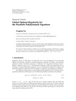
Functional conformation of P-gp induced by IINsFigure 1
Functional conformation of P-gp induced by IINs. Fluorescence profile of mAb UIC2 staining on MDR CEM-VBL10 cells
incubated in presence of the drug diluent (red histogram), 50 µg/ml of the indicated IINs (blue histogram), or the P-gp sub-
strate vinblastine (10 µg/ml, VBL). MAb MM4.17 staining was carried out in identical conditions in MDR CEM-VBL10 cells incu-
bated with drug diluent (red histogram) or 50 µg/ml of the RDS1974 (blue histogram).
Retrovirology 2007, 4:17 />Page 5 of 10
(page number not for citation purposes)
ity than verapamil, a drug that has been tested in clinical
trials to chemosensitize MDR tumours [36]; nevertheless,
in view of their relatively low in vitro cytotoxicity, this class
of IINs should be further investigated as potential chemo-
sensitizing compounds for P-gp expressing MDR
tumours.
Conclusion
IINs are among the most promising agents for the treat-
ment of HIV infection [26], with two of them being in an
advanced stage of clinical development [3-5]. To our
knowledge, this is the first study showing that IINs are
substrates for P-gp. However, we do not know whether
this property is shared by all IINs or is restricted to the
diketo acid class of IINs tested in this study [27], acting as
P-gp substrates by inducing the up-modulation of UIC2
epitope and inhibiting doxorubicin efflux in MDR CEM-
VBL100 cells (Table 2 and Fig 2). Concerning the com-
pounds that are under clinical trials, while the MK 0518
[3,4] is a chemically distinct compound, the GS-9137 [5]
is a quinolonyl diketoacid derivative comparable to the
molecules used in our study. Thus, with good approxima-
tion, for the GS-9137 we may hypothesize a similar pat-
tern of response as that observed for the DKA used in the
study. Further studies aimed at evaluating the interaction
of other clinically significant IINs with the P-gp system
will be needed to better address this question.
In our opinion, a successful P-gp modulation may add
further interest to this highly promising class of antiretro-
viral agents and open new perspectives for their clinical
use in fields other than HIV infection.
Materials and methods
Integrase Inhibitors and Chemicals
Nine quinolonyl diketoacid derivatives inhibiting the
strand transfer step of HIV-1 integration (IC50: 0.042–32
µM) [27] were used in this study. Verapamil (Isoptin) was
provided by BASF-Knoll (Milan, Italy), Vinblastine
(Velbe) by Eli Lilly (Paris, France) and Doxorubicin by
Farmitalia (Nerviano, Italy).
Antiviral activity and cytotoxicity
Anti-HIV activity was measured in the human T lymphoid
H9 cells. To this purpose, cells were cultured in RPMI
1640, supplemented with 2 mM L-glutamine, penicillin,
streptomycin and 10% fetal bovine serum (FBS), and
infected with the HTLVIIIB laboratory strain of HIV-1
virus (100000 TCID
50
– Tissue Culture Infective Dose- per
10
6
PBMC). After two hours of incubation, cells were
washed with medium, and cultured at 37°C (5000 cells/
well in 96-well microplates) for 3 days in presence of
medium and test compounds at concentrations ranging
from 50 µM to 0.1 µM. After 3 days p24 antigen concen-
Table 3: Induction of P-gp expression in CEM and CEMrev cell
lines exposed to IINs
Compound Concentration Days of
culture
% of P-gp expressing cells
CEM CEMrev
None 0 1–3
RDS 1974 10 µg/ml 104 0 25–30
RDS 1983 25 µg/ml 28 0 7–10
RDS 1984 25 µg/ml 28 0 15–18
RDS 1996 10 µg/ml 28 0 17–20
Vinblastine 10 ng/ml 28 ND* 30–35
104 ND* 60–70
ND, not done
* % of P-gp expressing cells could not be evaluated due to VBL
toxicity. Prolonged and stepwise VBL treatment induced high
percentage of P-gp expressing cells [31].
Table 2: Induction of functional conformation and drug transport inhibition exerted by other IINs in CEM MDR cells.
Compound Concentration µg/ml UIC2 epitope up-modulation (A) Doxorubicin efflux inhibition (B)
RDS 1992 25 NT +
50 + ++
100 ++ NT
RDS 1996 25 NT +
50 NT ++
100 NT NT
RDS 1997 25 NT +
50 + ++
100 ++ NT
RDS 2012 25 NT +
50 + ++
100 ++ NT
RDS 2022 25 NT +
50 + ++
100 ++ NT
A, modulation of UIC2 epitope was studied in CEM-VBL10 cells; B, the effect of IINs on the doxorubicin transport inhibition was analysed in CEM-
VBL100 cells; NT, not tested; the + or ++ symbols indicate arbitrary units in measuring and differentiating the level of UIC2 epitope modulation and
doxorubicin retention.
Retrovirology 2007, 4:17 />Page 6 of 10
(page number not for citation purposes)
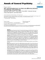
Drug transport inhibition mediated by IINsFigure 2
Drug transport inhibition mediated by IINs. Evaluation of efflux of the dye P-gp substrate doxorubicin in CEM-VBL100
MDR cells. Efflux was monitored in drug-free conditions (red histogram), in the presence of the potent P-gp blocker Verapamil
(2.5 µg/ml) (blue histogram) or following incubation with several IINs (RDS1974, RDS1981, RDS1983 and RDS1984) (green
histogram) at the indicated concentrations (range 1 µg/ml to 100 µg/ml).
Retrovirology 2007, 4:17 />Page 7 of 10
(page number not for citation purposes)
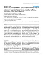
P-gp inhibition and MDR chemosensitizationFigure 3
P-gp inhibition and MDR chemosensitization. In (A), KB-V1 MDR cells were incubated at 37°C for 1 hr with 5 µg/ml
doxorubicin alone or in presence of 2.5 µg/ml verapamil or 25 µg/ml RDS 1984. After washing the cells were reincubated again
in identical conditions and doxorubicin efflux/retention were analysed in confocal microscopy after 1 h (panel a-c) and 3 hrs
(panel d-f). The natural efflux of doxorubicin P-gp mediated is shown in panel a and d, while the doxorubicin retention due to
the P-gp drug transport inhibition exerted by verapamil and RDS 1984 is shown in panel b-c (1 h incubation) or e-f (3 hrs incu-
bation). In (B), dose-response cytotoxicity to vinblastine in CEM-VBL100 MDR cells in presence of verapamil (2.5 µg/ml) or 10
µg/ml of the IINs RDS 1974, RDS 1981, RDS 1983 and RDS 1984 is shown. The values (formazan absorbance at 440 nm in
ELISA reader) were calculated as % of control cells cultured in presence of IINs only or verapamil. The mean of triplicate meas-
urements is shown; the SD was < 15% of each single value.
0
20
40
60
80
100
120
0.001 0.01 0.1 1 10 100
Vinblastine (µg/ml)
Vinblastine
RDS1974
RDS1981
RDS1983
RDS1984
Verapamil
A
B
Retrovirology 2007, 4:17 />Page 8 of 10
(page number not for citation purposes)
tration in the supernatants was measured by an ELISA
assay (Innotest HIV antigen mAb, Innogenetics NV Bel-
gium). Cell viability was determined by the trypan blue
exclusion method.
The 50% inhibitory drug concentration (IC50) and the
50% cytotoxic drug concentration (CC50) were calculated
by the median effect equation using Calcusyn Version 2.0
program (Biosoft Cambridge)
Cell lines
The multidrug resistant (MDR) variants CEM-VBL10 and
CEM-VBL100 cells were isolated by stepwise selection of
the parental drug sensitive CCRF-CEM (CEM) in the pres-
ence of increasing concentrations of VBL [31,32]. Cells
were grown under standard conditions for mammalian
cells cultured in suspension. The basic medium (BM) for
cell culturing consisted of RPMI-1640 supplemented with
10% foetal calf serum (FCS), L-glutamine (2 mM) penicil-
lin (100 U/mL) and streptomycin (100 U/mL). All these
components were purchased from Hyclone (Logan,
Utah). Identical BM, culture conditions and trypsin
(Hyclone) were used for the adherent MDR variant KB.V1
of the human oral epidermoid carcinoma KB cells [10]. To
test the ability of selected IINs (RDS 1974, RDS 1981, RDS
1984 and RDS 1996) to induce de novo expression of P-
glycoprotein, the drug-sensitive CEMrev cell line was
used; this cell line derives from the CEM-VBL10 MDR cell
line, cultured for more than 2 years in VBL-free medium
and expresses very low (1–3%) or undetectable amounts
of P-gp [32].
MDR efflux assay
CEM-VBL100 cells (1 × 10
6
) were loaded with doxoru-
bicin (10 µg/ml) in 1 ml of BM in the presence of a several
IINs (concentrations ranging from 50 µg/ml to 1 mg/ml)
or Verapamil (2.5 µg/ml) for 1 h at 37°C. The cells were
incubated with doxorubicin (10 µg/ml) only or drug dilu-
ent in parallel cultures. At the end of incubation, the cells
were washed in serum-free medium and resuspended in
BM in the presence of the IINs or Verapamil (drug diluent
was added in control samples) for a further 1 h at 37°C.
Finally, cells were washed twice with ice-cold PBS/FACS,
and analyzed in a flow cytometer (FACScan, Becton Dick-
inson, San Josè, CA).
Monoclonal antibodies and UIC-2 Shift assay
The anti CD4-FITC mAb was purchased from Vinci Bio-
chem, Firenze, Italy. The mAb UIC2 [30] was kindly pro-
vided by Dr. E. Mechetner (Chemicon Inc, Temecula, CA).
For determination of P-gp expression, the mAb MM4.17,
recognizing an extracellular P-gp epitope on intact/living
human MDR cells [28], was also used. Both UIC2 and
MM4.17 mAbs were used in a highly purified form.
The UIC2 shift assay was performed under physiological
conditions as previously described [29,33]. CEM-VBL10
cells (1 × 10
6
) were resuspended in 1 ml of PBS containing
2% FCS and allowed to equilibrate at 37°C in a water bath
for 10 min. The various IINs were added to samples (final
concentrations 100 or 50 µg/ml) and incubated for addi-
tional 15 min at 37°C with purified UIC2 mAb (final con-
centration 12.5 µg/ml). VBL (10 µg/ml), which is a well
known UIC2 shifting agent, and the drug diluents were
used as positive and negative controls, respectively. Cells
were then washed twice in ice-cold PBS containing 2%
FCS with 0.01 % sodium azide (Shift Stop Buffer, SSB),
stained on ice in SSB for additional 15 min with 5 µg/ml
of fluorescein -conjugated goat-antimouse antibody
(FITC-GAM, Cappel, West Chester, Pa, USA), washed
twice with ice cold PBS/FACS and maintained in ice until
flow cytometry analysis. The UIC2 shift is the difference
between UIC2 binding in the presence versus the absence
of the IINs under physiological conditions (37°C).
Flow cytometry and confocal microscopy
For confocal laser-scanning microscopy (CLSM) analyses,
KB-V1 adherent cells which express high level of MDR1 P-
glycoprotein [10] were grown in WillCo-dishes (WillCo
Wells B.V., Amsterdam, The Netherlands) for 24 hours.
For P-gp inhibition experiments, the cells were incubated
with 5 µg/ml doxorubicin for 1 h at 37°C in presence and
absence of the IIN RDS 1984 (25 µg/ml). After washing,
the cells were incubated for further 1 and 3 hrs at 37°C in
the same above described conditions to allow the efflux/
block of doxorubicin. CSLM observations were performed
using a Leica TCS 4D apparatus (Leica Lasertechnik
GmbH, Heidelberg, Germany), equipped with an argon-
krypton laser, 488 nm-dichroic splitter and LP515 long
pass filter. Image acquisition and processing were con-
ducted using the SCANware (Leica) and Adobe Pho-
toshop (Adobe Systems Inc., Mountain View, CA)
software programs.
MDR reversing
For the evaluation of the MDR reversing ability, CEM-
VBL100 cells in exponential phase of growth were col-
lected, extensively washed with warm RPMI-1640 and
resuspended at the concentration of 5 × 10
3
cells/ml in
BM alone, or in the presence of the IINs or Verapamil, as
appropriated. Then cells were seeded (in triplicate) in 96-
wells Costar plates (Costar, Rochester, NY) in which dif-
ferent VBL concentrations were previously added. Within
its inhibitory range, the drug decreased growth of all cell
lines proportionally to drug concentration. Cell prolifera-
tion was determined by adding 10 µg/well of PreMix WST-
1 (PreMix WST-1 cell proliferation kit, Vinci Biochem,
Firenze, Italy) to the cultures and measuring the absorb-
ance at about 440 nm in a microplate ELISA reader after 4
hrs incubation (48 hrs in total) [37]. The relative cell
Retrovirology 2007, 4:17 />Page 9 of 10
(page number not for citation purposes)
growth was calculated by applying the formula (En-E0)/
(Cn-C0) where E0 and En are the initial and after 48-treat-
ment absorbance values in the drug-containing cultures,
and C0 and Cn are the corresponding absorbance values
in the untreated control culture. The obtained dose-
response profile fulfilled the concentration inhibiting
growth by 50% (IC50).
Competing interests
The author(s) declare that they have no competing inter-
ests.
Authors' contributions
CM conceived and planned the biological approach of
this study, participated in the design and coordination of
the research and drafted the manuscript.
DML conceived and conducted all the cell biological
experiments to demonstrate the P-gp substrate activity of
the IINs.
MA carried out confocal microscopy studies for the visual-
ization of P-gp mediated activity of the IINs
VA purified and characterized at the biochemical level all
the mAbs used in this study and utilized for cell line phe-
notyping
CR collaborated in the design and synthesis of Integrase
Inhibitors
GCM and AM carried out the studies on antiviral activity
and cytotoxicity of the IINs in cell based assays.
CA was involved in revising the manuscript critically
DSR conceived and designed the IINs
PL coordinated and supervised the study, interpreted the
results and participated in drafting the manuscript
Acknowledgements
This work was supported by AIDS grants of Istituto Superiore di Sanità and
Italian Ministry of Health and partly by an ISS-NIH research grant.
We wish to thank Mr Marco Sabatini and Mrs Marina Tombesi for graphical
and technical support.
References
1. Vella S, Palmisano L: The global status of resistance to antiret-
roviral drugs. Clin Infect Dis 2005, 41(Suppl 4):S239-246.
2. Hazuda DJ, Young SD, Guare JP, Anthony NJ, Gomez RP, Wai JS,
Vacca JP, Handt L, Motzel SL, Klein HJ, Dornadula G, Danovich RM,
Witmer MV, Wilson KA, Tussey L, Schleif WA, Gabryelski LS, Jin L,
Miller MD, Casimiro DR, Emini EA, Shiver JW: Integrase inhibitors
and cellular immunity suppress retroviral replication in rhe-
sus macaques. Science 2004, 305:528-532.
3. Markowitz M, Morales-Ramirez JO, Nguyen B-Y, Kovacs CM, Steig-
bigel RT, Cooper DA, Liporace R, Schwartz R, isaacs R, Gilde LR,
Wenning L, Zhao J, Teppler H: Antiretroviral activity, pharma-
cokinetics, and tolerability of MK-0518 a novel inhibitor of
HIV-1 integrase, dosed as monotherapy for 10 days in treat-
ment-naúve HIV-1-infected individuals. J Acquir Immune Defic
Syndr 2006, 43:509-515.
4. Grinsztejn B, Nguyen BY, Katlama C, Gatell J, Lazzarin A, Vittecoq D,
Gonzalez C, Chen J, Isaacs R, the Protocol 005 Study Team: Potent
antiretroviral effect of MK-0518 a novel HIV-1 integrase
inhibitor, in patient with triple-class resistant virus. Proceed-
ings of the 13th Conference on Retroviruses and Opportunistic Infection,
Denver CO, 5–8 February 2006 Abstract LB 159 .
5. DeJesus E, Berger D, Markowitz M, Cohen C, Hawkins T, Ruane P,
Elion R, Farthing C, Zhong L, Cheng AK, McColl D, Kearney BP, for
the 183-0101 Study Team: Antiviral activity, pharmacokinetics,
and dose response of the HIV integrase inhibitor GS-9137
(JTK-303) in treatment-naive and treatment-experienced
patients. J Acquir Immune Defic Syndr 2006, 43:1-5.
6. Evans WE, McLeod HL: Pharmacogenomics – drug disposition,
drug targets, and side effects. N Engl J Med 2003, 348:538-549.
7. Schinkel AH: P-Glycoprotein, a gatekeeper in the blood-brain
barrier. Adv Drug Deliv Rev 1999, 36:179-194.
8. Huisman MT, Smit JW, Schinkel AH: Significance of P-glycopro-
tein for the pharmacology and clinical use of HIV protease
inhibitors. AIDS 2000, 14:237-242.
9. Kartner N, Riordan JR, Ling V: Cell surface P-glycoprotein asso-
ciated with multidrug resistance in mammalian cell lines. Sci-
ence 1983, 221:1285-1288.
10. Ueda K, Cornwell MM, Gottesman MM, Pastan I, Roninson IB, Ling V,
Riordan JR: The mdr1 gene, responsible for multidrug-resist-
ance, codes for P-glycoprotein. Biochem Biophys Res Commun
1986, 141:956-962.
11. Germann UA, Pastan I, Gottesman MM: P-glycoproteins: media-
tors of multidrug resistance. Semin Cell Biol 1993, 4:63-76.
12. Endicott JA, Ling V: The biochemistry of P-glycoprotein-medi-
ated multidrug resistance. Annu Rev Biochem 1989, 58:137-171.
13. Schinkel AH, Borst P: Multidrug resistance mediated by P-glyc-
oproteins. Semin Cancer Biol 1991, 2:213-226.
14. Kim RB, Fromm MF, Wandel C, Leake B, Wood AJ, Roden DM,
Wilkinson GR: The drug transporter P-glycoprotein limits oral
absorption and brain entry of HIV-1 protease inhibitors. J Clin
Invest 1998, 101:289-294.
15. Lee CG, Gottesman MM, Cardarelli CO, Ramachandra M, Jeang KT,
Ambudkar SV, Pastan I, Dey S: HIV-1 protease inhibitors are sub-
strates for the MDR1 multidrug transporter. Biochemistry
1998, 37:3594-3601.
16. Jones K, Hoggard PG, Sales SD, Khoo S, Davey R, Back DJ: Differ-
ences in the intracellular accumulation of HIV protease
inhibitors in vitro and the effect of active transport. Aids 2001,
15:675-681.
17. Washington CB, Duran GE, Man MC, Sikic BI, Blaschke TF: Interac-
tion of anti-HIV protease inhibitors with the multidrug
transporter P-glycoprotein (P-gp) in human cultured cells. J
Acquir Immune Defic Syndr Hum Retrovirol 1998, 19:203-209.
18. Perloff ES, Duan SX, Skolnik PR, Greenblatt DJ, von Moltke LL: Ata-
zanavir: effects on P-glycoprotein transport and CYP3A
metabolism in vitro. Drug Metab Dispos 2005, 33:764-770.
19. Chaudhary PM, Mechetner EB, Roninson IB: Expression and activ-
ity of the multidrug resistance P-glycoprotein in human
peripheral blood lymphocytes. Blood 1992, 80:2735-2739.
20. Klimecki WT, Futscher BW, Grogan TM, Dalton WS: P-glycopro-
tein expression and function in circulating blood cells from
normal volunteers. Blood 1994, 83:2451-2458.
21. Ludescher C, Pall G, Irschick EU, Gastl G: Differential activity of
P-glycoprotein in normal blood lymphocyte subsets. Br J Hae-
matol 1998, 101:722-727.
22. Thiebaut F, Tsuruo T, Hamada H, Gottesman MM, Pastan I, Willing-
ham MC: Cellular localization of the multidrug-resistance
gene product P-glycoprotein in normal human tissues. Proc
Natl Acad Sci USA 1987, 84:7735-7738.
23. Cordon-Cardo C, O'Brien JP, Boccia J, Casals D, Bertino JR, Melamed
MR: Expression of the multidrug resistance gene product (P-
glycoprotein) in human normal and tumor tissues. J Histo-
chem Cytochem 1990, 38:1277-1287.
24. Hoetelmans RM: Sanctuary sites in HIV-1 infection. Antivir Ther
1998, 3(Suppl 4):13-17.
Publish with BioMed Central and every
scientist can read your work free of charge
"BioMed Central will be the most significant development for
disseminating the results of biomedical research in our lifetime."
Sir Paul Nurse, Cancer Research UK
Your research papers will be:
available free of charge to the entire biomedical community
peer reviewed and published immediately upon acceptance
cited in PubMed and archived on PubMed Central
yours — you keep the copyright
Submit your manuscript here:
/>BioMedcentral
Retrovirology 2007, 4:17 />Page 10 of 10
(page number not for citation purposes)
25. Van Tellingen O: The importance of drug-transporting P-glyc-
oproteins in toxicology. Toxicol Lett 2001, 120:31-41.
26. Pommier Y, Johnson AA, Marchand C: Integrase inhibitors to
treat HIV/AIDS. Nat Rev Drug Discov 2005, 4:236-248.
27. Di Santo R, Costi R, Roux A, Artico M, Lavecchia A, Marinelli L,
Novellino E, Palmisano L, Andreotti M, Amici R, Galluzzo CM, Nen-
cioni L, Palamara AT, Pommier Y, Marchand C: Novel bifunctional
quinolonyl diketo acid derivatives as HIV-1 integrase inhibi-
tors: design, synthesis, biological activities, and mechanism
of action. J Med Chem 2006, 49:1939-1945.
28. Mechetner EB, Schott B, Morse BS, Stein WD, Druley T, Davis KA,
Tsuruo T, Roninson IB: P-glycoprotein function involves confor-
mational transitions detectable by differential immunoreac-
tivity. Proc Natl Acad Sci USA 1997, 94:12908-12913.
29. Nagy H, Goda K, Arceci R, Cianfriglia M, Mechetner E, Szabo G Jr: P-
Glycoprotein conformational changes detected by antibody
competition. Eur J Biochem 2001, 268:2416-2420.
30. Cianfriglia M, Willingham MC, Tombesi M, Scagliotti GV, Frasca G,
Chersi A: P-glycoprotein epitope mapping. I. Identification of
a linear human-specific epitope in the fourth loop of the P-
glycoprotein extracellular domain by MM4.17 murine mon-
oclonal antibody to human multi-drug-resistant cells. Int J
Cancer 1994, 56:153-160.
31. Cianfriglia M, Cenciarelli C, Tombesi M, Barca S, Mariani M, Morrone
S, Santoni A, Samoggia P, Alessio M, Malavasi F: Murine monoclonal
antibody recognizing a 90-kDa cell-surface determinant
selectively lost by multi-drug-resistant variants of CEM cells.
Int J Cancer 1990, 45:95-103.
32. Cenciarelli C, Currier SJ, Willingham MC, Thiebaut F, Germann UA,
Rutherford AV, Gottesman MM, Barca S, Tombesi M, Morrone S,
Ramoni C, Cianfriglia M: Characterization by somatic cell
genetics of a monoclonal antibody to the MDR1 gene prod-
uct (P-glycoprotein): determination of P-glycoprotein
expression in multi-drug-resistant KB and CEM cell variants.
Int J Cancer 1991, 47:533-543.
33. Dupuis ML, Flego M, Molinari A, Cianfriglia M: Saquinavir induces
stable and functional expression of the multidrug trans-
porter P-glycoprotein in human CD4 T-lymphoblastoid
CEMrev cells. HIV Med 2003, 4:338-345.
34. Jachez B, Cianfriglia M, Loor F: Modulation of human P-glycopro-
tein epitope expression by temperature and/or resistance-
modulating agents. Anticancer Drugs 1994, 5:655-665.
35. Gottesman MM, Fojo T, Bates SE: Multidrug resistance in cancer:
role of ATP-dependent transporters. Nat Rev Cancer 2002,
2:48-58.
36. Szakacs G, Paterson JK, Ludwig JA, Booth-Genthe C, Gottesman MM:
Targeting multidrug resistance in cancer. Nat Rev Drug Discov
2006, 5:219-234.
37. Skehan P, Storeng R, Scudiero D, Monks A, McMahon J, Vistica D,
Warren JT, Bokesch H, Kenney S, Boyd MR: New colorimetric
cytotoxicity assay for anticancer-drug screening. J Natl Cancer
Inst 1990, 82:1107-1112.