Báo cáo y học: "Discovery of a new human T-cell lymphotropic virus (HTLV-3) in Central Africa" potx
Bạn đang xem bản rút gọn của tài liệu. Xem và tải ngay bản đầy đủ của tài liệu tại đây (744.22 KB, 4 trang )
BioMed Central
Page 1 of 4
(page number not for citation purposes)
Retrovirology
Open Access
Short report
Discovery of a new human T-cell lymphotropic virus (HTLV-3) in
Central Africa
Sara Calattini
†1
, Sébastien Alain Chevalier
†1
, Renan Duprez
1
,
Sylviane Bassot
1
, Alain Froment
2
, Renaud Mahieux
†1
and Antoine Gessain*
†1
Address:
1
Unité d'Epidémiologie et Physiopathologie des Virus Oncogènes, Institut Pasteur, 28 rue du Dr Roux, 75015 Paris, France and
2
Laboratoire ERMES, IRD, Technoparc, Orléans cedex 2, France
Email: Sara Calattini - ; Sébastien Alain Chevalier - ; Renan Duprez - ;
Sylviane Bassot - ; Alain Froment - ; Renaud Mahieux - ;
Antoine Gessain* -
* Corresponding author †Equal contributors
Abstract
Human T-cell Leukemia virus type 1 (HTLV-1) and type 2 (HTLV-2) are pathogenic retroviruses
that infect humans and cause severe hematological and neurological diseases. Both viruses have
simian counterparts (STLV-1 and STLV-2). STLV-3 belongs to a third group of lymphotropic viruses
which infect numerous African monkeys species. Among 240 Cameroonian plasma tested for the
presence of HTLV-1 and/or HTLV-2 antibodies, 48 scored positive by immunofluorescence.
Among those, 27 had indeterminate western-blot pattern. PCR amplification of pol and tax regions,
using HTLV-1, -2 and STLV-3 highly conserved primers, demonstrated the presence of a new
human retrovirus in one DNA sample. tax (180 bp) and pol (318 bp) phylogenetic analyses
demonstrated the strong relationships between the novel human strain (Pyl43) and STLV-3 isolates
from Cameroon. The virus, that we tentatively named HTLV-3, originated from a 62 years old
Bakola Pygmy living in a remote settlement in the rain forest of Southern Cameroon. The plasma
was reactive on MT2 cells but was negative on C19 cells. The HTLV 2.4 western-blot exhibited a
strong reactivity to p19 and a faint one to MTA-1. On the INNO-LIA strip, it reacted faintly with
the generic p19 (I/II), but strongly to the generic gp46 (I/II) and to the specific HTLV-2 gp46. The
molecular relationships between Pyl43 and STLV-3 are thus not paralleled by the serological
results, as most of the STLV-3 infected monkeys have an "HTLV-2 like" WB pattern. In the context
of the multiple interspecies transmissions which occurred in the past, and led to the present-day
distribution of the PTLV-1, it is thus very tempting to speculate that this newly discovered human
retrovirus HTLV-3 might be widespread, at least in the African continent.
Findings
Three types of Primate T-cell lymphotropic viruses
(PTLVs) have been discovered so far in primates [1]. While
two of them i.e. PTLV-1 and PTLV-2 include human
(HTLV-1, HTLV-2) and simian (STLV-1, STLV-2) viruses,
the third type (STLV-3) consists only, so far, of simian
strains. Sequence comparisons of STLV-3 proviruses indi-
cated that these strains are highly divergent from HTLV-1
(60% nucleotide similarity), HTLV-2 (62%), or STLV-2
(62%) prototype sequences. In all phylogenetic analyses,
Published: 09 May 2005
Retrovirology 2005, 2:30 doi:10.1186/1742-4690-2-30
Received: 20 April 2005
Accepted: 09 May 2005
This article is available from: />© 2005 Calattini et al; licensee BioMed Central Ltd.
This is an Open Access article distributed under the terms of the Creative Commons Attribution License ( />),
which permits unrestricted use, distribution, and reproduction in any medium, provided the original work is properly cited.
Retrovirology 2005, 2:30 />Page 2 of 4
(page number not for citation purposes)
STLV-3 viruses cluster in a highly supported group, indi-
cating an evolutionary lineage independent from PTLV-1
and PTLV-2. Nevertheless, STLV-3 lineage is composed of
at least three subtypes that are corresponding more or less
to the geographical origin of the virus (East, West or Cen-
tral Africa) [2-9]. Most of the viruses belonging to the
PTLV-1 type cannot be separated into distinct phyloge-
netic lineages according to their species of origin. Their
intermixing has therefore been inferred as an evidence for
past or recent interspecies transmission episodes. The
hypothesis of viral transmission from monkeys to
humans is supported by an increasing number of observa-
tions [1]. Thus, it has been proposed that HTLV strains
related to STLV-3 might infect human populations living
in areas where STLV-3 is present.
Cameroon has a remarkable diversity of retroviruses. All
the subtypes of HIV-1 group M (A to H) are present, sub-
type-recombinant strains co-circulate, and HIV-1 groups
O and N have been reported. Besides, HTLV-1 subtypes B
and D as well as HTLV-2 type A and B are also present in
Cameroonian individuals, while STLV-1 and STLV-3
strains have been isolated from several non-human pri-
mates (NHPs) species living in this region [3,4,8]. We
therefore conducted a study to search for HTLV variants in
Cameroonian individuals with HTLV-1/2 indeterminate
serology. This survey was approved by both the national
(Cameroon Ministry of Health and their National Ethics
committee) and local authorities (village chief) with
information to each participant. An oral informed con-
sent was obtained from each participant (adults or parents
for minors). A series of 240 blood samples was obtained
from Bakola (n = 64) and Baka (n = 65) Pygmies, while
others (n = 111) were obtained from Bantous (mainly
from the Fang, Mvae and Ngumba tribes). All these indi-
viduals (117 women and 123 men, mean age 44, range
10–75 years) live in remote villages in the rain forest area
of the Southern part of Cameroon.
The 240 plasma were tested at a 1/40 dilution for the pres-
ence of HTLV-1/2 antibodies with a highly sensitive
immunofluorescence test (IF), that uses MT2 and C19 as
HTLV-1 and HTLV-2 viral antigen producing cells respec-
tively. This test also allows the detection of STLV-3 posi-
tive samples [4,5]. The 48 plasma that were IF reactive on
MT2, C19 or both, were further tested by western blot
(WB HTLV BLOT 2.4; Genelabs Diagnostics, Singapore).
Among the 48 samples tested, 4 and 11 WB patterns were
very evocative of HTLV-1 and HTLV-2 infection respec-
tively, while 27 exhibited diverse HTLV incomplete pat-
terns, including some HTLV-1 indeterminate gag profile
(HGIP). Six samples were WB negative. High-molecular
weight DNA was extracted from the 48 blood samples and
was first subjected to PCR using human β-globin specific
primers, to ensure that DNA was amplifiable. They were
then subjected to two series of PCR using degenerated tax
and pol primers designed on highly conserved regions that
are common to all PTLVs. The tax primers are the follow-
ing: SCTaxoutse: 5'-CTHTAYGGRTACCCHGTCTACGT-3'
and SCTaxoutas: 5'-AGGGGAGBCGAGGGATAAGG-3'
corresponding to nucleotides 7279 to 7301 and 7455 to
7474 respectively of the prototype STLV-3
PHA969
sequence
(GenBank accession number Y07616). The pol primers are
SCPOL1outse: 5'-TTAAACCDGARCGCCTCCAGGC-3' (nt
2485 to 2506) SCPOL1outas: 5'-GGDGTDCCYTTRGA-
GACCCA-3' (nt 3201 to 3220) and SCPOL1inse: 5'-TAY-
HHAGGRCCAGGMAATAACCC-3' (nt 2556 to 2578).
HTLV-1 and HTLV-2 tax sequences were obtained for 4
and 11 samples which exhibited complete HTLV-1 and
HTLV-2 WB profiles respectively, but none of the WB
indeterminate sample gave a PCR signal. Consistent
results were obtained for these HTLV-1 and HTLV-2
strains with the pol semi-nested PCR. However, a faint
band (665 bp) was also obtained for one sample (Pyl43),
which was previously found to be tax PCR negative.
Sequencing of this fragment indicated the presence of an
HTLV pol sequence that is highly related to STLV-3 strains
(86.6% to 99.2% nucleotide identity). Based on an align-
ment of different STLV-3 sequences, a tax semi-nested
PCR was then designed using SCTaxoutse (see above) and
Mac4 followed by Mac2 and Mac4 as inner primers [10].
This allowed the amplification of a 279 bp fragment
which was also found to be highly homologous to STLV-
3 strains (92.4% to 99.6% nucleotide identity). We did
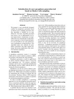
two phylogenetic analyses (tax and pol) with the neighbor
joining method. Assessment of a 180-bp tax sequence
(Figure 1) or of a 665-bp pol region (data not shown)
demonstrated a strong relationship between Pyl43 and
STLV-3 strains from Cameroon.
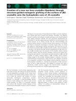
The HTLV-3 sample originated from a 62 years old Bakola
Pygmy living in a remote settlement in the ocean depart-
ment of Southern Cameroon. His plasma was reactive on
MT2 cells (titer: 1/320) but was negative on C19 cells. The
HTLV BLOT 2.4 WB [11] exhibited a strong reactivity to
p19 and a faint one to MTA-1 (Figure 2A). On the INNO-
LIA strip (Innogenetics, Ghent, Belgium) [12], it reacted
faintly (+/-) with the generic p19 (I/II), but strongly to the
generic env gp46 (I/II) and to the specific HTLV-2 gp46
(Figure 2B). Surprisingly, the close molecular relationship
between Pyl43 and STLV-3 is thus not paralleled by the
serological results, as most of the STLV-3 infected mon-
keys have an "HTLV-2 like" WB pattern (p24 > to p19 with
or without K55) (Figure 2A, lanes 3–4) [2-9].
In conclusion, we have demonstrated in this report the
presence of a new human retrovirus in the peripheral
blood cells of a Central African native. This virus is closely
related to STLV-3. In the context of multiple interspecies
Retrovirology 2005, 2:30 />Page 3 of 4
(page number not for citation purposes)
transmissions that have occurred in the past and led to the
present-day distribution of the PTLV-1 [1], we suggest that
HTLV-3 might be widespread, throughout the African
continent. HTLV-3 infection seems to be reflected by an
HTLV indeterminate serological WB pattern. This raises an
important public health question regarding the effective-
ness of the current commercially available screening and
confirmation tests for detecting this new HTLV type. Key
research priorities are now to investigate the transmission
modes of this virus as well as possible pathogenic
associations.
HTLV-3 is closely related to STLV-3Figure 1
HTLV-3 is closely related to STLV-3. Unrooted phylo-
genetic tree generated with the Neighbor-joining method,
performed in the PAUP program (v4.0b10), on a 180 bp frag-
ment of the tax gene using all full length PTLV-1/2 available
sequences and all published STLV-3 tax sequences. The
PTLV-1/2/3 strains, including (in bold), the novel sequence
generated in this work (Pyl43), were aligned with the
DAMBE program (version 4.2.13). The final alignment was
submitted to the Modeltest program (version 3.6) to select,
according to the Akaike Information Criterion (AIC), the
best model to apply to phylogenetic analyses. The selected
model was the TrN+G. Bootstrap support (1,000 replicates)
is noted on the branches of the tree. The branch lengths are
drawn to scale, with the bar indicating 0.1 nucleotide
replacement per site.
Serological pattern of the person infected by the HTLV-3 Pyl43 strainFigure 2
Serological pattern of the person infected by the
HTLV-3 Pyl43 strain. (A) Western Blot from Genelabs
Diagnostics (HTLV BLOT 2.4 version) and (B) a line immu-
noassay (INNO-LIA HTLV confirmation Immunogenetics).
The HTLV 2.4 western blot kit is based on strips incorporat-
ing HTLV-1/2 native viral antigens (originating from HTLV-1
infected cells) to which HTLV-1 (MTA-1) or HTLV-2 (K55)
gp46s or HTLV-1 and HTLV-2 (GD21) gp21 recombinant
proteins have been added [11]. The INNO LIA kit uses only
recombinant antigens and synthetic peptides derived from
both HTLV-1 and HTLV-2 proteins sequences. Whereas gag
p19 I/II corresponds both to a recombinant protein and syn-
thetic peptides being recognized by anti HTLV-1 and HTLV-2
immune sera, env gp46 I/II corresponds only to synthetic
peptides recognized by anti HTLV-1 and HTLV-2 immune
sera. env gp46 II corresponds to synthetic peptides specific of
HTLV-2 [12]. (A, B) Lane 1: HTLV-1 positive control; lane 2:
HTLV-2 positive control; lane 3: STLV-3 positive control
(STLV-3
604
strain); lane 4: STLV-3 positive control (STLV-
3
F3
); lane 5: HTLV-1/2 negative control; lane 6: plasma from
the person infected by HTLV-3 (Pyl43 strain).
Publish with Bio Med Central and every
scientist can read your work free of charge
"BioMed Central will be the most significant development for
disseminating the results of biomedical research in our lifetime."
Sir Paul Nurse, Cancer Research UK
Your research papers will be:
available free of charge to the entire biomedical community
peer reviewed and published immediately upon acceptance
cited in PubMed and archived on PubMed Central
yours — you keep the copyright
Submit your manuscript here:
/>BioMedcentral
Retrovirology 2005, 2:30 />Page 4 of 4
(page number not for citation purposes)
List of abbreviations used
PTLV: Primate T Lymphotropic Viruses
HTLV: Human T Cell Lymphotropic Virus
PCR: Polymerase Chain Reaction
WB: western-blot
NHPs: Non Human Primates
HGIP: HTLV Gag Indeterminate Profile
Nucleotide accession number
The tax and pol accession number for the sequences deter-
mined in this study are: [GenBank:DQ020492
,
GenBank:DQ020493
] respectively.
Competing interests
The author(s) declare that they have no competing
interests.
Authors' contributions
SC and SAC performed the laboratory work. SB did the
serological assay and RD the phylogenetic analyses. AF
and AG organized and performed the field studies, AG
and RM designed, implemented and coordinated the
study, wrote the manuscript. All authors have read and
approved the manuscript.
Note
Wolfe et al. recently reported in an abstract the presence of
two novel HTLV viruses [13]. Whether or not these viruses
are related to the new strain described here (HTLV-3
Pyl43) remains to be determined by further comparative
studies.
Acknowledgements
This work was supported by a grant from l'Association de Recherche sur
le Cancer (ARC # 4781) to RM and fellowships from le Ministère de la
Recherche to SAC and Virus Cancer Prévention association to SC. RM is
supported by INSERM. SC and SAC contributed equally to the laboratory
work. RM and AG share senior authorship on this work. We also thank Dr
Timothy Stinear for his critical comments.
References
1. Slattery JP, Franchini G, Gessain A: Genomic evolution, patterns
of global dissemination, and interspecies transmission of
human and simian T-cell leukemia/lymphotropic viruses.
Genome Res 1999, 9:525-540.
2. Goubau P, Van Brussel M, Vandamme AM, Liu HF, Desmyter J: A pri-
mate T-lymphotropic virus, PTLV-L, different from human
T-lymphotropic viruses types I and II, in a wild-caught
baboon (Papio hamadryas). Proc Natl Acad Sci U S A 1994,
91:2848-2852.
3. Courgnaud V, Van Dooren S, Liegeois F, Pourrut X, Abela B, Loul S,
Mpoudi-Ngole E, Vandamme A, Delaporte E, Peeters M: Simian T-
cell leukemia virus (STLV) infection in wild primate popula-
tions in Cameroon: evidence for dual STLV type 1 and type
3 infection in agile mangabeys (Cercocebus agilis). J Virol 2004,
78:4700-4709.
4. Meertens L, Mahieux R, Mauclere P, Lewis J, Gessain A: Complete
sequence of a novel highly divergent simian T-cell lympho-
tropic virus from wild-caught red-capped mangabeys (Cer-
cocebus torquatus) from Cameroon: a new primate T-
lymphotropic virus type 3 subtype. J Virol 2002, 76:259-268.
5. Meertens L, Gessain A: Divergent simian T-cell lymphotropic
virus type 3 (STLV-3) in wild-caught Papio hamadryas papio
from Senegal: widespread distribution of STLV-3 in Africa. J
Virol 2003, 77:782-789.
6. Meertens L, Shanmugam V, Gessain A, Beer BE, Tooze Z, Heneine W,
Switzer WM: A novel, divergent simian T-cell lymphotropic
virus type 3 in a wild-caught red-capped mangabey (Cer-
cocebus torquatus torquatus) from Nigeria. J Gen Virol 2003,
84:2723-2727.
7. Takemura T, Yamashita M, Shimada MK, Ohkura S, Shotake T, Ikeda
M, Miura T, Hayami M: High prevalence of simian T-lympho-
tropic virus type L in wild ethiopian baboons. J Virol 2002,
76:1642-1648.
8. Van Dooren S, Salemi M, Pourrut X, Peeters M, Delaporte E, Van
Ranst M, Vandamme AM: Evidence for a second simian T-cell
lymphotropic virus type 3 in Cercopithecus nictitans from
Cameroon. J Virol 2001, 75:11939-11941.
9. Van Dooren S, Shanmugam V, Bhullar V, Parekh B, Vandamme AM,
Heneine W, Switzer WM: Identification in gelada baboons
(Theropithecus gelada) of a distinct simian T-cell lympho-
tropic virus type 3 with a broad range of Western blot
reactivity. J Gen Virol 2004, 85:507-519.
10. Mahieux R, Pecon-Slattery J, Gessain A: Molecular characteriza-
tion and phylogenetic analyses of a new, highly divergent
simian T-cell lymphotropic virus type 1 (STLV-1marc1) in
Macaca arctoides. J Virol 1997, 71:6253-6258.
11. Varma M, Rudolph DL, Knuchel M, Switzer WM, Hadlock KG, Velli-
gan M, Chan L, Foung SK, Lal RB: Enhanced specificity of trun-
cated transmembrane protein for serologic confirmation of
human T-cell lymphotropic virus type 1 (HTLV-1) and
HTLV-2 infections by western blot (immunoblot) assay con-
taining recombinant envelope glycoproteins. J Clin Microbiol
1995, 33:3239-3244.
12. Zrein M, Louwagie J, Boeykens H, Govers L, Hendrickx G, Bosman F,
Sablon E, Demarquilly C, Boniface M, Saman E: Assessment of a
new immunoassay for serological confirmation and discrim-
ination of human T-cell lymphotropic virus infections. Clin
Diagn Lab Immunol 1998, 5:45-49.
13. Wolfe N, Heneine W, Carr JK, Garcia A, Shanmugam V, Tamoufe U,
Torimiro J, Prosser A, LeBreton M, Mpoudi-Ngole E, Mccutchan F,
Birx DL, Folks T, Burke DS, Switzer WM: Discovery of New
Human T-lymphotropic Viruses Reveals Frequent and
Ongoing Zoonotic Retrovirus Introductions. Conference on Ret-
roviruses and Opportunistic Infections 2005. roconfer
ence.org/2005/cd/Abstracts/25714.htm. Boston, Massachusetts, USA