Báo cáo y học: "Bench-to-bedside review: The value of cardiac biomarkers in the intensive care patient" pdf
Bạn đang xem bản rút gọn của tài liệu. Xem và tải ngay bản đầy đủ của tài liệu tại đây (418.91 KB, 9 trang )
Page 1 of 9
(page number not for citation purposes)
Available online />Abstract
The use of cardiac biomarkers in the intensive care setting is
gaining increasing popularity. There are several reasons for this
increase: there is now the facility for point-of-care biomarker
measurement providing a rapid diagnosis; biomarkers can be used
as prognostic tools; biomarkers can be used to guide therapy; and,
compared with other methods such as echocardiography, the
assays are easier and much more affordable. Two important
characteristics of the ideal biomarker are disease specificity and a
linear relationship between the serum concentration and disease
severity. These characteristics are not present, however, in the
majority of biomarkers for cardiac dysfunction currently available.
Those clinically useful cardiac biomarkers, which naturally received
the most attention, such as troponins and B-type natriuretic
peptide, are not as specific as was originally thought. In the
intensive care setting, it is important for the user to understand the
degree of specificity of these biomarkers and that the interpretation
of the results should always be guided by other clinical information.
The present review summarizes the available biomarkers for
different cardiac conditions. Potential biomarkers under evaluation
are also briefly discussed.
Introduction
Nearly 30% of patients admitted to a general intensive care
unit (ICU) have underlying cardiac diseases, and approxi-
mately one-half of these 30% are admitted to the ICU with
cardiac problems as the primary cause [1,2]. The latter group
is mainly comprised of patients with acute myocardial infarc-
tion, acute heart failure (HF) or cardiogenic shock. Pulmonary
embolism, sepsis-related cardiac dysfunction and arrhythmias
are also commonly found in the ICU.
The diagnosis of cardiac problem can be a difficult task in the
ICU, partly due to the nonspecificity of clinical signs and
symptoms. Prompt treatment can reduce mortality and
improve patient outcome, and therefore the value of rapidly
identifying the problem and assessment of the condition
cannot be understated. Although the introduction of intensive
care echocardiography has made the diagnoses easier,
diagnoses based on echocardiography alone are not always
sufficient and the application requires ready availability of
skilled operators [3]. For example, while an enlarged right
ventricle denotes pressure or volume overloading, echo-
cardiography sheds little light on the etiology. Proper diagnosis
requires the incorporation of various clinical information
including medical history, physical examination, electro-
cardiography, chest X-ray scans and, recently, biomarker levels.
Biomarkers offer certain advantages over other diagnostic
tools. First, biomarkers can help clinicians efficiently formulate
differential diagnoses. Second, as biomarker levels often
correlate with the severity of the disease, they can be used to
guide therapy. Third, some of the biomarkers can provide
prognostic values. The earliest type of cardiac biomarkers
was cardiac enzymes, the uses of which were restricted to
the diagnosis of acute myocardial infarction (cardiac
necrosis). The discovery of new cardiac biomarkers and the
increased sensitivity of the assays have extended the
boundary of applications, for example, to the detection of
other cardiac pathophysiological processes such as pump
failure and right ventricular pressure overload secondary to
pulmonary emboli. The present review summarizes the
findings of some cardiac biomarkers and examines their
usefulness in the ICU.
Detection of cardiac dysfunction in the ICU
Traditionally, the intensivist has relied on medical history,
physical examination and basic investigations such as the
electrocardiogram and the chest X-ray scan to detect cardiac
dysfunction. Occasionally, invasive measurements such as
the pulmonary artery catheter will be employed. Although
echocardiography can play a major role, the limited availability
Review
Bench-to-bedside review: The value of cardiac biomarkers in the
intensive care patient
Anthony S McLean, Stephen J Huang and Mark Salter
Department of Intensive Care Medicine, Nepean Hospital, University of Sydney, Sydney, NSW 2750, Australia
Corresponding author: Anthony S McLean,
Published: 2 June 2008 Critical Care 2008, 12:215 (doi:10.1186/cc6880)
This article is online at />© 2008 BioMed Central Ltd
BNP = B-type natriuretic peptide; CK-MB = creatine kinase-myocardial band; CRP = C-reactive protein; cTn = cardiac troponins; cTnI = cardiac
troponin I; cTnT = cardiac troponin T; HF = heart failure; H-FABP = heart-type fatty acid binding protein; ICU = intensive care unit; IL = interleukin;
SRMD = sepsis-related myocardial dysfunction; TNF = tumor necrosis factor.
Page 2 of 9
(page number not for citation purposes)
Critical Care Vol 12 No 3 McLean et al.
in many ICUs prompts the need for a simpler method to
detect cardiac dysfunction. Serum biomarkers seem able to
fulfill this role, and some have been evaluated for uses in
myocardial ischemia and necrosis, acute decompensating
HF, reversible myocardial depression, valvular disease and
pulmonary embolus.
Acute heart failure
Nearly 5 million people in the United States and at least 10
million people in Europe have HF. In the United States, HF
accounted for at least 20% of all hospital admissions for
patients over 65 years old [4]. From 1989 to 2003,
approximately 14,000 patients were diagnosed with HF in
New South Wales, Australia each year [5].
B-type natriuretic peptide
B-type natriuretic peptide (BNP) is a 32-amino-acid peptide
secreted mainly by the cardiac ventricles in response to
pressure or volume overloading (ventricular stretch) [6]. BNP
causes diuresis and natriuresis by decreasing tubular salt and
water reuptake, increasing the glomerular filtration rate and
inhibiting angiotensin action on the proximal tubule [7]. BNP
also induces vasodilatation, thereby reducing afterload [8].
The peptide therefore plays an important role in the
maintenance of circulatory homeostasis and serves to protect
the cardiovascular system from volume overload. BNP has
been used to differentiate cardiac causes of dyspnea from
pulmonary causes in the emergency setting [9].
A number of clinical and epidemiology studies have demon-
strated the relationship between HF and BNP or N-terminal-
proBNP [10-12]. BNP is now commonly used to assist the
diagnosis of HF, and has been endorsed as a useful
diagnostic marker for HF [13,14]. In the Breathing Not Properly
study, a plasma BNP level >100 pg/ml was demonstrated to
predict congestive HF (sensitivity = 90%, specificity = 73%)
[15]. BNP fails to correlate with the New York Heart
Association class of dyspnea, however, and does not predict
the severity of HF [16]. BNP is elevated in a number of
conditions and is not specific to heart failure (Table 1).
Considering the consistent high negative predictive values,
BNP is most useful as a rule-out tool clinically.
In the ICU, plasma BNP concentrations are increased in
patients with different types of cardiac dysfunction, including
heart failure, left ventricular diastolic dysfunction, right
ventricular pressure overload, and valvular stenosis. A BNP
level >144 pg/ml predicts cardiac dysfunction with high
sensitivity (92%) and high specificity (86%) [2]. As BNP is
increased in a variety of cardiac conditions, it offers little help
in differential diagnosis and has low specificity for detecting
specific cardiac disease such as heart failure [2]. BNP levels
are also found to be significantly confounded by age, gender
and fluid loading [1,17,18]. Owing to its high negative
predictive value, BNP is best used for ruling out cardiac
dysfunction. Since BNP is increased in various cardiac
conditions, the use of BNP as a specific diagnostic tool for
HF cannot be recommended in the ICU.
Troponins
Although cardiac troponins (cTn) were initially used as serum
markers for myocardial infarction, it is now known that cTn
were also elevated in patients with HF even in the absence of
overt ischemia [19,20]. The percentage of HF patients with
elevated cTn could be as high as 45% [21]. The mechanism
for this elevation is believed to be due to ongoing myocyte
injury and the progressive loss of cardiac myocytes, hence
releasing cTn into the circulation [22,23].
As a diagnostic tool for HF, however, cTn lack both sensitivity
and specificity. cTn are more useful as a prognostic tool.
Increased serum cTn, either cardiac troponin I (cTnI) or
cardiac troponin T (cTnT), in patients with HF have been
demonstrated to be associated with increased risks of
cardiac events, rehospitalization and mortality [19,21,24,25].
Other potential heart failure markers
IL-18 is a member of the IL-1 family and possesses pro-
inflammatory functions. IL-18 induces TNFα and IL-6. Circu-
lating IL-18 is markedly increased in patient with congestive
HF, and is decreased with inotropic treatment [26,27]. As
plasma IL-18 levels decrease with improving clinical status,
IL-18 can be used as a surrogate for guided therapy [27].
Noteworthy, however, is the fact that IL-18 is also elevated in
ischemic heart disease [28].
Carbohydrate antigen 125 was originally used as a tumor
marker but was later also found to be increased in patients
Table 1
Conditions or factors commonly associated with B-type
natriuretic peptide or N-terminal-pro-B-type natriuretic peptide
elevations
Age
Arrhythmias
Cardiomyopathy: hypertrophic, ischemic, or dilated
Congestive heart failure
Coronary artery disease
Gender
Hypertension
Left ventricular diastolic dysfunction
Pulmonary embolism
Renal failure
Right heart failure
Right ventricular overloading: fluid, or pressure overloading
Sepsis or septic shock
Sepsis-related myocardial dysfunction
Page 3 of 9
(page number not for citation purposes)
with HF [29,30]. Serum carbohydrate antigen 125 correlates
with clinical status (New York Heart Association class), and
correlates weakly with right atrial pressure, right ventricular
systolic pressure and pulmonary artery wedge pressure
[31,32]. Interestingly, carbohydrate antigen 125 does not
seem to correlate with most of the echocardiographic left
ventricular systolic and diastolic function parameters [31,32].
The fact that carbohydrate antigen 125 is also increased in
isolated right heart failure, pericardial effusion and renal
dysfunction precludes its use as a diagnostic tool for HF
[33,34], although its significant reduction with aggressive
treatment may render it a surrogate marker [31].
Cardiac injury and necrosis
Creatine kinase-myocardial band
Irreversible myocardial necrosis is the landmark of acute
myocardial infarction. Myocardial injury leads to the release of
specific cytosolic substances that can be used as a marker
for injury. Creatine kinase-myocardial band (CK-MB) is an
enzyme present primarily in cardiac muscles [35]. The
enzyme is released rapidly (within 4 to 6 hours) into the
circulation after the onset of infarction. It peaks at 24 hours,
and returns to normal levels by 36 to 72 hours [36]. CK-MB
is not cardiospecific, however, and skeletal muscle injury can
increase its circulatory level [37]. Other uses of CK-MB
include estimating the infarct time, the infarct size and
expansion, and reinfarction [38].
Troponins
Troponin T and troponin I are part of the contractile apparatus
of striated muscle, including the cardiac myocytes. cTnT and
cTnI are the most specific and sensitive markers of myocardial
injury, and there is no clinical difference between cTnT and
cTnI for diagnosing cardiac necrosis [39]. The trigger for cTn
release is necrosis, and cTn assays can detect as little as 1 g
myocardial necrosis [40]. cTn begin to increase within 2 to
4 hours after onset of symptoms, and remain elevated for
days. Early release is believed to be attributable to the
cytosolic pool, and later release attributable to the structural
pool. cTn are particularly useful in determining whether a given
event is acute, chronic or reinfarction by observing if the level
is increasing or re-elevating.
Not only are cTn elevated in patients with acute and chronic
cardiovascular disease, but also in patients with non-
cardiovascular disease. Studies in both symptomatic and
asymptomatic patients have shown that renal failure is
associated with chronic elevations of cTn [41]. Sepsis or
pulmonary embolism can also independently increase cTn
[42]. Other causes of cTn elevation include trauma,
pericarditis, HF, hypertension, and inflammatory diseases
(Table 2) [43]. Encountering patients with elevated cTn
without apparent causes is also not infrequent. There are a
number of reasons for this, including the high sensitivities of
the new-generation assays, the use of low cutoff points and
the imprecision of the assays. In view of this uncertainty,
serial testing has been recommended to improve specificity
[44]. A single measurement of cTn, albeit elevated, does not
reflect the mechanism of myocardial damage and should not
be used alone to diagnose myocardial infarction. cTn,
however, is still useful in predicting outcomes in patients with
or without acute coronary syndromes [45,46].
Heart-type fatty acid binding protein
Heart-type fatty acid binding protein (H-FABP) is a small
cytosolic protein found in cardiomyocytes responsible for
fatty acid transportation [47]. H-FABP is rapidly released into
the circulation following myocardial injury, and is detectable
within 2 to 3 hours of the onset of clinical symptoms [48].
The diagnostic sensitivity of H-FABP for acute myocardial
infarction in the superacute phase (within the first 3 hours) is
93.1%, which is higher than that for CK-MB and for cTn. The
specificity, however, is lower than that of cTn (64.3%) [49].
In a study involving 108 patients with acute ischemic-type
chest pain admitted to a mobile intensive care unit, H-FABP
showed a better sensitivity to identify myocardial infarction
than cTnI, myoglobin and CK-MB. In patients with normal
prehospital cTnI levels and no ST-elevation (n = 63), a
positive H-FABP test had 83.3% sensitivity and 93.3%
specificity for predicting evolving myocardial infarction [50].
H-FABP also offers better sensitivity than cTnT for detecting
ongoing myocardial damage in congestive HF [51]. Elevated
serum H-FABP is associated with an increased risk of death
and major cardiac events in patients with acute coronary
syndromes despite negative serum cTn and BNP [52].
Available online />Table 2
Conditions commonly associated with cardiac troponin
elevations
Arrhythmias
Congestive heart failure
Coronary artery disease
Coronary vasospasm
Critically ill patient
Hypertension
Myocarditis
Pericarditis, acute
Pulmonary embolism
Pulmonary hypertension, severe
Renal failure
Sepsis/septic shock
Sepsis-related myocardial dysfunction
Systemic inflammatory diseases
Takotsubo cardiomyopathy
Trauma
Inflammatory markers of atherosclerotic plaque
Inflammation plays a key role in coronary artery disease [53].
All stages of plaque development and eventual rupture
leading to acute coronary syndromes can be considered an
inflammatory response [54]. The detection of key molecules
involved in the atherosclerotic inflammatory cascade there-
fore offers an attractive approach for detecting cardiac
ischemia and predicting outcomes [55].
C-reactive protein
C-reactive protein (CRP) is produced mainly in the liver and is
believed to have a direct role in the pathophysiology of
atherosclerosis. CRP enhances macrophage uptake of low-
density lipoprotein and contributes to foam cell formation.
The protein also causes plaque instability, induces adhesion
molecule expression, and associates with endothelial dys-
function [56,57]. CRP was elevated in patients with unstable
angina but not in those with variant angina caused by
vasospasm, indicating that CRP is associated with inflam-
mation in the coronary artery rather than in the ischemic
myocardium [58]. CRP was also increased in other inflam-
matory conditions such as acute injury, infection, and chronic
renal failure [59,60]. High levels of CRP in unstable angina
are associated with worsening outcome [61].
Interleukins
IL-6, a proinflammatory cytokine produced by macrophages in
atherosclerotic plaque, induces hepatic synthesis of all the
acute phase proteins, including CRP [54,62]. Elevated IL-6
was associated with a 3.5-fold increase in 1-year mortality in
patients with acute coronary syndrome [63]. Healthy
individuals with high IL-6 also had an increased risk for future
myocardial infarction [64]. One should bear in mind, however,
that IL-6 is unlikely to be helpful in differentiating diseases
because it is an inflammatory cytokine that is elevated in
many diseases, and in almost any inflammatory disease. As
such, IL-6 is not specific enough to be used as a diagnostic
tool.
IL-18 is also a proinflammatory cytokine that is highly
expressed in atherosclerotic plaque (macrophages). Signifi-
cantly higher levels of IL-18 mRNA were found in sympto-
matic (unstable) plaque than in asymptomatic (stable) plaque,
suggesting IL-18 destabilizes atherosclerotic plaque leading
to ischemic syndromes [65,66]. IL-18 was a strong predictor
of death from cardiovascular causes in patients with coronary
artery disease [67]. Owing to its high level in HF, IL-18 is not
suitable for selectively diagnosing ischemic heart disease.
Sepsis-related myocardial dysfunction
Sepsis-related myocardial dysfunction (SRMD) refers to the
transient depression in left ventricular function in patients with
sepsis [68]. SRMD is a common complication, occurring in
up to 50% of septic patients, and early recognition and
aggressive supportive therapy are mandatory as the mortality
in these patients is high [69].
B-type natriuretic peptide
Patients with severe sepsis or septic shock had elevated
BNP levels [1,2,70]. BNP correlated with the cardiac index in
patients with septic shock, and levels were higher in those
with reduced left ventricular function [71,72]. Our recent
study found that patients with severe sepsis or septic shock
had higher BNP than normal levels regardless of cardiac
function. Interestingly, differentiation of septic patients with or
without SRMD with BNP alone was proved not practical as
both populations demonstrated similar levels of BNP [73].
Given the number of confounding factors of BNP in this
setting, the specific use of BNP in diagnosing SRMD is not
recommended at this stage [2,74].
Cardiac troponins
cTn levels have been shown to be associated with SRMD
[75,76]. Neither myocardial ischemia nor necrosis (irrever-
sible damage) could fully explain the elevated cTn levels
observed in SRMD [77]. It is postulated that a transient
(reversible) increase in membrane permeability of the cardio-
myocytes in SRMD, together with intracellular degradation of
troponin I, was responsible for the increased cTn levels
[78,79]. The use of cTn as a diagnostic tool for SRMD is
again limited by its low specificity.
Pulmonary embolism
cTn and BNP were elevated in patients with pulmonary
embolism, and could be the result of right ventricular overload
or dysfunction secondary to pulmonary hypertension [80,81].
About 70% of patients with pulmonary embolism had
elevated cTnI, and was significantly associated with right
ventricular dysfunction [80]. BNP and N-terminal-proBNP
were also found to be elevated in pulmonary embolism, but
only in patients with concomitant right ventricular dysfunction
[82]. BNP concentrations were found proportional to the
severity of embolism, probably due to the increasing degree
of right ventricular stress [83].
In a recent single-centered small study, it was observed that
patients with elevated H-FABP on admission had a higher risk
of developing major pulmonary embolism-related complica-
tions [84]. H-FABP was also found to have a better
discriminatory ability for pulmonary embolism-related com-
plications than cTnT and N-terminal-proBNP [84].
Other potential cardiac biomarkers
Ischemia-modified albumin
The ability of human serum albumin to bind cobalt is reduced
in myocardial ischemia [85,86]. Using blood samples collec-
ted within 2 hours of arrival at the Emergency Department,
ischemia-modified albumin (noncobalt-binding albumin) was
found to be increased in patients with unstable angina
(sensitivity = 91%) [87]. The sensitivities, however, were
lower for detecting myocardial infarction. Muscle ischemia,
low albumin levels and physical exercise have all been shown
to affect ischemia-modified albumin levels [88-90].
Critical Care Vol 12 No 3 McLean et al.
Page 4 of 9
(page number not for citation purposes)
Whole blood choline
Choline is released by cleavage of membrane phospholipids
by phospholipase D. Whole blood choline and plasma choline
concentrations increase rapidly after activation of phos-
pholipase D in acute coronary syndromes [91]. Whole blood
choline and plasma choline are significant and independent
predictors of major cardiac events in admission cTnT-negative
patients [92]. Both cholines are predictive for events related to
tissue ischemia, and are independent of other known factors
such as age, gender, prior myocardial infarction, coronary risk
factors and the electrocardiogram [92].
CD154
The soluble CD40 ligand, now known as CD154, is found
both on the cell surface and in soluble form. CD154 is a
platelet-derived inflammatory cytokine and can be found on
lymphocytes and the endothelial surface. Interaction with the
CD40 receptor leads to B-cell activation and induction of
other inflammatory markers, such as cell adhesion molecules,
cytokines and chemokines [93]. In patients with HF, the
abundance of CD154 on platelets is increased and
correlates with New York Heart Association classification
[94]. Elevated CD154 levels independently predict cardio-
vascular events and death [95].
Urocortin
Urocortin, like BNP, is a cardioprotective peptide and can be
found in the brain and in the heart [96]. Urocortin increases
myocardial contractility, induces vasodilatation, and possesses
antiapoptotic and anti-inflammatory activities [97,98]. In
patients with HF, urocortin is associated with left ventricular
dysfunction [99]. Studies involving humans are limited, and
more research is needed before urocortin can be used as a
biomarker.
Myeloperoxidase
Myeloperoxidase, a proinflammatory enzyme involved in low-
density lipoprotein oxidation, is significantly elevated in HF
patients [100]. Elevated plasma myeloperoxidase levels in HF
subjects were associated with worsening conditions [101]. In
the emergency setting, myeloperoxidase predicts the risk of
myocardial infarction in patients with chest pain even in the
absence of cardiac necrosis [102].
Multimarker approach
The reliance on a single biomarker for diagnostic or prognostic
purpose has in many cases proven unsatisfactory. A number
of studies have demonstrated that the value of using
biomarkers for diagnosis or prognosis could be more apparent
if several biomarkers were used together. For example, when
CRP was used in conjunction with BNP or cTn in the
emergency and cardiology settings, the prognostic value was
better than each biomarker used singly [103,104]. Similarly,
the combination of cTnT, electrocardiogram and ischemia-
modified albumin could identify 95% of patients whose chest
pain was attributable to ischemic heart disease [87,105].
Intensive care unit
A number of cardiac biomarkers are now commonly used in
the ICU; in particular, cTn, CRP, and CK-MB. cTn are known
to be increased in intensive care patients, and are not
confined to patients with cardiac injury or acute coronary
syndromes [106-109]. Nonthrombotic cardiac conditions, as
well as noncardiac conditions, are also associated with
increased cTn levels (Table 2). The presence of elevated cTn
per se is not sufficient to diagnose cardiac injury [110,111].
Based on the data provided by Lim and colleagues [111], the
Bayesian probability that a critically ill patient with an
increased troponin level will have cardiac injury (myocardial
infarction) is between 0.5 and 0.6; that is, the chance of
prediction is only slightly better than tossing a coin.
Although CRP has been used as a cardiac marker in the
emergency or cardiology settings, it is not normally used as a
cardiac biomarker in the ICU. CRP is instead used as an acute
phase inflammatory marker to assist the diagnosis of infection
[112,113]. In a heterogeneous ICU population, elevated
concentrations of serum CRP on ICU admission were
correlated with an increased risk of organ failure and death
[114]. To date, we are not aware of any study demonstrating
the usefulness of CRP as a cardiac biomarker in the intensive
care setting.
BNP is also a promising biomarker for use in the ICU, but its
application is confined mainly to screening purposes. Appli-
cations in the area of differential diagnosis, guiding treatment
as well as prognosis are still developing.
Given the comorbidities, aggressive treatments and the lack
of specificity and sensitivity of a single cardiac marker, it is
probable that the intensive care setting will benefit from the
multimarker approach. The development of a multimarker
approach for ICU use, however, should be distinctive; the
question of which biomarkers are the best to use will require
further research.
Conclusion
There is no doubt that cardiac biomarkers play an important
role in providing additional information for differential
diagnosis in the ICU. This additional information, depending
on the biomarker(s) used, may include the presence or
absence of cardiac disease, cardiac injury, atherosclerotic
plaque, or pulmonary embolism (Fig. 1). While most
information could be obtained from detailed clinical
investigations, such as echocardiography, angiography and
other hemodynamic assessments, the biomarker approach
provides quick information and adds value to the diagnostic
process. The helpfulness of the biomarker information will
depend on the way in which it is used (for example, sampling
time, the cutoff points chosen), the clinician’s belief and
approach, as well as the clinical context. The main attractions
of using biomarkers are the close link between the
Available online />Page 5 of 9
(page number not for citation purposes)
pathophysiology and the biomarkers, the rapid appearance of
the biomarkers, the correlation between the biomarkers and
the severity of the disease, the provision of prognosis, and
the ease of performing the test.
The use of cardiac biomarkers in the ICU continues to evolve
with new findings. Ideally, the biomarkers should be specific
for cardiac diseases, but this is both theoretically and
practically impossible due to the sharing of common
biochemical or immunological pathways of the pathophysio-
logical processes. Despite most of the biomarkers lacking
sensitivity and specificity, this should not prevent biomarkers
being used in a clinically useful way. Clinicians need to be
aware of the biomarkers’ limitations, and should interpret
them within the clinical context. A multimarker approach may
prove a valuable approach in the future for the ICU.
Competing interests
The authors declare that they have no competing interests.
References
1. McLean AS, Huang SJ, Nalos M, Tang B, Stewart DE: The con-
founding effects of age, gender, serum creatinine, and elec-
trolyte concentrations on plasma B-type natriuretic peptide
concentrations in critically ill patients. Crit Care Med 2003, 31:
2611-2618.
2. McLean AS, Tang B, Nalos M, Huang SJ, Stewart DE: Increased
B-type natriuretic peptide (BNP) level is a strong predictor for
cardiac dysfunction in intensive care unit patients. Anaesth
Intensive Care 2003, 31:21-27.
3. McLean AS, Huang SJ: Intensive care echocardiography. In
2006 Yearbook of Intensive Care and Emergency Medicine.
Edited by Vincent J-L. Berlin: Springer-Verlag; 2006:131-141.
4. Jessup M, Brozena S: Heart failure. N Engl J Med 2003, 348:
2007-2018.
5. McLean AS, Eslick GD, Coats AJ: The epidemiology of heart
failure in Australia. Int J Cardiol 2007, 118:370-374.
6. Vanderheyden M, Goethals M, Verstreken S, De Bruyne B, Muller
K, Van Schuerbeeck E, Bartunek J: Wall stress modulates brain
natriuretic peptide production in pressure overload cardiomy-
opathy. J Am Coll Cardiol 2004, 44:2349-2354.
7. Houben AJ, van der Zander K, de Leeuw PW: Vascular and renal
actions of brain natriuretic peptide in man: physiology and
pharmacology. Fundam Clin Pharmacol 2005, 19:411-419.
8. van der Zander K, Houben AJ, Kroon AA, de Leeuw PW: Effects
of brain natriuretic peptide on forearm vasculature: compari-
son with atrial natriuretic peptide. Cardiovasc Res 1999, 44:
595-600.
9. Maisel AS, McCord J, Nowak RM, Hollander JE, Wu AH, Duc P,
Omland T, Storrow AB, Krishnaswamy P, Abraham WT, Clopton
P, Steg G, Aumont MC, Westheim A, Knudsen CW, Perez A,
Kamin R, Kazanegra R, Herrmann HC, McCullough PA: Bedside
B-type natriuretic peptide in the emergency diagnosis of
heart failure with reduced or preserved ejection fraction.
Results from the Breathing Not Properly Multinational Study. J
Am Coll Cardiol 2003, 41:2010-2017.
Critical Care Vol 12 No 3 McLean et al.
Page 6 of 9
(page number not for citation purposes)
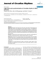
Figure 1
Common cardiac conditions encountered in the intensive care unit and the related biomarkers. Note the lack of specificity of some biomarkers.
BNP, B-type natriuretic peptide; CA125, carbohydrate antigen 125; CD154, soluble CD40 ligand; CK-MB, creatine kinase-myocardial band; CRP,
C-reactive protein; cTn, cardiac troponins; ICU, intensive care unit; IL, interleukin; IMA, ischemia-modified albumin; HFABP, heart-type fatty acid
binding protein.
10. Davis M, Espiner E, Richards G, Billings J, Town I, Neill A,
Drennan C, Richards M, Turner J, Yandle T: Plasma brain natri-
uretic peptide in assessment of acute dyspnoea. Lancet 1994,
343:440-444.
11. McDonagh TA, Robb SD, Murdoch DR, Morton JJ, Ford I, Morri-
son CE, Tunstall-Pedoe H, McMurray JJ, Dargie HJ: Biochemical
detection of left-ventricular systolic dysfunction. Lancet 1998,
351:9-13.
12. Januzzi JL, Jr, Camargo CA, Anwaruddin S, Baggish AL, Chen AA,
Krauser DG, Tung R, Cameron R, Nagurney JT, Chae CU, Lloyd-
Jones DM, Brown DF, Foran-Melanson S, Sluss PM, Lee-
Lewandrowski E, Lewandrowski KB: The N-terminal Pro-BNP
investigation of dyspnea in the emergency department
(PRIDE) study. Am J Cardiol 2005, 95:948-954.
13. Cowie MR, Struthers AD, Wood DA, Coats AJ, Thompson SG,
Poole-Wilson PA, Sutton GC: Value of natriuretic peptides in
assessment of patients with possible new heart failure in
primary care. Lancet 1997, 350:1349-1353.
14. Remme WJ, Swedberg K: Guidelines for the diagnosis and
treatment of chronic heart failure. Eur Heart J 2001, 22:1527-
1560.
15. McCullough PA, Nowak RM, McCord J, Hollander JE, Herrmann
HC, Steg PG, Duc P, Westheim A, Omland T, Knudsen CW,
Storrow AB, Abraham WT, Lamba S, Wu AH, Perez A, Clopton P,
Krishnaswamy P, Kazanegra R, Maisel AS: B-type natriuretic
peptide and clinical judgment in emergency diagnosis of
heart failure: analysis from Breathing Not Properly (BNP)
Multinational Study. Circulation 2002, 106:416-422.
16. Maisel A, Hollander JE, Guss D, McCullough P, Nowak R, Green
G, Saltzberg M, Ellison SR, Bhalla MA, Bhalla V, Clopton P, Jesse
R: Primary results of the Rapid Emergency Department Heart
Failure Outpatient Trial (REDHOT). A multicenter study of B-
type natriuretic peptide levels, emergency department deci-
sion making, and outcomes in patients presenting with
shortness of breath. J Am Coll Cardiol 2004, 44:1328-1333.
17. McLean AS, Huang SJ: The applications of B-type natriuretic
peptide measurement in the intensive care unit. Curr Opin Crit
Care 2005, 11:406-412.
18. McLean AS, Poh G, Huang SJ: The effects of acute fluid
loading on plasma B-type natriuretic peptide levels in a septic
shock patient. Anaesth Intensive Care 2005, 33:528-530.
19. Nellessen U, Goder S, Schobre R, Abawi M, Hecker H, Tschoke
S: Serial analysis of troponin I levels in patients with ischemic
and nonischemic dilated cardiomyopathy. Clin Cardiol 2006,
29:219-224.
20. Wallace TW, Abdullah SM, Drazner MH, Das SR, Khera A,
McGuire DK, Wians F, Sabatine MS, Morrow DA, de Lemos JA:
Prevalence and determinants of troponin T elevation in the
general population. Circulation 2006, 113:1958-1965.
21. Liu Z, Cui L, Wang Y, Guo Y: Cardiac troponin I and ventricular
arrhythmia in patients with chronic heart failure. Eur J Clin
Invest 2006, 36:466-472.
22. Matsumori A, Kawai C, Yamada T, Ohkusa T, Morishima S, Tamaki
N, Watanabe Y, Yonekura Y, Endo K, Konishi J, Yoshida A: Mech-
anism and significance of myocardial uptake of antimyosin
antibody in myocarditis and cardiomyopathy: clinical and
experimental studies. Clin Immunol Immunopathol 1993, 68:
215-219.
23. Sato Y, Kita T, Takatsu Y, Kimura T: Biochemical markers of
myocyte injury in heart failure. Heart 2004, 90:1110-1113.
24. Latini R, Masson S, Anand IS, Missov E, Carlson M, Vago T,
Angelici L, Barlera S, Parrinello G, Maggioni AP, Tognoni G, Cohn
JN: Prognostic value of very low plasma concentrations of tro-
ponin T in patients with stable chronic heart failure. Circulation
2007, 116:1242-1249.
25. Nishio Y, Sato Y, Taniguchi R, Shizuta S, Doi T, Morimoto T,
Kimura T, Kita T: Cardiac troponin T vs other biochemical
markers in patients with congestive heart failure. Circ J 2007,
71:631-635.
26. Mallat Z, Heymes C, Corbaz A, Logeart D, Alouani S, Cohen-Solal
A, Seidler T, Hasenfuss G, Chvatchko Y, Shah AM, Tedgui A: Evi-
dence for altered interleukin 18 (IL)-18 pathway in human
heart failure. FASEB J 2004, 18:1752-1754.
27. White M, Ducharme A, Ibrahim R, Whittom L, Lavoie J, Guertin
MC, Racine N, He Y, Yao G, Rouleau JL, Schiffrin EL, Touyz RM:
Increased systemic inflammation and oxidative stress in
patients with worsening congestive heart failure: improve-
ment after short-term inotropic support. Clin Sci (Lond) 2006,
110:483-489.
28. Pomerantz BJ, Reznikov LL, Harken AH, Dinarello CA: Inhibition
of caspase 1 reduces human myocardial ischemic dysfunc-
tion via inhibition of IL-18 and IL-1
ββ
. Proc Natl Acad Sci USA
2001, 98:2871-2876.
29. Bast RC, Jr, Klug TL, St John E, Jenison E, Niloff JM, Lazarus H,
Berkowitz RS, Leavitt T, Griffiths CT, Parker L, Zurawski VR Jr,
Knapp RC: A radioimmunoassay using a monoclonal antibody
to monitor the course of epithelial ovarian cancer. N Engl J
Med 1983, 309:883-887.
30. Nagele H, Bahlo M, Klapdor R, Schaeperkoetter D, Rodiger W:
CA 125 and its relation to cardiac function. Am Heart J 1999,
137:1044-1049.
31. D’Aloia A, Faggiano P, Aurigemma G, Bontempi L, Ruggeri G,
Metra M, Nodari S, Dei Cas L: Serum levels of carbohydrate
antigen 125 in patients with chronic heart failure: relation to
clinical severity, hemodynamic and Doppler echocardio-
graphic abnormalities, and short-term prognosis. J Am Coll
Cardiol 2003, 41:1805-1811.
32. Kouris NT, Zacharos ID, Kontogianni DD, Goranitou GS, Sifaki
MD, Grassos HE, Kalkandi EM, Babalis DK: The significance of
CA125 levels in patients with chronic congestive heart failure.
Correlation with clinical and echocardiographic parameters.
Eur J Heart Fail 2005, 7:199-203.
33. Mathew B, Bhatia V, Mahy IR, Ahmed I, Francis L: Elevation of
the tumor marker CA125 in right heart failure. South Med J
2004, 97:1013-1014.
34. Seo T, Ikeda Y, Onaka H, Hayashi T, Kawaguchi K, Kotake C, Toda
T, Kobayashi K: Usefulness of serum CA125 measurement for
monitoring pericardial effusion. Jpn Circ J 1993, 57:489-494.
35. van der Veen KJ, Willebrands AF: Isoenzymes of creatine phos-
phokinase in tissue extracts and in normal and pathological
sera. Clin Chim Acta 1966, 13:312-316.
36. Sobel BE, Shell WE: Serum enzyme determinations in the
diagnosis and assessment of myocardial infarction. Circula-
tion 1972, 45:471-482.
37. Collinson PO, Chandler HA, Stubbs PJ, Moseley DS, Lewis D,
Simmons MD: Measurement of serum troponin T, creatine
kinase MB isoenzyme, and total creatine kinase following
arduous physical training. Ann Clin Biochem 1995, 32:450-
453.
38. Panteghini M: Acute coronary syndrome: biochemical strate-
gies in the troponin era. Chest 2002, 122:1428-1435.
39. Jaffe AS, Ravkilde J, Roberts R, Naslund U, Apple FS, Galvani M,
Katus H: It’s time for a change to a troponin standard. Circula-
tion 2000, 102:1216-1220.
40. Alpert JS, Thygesen K, Antman E, Bassand JP: Myocardial infarc-
tion redefined – a consensus document of The Joint Euro-
pean Society of Cardiology/American College of Cardiology
Committee for the redefinition of myocardial infarction. J Am
Coll Cardiol 2000, 36:959-969.
41. De Zoysa JR: Cardiac troponins and renal disease. Nephrology
2004, 9:83-88.
42. Wu AH, Jaffe AS: The clinical need for high-sensitivity cardiac
troponin assays for acute coronary syndromes and the role
for serial testing. Am Heart J 2008, 155:208-214.
43. Donnino MW, Karriem-Norwood V, Rivers EP, Gupta A, Nguyen
HB, Jacobsen G, McCord J, Tomlanovich MC: Prevalence of ele-
vated troponin I in end-stage renal disease patients receiving
hemodialysis. Acad Emerg Med 2004, 11:979-981.
44. Jaffe AS, Babuin L, Apple FS: Biomarkers in acute cardiac
disease: the present and the future. J Am Coll Cardiol 2006,
48:1-11.
45. Hamm CW, Giannitsis E, Katus HA: Cardiac troponin elevations
in patients without acute coronary syndrome. Circulation
2002, 106:2871-2872.
46. Aviles RJ, Askari AT, Lindahl B, Wallentin L, Jia G, Ohman EM,
Mahaffey KW, Newby LK, Califf RM, Simoons ML, Topol EJ,
Berger P, Lauer MS: Troponin T levels in patients with acute
coronary syndromes, with or without renal dysfunction. N Engl
J Med 2002, 346:2047-2052.
47. Glatz JF, Paulussen RJ, Veerkamp JH: Fatty acid binding pro-
teins from heart. Chem Phys Lipids 1985, 38:115-129.
48. Tanaka T, Hirota Y, Sohmiya K, Nishimura S, Kawamura K: Serum
and urinary human heart fatty acid-binding protein in acute
myocardial infarction. Clin Biochem 1991, 24:195-201.
Available online />Page 7 of 9
(page number not for citation purposes)
49. Tanaka T, Sohmiya K, Kitaura Y, Takeshita H, Morita H, Ohkaru Y,
Asayama K, Kimura H: Clinical evaluation of point-of-care-
testing of heart-type fatty acid-binding protein (H-FABP) for
the diagnosis of acute myocardial infarction. J Immunoassay
Immunochem 2006, 27:225-238.
50. Ecollan P, Collet JP, Boon G, Tanguy ML, Fievet ML, Haas R,
Bertho N, Siami S, Hubert JC, Coriat P, Montalescot G: Pre-hos-
pital detection of acute myocardial infarction with ultra-rapid
human fatty acid-binding protein (H-FABP) immunoassay. Int
J Cardiol 2007, 119:349-354.
51. Niizeki T, Takeishi Y, Arimoto T, Takabatake N, Nozaki N, Hirono
O, Watanabe T, Nitobe J, Harada M, Suzuki S, Koyama Y, Kita-
hara T, Sasaki T, Kubota I: Heart-type fatty acid-binding protein
is more sensitive than troponin T to detect the ongoing
myocardial damage in chronic heart failure patients. J Card
Fail 2007, 13:120-127.
52. O’Donoghue M, de Lemos JA, Morrow DA, Murphy SA, Buros JL,
Cannon CP, Sabatine MS: Prognostic utility of heart-type fatty
acid binding protein in patients with acute coronary syn-
dromes. Circulation 2006, 114:550-557.
53. Libby P: Inflammation in atherosclerosis. Nature 2002, 420:
868-874.
54. Hansson GK: Inflammation, atherosclerosis, and coronary
artery disease. N Engl J Med 2005, 352:1685-1695.
55. Larsson PT, Hallerstam S, Rosfors S, Wallen NH: Circulating
markers of inflammation are related to carotid artery athero-
sclerosis. Int Angiol 2005, 24:43-51.
56. Pasceri V, Cheng JS, Willerson JT, Yeh ET: Modulation of C-
reactive protein-mediated monocyte chemoattractant protein-
1 induction in human endothelial cells by anti-atherosclerosis
drugs. Circulation 2001, 103:2531-2534.
57. Tanaka A, Shimada K, Sano T, Namba M, Sakamoto T, Nishida Y,
Kawarabayashi T, Fukuda D, Yoshikawa J: Multiple plaque
rupture and C-reactive protein in acute myocardial infarction.
J Am Coll Cardiol 2005, 45:1594-1599.
58. Liuzzo G, Biasucci LM, Rebuzzi AG, Gallimore JR, Caligiuri G,
Lanza GA, Quaranta G, Monaco C, Pepys MB, Maseri A: Plasma
protein acute-phase response in unstable angina is not
induced by ischemic injury. Circulation 1996, 94:2373-2380.
59. Wong CK, Szeto CC, Chan MH, Leung CB, Li PK, Lam CW: Ele-
vation of pro-inflammatory cytokines, C-reactive protein and
cardiac troponin T in chronic renal failure patients on dialysis.
Immunol Invest 2007, 36:47-57.
60. Deodhar SD: C-reactive protein: the best laboratory indicator
available for monitoring disease activity. Cleveland Clin J Med
1989, 56:126-130.
61. Lindahl B, Toss H, Siegbahn A, Venge P, Wallentin L: Markers of
myocardial damage and inflammation in relation to long-term
mortality in unstable coronary artery disease. FRISC Study
Group. Fragmin during Instability in Coronary Artery Disease.
N Engl J Med 2000, 343:1139-1147.
62. Schieffer B, Schieffer E, Hilfiker-Kleiner D, Hilfiker A, Kovanen PT,
Kaartinen M, Nussberger J, Harringer W, Drexler H: Expression
of angiotensin II and interleukin 6 in human coronary athero-
sclerotic plaques: potential implications for inflammation and
plaque instability. Circulation
2000, 101:1372-1378.
63. Lindmark E, Diderholm E, Wallentin L, Siegbahn A: Relationship
between interleukin 6 and mortality in patients with unstable
coronary artery disease: effects of an early invasive or nonin-
vasive strategy. JAMA 2001, 286:2107-2113.
64. Ridker PM, Rifai N, Stampfer MJ, Hennekens CH: Plasma con-
centration of interleukin-6 and the risk of future myocardial
infarction among apparently healthy men. Circulation 2000,
101:1767-1772.
65. Mallat Z, Corbaz A, Scoazec A, Besnard S, Leseche G,
Chvatchko Y, Tedgui A: Expression of interleukin-18 in human
atherosclerotic plaques and relation to plaque instability. Cir-
culation 2001, 104:1598-1603.
66. Zirlik A, Abdullah SM, Gerdes N, MacFarlane L, Schonbeck U,
Khera A, McGuire DK, Vega GL, Grundy S, Libby P, de Lemos JA:
Interleukin-18, the metabolic syndrome, and subclinical ather-
osclerosis: results from the Dallas Heart Study. Arterioscler
Thromb Vasc Biol 2007, 27:2043-2049.
67. Blankenberg S, Tiret L, Bickel C, Peetz D, Cambien F, Meyer J,
Rupprecht HJ: Interleukin-18 is a strong predictor of cardio-
vascular death in stable and unstable angina. Circulation
2002, 106:24-30.
68. Parker MM, Shelhamer JH, Bacharach SL, Green MV, Natanson
C, Frederick TM, Damske BA, Parrillo JE: Profound but
reversible myocardial depression in patients with septic
shock. Ann Int Med 1984, 100:483-490.
69. Annane D, Bellissant E, Cavaillon JM: Septic shock. Lancet
2005, 365:63-78.
70. Cuthbertson BH, Patel RR, Croal BL, Barclay J, Hillis GS: B-type
natriuretic peptide and the prediction of outcome in patients
admitted to intensive care. Anaesthesia 2005, 60:16-21.
71. Witthaut R, Busch C, Fraunberger P, Walli A, Seidel D, Pilz G,
Stuttmann R, Speichermann N, Verner L, Werdan K: Plasma
atrial natriuretic peptide and brain natriuretic peptide are
increased in septic shock: impact of interleukin-6 and sepsis-
associated left ventricular dysfunction. Intensive Care Med
2003, 29:1696-1702.
72. Charpentier J, Luyt CE, Fulla Y, Vinsonneau C, Cariou A, Grabar
S, Dhainaut JF, Mira JP, Chiche JD: Brain natriuretic peptide: a
marker of myocardial dysfunction and prognosis during
severe sepsis. Crit Care Med 2004, 32:660-665.
73. McLean AS, Huang SJ, Hyams S, Poh G, Nalos M, Pandit R, Balik
M, Tang B, Seppelt I: Prognostic values of B-type natriuretic
peptide in severe sepsis and septic shock. Crit Care Med
2007, 35:1019-1026.
74. McLean AS, Huang SJ: The applications of B-type natriuretic
peptide measurement in the intensive care unit. Curr Opin Crit
Care 2005, 11:406-412.
75. ver Elst KM, Spapen HD, Nguyen DN, Garbar C, Huyghens LP,
Gorus FK: Cardiac troponins I and T are biological markers of
left ventricular dysfunction in septic shock. Clin Chem 2000,
46:
650-657.
76. Mehta NJ, Khan IA, Gupta V, Jani K, Gowda RM, Smith PR:
Cardiac troponin I predicts myocardial dysfunction and
adverse outcome in septic shock. Int J Cardiol 2004, 95:13-17.
77. Cunnion RE, Schaer GL, Parker MM, Natanson C, Parrillo JE: The
coronary circulation in human septic shock. Circulation 1986,
73:637-644.
78. Ammann P, Fehr T, Minder EI, Gunter C, Bertel O: Elevation of
troponin I in sepsis and septic shock. Intensive Care Med
2001, 27:965-969.
79. Wu AH, Feng YJ, Moore R, Apple FS, McPherson PH, Buechler
KF, Bodor G: Characterization of cardiac troponin subunit
release into serum after acute myocardial infarction and com-
parison of assays for troponin T and I. American Association
for Clinical Chemistry Subcommittee on cTnI Standardization.
Clin Chem 1998, 44:1198-1208.
80. Amorim S, Dias P, Rodrigues RA, Araujo V, Macedo F, Maciel MJ,
Goncalves FR: Troponin I as a marker of right ventricular dys-
function and severity of pulmonary embolism. Rev Port Cardiol
2006, 25:181-186.
81. Pruszczyk P: N-terminal pro-brain natriuretic peptide as an
indicator of right ventricular dysfunction. J Card Fail 2005, 11:
S65-S69.
82. Pruszczyk P, Kostrubiec M, Bochowicz A, Styczynski G, Szulc M,
Kurzyna M, Fijalkowska A, Kuch-Wocial A, Chlewicka I, Torbicki A:
N-terminal pro-brain natriuretic peptide in patients with acute
pulmonary embolism. Eur Respir J 2003, 22:649-653.
83. Kruger S, Graf J, Merx MW, Koch KC, Kunz D, Hanrath P,
Janssens U: Brain natriuretic peptide predicts right heart
failure in patients with acute pulmonary embolism. Am Heart J
2004, 147:60-65.
84. Renaud B, Ngako A: Heart-type fatty acid-binding proteins (H-
FABP): a reliable tool for initial risk stratification of pulmonary
embolism? Eur Heart J 2007, 28:146-147.
85. Bar-Or D, Lau E, Rao N, Bampos N, Winkler JV, Curtis CG:
Reduction in the cobalt binding capacity of human albumin
with myocardial ischemia [abstract]. Ann Emerg Med 1999;
34:S56.
86. Bar-Or D, Lau E, Winkler JV: A novel assay for cobalt–albumin
binding and its potential as a marker for myocardial ischemia-
a preliminary report. J Emerg Med 2000, 19:311-315.
87. Sinha MK, Roy D, Gaze DC, Collinson PO, Kaski JC: Role of
‘ischemia modified albumin’, a new biochemical marker of
myocardial ischaemia, in the early diagnosis of acute coro-
nary syndromes. Emerg Med J 2004, 21:29-34.
88. Refaai MA, Wright RW, Parvin CA, Gronowski AM, Scott MG,
Eby CS: Ischemia-modified albumin increases after skeletal
muscle ischemia during arthroscopic knee surgery. Clin Chim
Critical Care Vol 12 No 3 McLean et al.
Page 8 of 9
(page number not for citation purposes)
Acta 2006, 366:264-268.
89. Gaze DC, Crompton L, Collinson P: Ischemia-modified albumin
concentrations should be interpreted with caution in patients
with low serum albumin concentrations. Med Princ Pract 2006,
15:322-324.
90. Lippi G, Brocco G, Salvagno GL, Montagnana M, Dima F, Guidi
GC: High-workload endurance training may increase serum
ischemia-modified albumin concentrations. Clin Chem Lab
Med 2005, 43:741-744.
91. Danne O, Mockel M, Lueders C, Mugge C, Zschunke GA, Lufft H,
Muller C, Frei U: Prognostic implications of elevated whole
blood choline levels in acute coronary syndromes. Am J
Cardiol 2003, 91:1060-1067.
92. Danne O, Lueders C, Storm C, Frei U, Mockel M: Whole blood
choline and plasma choline in acute coronary syndromes:
prognostic and pathophysiological implications. Clin Chim
Acta 2007, 383:103-109.
93. Schonbeck U, Mach F, Libby P: CD154 (CD40 ligand). Int J
Biochem Cell Biol 2000, 32:687-693.
94. Stumpf C, Lehner C, Eskafi S, Raaz D, Yilmaz A, Ropers S,
Schmeisser A, Ludwig J, Daniel WG, Garlichs CD: Enhanced
levels of CD154 (CD40 ligand) on platelets in patients with
chronic heart failure. Eur J Heart Fail 2003, 5:629-637.
95. Varo N, de Lemos JA, Libby P, Morrow DA, Murphy SA, Nuzzo R,
Gibson CM, Cannon CP, Braunwald E, Schonbeck U: Soluble
CD40L: risk prediction after acute coronary syndromes. Circu-
lation 2003, 108:1049-1052.
96. Nishikimi T, Miyata A, Horio T, Yoshihara F, Nagaya N, Takishita S,
Yutani C, Matsuo H, Matsuoka H, Kangawa K: Urocortin, a
member of the corticotropin-releasing factor family, in normal
and diseased heart. Am J Physiol 2000, 279:H3031-H3039.
97. Agnello D, Bertini R, Sacco S, Meazza C, Villa P, Ghezzi P: Corti-
costeroid-independent inhibition of tumor necrosis factor pro-
duction by the neuropeptide urocortin. Am J Physiol 1998,
275:E757-E762.
98. Rademaker MT, Charles CJ, Espiner EA, Fisher S, Frampton CM,
Kirkpatrick CM, Lainchbury JG, Nicholls MG, Richards AM, Vale
WW: Beneficial hemodynamic, endocrine, and renal effects of
urocortin in experimental heart failure: comparison with
normal sheep. J Am Coll Cardiol 2002, 40:1495-1505.
99. Ng LL, Loke IW, O’Brien RJ, Squire IB, Davies JE: Plasma uro-
cortin in human systolic heart failure. Clin Sci (Lond) 2004,
106:383-388.
100. Tang WH, Brennan ML, Philip K, Tong W, Mann S, Van Lente F,
Hazen SL: Plasma myeloperoxidase levels in patients with
chronic heart failure. Am J Cardiol 2006, 98:796-799.
101. Tang WH, Tong W, Troughton RW, Martin MG, Shrestha K,
Borowski A, Jasper S, Hazen SL, Klein AL: Prognostic value and
echocardiographic determinants of plasma myeloperoxidase
levels in chronic heart failure. J Am Coll Cardiol 2007, 49:
2364-2370.
102. Brennan ML, Penn MS, Van Lente F, Nambi V, Shishehbor MH,
Aviles RJ, Goormastic M, Pepoy ML, McErlean ES, Topol EJ,
Nissen SE, Hazen SL: Prognostic value of myeloperoxidase in
patients with chest pain. N Engl J Med 2003, 349:1595-1604.
103. Stolker JM, Rich MW: The combination of B-type natriuretic
peptide and C-reactive protein provides incremental prognos-
tic value among older patients referred for cardiac catheteri-
zation. Am J Geriatr Cardiol 2007, 16:229-235.
104. Foussas SG, Zairis MN, Makrygiannis SS, Manousakis SJ, Anas-
tassiadis FA, Apostolatos CS, Patsourakos NG, Glyptis MP,
Papadopoulos JK, Xenos DC, Adamopoulou EN, Olympios CD,
Argyrakis SK: The significance of circulating levels of both
cardiac troponin I and high-sensitivity C reactive protein for
the prediction of intravenous thrombolysis outcome in
patients with ST-segment elevation myocardial infarction.
Heart 2007, 93:952-956.
105. Peacock F, Morris DL, Anwaruddin S, Christenson RH, Collinson
PO, Goodacre SW, Januzzi JL, Jesse RL, Kaski JC, Kontos MC,
Lefevre G, Mutrie D, Sinha MK, Uettwiller-Geiger D, Pollack CV:
Meta-analysis of ischemia-modified albumin to rule out acute
coronary syndromes in the emergency department. Am Heart
J 2006, 152:253-262.
106. Guest TM, Ramanathan AV, Tuteur PG, Schechtman KB, Laden-
son JH, Jaffe AS: Myocardial injury in critically ill patients. A fre-
quently unrecognized complication. JAMA 1995, 273:
1945-1949.
107. Noble JS, Reid AM, Jordan LV, Glen AC, Davidson JA: Troponin I
and myocardial injury in the ICU. Br J Anaesth 1999, 82:41-46.
108. King DA, Codish S, Novack V, Barski L, Almog Y: The role of
cardiac troponin I as a prognosticator in critically ill medical
patients: a prospective observational cohort study. Crit Care
2005, 9:R390-R395.
109. Ammann P, Maggiorini M, Bertel O, Haenseler E, Joller-Jemelka
HI, Oechslin E, Minder EI, Rickli H, Fehr T: Troponin as a risk
factor for mortality in critically ill patients without acute coro-
nary syndromes. J Am Coll Cardiol 2003, 41:2004-2009.
110. Lim W, Qushmaq I, Cook DJ, Crowther MA, Heels-Ansdell D,
Devereaux PJ: Elevated troponin and myocardial infarction in
the intensive care unit: a prospective study. Crit Care 2005, 9:
R636-R644.
111. Lim W, Cook DJ, Griffith LE, Crowther MA, Devereaux PJ: Ele-
vated cardiac troponin levels in critically ill patients: preva-
lence, incidence, and outcomes. Am J Crit Care 2006, 15:
280-288.
112. Povoa P, Coelho L, Almeida E, Fernandes A, Mealha R, Moreira P,
Sabino H: C-reactive protein as a marker of infection in criti-
cally ill patients. Clin Microbiol Infect 2005, 11:101-108.
113. Sierra R, Rello J, Bailen MA, Benitez E, Gordillo A, Leon C,
Pedraza S: C-reactive protein used as an early indicator of
infection in patients with systemic inflammatory response
syndrome. Intensive Care Med 2004, 30:2038-2045.
114. Lobo SM, Lobo FR, Bota DP, Lopes-Ferreira F, Soliman HM,
Melot C, Vincent JL: C-reactive protein levels correlate with
mortality and organ failure in critically ill patients. Chest 2003,
123:2043-2049.
Available online />Page 9 of 9
(page number not for citation purposes)