Báo cáo y học: " Differential temporal profile of lowered blood glucose levels (3.5 to 6.5 mmol/l versus 5 to 8 mmol/l) in patients with severe traumatic brain injury" docx
Bạn đang xem bản rút gọn của tài liệu. Xem và tải ngay bản đầy đủ của tài liệu tại đây (457.88 KB, 13 trang )
Open Access
Available online />Page 1 of 13
(page number not for citation purposes)
Vol 12 No 4
Research
Differential temporal profile of lowered blood glucose levels (3.5
to 6.5 mmol/l versus 5 to 8 mmol/l) in patients with severe
traumatic brain injury
Regula Meier
1
, Markus Béchir
1
, Silke Ludwig
1
, Jutta Sommerfeld
1
, Marius Keel
2
, Peter Steiger
1
,
Reto Stocker
1
and John F Stover
1
1
Surgical Intensive Care Medicine, University Hospital Zuerich, Raemistrasse 100, CH 8091 Zuerich, Switzerland
2
Department of Surgery, Division of Trauma Surgery, University Hospital Zuerich, Raemistrasse 100, CH 8091 Zuerich, Switzerland
Corresponding author: John F Stover,
Received: 28 May 2008 Revisions requested: 23 Jun 2008 Revisions received: 14 Jul 2008 Accepted: 4 Aug 2008 Published: 4 Aug 2008
Critical Care 2008, 12:R98 (doi:10.1186/cc6974)
This article is online at: />© 2008 2008 Meier et al.; licensee BioMed Central Ltd.
This is an open access article distributed under the terms of the Creative Commons Attribution License ( />),
which permits unrestricted use, distribution, and reproduction in any medium, provided the original work is properly cited.
Abstract
Introduction Hyperglycaemia is detrimental, but maintaining
low blood glucose levels within tight limits is controversial in
patients with severe traumatic brain injury, because decreased
blood glucose levels can induce and aggravate underlying brain
injury.
Methods In 228 propensity matched patients (age, sex and
injury severity) treated in our intensive care unit (ICU) from 2000
to 2004, we retrospectively evaluated the influence of different
predefined blood glucose targets (3.5 to 6.5 versus 5 to 8
mmol/l) on frequency of hypoglycaemic and hyperglycaemic
episodes, insulin and norepinephrine requirement, changes in
intracranial pressure and cerebral perfusion pressure, mortality
and length of stay on the ICU.
Results Mortality and length of ICU stay were similar in both
blood glucose target groups. Blood glucose values below and
above the predefined levels were significantly increased in the
3. 5 to 6.5 mmol/l group, predominantly during the first week.
Insulin and norepinephrine requirements were markedly
increased in this group. During the second week, the incidences
of intracranial pressure exceeding 20 mmHg and infectious
complications were significantly decreased in the 3.5 to 6.5
mmol/l group.
Conclusion Maintaining blood glucose within 5 to 8 mmol/l
appears to yield greater benefit during the first week. During the
second week, 3.5 to 6.5 mmol/l is associated with beneficial
effects in terms of reduced intracranial hypertension and
decreased rate of pneumonia, bacteraemia and urinary tract
infections. It remains to be determined whether patients might
profit from temporally adapted blood glucose limits, inducing
lower values during the second week, and whether concomitant
glucose infusion to prevent hypoglycaemia is safe in patients
with post-traumatic oedema.
Introduction
After severe traumatic brain injury (TBI), secondary brain dam-
age related to activated local cascades as well as systemic
influences can compromise regenerative and reparative proc-
esses, thereby increasing morbidity and mortality. Within this
context, elevated blood glucose concentrations at admission
and during intensive care exceeding 9.4 mmol/l (170 mg/dl)
are associated with increased mortality [1,2] and morbidity [3-
5] compared with normoglycaemic patients. Consequently, it
appears logical to correct and maintain blood glucose levels at
lower yet controllable values in order to prevent and counter-
act hyperglycaemia-induced mitochondrial damage, sustained
cytotoxic oxidative stress, impaired neutrophil function and
reduced phagocytosis, as well as impaired intracellular bacte-
ricidal and opsonic activity [6].
As recently shown by van den Berghe and colleagues [7],
maintaining blood glucose levels at low levels ranging from 4.4
to 6.1 mmol/l (80 to 110 mg/dl), as compared with concentra-
tions exceeding 12 mmol/l (220 mg/dl), appears to be
CPP = cerebral perfusion pressure; CRP = C-reactive protein; GLUT = glucose transporter; ICP = intracranial pressure; ICU = intensive care unit;
TBI = traumatic brain injury.
Critical Care Vol 12 No 4 Meier et al.
Page 2 of 13
(page number not for citation purposes)
beneficial for surgical and medical patients requiring intensive
care treatment longer than 3 days. Overall, this approach sig-
nificantly reduced morbidity and mortality, and prevented criti-
cal illness polyneuropathy, bacteraemia, anaemia, acute renal
failure and hyperbilirubinaemia. These benefits ultimately cul-
minated reduced length of hospitalization, duration of ventila-
tion and substantially lowered costs [7].
Patients with various types of traumatic and nontraumatic
brain lesions also appear to profit from this approach [8]. How-
ever, this reduced infection rate and mortality could not be
reproduced by Bilotta and colleagues [9] in their prospective
randomized trial conducted in brain-injured patients employing
a similar study design to that used by van den Berghe and col-
leagues [7].
Following the results published by van den Berghe and col-
leagues [7], targeted blood glucose levels were lowered from
5 to 8 mmol/l (90 to 144 mg/dl) to 3.5 to 6.5 mmol/l (63 to
117 mg/dl) at our institution, with the aim being to reduce cel-
lular insults related to high blood glucose levels and concomi-
tantly to promote insulin-mediated nonglycaemic protective
effects related to the anti-apoptotic and anti-inflammatory
effects of normoglycaemia.
Recently, implementation of these tightly controlled blood glu-
cose levels was criticized in brain-injured patients because of
the resulting increased risk for hypoglycaemic episodes,
which promote an increase in extracellular glutamate and
signs of metabolic derangement, reflected by an increased
lactate/pyruvate ratio [10]. Absolute as well as relative
decreases in blood glucose concentrations below 5 mmol/l
were consistently associated with spontaneous cortical depo-
larizations under both experimental and clinical conditions [11-
14]. These alterations with and without excessive correction of
hypoglycaemic values are in turn feared to induce secondary
brain injury, thereby possibly offsetting anticipated neuropro-
tection in these patients.
The main hypothesis of the present study was that maintaining
arterial blood glucose between 3.5 to 6.5 mmol/l, as com-
pared with 5 to 8 mmol/l, significantly decreases mortality and
reduces rates of infectious complications. Based on this hypo-
thesis, primary end-points were intensive care unit (ICU) mor-
tality, and rates of pneumonia, bacteraemia and urinary tract
infections. In addition, we investigated the impact of maintain-
ing blood glucose levels within low and tight limits on fluctua-
tions in blood glucose values, insulin and norepinephrine
requirements, alterations in intracranial pressure (ICP), length
of stay on the ICU, and signs of inflammation in patients with
severe TBI. For this, we retrospectively compared 114 propen-
sity-matched patients in whom blood glucose levels were
maintained between 3.5 to 6.5 mmol/l with 114 patients with
a blood glucose target between 5 to 8 mmol/l. Patients were
matched with respect to age, sex, and type, number and sever-
ity of injuries.
Materials and methods
Following approval by the local ethics committee, which
waived the need for written informed consent for this retro-
spective study, patient records from a total of 320 patients
treated on our ICU from 2000 to 2004 were reviewed. In the
years 2000 to 2002, blood glucose levels were maintained
between 4 and 8 mmol/l. Thereafter, blood glucose limits were
reduced to 3.5 to 6.5 mmol/l during the years 2002 to 2004.
Following exclusion of 92 patients (29%), 228 propensity-
matched critically ill patients suffering from severe TBI were
eligible for subsequent analysis aimed at comparing the influ-
ence of blood glucose levels maintained between 3.5 to 6.5
mmol/l versus 5 to 8 mmol/l (Figure 1).
Propensity-matched patients
To increase comparability between patients who were treated
sequentially (2000 to 2002 and 2002 to 2004) with different
blood glucose limits, patients were matched according to age,
sex, injury types and severity of underlying injuries based on
the Injury Severity Score, determined after admission to the
emergency room of the University Hospital Zuerich. This
allowed us to minimize the impact of uncontrolled influences
that can occur over a 4-year period.
Inclusion criteria
Patients had to be treated on our ICU for longer than 24 hours.
All patients were required to have had an ICP probe placed
within the first 8 hours after injury.
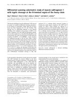
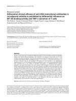
Figure 1
Study descriptionStudy description. Presented is a flow chart showing inclusion of 228
patients and exclusion of 92 patients suffering from severe traumatic
brain injury subjected to two different blood glucose targets, namely
3.5 to 6.5 mmol/l versus 5 to 8 mmol/l, over a period of 4 years. The
main hypothesis as well as primary and secondary end-points are
shown.
Available online />Page 3 of 13
(page number not for citation purposes)
Exclusion criteria
Patients who died within the first 24 hours after injury and
those in whom an ICP probe was not inserted (low or high
severity of injury) were not included. Patients with incomplete
data were excluded as well.
Standardized critical care
All patients were treated using a standardized protocol. Anal-
gesia and sedation were maintained with fentanyl (Sintenyl
®
)
and midazolam (Dormicum
®
). If required, muscle relaxation
was induced with pancuronium. Haemodynamic stability was
maintained by fluid and vasopressor administration and
adapted to maintain cerebral perfusion pressure (CPP)
between 70 and 90 mmHg. Increased analgesia, sedation,
CPP, controlled hyperventilation and cerebral spinal fluid
release in patients with external ventricular drainage were
employed to maintain ICP levels below 20 mmHg. Lung pro-
tective ventilation was maintained by keeping peak inspiratory
pressure below 35 mbar. Enteral nutrition was begun within
the first 12 hours and controlled by means of indirect calo-
rimety at least twice weekly. Continuously infused insulin was
tapered according to the measured blood glucose levels. Con-
trary to the protocol used by van den Berghe and colleagues
[7], we did not routinely infuse glucose in our patients because
of concern that increased post-traumatic brain oedema forma-
tion might result. Glucose was only infused in case of hypogly-
caemia under 1.5 mmol/l. Blood glucose levels were
determined using the blood gas analyzer ABL 825 Flex (Radi-
ometer, Copenhagen, Denmark) at least every 4 hours or at
shorter intervals, depending on the clinical situation and the
determined blood glucose level, in order to avoid hypoglycae-
mic and hyperglycemic episodes. Hypoglycaemia was defined
at blood glucose levels under 2.5 mmol/l, whereas hypergly-
caemia was defined at blood glucose concentrations above
10 mmol/l.
Investigated parameters
The data bank (Microsoft Excel
®
and Microsoft Access
®
;
Microsoft Inc., Redmond WA, USA) consisted of values that
were determined at 4-hour intervals: blood glucose, infused
insulin and norepinephrine dose, as well as ICP and CPP lev-
els. In addition, mortality, length of ICU stay, positive blood cul-
tures and positive tracheobronchial secretions, as well as
changes in maximal leukocytes, C-reactive protein (CRP) and
interleukin-6 (IL-6), were recorded. This resulted in a total of
58,794 values in all patients and an average of 258 values per
patient.
Values assessed at 4-hour intervals or once daily were used to
determine changes in the individual parameters over time and
to calculate absolute and relative frequencies within prede-
fined clusters.
The database was constructed by entering data in predefined
columns within a Microsoft Excel
®
sheet for every individual
patient. Then, all individual sheets were transferred to one
Microsoft Excel
®
sheet, which contained data for all patients.
This Microsoft Excel
®
sheet was then imported into a Micro-
soft Access
®
database. Data were entered by RM, SL and JS,
and checked for plausibility and correctness by JFS and SL;
after an automated search for incorrect outliers within each
column, these values were then corrected by referring to the
original patient records.
Relative frequency was determined by first assessing the
absolute number of values found within predefined clusters,
followed by expressing the number of values or incidences per
predefined cluster as a percentage of the absolute number of
all values of a certain parameter, for instance arterial blood
glucose.
Blood glucose variability was assessed by calculating the
arithmetic difference compared with the previous arterial
blood glucose value.
Statistical analysis
Changes over time and between groups were evaluated for
statistically significant difference using the Mann-Whitney rank
sum test and analysis of variance on ranks. Survival probability
was determined by log-rank analysis (Kaplan-Meier survival
analysis with surviving patients subjected to censoring). P <
0.05 was considered to represent statistical significance. Sta-
tistical analysis was performed using SigmaStat
®
3.5; figures
were created using SigmaPlot
®
10.0 (SYSTAT Software Inc.,
Swtizerland)
Results
Demographic data and mortality
Propensity-matched patients (Table 1) within the 3.5 to 6.5
mmol/l blood glucose group exhibited a nonsignificant trend
toward an increased mortality rate during the first 2 weeks
compared with the 5 to 8 mmol/l group (Table 1 and Figure 2).
Overall mortality rates were 25% versus 19% (3.5 to 6.5
mmol/l versus 5 to 8 mmol/l). There was no significant differ-
ence between groups.
Influence of additional injuries
Presence, type and degree of intracranial and extracranial inju-
ries had no statistically significant influence (data not shown).
Thus, TBI patients with and without additional injuries were
combined for subsequent analysis.
Changes in blood glucose levels
Overall, calculated relative frequencies in blood glucose val-
ues (number of values per pre- defined cluster expressed in
percent of the total number) exhibited a normal distribution in
surviving and deceased patients, regardless of treatment
group, with maximal values at 5 to 5.9 mmol/l (5.9 ± 0.02
mmol/l) versus 6 to 6.9 mmol/l (6.8 ± 0.01 mmol/l) in the blood
glucose targets 3.5 to 6.5 mmol/l and 5 to 8 mmol/l,
Critical Care Vol 12 No 4 Meier et al.
Page 4 of 13
(page number not for citation purposes)
respectively (Figure 3). The majority of blood glucose levels
remained within the targeted blood glucose limits in surviving
and deceased patients, irrespective of blood glucose target
(Figure 3). Blood glucose levels below the lower limits (3.5
and 5 mmol/l, respectively) and above the upper limit (> 6.5
and > 8 mmol/l but remaining < 10 mmol/l) were predomi-
nantly found in the 3.5 to 6.5 mmol/l group (Figure 4, and
Tables 2 and 3).
The overlapping blood glucose levels result from maintaining
arterial blood glucose levels within predefined tight limits of
3.5 to 6.5 mmol/l and 5 to 8 mmol/l. In both groups insulin was
administered to reach the predefined glucose limits. The
resulting overlapping range is 5 to 6.5 mmol/l. In surviving as
well as deceased patients treated within the 3.5 to 6.5 mmol/
l target, 52% of arterial blood glucose values were overlapping
whereas 41% of arterial blood glucose values were overlap-
ping in the 5 to 8 mmol/l target.
Severely hypoglycaemic values under 2.5 mmol/l were rare but
mainly occurred in the 3.5 to 6.5 mmol/l rather than in the 5 to
8 mmol/l group (0.27% versus 0.027%; P > 0.001), corre-
sponding to 14 versus three patients (12% versus 2.6%; P <
0.001). Hypoglycaemia mainly occurred during the first week
(77%). Hyperglycaemic values exceeding 10 mmol/l were
found in fewer than 3% of all measured blood glucose values,
being significantly decreased in surviving patients within the
3.5 to 6.5 mmol/l group (Figure 4) and mainly encountered
during the first week (75%).
Blood glucose variability
In surviving patients blood glucose variability, determined by
subtracting arterial blood glucose from previous values, was
significantly greater in the 3.5 to 6.5 mmol/l group for blood
glucose levels below the lower limit (3.5 mmol/l versus 5
mmol/l): -3.7 ± 0.2 versus -2.5 ± 0.4 (Mann-Whitney rank-sum
test; P = 0.006). This was also the case for blood glucose lev-
els within the limits (3.5 to 6.5 mmol/l versus 5 to 8 mmol/l): -
0.43 ± 0.02 versus -0.22 ± 0.01 (Mann-Whitney rank-sum
test; P < 0.001). For glucose levels exceeding the upper limit
(6.5 mmol/l versus 8 mmol/l) there was no significant differ-
ence (1.4 ± 0.04 versus 1.4 ± 0.06; not significant).
In patients who died blood glucose variability was significantly
different only for blood glucose levels within the predefined
limits 3.5 to 6.5 mmol/l versus 5 to 8 mmol/l: -0.4 ± 0.05
versus -0.25 ± 0.03 (Mann-Whitney rank-sum test; P =
0.026). Below the lower and above the upper limit, there was
no significant difference in blood glucose variability (below the
lower limit [3.5 mmol/l versus 5 mmol/l]: -3.3 ± 0.6 versus -2.5
± 0.6, not significant; above the upper limit [6.5 mmol/l versus
8 mmol/l]: 1.6 ± 0.1 versus 1.4 ± 0.1, not significant).
Incidences and time points of decreased blood glucose
levels
In surviving patients within the 3.5 to 6.5 mmol/l group there
was a significant increase in two and three or more episodes
of blood glucose levels below the lower limit as compared with
the 5 to 8 mmol/l group (Table 2). These incidences predomi-
nantly occurred during the first week in the 3.5 to 6.5 mmol/l
group (Table 2).
In deceased patients, reduced blood glucose levels below the
lower limit were mainly encountered during the first week
(Table 2).
Incidences and time points of elevated blood glucose
levels
In surviving patients and those who died within the 3.5 to 6.5
mmol/l group, there was a significant rightward shift toward
increased frequency of sustained episodes of blood glucose
levels exceeding the upper limit (Table 3), which was predom-
inantly encountered during the first week.
Changes in administered insulin and norepinephrine
Throughout the study period, surviving patients within the 3.5
to 6.5 mmol/l group (Figure 5a) required significantly more
insulin (3.2 ± 0.04 versus 1.2 ± 0.03 units/hour; P < 0.001;
Figure 5b) and norepinephrine (8.3 ± 0.1 versus 4.4 ± 0.08
μg/minute; P < 0.001; Figure 5c) compared with the 5 to 8
mmol/l group. This was less pronounced in the deceased
patients.
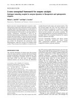
Figure 2
Survival during the first 2 weeksSurvival during the first 2 weeks. The Kaplan-Meier survival curve illus-
trates a trend toward increased mortality during the first 2 weeks in
patients subjected to blood glucose target of 3.5 to 6.5 mmol/l com-
pared with 5 to 8 mmol/l.
Available online />Page 5 of 13
(page number not for citation purposes)
Table 1
Demographic data
Parameters Blood glucose 3.5 to 6.5 mmol/l Blood glucose 5 to 8 mmol/l
Number of patients 114 114
Men (n [%]) 87 (76%) 87 (76%)
Women (n [%]) 27 (24%) 27 (24%)
Isolated TBI (n)4040
Multiple injuries (n)74 74
Age (years; mean [range]) 41 (18–81) 38 (18–81)
AIS head (mean [range]) 25 (9–36) 25 (9–36)
AIS without head (mean [range]) 16 (1–55) 16 (1–55)
ISS (mean [range]) 34 (16–54) 34 (12–67)
Injured organs (n [range]) 2 (1–5) 2 (1–5)
Initial GCS (mean [range]) 11 (3–15) 10 (3–15)
CT lesions (n [%])
EDH 4 (3.5%) 3 (2.6%)
SDH 8 (7%) 12 (10.5%)
Contusions 15 (13.2%) 15 (13.2%0
Generalized oedema 7 (6.1%) 9 (7.9%
tSAH 3 (2.6%) 6 (5.3%)
mixed lesions 77 (67.5%) 69 (60.5%)
Surgery (%)
ICP probe 100% 100%
Fractures 28% 31%
Craniectomy 4% 4%
ICP > 20 mmHg (n [%]) 27 (24%) 33 (29%)
Nonsurvivors (n [%]) 12 (41%) 11 (50%)
Survivors (n [%]) 22 (26%) 17 (18%)
Arterial hypotension (SBP < 90 mmHg; n)1 1
Mortality (n/n [%]) 29/114 (25%) 22/114 (19%)
Week 1 12/114 (11%) 8/114 (7%)
Week 2 9/89 (10%) 5/86 (6%)
Week 3 8/57 (14%) 9/57 (16%)
ICU length (days; median [range])
Survivors 17 (2–48) 15 (2–52)
Deceased 9 (2–23) 11 (2–43)
Demographic data in 228 propensity-matched patients with severe traumatic brain injury (TBI) subjected to two different blood glucose targets:
3.5 to 6.5 mmol/l versus 5 to 8 mmol/l. AIS, abbreviated injury score; CT, computed tomography; EDH, epidural haematoma; GCS, Glasgow
Coma Scale; ICP, intracranial pressure; ICU, intensive care unit; ISS, injury severity score; SDH, subdural haematoma; tSAH, traumatic
subarachnoid haemorrhage; BP = blood pressure.
Critical Care Vol 12 No 4 Meier et al.
Page 6 of 13
(page number not for citation purposes)
Changes in intracranial pressure and cerebral perfusion
pressure
In surviving patients with targeted blood glucose levels
between 3.5 and 6.5 mmol/l, ICP was significantly increased
during the first week (14 ± 0.1 mmHg versus 12 ± 0.1 mmHg;
P < 0.001) and significantly decreased during the third week
compared with the 5 to 8 mmol/l group (15 ± 0.1 mmHg ver-
sus 17 ± 0.1 mmHg; P < 0.001; Figure 5d). Overall, deceased
patients exhibited significantly increased ICP levels compared
with surviving patients. In the deceased patients, elevated ICP
levels were also significantly reduced in the 3.5 to 6.5 mmol/l
group versus the 5 to 8 mmol/l group during the third week (22
± 1 versus 28 ± 1 mmHg; P = 0.046; Figure 5d).
Overall, the incidence of elevated ICP of 20 mmHg or greater
was comparable in the two blood glucose target groups and
corresponding subgroups (survival versus death; 3.5 to 6.5
mmol/l versus 5 to 8 mmol/l: survivors 31% versus 40%;
deceased 69% versus 60%)]. From the second week, how-
ever, the incidence of ICP of 20 mmHg or greater was signifi-
cantly decreased in the patients who died within the low blood
glucose target group (3.5 to 6.5 mmol/l versus 5 to 8 mmol/l:
24% versus 35% [week 2] and 23% versus 33% [week 3]). In
surviving patients there was no difference.
Overall, CPP was maintained between 70 and 90 mmHg,
without a clear influence of the different target blood glucose
levels in surviving patients and those who died (data not
shown).
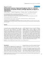
Figure 3
Arterial blood glucose levelsArterial blood glucose levels. Presented are histograms showing distri-
bution of arterial blood glucose levels within predefined clusters in sur-
viving patients (upper panel) and patients who died (lower panel)
treated within the 3.5 to 6.5 mmol/l (black columns) and 5 to 8 mmol/l
(white columns) blood glucose targets.
Figure 4
Frequencies of arterial blood glucose within target rangeFrequencies of arterial blood glucose within target range. Shown are
the relative frequencies of arterial blood glucose concentrations within
the specified ranges, determined at 4-hour intervals. The frequencies of
blood glucose levels below and above the predefined blood glucose
target values were significantly increased in the 3.5 to 6.5 mmol/l com-
pared with the 5 to 8 mmol/l group in the surviving patients (upper
panel) and the patients who died (lower panel). In both groups, the
majority of blood glucose values were within the target range. *P <
0.05, Mann-Whitney rank-sum test.
Available online />Page 7 of 13
(page number not for citation purposes)
Table 2
Episodes of blood glucose levels below the lower limit
Survival status Parameters Blood glucose 3.5 to 6.5 mmol/l Blood glucose 5 to 8 mmol/l
Survived Blood glucose (mmol/l; median [range]) 3.2 (0.7–3.4); NS 3.5 (1.5–3.9)
Blood glucose < lower limit (n [%]) 47/85 (55%)* 24/92 (26%)
Episodes (median [range]) 1 (1–6) 1 (1–11)
Time point of occurrence (%)
Week 1 55%* 28%
Week 2 24% 36%
Week 3 21% 36%
Died Blood glucose (mmol/l; median [range]) 2.7 (0.6–3.4); NS 3.7 (3.1–3.9)
Blood glucose < lower limit (n [%]) 12/29 (41%)* 6/22 (27%)
Episodes (median [range]) 1.5 (1–6) 1.5 (1–3)
Time point of occurrence (%)
Week 1 95%* 50%
Week 2 5% 30%
Week 3 0 20%
Shown are episodes of blood glucose levels below the lower limit in surviving patients and those who died within predefined blood glucose
groups. Decreased blood glucose was predominantly encountered in the low blood glucose group during the first week. *P < 0.05, Whitney-
Mann rank-sum test. NS, not significant.
Table 3
Episodes of blood glucose levels exceeding the upper limit
Survival status Parameters Blood glucose 3.5 to 6.5 mmol/l Blood glucose 5 to 8 mmol/l
Survived Blood glucose (mmol/l; median [range]) 7.3 (6.6–14.8)* 8.7 (8.1–18.1)
Blood glucose < lower limit (n [%]) 81/85 (95%) 89/92 (97%)
Episodes (median [range]) 17 (2–75)* 8.5 (1–85)
Time point of occurrence (%)
Week 1 55% 50%
Week 2 24% 27%
Week 3 21% 23%
Died Blood glucose (mmol/l; median [range]) 2.7 (0.6–3.4); NS 3.7 (3.1–3.9)
Blood glucose < lower limit (n [%]) 28/29 (97%) 20/22 (91%)
Episodes (median [range]) 11.5 (1–31)* 6.5 (1–61)
Time point of occurrence (%)
Week 1 78%* 47%
Week 2 16% 18%
Week 3 6% 35%
Episodes of blood glucose levels exceeding the upper limit in surviving patient and those who died within the two predefined blood glucose
groups. Increased incidences in elevated blood glucose levels were predominantly encountered in the low blood glucose group during the first
week. *P < 0.05, Whitney-Mann rank-sum test.
Critical Care Vol 12 No 4 Meier et al.
Page 8 of 13
(page number not for citation purposes)
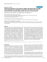
Figure 5
Changes in arterial blood glucose, insulin and norepinephrine dose, and ICPChanges in arterial blood glucose, insulin and norepinephrine dose, and ICP. Shown are changes in arterial blood glucose, insulin and norepine-
phrine dose, and intracranial pressure (ICP) in surviving patients and in those who died, within the different blood glucose target groups over time.
(a) Arterial blood glucose levels were significanlty decreased in both surviving and deceased patients in the 3.5 to 6.5 group. (b) Insulin requirement
was significantly increased in the 3.5 to 6.5 mmol/l group. (c) Within the 3.5 to 6.5 mmol/l group, surviving patients and those who died required sig-
nificantly greater amounts of norepinephrine. (d) ICP was significantly increased in the 3.5 to 6.5 mmol/l group during the first week in surviving
patients, followed by a significant decrease during the subsequent weeks. Patients who died exhibited a significantly increased ICP in the first week,
irrespective of blood glucose target. In the third week, however, ICP was significantly increased in the 5 to 8 mmol/l group. *P < 0.05, analysis of
variance on ranks.
Available online />Page 9 of 13
(page number not for citation purposes)
Impact of blood glucose diverging from the anticipated
blood glucose targets
Higher blood glucose levels were associated with higher insu-
lin requirement. Overall, blood glucose values above the upper
limit or below the lower limit were not associated with an
increase in ICP or a decrease in CPP (data not shown).
Caloric intake
Average daily total caloric intake was comparable in both
groups (3.5 to 6.5 mmol/l versus 5 to 8 mmol/l): 1,965 ± 38
versus 2,049 ± 35 kcal. There was no significant difference
between the two groups on any given day.
Bacteraemia, urinary tract infection, positive
tracheobronchial secretions and blood inflammation
parameters
Overall there was no statistically significant difference in rate
of pneumonia between the two blood glucose groups. How-
ever, bacteraemia (25% versus 18%; relative difference:
+28%), and urinary tract infections (22% versus 16%; relative
difference: +27%) were significantly increased in patients
within the 3.5 to 6.5 mmol/l group.
Over time, the rate of bacteraemia was not significantly differ-
ent between the two blood glucose groups. The rate of pneu-
monia was significantly reduced in the third week in surviving
and deceased patients within the 3.5 to 6.5 mmol/l group as
compared with the 5 to 8 mmol/l group (18% versus 26%; -
44%; P < 0.005). The rate of urinary tract infections was sig-
nificantly decreased in the second week (26% versus 53%; -
51%; P < 0.005) followed by a significant increase in the third
week (48% versus 24%; +50%; P < 0.005) in patients within
the 3.5 to 6.5 mmol/l group as compared with the 5 to 8 mmol/
l group.
Within the 3.5 to 6.5 mmol/l group, bacteraemia was signifi-
cantly less likely to be caused by Gram-positive bacteria (62%
versus 78%; -26%; P < 0.05), whereas urinary tract infections
were significantly more likely to be caused by Gram-positive
bacteria (30% versus 17%; relative difference: +43%; P <
0.005) compared with the 5 to 8 mmol/l group. Gram-negative
bacteria exhibited a similar rate in the two glucose groups. Tra-
cheobronchial cultures revealed a similar distribution in Gram-
positive and Gram-negative bacteria.
There were no differences in maximal leukocyte, CRP and IL-
6 levels between the predefined blood glucose groups (data
not shown).
Discussion
In 228 propensity-matched patients suffering from severe TBI,
the target blood glucose concentration of 3.5 to 6.5 mmol/l
was associated with a trend toward increased mortality during
the first 2 weeks, markedly increased frequency of hypogly-
caemic and hyperglycaemic episodes, significantly elevated
ICP during the first week, and markedly increased insulin and
norepinephrine requirement compared with patients with a
blood glucose target of 5 to 8 mmol/l. From the second week,
however, decreased ICP and reduced rate of infectious com-
plications prevailed in the 3.5 to 6.5 mmol/l group compared
with the 5 to 8 mmol/l target group.
While a slightly higher blood glucose target (5 to 8 mmol/l)
appears to be more beneficial during the first week, lower
blood glucose levels (3.5 to 6.5 mmol/l) perhaps should be
implemented during the first week.
Limitations of this retrospective study
The present retrospective study is weakened by its lack of
controlling for clinically important interventions, because
investigated parameters were 'only' documented in 4-hour
intervals or once daily. Thus, this approach is unfortunately
likely to miss potentially important alterations that might have
occurred within the 4-hour intervals. In addition, the present
data do not allow us to assess the impact of speed and mag-
nitude of blood glucose level correction, which might also be
disadvantageous. To avoid this methodological setback, con-
tinuous recording and painstaking documentation of important
events is required; this, however, is time consuming and diffi-
cult in the daily routine.
Our assimilation of patients recruited during sequential time
periods (2000 to 2002 versus 2002 to 2004) by pre-defining
age, sex, as well as presence and severity of additional injuries
allowed us to control for certain baseline variables, thereby
enhancing the quality of our retrospective analysis of pooled
data within post hoc defined clusters. Normalization of the
data by calculating relative frequencies within predefined clus-
ters helps to compare patient groups and permits determina-
tion of the potential impact of blood glucose targets. However,
we cannot exclude the possibility that improved awareness
and knowledge, which clearly develop over time, might also
have influenced basic treatment and could have blurred rele-
vant differences.
Owing to differences in individual clinical course and different
durations of hospitalization, patients exhibit different values for
the various parameters; this may account for the reduced
number of values recorded the third week, especially in the
patients who died. Thus, we obtained the greatest statistical
power within the first and second weeks.
The chosen blood glucose targets are overlapping. Thus, the
close proximity of the upper and lower limits of the two blood
glucose targets, namely 6.5 and 5 mmol/l, might have
obscured an even more significant impact, as in the study pub-
lished by van den Berghe and colleagues [7], when larger dif-
ferences were studied under 6.1 mmol/l versus under 12
mmol/l. However, in reality, even in that prospective study, the
difference between low and high blood glucose target groups
Critical Care Vol 12 No 4 Meier et al.
Page 10 of 13
(page number not for citation purposes)
(< 6.1 versus < 12 mmol/l) was much smaller, being on aver-
age 5.6 mmol/l versus 8.9 mmol/l, with similar initial blood
glucose values [7,8]. The rate of overlapping blood glucose
values, however, was not reported [7,8,15].
The overlapping values resulting from insulin administration,
and which are a reflection of the meticulous attention given to
adhering to the predefined blood glucose targets in both
groups, appear to have reduced the impact in the present
study. However, the significant differences in primary end-
points, glucose variability and extreme blood glucose values
show that the predefined blood glucose targets are of patho-
physiologic relevance, despite overlapping of blood glucose
values. Within this context, patients within the 3.5 to 6.5 mmol/
l group were metabolically less stable, as reflected by the
higher incidence of hypoclycaemic and hyperglycaemic
vlaues. Apparently, the chosen lower limit of 3.5 mmol/l predis-
poses to hypoglycaemic complications in the face of sup-
pressed hormonal counterregulation. However, as was
recently demonstrated by McMullin and colleagues [14], who
compared the target range 5 to 7 mmol/l versus 8 to 10 mmol/
l, similar difficulties were encountered even at higher blood
glucose targets.
Blood glucose and secondary brain damage
TBI is characterized by regionally and temporally altered glu-
cose metabolism caused by altered cellular demands and
functional disturbances. These changes are not restricted to
the site of injury [16,17] and can persist for up to several
months in patients with moderate to severe TBI [18-21].
In face of the limited cerebral energetic reserves, with marginal
cerebral availibility of glycogen, glucose is the predominant
fuel for neuronal and glial activities [22]. To ensure adequate
glucose supply in the face of increased glucose consumption,
cerebral glucose uptake occurs independently of insulin via
specific endothelial/glial (glucose transporter [GLUT]1) and
neuronal (GLUT3) glucose transporters, which have different
transport characteristics. In this context, GLUT1 (with its inter-
mediate Michaelis constant of 5 to 7 mmol/l) and GLUT3 (with
its low Michaelis constant of 1.6 mmol/l) ensure neuronal glu-
cose uptake even during hypoglycaemia [23]. Nevertheless,
any decrease in blood glucose levels, such as those observed
in the present study, predisposes the patient to risk for reach-
ing the lower glucose transportation rate, especially in
endothelial/glial glucose transporters, which can be aggra-
vated by concomitant impaired perfusion and sustained glyco-
lysis [24] or altered enzymatic activity [20,21]. This, in turn,
increases the risk for additional injury. In this regard, a
decrease in blood glucose levels below 8 mmol/l was associ-
ated with an increase in extracellular cerebral lactate, meas-
ured using microdialysis, which coincided with a significant
elevation in perischaemic cortical depolarizations [12]. A
dramatic increase in perischaemic cortical depolarizations
was observed when blood glucose levels dropped below 6
mmol/l [11-13]. By implementing low blood glucose levels
(such as 3.5 to 6.5 mmol/l [present study] or 4.4 to 6.1 mmol/
l [7]), we are actively risking progressive and additional sec-
ondary insults, which could aggravate underlying structural
and functional damage. Evidence for such a process was pro-
vided by Vespa and colleagues [25], who reported a signifi-
cant increase in glutamate and lactate/pyruvate ratio during
intensive insulin therapy with arterial blood glucose levels
ranging from 5 to 6.7 mmol/l versus 6.7 to 8.3 mmol/l.
In addition, hypoglycaemia combined with insufficient tissue
oxygenation predisposes the brain to aggravated damage
induced by subsequent hyperglycaemia [26]. The significant
increase in ICP and elevated requirement for norepinephrine
to maintain CPP above 70 mmHg observed in the present
analysis could reflect ongoing alterations within the injured
brain, possibly induced by maintaining blood glucose levels
between 3.5 and 6.5 mmol/l, because this range is close to
the threshold for inducing cortical spreading depressions with
subsequent oedema progression [13]. The significant
increase in ICP coincided with an increase in hypoglycaemic
values, which were predominantly observed during the first
week. Because the majority of pathological cascades are acti-
vated within the first week, any additional insults, such as hyp-
ogycaemia, hyperglycemia and changing blood glucose
values, should be avoided to prevent secondary brain damage.
Apart from hypoglycaemia-induced damage, hyperglycaemia
is also a feared complication for its detrimental effects. In this
context, hyperglycaemia has the following effects [27-29]: it
impairs cerebral perfusion because of cellular swelling or
neutralization of nitric oxide by free radical production; it pro-
motes local tissue acidosis; it induces oxidative stress with
subsequent mitochondrial damage and impaired oxidative
phosphorylation; it promotes glutamate-driven increase in
intracellular calcium concentrations; it induces microcircula-
tory damage and blood-brain barrier disruption because of ele-
vated inflammation with sustained cerebral leukocyte
adherence and invasion, and production of matrix metallopro-
teinase-9; and it interferes with transcription processes.
The general consensus is to avoid blood glucose levels
exceeding 10 mmol/l, because they are associated with neu-
rologic deterioration [23]. In the present study, dangerously
elevated blood glucose levels exceeding 10 mmol/l were
observed in fewer than 3% of all blood glucose values. Neither
these hyperglycaemic nor the hypoglycaemic values were
associated with signs of cerebral worsening (increased ICP or
decreased CPP).
Pharmacodynamic effects of insulin
Insulin is known for its anabolic effects, which promote lipo-
genesis and protein synthesis mediated by uptake of glucose
and amino acids. In addition, insulin inhibits hyperglycaemia-
induced oxidative cell damage [6,7,27], thereby positively
Available online />Page 11 of 13
(page number not for citation purposes)
influencing various intracellular signaling cascades [30].
These effects are viewed as the pharmacodynamic basis of
improved organ function: decreased renal failure [31],
reduced ventilatory support [32], decreased infection rate
[33] and reduced transfusion requirements [31]. They contrib-
ute to shortened length of ICU stay [32], improved recovery
and decreased mortality [32] in critically ill patients [7,8].
These positive findings are in contrast to the present findings
of sustained haemodynamic instability and increased infec-
tions (bacteraemia and urinary tract infections) in patients
subjected to the low blood glucose target (3.5 to 6.5 mmol/l
versus 5 to 8 mmol/l). This, of course, could result from differ-
ences in policy concerning volume management (type, dura-
tion, or predefined targets), catheter placement (duration
before renewal or removal) and administration of antibiotics
(type, duration and start), because we maintained similar
blood glucose levels using similar insulin doses as were
reported by van den Berghe and coworkers [7,8]. At the a cel-
lular level, insulin attenuates norepinephrine-mediated vaso-
constriction [34], which could explain the increased
norepinephrine requirement in patients within the 3.5 to 6.5
mmol/l group. In addition, attenuated respiratory burst [35]
and decreased chemokinesis [36] in neutrophils caused by
insulin-mediated decreased blood glucose levels contribute to
impaired cellular defense mechanisms, thereby promoting
infections, as was observed in the present study in terms of
increased bacteraemia and urinary tract infections. A concen-
tration-dependent effect in critically ill patients requires
investigation.
Control of blood glucose levels
Implementing a strict and tight control of blood glucose levels
by intensified insulin administration has been shown to result
in more rapid correction, more stable blood glucose concen-
trations and longer maintenance within the predefined target
range as compared with controls [37]. This, however, requires
steady parenteral or enteral supply along with nutrients, and
tight control to prevent hypoglycaemic episodes, which are
feared for their cerebrotoxic effects.
van den Berghe and colleagues [7] used a particular protocol,
which includes continuous infusion of glucose (9 g/hour),
intake of sufficient calories (19 kcal/kg per hour) during con-
tinuous insulin administration (0.04 units/kg per hour). In our
patients subjected to a blood glucose target of 3.5 to 6.5
mmol/l, a similar insulin dose was administered to that
reported by van den Berghe and coworkers [7,8]. Apparently,
glucose must be co-infused to prevent and correct insulin-
induced hypoglycaemia. This, however, was not done in our
patients with severe TBI, because infusing glucose results in
administration of 'free' water, which could aggravate brain
oedema formation because glucose and free water can diffuse
more easily and rapidly through the blood-brain barrier com-
pared with crystalloids.
Regardless of strategy, maintaining glucose within predefined
limits can be difficult, with hypoglycaemic episodes ranging
from 12% to 40% in patients subjected to intensified insulin
treatment compared with 1.2% to 7.4% in the conventional
group [7-9,14,33,38-40]. This is in line with the observed glo-
bal incidence of hypoglycaemic values (< 2.5 mmol/l), occur-
ring in 12% (14/114 patients; 3.5 to 6.5 mmol/l group) versus
2.6% (3/114 patients; 5 to 8 mmol/l group) in the presently
investigated 228 patients suffering from severe TBI. These
data also clearly show that intermittent control in 4-hour inter-
vals is insufficient to prevent increased or decreased blood
glucose levels. Continuous control would yield superior
results. However, an adequate procedure has not yet been
introduced into clinical routine practice.
Are there different requirements for specific time points
following severe TBI?
In daily clinical routine, patients undergo different phases that
require different types and degrees of interventions. Although
haemodynamic instability and systemic inflammation prevail
during the first week, infections with a new inflammatory
challenge develop during the second week. Some patients
again recover without additional complications. Thus, an indi-
vidual strategy might be required to maintain metabolic stabil-
ity and prevent additional insults related to blood glucose
deviations that might result in increased mortality. These
dynamic and time-dependent processes could also influence
the threshold for additional cell damage over time.
Based on the present data (ICP, length of ICU stay, infections,
mortality, and hypoglycaemic and hyperglycemic episodes),
patients do not profit from blood glucose levels maintained
between 3.5 and 6.5 mmol/l during the first week. From the
second week, however, lowered blood glucose levels could
prove beneficial because ICP levels were significantly
decreased compared with patients in whom blood glucose
levels were maintained between 5 and 8 mmol/l. However, ICP
values were in a similar range, making it difficult to assess pos-
itive effects at the bedside in real time. Such a possible time-
dependent pattern is also suggested by the findings reported
by van den Berghe and coworkers [7], wo reported that
patients only profit from lowered blood glucose levels
achieved using an intensified insulin therapy if they require ICU
treatment longer than 3 days (medical ICU patients) or 5 days
(surgical ICU patients) [15]. In fact, mortality was significantly
increased in medical patients remaining on the ICU for fewer
than 3 days [15]. In critically ill patients suffering from isolated
TBI and subjected to low blood glucose levels (average 5.6 ±
0.5 mmol/l), mortality on the ICU at 6 and 12 months was
increased (23% versus 18%, 48% versus 30%, respectively)
[8].
This could be in favour of a certain beneficial effect of elevated
blood glucose levels during the early phase following a
defined insult, as suggested by Ghandi and colleagues [41]
Critical Care Vol 12 No 4 Meier et al.
Page 12 of 13
(page number not for citation purposes)
and as is also seen under in vitro conditions where short-term
hyperglycaemia (15 to 60 mmol/l) is protective in cardiac myo-
cytes, astrocytes and neurons [42-44].
Future studies are required to investigate dynamic adaptation
of blood glucose targets over time.
Conclusion
Based on our retrospective analysis, revealing a significant
increase in hypoclycaemic and hyperglycemic episodes, as
well as elevated insulin and norepinephrine requirements, we
cannot recommend maintaining blood glucose levels between
3.5 and 6.5 mmol/l during the first week after severe TBI. Main-
taining arterial blood glucose between 5 and 8 mmol/l is more
favourable during the first week. However, significant
decreases in ICP, including intracranial hypertension exceed-
ing 20 mmHg, as well as reduced infectious complications
during the second week are in favour of the lower arterial
blood glucose levels maintained between 3.5 and 6.5 mmol/l.
It remains to be determined whether a temporally adapted
blood glucose target might be required. For this, however, an
optimal blood glucose level must be defined. In addition, sur-
veillance of changes in blood glucose needs to be improved,
preferably using continuous measurements, because intermit-
tent analysis is limited by its risk for delayed assessment and
correction of hypoglycaemic and hyperglycemic episodes.
Furthermore, downstream monitoring of metabolic impairment,
as indicated by parameters such as cerebral lactate, lactate/
pyruvate ratio, glucose/lactate ratio and glutamate level, is
indispensible in identifying adequate and optimal blood glu-
cose level, which is required for subsequent decision making
and treatment.
Competing interests
The authors declare that they have no competing interests.
Authors' contributions
RM collected most of the data, drafted parts of the manuscript
and performed graphical analysis. MB helped analyzing and
interpreting the data, and drafted parts of the manuscript. SL
and JS were responsible for data collection and upkeeping of
the databank. MK, PS and RS helped to analyze and interpret
the data. JFS conceived the study design, collected some of
the data, performed graphical and statistical analyses, and
drafted parts of the manuscript.
Acknowledgements
The help of the nursing staff in collecting clinical data is gratefully
acknowledged. This study was supported in parts by grants from the
SUVA Fonds to JFS and RS.
References
1. Van Beek JG, Mushkudiani NA, Steyerberg EW, Butcher I,
McHugh GS, Lu J, Marmarou A, Murray GD, Maas AI: Prognostic
value of admission laboratory parameters in traumatic brain
injury: results from the IMPACT study. J Neurotrauma 2007,
24:315-328.
2. Jeremitsky E, Omert LA, Dunham CM, Wilberger J, Rodriguez A:
The impact of hyperglycemia on patients with severe brain
injury. J Trauma 2005, 58:47-50.
3. Gale SC, Sicoutris C, Reilly PM, Schwab CW, Gracias VH: Poor
glycemic control is associated with increased mortality in crit-
ically ill trauma patients. Am Surg 2007, 73:454-460.
4. Longstreth WT Jr, Inui TS: High blood glucose level on hospital
admission and poor neurological recovery after cardiac arrest.
Ann Neurol 1984, 15:59-63.
5. Capes SE, Hunt D, Malmberg K, Gerstein HC: Stress hypergly-
caemia and increased risk of death after myocardial infarction
in patients with and without diabetes: a systematic overview.
Lancet 2000, 355:773-778.
6. Vanhorebeek I, Langouche L, Berghe G Van den: Tight blood glu-
cose control: what is the evidence? Crit Care Med 2007,
35:S496-S502.
7. Berghe G van den, Wouters P, Weekers F, Verwaest C, Bruyn-
inckx F, Schetz M, Vlasselaers D, Ferdinande P, Lauwers P, Bouil-
lon R: Intensive insulin therapy in the critically ill patients. N
Engl J Med 2001, 345:1359-1367.
8. Berghe G Van den, Schoonheydt K, Becx P, Bruyninckx F, Wout-
ers PJ: Insulin therapy protects the central and peripheral nerv-
ous system of intensive care patients. Neurology 2005,
64:1348-1353.
9. Bilotta F, Caramia R, Cernak I, Paoloni FP, Doronzio A, Cuzzone V,
Santoro A, Rosa G: Intensive insulin therapy after severe trau-
matic brain injury: a randomized clinical trial. Neurocrit Care
2008 in press.
10. Vespa PM, McArthur D, O'Phelan K, Glenn T, Etchepare M, Kelly
D, Bergsneider M, Martin NA, Hovda DA: Persistently low extra-
cellular glucose correlates with poor outcome 6 months after
human traumatic brain injury despite a lack of increased lac-
tate: a microdialysis study. J Cereb Blood Flow Metab 2003,
23:865-877.
11. Strong AJ, Smith SE, Whittington DJ, Meldrum BS, Parsons AA,
Krupinski J, Hunter AJ, Patel S, Robertson C: Factors influencing
Key messages
• Maintaining low blood glucose concentrations between
3.5 and 6.5 mmol/l, as compared with 5 to 8 mmol/l,
increases rate of hypoglycaemic and hyperglycaemic
values, especially during the first week.
• The need for norepinephrine to maintain stable CPP
level is significantly increased when the target blood
glucose level is low.
• The rates of bacteraemia and urinary tract infection are
significantly increased when reducing blood glucose
levels to 3.5 to 6.5 mmol/l as compared with 5 to 8
mmol/l during the first week, followed by a significant
decrease in the second week.
• Temporal profile of decreased ICP suggests that blood
glucose levels maintained between 3.5 and 6.5 mmol/l
could be of benefit during the second week, reflected
by decreased incidence of ICP exceeding 20 mmHg.
• Future trials must determine a characteristic temporal
profile of specific arterial blood glucose targets,
because the present study suggests less favourable
effects of low blood glucose levels (3.5 to 6.5 mmol/l)
during the first week, followed by more favourable
effects as of the second week compared with the blood
glucose target of 5 to 8 mmol/l.
Available online />Page 13 of 13
(page number not for citation purposes)
the frequency of fluorescence transients as markers of peri-
infarct depolarizations in focal cerebral ischemia. Stroke 2000,
31:214-222.
12. Hopwood SE, Parkin MC, Bezzina EL, Boutelle MG, Strong AJ:
Transient changes in cortical glucose and lactate levels asso-
ciated with peri-infarct depolarisations, studied with rapid-
sampling microdialysis. J Cereb Blood Flow Metab 2005,
25:391-401.
13. Strong AJ, Hartings JA, Dreier JP: Cortical spreading depres-
sion: an adverse but treatable factor in intensive care? Curr
Opin Crit Care 2007, 13:126-133.
14. McMullin J, Brozek J, McDonald E, Clarke F, Jaeschke R, Heels-
Ansdell D, Leppert R, Foss A, Cook D: Lowering of glucose in
critical care: a randomized pilot trial. J Crit Care 2007,
22:112-118.
15. Berghe G Van den, Wilmer A, Hermans G, Meersseman W, Wout-
ers PJ, Milants I, Van Wijngaerden E, Bobbaers H, Bouillon R:
Intensive insulin therapy in the medical ICU. N Engl J Med
2006, 354:449-461.
16. Thomale UW, Griebenow M, Mautes A, Beyer TF, Dohse NK,
Stroop R, Sakowitz OW, Unterberg AW, Stover JF: Heterogene-
ous regional and temporal energetic impairment following
controlled cortical impact injury in rats. Neurol Res 2007,
29:594-603.
17. Nelson DW, Bellander BM, Maccallum RM, Axelsson J, Alm M,
Wallin M, Weitzberg E, Rudehill A: Cerebral microdialysis of
patients with severe traumatic brain injury exhibits highly indi-
vidualistic patterns as visualized by cluster analysis with self-
organizing maps. Crit Care Med 2004, 32:2428-2436.
18. Kato T, Nakayama N, Yasokawa Y, Okumura A, Shinoda J, Iwama
T: Statistical image analysis of cerebral glucose metabolism in
patients with cognitive impairment following diffuse traumatic
brain injury. J Neurotrauma 2007, 24:919-926.
19. Glenn TC, Kelly DF, Boscardin WJ, McArthur DL, Vespa P, Oertel
M, Hovda DA, Bergsneider M, Hillered L, Martin NA: Energy dys-
function as a predictor of outcome after moderate or severe
head injury: indices of oxygen, glucose, and lactate
metabolism. J Cereb Blood Flow Metab 2003, 23:1239-1250.
20. Hattori N, Huang SC, Wu HM, Liao W, Glenn TC, Vespa PM,
Phelps ME, Hovda DA, Bergsneider M: Acute changes in
regional cerebral
18
F-FDG kinetics in patients with traumatic
brain injury. J Nucl Med 2004, 45:775-783.
21. Wu HM, Huang SC, Hattori N, Glenn TC, Vespa PM, Yu CL,
Hovda DA, Phelps ME, Bergsneider M: Selective metabolic
reduction in gray matter acutely following human traumatic
brain injury. J Neurotrauma 2004, 21:149-161.
22. Bartnik BL, Hovda DA, Lee PW: Glucose metabolism after trau-
matic brain injury: estimation of pyruvate carboxylase and
pyruvate dehydrogenase flux by mass isotopomer analysis. J
Neurotrauma 2007, 24:181-194.
23. Oddo M, Schmidt JM, Mayer SA, Chioléro RL: Glucose control
after severe brain injury. Curr Opin Clin Nutr Metab Care 2008,
11:134-139.
24. Kelly DF, Kozlowski DA, Haddad E, Echiverri A, Hovda DA, Lee
SM: Ethanol reduces metabolic uncoupling following experi-
mental head injury. J Neurotrauma 2000, 17:261-272.
25. Vespa P, Boonyaputthikul R, McArthur DL, Miller C, Etchepare M,
Bergsneider M, Glenn T, Martin N, Hovda D: Intensive insulin
therapy reduces microdialysis glucose values without altering
glucose utilization or improving the lactate/pyruvate ratio
after traumatic brain injury. Crit Care Med 2006, 34:850-856.
26. de Courten-Myers GM, Xi G, Hwang JH, Dunn RS, Mills AS, Hol-
land SK, Wagner KR, Myers RE: Hypoglycemic brain injury:
potentiation from respiratory depression and injury aggrava-
tion from hyperglycemic treatment overshoots. J Cereb Blood
Flow Metab 2000, 20:82-92.
27. Erol A: Insulin resistance is an evolutionarily conserved physi-
ological mechanism at the cellular level for protection against
increased oxidative stress. Bioessays 2007, 29:811-818.
28. Garg R, Chaudhuri A, Munschauer F, Dandona P: Hyperglycemia,
insulin, and acute ischemic stroke: a mechanistic justification
for a trial of insulin infusion therapy. Stroke 2006, 37:267-273.
29. Kinoshita K, Kraydieh S, Alonso O, Hayashi N, Dietrich WD: Effect
of posttraumatic hyperglycemia on contusion volume and
neutrophil accumulation after moderate fluid-percussion
brain injury in rats. J Neurotrauma 2002, 19:681-692.
30. Hui L, Pei DS, Zhang QG, Guan QH, Zhang GY: The neuropro-
tection of insulin on ischemic brain injury in rat hippocampus
through negative regulation of JNK signaling pathway by
PI3K/Akt activation. Brain Res 2005, 1052:1-9.
31. Krinsley JS: Effect of an intensive glucose management proto-
col on the mortality of critically ill adult patients. Mayo Clin
Proc 2004, 79:992-1000.
32. Quinn JA, Snyder SL, Berghoff JL, Colombo CS, Jacobi J: A prac-
tical approach to hyperglycemia management in the intensive
care unit: evaluation of an intensive insulin infusion protocol.
Pharmacotherapy 2006, 26:1410-1420.
33. Grey NJ, Perdrizet GA: Reduction of nosocomial infections in
the surgical intensive-care unit by strict glycemic control.
Endocr Pract 2004, 10(suppl 2):46-52.
34. De Ciuceis C, Rizzoni D, Porteri E, Boari GE, Zani F, Miclini M,
Tiberio GA, Giulini SM, Paiardi S, Rizzardi N, Platto C, Agabiti-
Rosei E: Effects of insulin on endothelial and contractile func-
tion of subcutaneous small resistance arteries of hypertensive
and diabetic patients. J Vasc Res 2008, 45:512-520.
35. Thomson GA, Fisher BM, Gemmell CG, MacCuish AC, Gallacher
SJ: Attenuated neutrophil respiratory burst following acute
hypoglycaemia in diabetic patients and normal subjects. Acta
Diabetol 1997, 34:253-256.
36. Oldenborg PA, Sehlin J: The glucose concentration modulates
N-formyl-methionyl-leucyl-phenylalanine (fMet-Leu-Phe)-
stimulated chemokinesis in normal human neutrophils. Biosci
Rep 1999, 19:511-523.
37. Kanji S, Singh A, Tierney M, Meggison H, McIntyre L, Hebert PC:
Standardization of intravenous insulin therapy improves the
efficiency and safety of blood glucose control in critically ill
adults. Intensive Care Med 2004, 30:804-810.
38. Vriesendorp TM, van Santen S, DeVries JH, de Jonge E, Rosendaal
FR, Schultz MJ, Hoekstra JB: Predisposing factors for hypogly-
cemia in the intensive care unit. Crit Care Med 2006,
34:96-101.
39. Clayton SB, Mazur JE, Condren S, Hermayer KL, Strange C: Eval-
uation of an intensive insulin protocol for septic patients in a
medical intensive care unit. Crit Care Med 2006,
34:2974-2978.
40. Cochran A, Davis L, Morris SE, Saffle JR: Safety and efficacy of
an intensive insulin protocol in a burn-trauma intensive care
unit. J Burn Care Res 2008, 29:187-191.
41. Gandhi GY, Nuttall GA, Abel MD, Mullany CJ, Schaff HV, O'Brien
PC, Johnson MG, Williams AR, Cutshall SM, Mundy LM, Rizza RA,
McMahon MM: Intensive intraoperative insulin therapy versus
conventional glucose management during cardiac surgery: a
randomized trial. Ann Intern Med 2007, 146:233-243.
42. Malliopoulou V, Xinaris C, Mourouzis I, Cokkinos AD, Katsilambros
N, Pantos C, Kardami E, Cokkinos DV: High glucose protects
embryonic cardiac cells against simulated ischemia. Mol Cell
Biochem 2006, 284:87-93.
43. Kelleher JA, Chan PH, Chan TY, Gregory GA: Modification of
hypoxia-induced injury in cultured rat astrocytes by high levels
of glucose. Stroke 1993, 24:855-863.
44. Shi H, Liu KJ: Effects of glucose concentration on redox status
in rat primary cortical neurons under hypoxia. Neurosci Lett
2006, 410:57-61.