Báo cáo y học: "Pressure support ventilation attenuates ventilator-induced protein modifications in the diaphragm" doc
Bạn đang xem bản rút gọn của tài liệu. Xem và tải ngay bản đầy đủ của tài liệu tại đây (413.87 KB, 9 trang )
Open Access
Available online />Page 1 of 9
(page number not for citation purposes)
Vol 12 No 5
Research
Pressure support ventilation attenuates ventilator-induced
protein modifications in the diaphragm
Emmanuel Futier
1
, Jean-Michel Constantin
1
, Lydie Combaret
2
, Laurent Mosoni
2
, Laurence Roszyk
3
,
Vincent Sapin
3
, Didier Attaix
2
, Boris Jung
4
, Samir Jaber
4
and Jean-Etienne Bazin
1
1
General Intensive Care Unit, Hotel-Dieu Hospital, University Hospital of Clermont-Ferrand, Boulevard L. Malfreyt, Clermond-Ferrand, 63058, France
2
Human Nutrition Research Center of Clermont-Ferrand, Nutrition and Protein Metabolism Unit, Institut National de la Recherche Agronomique, Route
de Theix, Ceyrat, 63122 France
3
Department of Biochemistry, University Hospital of Clermont-Ferrand, Boulevard L. Malfreyt, Clermont-Ferrand, 63000, France
4
SAR B, Saint-Eloi Hospital, University Hospital of Montpellier, Avenue Augustin Fliche, Montpellier, 34000, France
Corresponding author: Jean-Michel Constantin,
Received: 25 May 2008 Revisions requested: 19 Jun 2008 Revisions received: 31 Jul 2008 Accepted: 11 Sep 2008 Published: 11 Sep 2008
Critical Care 2008, 12:R116 (doi:10.1186/cc7010)
This article is online at: />© 2008 Futier et al.; licensee BioMed Central Ltd.
This is an open access article distributed under the terms of the Creative Commons Attribution License ( />),
which permits unrestricted use, distribution, and reproduction in any medium, provided the original work is properly cited.
Abstract
Introduction Controlled mechanical ventilation (CMV) induces
profound modifications of diaphragm protein metabolism,
including muscle atrophy and severe ventilator-induced
diaphragmatic dysfunction. Diaphragmatic modifications could
be decreased by spontaneous breathing. We hypothesized that
mechanical ventilation in pressure support ventilation (PSV),
which preserves diaphragm muscle activity, would limit
diaphragmatic protein catabolism.
Methods Forty-two adult Sprague-Dawley rats were included in
this prospective randomized animal study. After intraperitoneal
anesthesia, animals were randomly assigned to the control
group or to receive 6 or 18 hours of CMV or PSV. After sacrifice
and incubation with
14
C-phenylalanine, in vitro proteolysis and
protein synthesis were measured on the costal region of the
diaphragm. We also measured myofibrillar protein carbonyl
levels and the activity of 20S proteasome and
tripeptidylpeptidase II.
Results Compared with control animals, diaphragmatic protein
catabolism was significantly increased after 18 hours of CMV
(33%, P = 0.0001) but not after 6 hours. CMV also decreased
protein synthesis by 50% (P = 0.0012) after 6 hours and by
65% (P < 0.0001) after 18 hours of mechanical ventilation.
Both 20S proteasome activity levels were increased by CMV.
Compared with CMV, 6 and 18 hours of PSV showed no
significant increase in proteolysis. PSV did not significantly
increase protein synthesis versus controls. Both CMV and PSV
increased protein carbonyl levels after 18 hours of mechanical
ventilation from +63% (P < 0.001) and +82% (P < 0.0005),
respectively.
Conclusions PSV is efficient at reducing mechanical
ventilation-induced proteolysis and inhibition of protein
synthesis without modifications in the level of oxidative injury
compared with continuous mechanical ventilation. PSV could be
an interesting alternative to limit ventilator-induced
diaphragmatic dysfunction.
Introduction
Controlled mechanical ventilation (CMV) has been shown to
induce muscle atrophy and to alter diaphragm contractile
properties [1-6], leading to early and severe ventilator-induced
diaphragm dysfunction (VIDD) that has been implicated in
weaning failure [7,8]. Although weaning failure may be due to
numerous factors, diaphragm dysfunction induced by mechan-
ical ventilation (MV) probably plays an important role. Indeed,
animal studies reveal that 18 hours of CMV results in diaphrag-
matic contractile dysfunction and atrophy [9]. Moreover, the
combination of 18 to 69 hours of complete diaphragmatic
inactivity and MV results in marked atrophy of human dia-
phragm myofibers [1].
The mechanisms of VIDD have not been fully elucidated. Mus-
cle atrophy, oxidative stress, and structural injury have been
documented after CMV [7]. Muscle proteolysis is a highly reg-
ulated process accomplished by at least three different
14
C-Phe:
14
C-phenylalanine; AAF: alanine-alanine-phenylalanine; AMC: 7-amino-4-methylcoumarin; CMV: controlled mechanical ventilation; DNPH:
2,4-dinitrophenylhydrazones; DTT: dithiothreitol; FiO
2
: fraction of inspired oxygen; LLVY: leucine-leucine-valine-tyrosine; MV: mechanical ventilation;
PSV: pressure support ventilation; TCA: trichloroacetic acide; TPPII: tripeptidylpeptidase II; VIDD: ventilator-induced diaphragm dysfunction.
Critical Care Vol 12 No 5 Futier et al.
Page 2 of 9
(page number not for citation purposes)
proteolytic systems: the ubiquitin-proteosome pathway, the
Ca
2+
-dependent system, and the lysosomal system. All three
proteolytic systems have been shown to be implicated in the
increased diaphragmatic proteolysis observed after CMV, as
indicated by changes in the gene expression profile of several
proteolytic enzymes [10]. Muscle atrophy is not due only to an
increase in proteolysis. Shanely and colleagues [11] have
shown that CMV induced a rapid decreased synthesis of dia-
phragmatic mixed muscle protein and myosin heavy chain pro-
tein. Indeed, within the first 6 hours of MV, mixed muscle
protein synthesis decreased by 30% and myosin heavy chain
protein synthesis decreased by 65% [11].
MV-induced oxidative stress is also an important contributor to
both MV-induced proteolysis and contractile dysfunction.
Indeed, Shanely and colleagues [2] have shown that MV is
associated with a rapid onset of protein oxidation in diaphragm
fibers. This is significant because oxidative stress has been
shown to promote disuse muscle atrophy [12] and has been
directly linked to activation of the ubiquitin-proteasome system
of proteolysis [13]. The precise contribution of each factor to
the development of VIDD and their kinetic of apparition has yet
to be defined.
Although it was demonstrated that CMV exerted several dele-
terious effects on the diaphragm, only few protective counter-
measures have been developed to minimize CMV-induced
diaphragm dysfunction and atrophy. Administration of the anti-
oxidant Trolox has been shown to prevent CMV-induced dia-
phragm contractile impairments and to retard proteolysis [14].
Administration of the protease inhibitor leupeptin concomi-
tantly with MV prevented the apparition of VIDD in rats after 24
hours of MV [15]. Intermittent spontaneous breathing during
the course of CMV has been shown to protect the diaphragm
against the deleterious effects of CMV [16].
In clinical practice, spontaneous breathing increases work of
breathing and patients often need positive pressure ventilation
to improve gas exchange [17]. The spontaneous breathing
period during CMV is not always the best issue for critical care
patients. In contrast, pressure support ventilation (PSV) is effi-
cient for patients with acute respiratory failure and/or chronic
obstructive pulmonary disease, even if they are anesthetized
[18-20]. PSV allows diaphragmatic activity with positive pres-
sure ventilation [21,22]. We hypothesized that PSV-associ-
ated preservation of respiratory muscle activity would induce
less diaphragmatic catabolic damage as shown by modifica-
tions of proteolytic and protein synthesis activities and oxida-
tive injury.
Materials and methods
Animals and experimental design
This study was performed in accordance with the recommen-
dations of the National Research Council's Guide for the Care
and Use of Laboratory Animals [23]. This experiment was
approved by the University of Clermont-Ferrand animal use
committee. Forty-two adult male Sprague-Dawley rats (250 g)
were individually housed and fed rat chow and water ad libi-
tum and were maintained on a 12-hour light/dark photoperiod
for 1 week before initiation of these experiments. Animals were
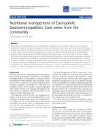
randomly assigned to 6 or 18 hours of CMV or PSV with 21%
O
2
(Figure 1). All surgical procedures were performed using
aseptic techniques. After reaching a surgical plane of anesthe-
sia (sodium pentobarbital, 50 mg/kg of body weight, intraperi-
toneal), animals were weighed and tracheostomized. The
jugular vein was cannulated for the infusion of saline and
sodium pentobarbital (5 mg/kg of body weight per hour). Body
fluid homeostasis was maintained by administration of 2 mL/
kg per hour intravenous electrolyte solution. The carotid artery
was cannulated for measurement of arterial blood pressure,
pH, and blood gas tensions (GEMpremier-3000 system;
Instrumentation Laboratory, Lexington, MA, USA). Heart rate
and electrical activity of the heart were monitored via a lead II
electrocardiogram using needle electrodes placed subcutane-
ously. Throughout the ventilation period, animals received
enteral nutrition (via a nasogastric tube) using the AIN-76
rodent diet with a nutrient composition of proteins, lipids, car-
bohydrates, and vitamins which provided an isocaloric diet
(Research Diets, Inc., Brunswick, NJ, USA). Body temperature
was monitored (rectal thermometer) and maintained at 37°C ±
Figure 1
Schematic illustration of the experimental design usedSchematic illustration of the experimental design used.
Available online />Page 3 of 9
(page number not for citation purposes)
1°C with a recirculating heating blanket. Continuing care dur-
ing the experimental period included expressing the bladder,
removing upper airway mucus, lubricating the eyes, rotating
the animal, and passive movements of the limbs. Animals (both
CMV and PSV) were regularly rotated to prevent atelectasis,
to limit mechanical constraints, and to maintain ventilation/per-
fusion ratio homogeneity.
Protocol for control mechanical ventilation group
Immediately after inclusion, animals were mechanically venti-
lated using a volume-driven ventilator (Rodent Ventilator
model 683; Harvard Apparatus, Holliston, MA, USA) for 6
hours (group 1) or 18 hours (group 2). The tidal volume was
10 mL/kg of body weight and the respiratory rate was 80
breaths per minute, with a fraction of inspired oxygen (FiO
2
) of
21% but without positive end-expiratory pressure. These ven-
tilatory conditions resulted in complete diaphragmatic inactiv-
ity and prevented noxious effects of a hypercapnia on the
muscular contractile properties [2,3,24,25]. At the end of the
experimental period, each animal was weighed, and the costal
diaphragm was rapidly dissected and frozen in liquid nitrogen.
Samples were stored at -80°C until subsequent assay (except
for samples in which protein synthesis and proteolysis were
analyzed, which were treated as described below). At the
same time, arterial blood was obtained for culture.
Protocol for pressure support ventilation group
Animals were also anesthetized and mechanically ventilated
for 6 hours (group 4) or 18 hours (group 5) as described
above (model PSV ventilator DARHD01; IFMA, Aubière,
France). The level of pressure support applied, determined
during preliminary studies, allowed a minute volume of 200 ±
10 mL/minute (respiratory rate of 80 ± 10 breaths per minute
and FiO
2
of 21%). The range of pressure support used was 5
to 7 cm H
2
O. The ventilator had a pressure trigger. The expir-
atory trigger was fixed at 25% of peak inspiratory flow, and the
maximum inspiratory time was set at 1 second. The ventilator
did not have back-up ventilation. If the animal was not trigger-
ing, no pressure was released. Continuing care during the
experiment was also applied as above. At the end of the exper-
imental period, the costal diaphragm was rapidly removed, dis-
sected, and frozen in liquid nitrogen. Samples were stored at
-80°C.
Protocol for control animals
Control animals (group 3) were free of intervention before
inclusion (not mechanically ventilated). These animals were
anesthetized and their diaphragms were rapidly dissected, fro-
zen, and stored at -80°C until subsequent assay. Because of
the biochemical constraints (variability of the solutions of
Krebs-Henselheit), each day of experimentation required a
control animal.
Tissue removal and storage
At the appropriate times (6 or 18 hours), the entire diaphragm,
costal and crural, was removed, dissected, and weighed. All
biochemical studies were conducted using the costal region
of the diaphragm. Samples were rapidly frozen in liquid nitro-
gen and stored at -80°C until assay.
Biochemical assays
Measurement of protein turnover in vitro
Proteolysis and protein synthesis were measured on the costal
region of the diaphragm (approximately 250 mg). Diaphrag-
matic protein synthesis was evaluated by measurement of
14
C-
phenylalanine (
14
C-Phe) incorporation into diaphragm strips
as described previously by Tischler and colleagues [26]. Dia-
phragmatic protein breakdown was measured by evaluation of
the rate of tyrosine release from diaphragm samples according
to the fluorimetric method of Waalkes and Udenfriend [27].
The rationale for this technique is that tyrosine is neither syn-
thesized nor degraded by skeletal muscle and is suited as a
marker of whole protein degradation [26]. Diaphragm samples
were quickly removed from each experimental animal and pre-
incubated at 37°C in Krebs-Henselheit bicarbonate buffer
equilibrated with 95% O
2
and 5% CO
2
, containing 5 mM glu-
cose, 0.2 U/mL insulin, 0.17 mM leucine, 0.10 mM isoleucine,
and 0.20 mM valine to improve protein balance [26]. After a
30-minute preincubation period, muscles were transferred to
a fresh medium of similar composition but containing 0.5 mM
14
C-Phe (Amersham Corporation, now part of GE Healthcare,
Little Chalfont, Buckinghamshire, UK) to measure the rate of
protein synthesis. The muscles were incubated for an addi-
tional 1-hour period. The rate of protein synthesis was deter-
mined by incubating muscles in a medium containing 0.5 mM
14
C-Phe with a specific radioactivity in the medium of 1,500
disintegrations per minute per nanomole as described previ-
ously [28]. Tissues were homogenized in 10% trichloroacetic
acid and hydrolyzed in 1 M NaOH at 37°C. Tissue protein
mass was determined using the bicinchoninic acid procedure
[29]. Rates of phenylalanine incorporation were converted into
tyrosine equivalents, as described previously [26], and
expressed as nanomoles of tyrosine incorporated per milli-
gram of muscle per hour. Muscle protein content was meas-
ured according to the bicinchoninic acid procedure. Rates of
protein breakdown were measured by following the rates of
tyrosine release into the medium. At the completion of the
incubation period, tyrosine concentrations were assayed by
the fluorimetric method of Waalkes and Udenfriend [27]. The
rates of total protein degradation were calculated by adding
the rate of protein synthesis and the net rate of tyrosine release
into the medium [28,30]. Rates of protein turnover were
expressed in nanomoles of tyrosine per milligram of protein per
hour [30].
Measurement of proteasome proteolytic activities
On the controlateral costal diaphragm, proteins from skeletal
muscle samples were homogenized in ice-cold buffer (pH 7.5)
Critical Care Vol 12 No 5 Futier et al.
Page 4 of 9
(page number not for citation purposes)
containing 50 mM Tris, 250 mM sucrose, 10 mM ATP, 5 mM
MgCl
2
, 1 mM dithiothreitol (DTT), and protease inhibitors (10
μg/mL of antipain, aprotinin, leupeptin, and pepstatin A and 20
μM PMSF [phenylmethylsulphonylfluoride]). The proteasomes
were isolated by three sequential centrifugations as described
previously [31-33]. The final pellet was resuspended in buffer
containing 50 mM Tris (pH 7.5), 5 mM MgCl
2
, and 20% glyc-
erol. The protein content of the proteasome preparation was
determined according to Lowry and colleagues [34]. Chymot-
rypsin-like activity of the proteasome and the tripeptidylpepti-
dase II (TPPII) activity were determined by measuring the
hydrolysis of the fluorogenic substrates succinyl-Leu-Leu-Val-
Tyr-7-amino-4-methylcoumarin (LLVY-AMC) and Ala-Ala-Phe-
AMC (AAF-AMC). To measure peptidase activity, 15 μL of the
extract was added to 60 μL of medium containing 50 mM Tris
(pH 8.0), 10 mM MgCl
2
, 1 mM DTT, 2 U apyrase, and 300 μM
LLVY-AMC or 300 μM AAF-AMC. The activities were deter-
mined by measuring the accumulation of the fluorogenic cleav-
age product (methylcoumaryl-AMC) using a luminescence
spectrometer FLX 800 (BioTek Instruments, Inc., Winooski,
VT, USA). Fluorescence was measured continuously during
45 minutes at a 380-nm excitation wavelength and a 440-nm
emission wavelength. The difference between arbitrary fluo-
rescence units recorded with or without 40 μM of the protea-
some inhibitor MG132 (Affiniti Research Projects Limited,
Exeter, Devon, UK) or 100 μM of the TPPII inhibitor AAF-chlo-
romethylketone (Sigma-Aldrich, St. Louis, MO, USA) in the
reaction medium was calculated, and the final data were cor-
rected by the amount of protein in the reaction. The time
course for the accumulation of AMC after hydrolysis of the
substrate was analyzed by linear regression to calculate activ-
ities (for example, the slopes of best fit of accumulated AMC
versus time). Different kinetics were performed to individually
measure the chymotrypsin-like activity of the proteasome and
the TPPII activity.
Measurement of diaphragm oxidative injury
Myofibrillar protein carbonyl content was determined accord-
ing to Fagan and colleagues [35] with slight modifications.
Briefly, myofibrillar proteins were purified, treated with HCl-
acetone to remove interfering chromophores, and protein car-
bonyl content was then measured using 2,4-dinitrophenylhy-
drazones (DNPH). Following DNPH treatment, samples were
subjected to successive washings with trichloroacetic acide
(TCA) 30%, TCA 10%, and four washes with ethanol/ethylac-
etate (1:1). The pellet was solubilized with 6 M guanidine
hydrochloride and 20 M potassium phosphate (pH 2.3)
through incubation at 50°C during 30 minutes. After centrifu-
gation (800 g for 10 minutes at 20°C), absorbances at 280
and 380 nm were measured on the supernatant to determine
protein and carbonyl content, respectively. Protein content
was calculated using a calibration curve and carbonyl content
using the absorption coefficient 22,000/M-cm.
Statistical analysis
A two-way analysis of variance (StatView
®
, version 5.0; SAS
Institute Inc., Cary, NC, USA) with time (6 versus 18 hours) as
one factor and modality (PSV versus CMV versus control) as
the other factor was used. When appropriate, a post hoc pro-
tected least squares difference Fisher test was used. Values
are mean ± standard deviation in the text and mean ± standard
error of the mean in the tables and graphs. Statistical signifi-
cance was defined a priori as a P value of less than 0.05.
Results
Systemic and biologic response to mechanical
ventilation
The principal biologic parameters are summarized in Table 1.
Blood gas/pH and cardiovascular homeostases were main-
tained constant in all animals during CMV and PSV. There
were no significant differences in total body mass between
groups and no group experienced a significant loss of body
mass, indicating adequate hydration and nutrition during the
experimental period (Table 2). All animals urinated and experi-
enced intestinal transit during the experimental period. All
blood cultures were negative for bacteria and none of the ani-
mals demonstrated sepsis signs.
In vitro proteolysis
Compared with control animals, diaphragmatic protein catab-
Table 1
Systemic and biologic response to mechanical ventilation
Biologic parameters Control CMV at 6 hours CMV at 18 hours PSV at 6 hours PSV at 18 hours
pH 7.38 ± 0.02 7.42 ± 0.04 7.40 ± 0.05 7.39 ± 0.02 7.43 ± 0.01
PaO
2
/FiO
2
, mm Hg 360 ± 50 380 ± 40 350 ± 30 360 ± 40 370 ± 20
PaCO
2
, mm Hg 38 ± 3 40 ± 2 38 ± 3 40 ± 5 40 ± 3
MAP, mm Hg 90 ± 10 95 ± 15 97 ± 12 100 ± 10 95 ± 15
Na
+
, mmol/L 135 ± 2 138 ± 5 135 ± 3 140 ± 5 138 ± 4
K
+
, mmol/L 4.20 ± 0.1 4.0 ± 0.3 4.10 ± 0.2 3.90 ± 0.3 4.20 ± 0.2
Fraction of inspired oxygen (FiO
2
) is 21%. CMV, controlled mechanical ventilation; MAP, mean arterial pressure; PaCO
2
, arterial partial pressure
of carbon dioxide; ]PaO
2
, arterial partial pressure of oxygen; PSV, pressure support ventilation.
Available online />Page 5 of 9
(page number not for citation purposes)
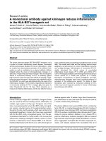
olism was significantly increased after 18 hours of CMV (33%,
P = 0.0001) but not after 6 hours (Figure 2). There was a 36%
increase in proteolysis between 6 and 18 hours of CMV (P =
0.0003). Compared with CMV, 6 and 18 hours of PSV
showed no significant increase in proteolysis. Moreover, dura-
tion of PSV had no effect on total proteolysis evolution (4.18
± 0.20 and 4.23 ± 0.12 nmol of tyrosine per milligram of pro-
tein per hour after 6 and 18 hours, respectively). Both chymo-
trypsin-like and tripeptydyl-peptidase 20S proteasome
activities were increased after 18 hours of CMV (+50% versus
controls and +45% versus CMV 6 hours). PSV did not
increase 20S proteasome activities, regardless of the ventila-
tion duration (6 or 18 hours).
In vitro protein synthesis
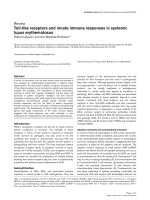
Compared with control animals, CMV decreased diaphrag-
matic protein synthesis by 50% (P = 0.0012) after 6 hours
and by 65% (P < 0.0001) after 18 hours of MV (Figure 3). The
difference between 6 and 18 hours of CMV was 30%, which
was not statistically significant. No variation of protein synthe-
sis was observed during PSV. After 18 hours of MV, CMV
showed a 94% reduction in protein synthesis compared with
PSV (P = 0.0002).
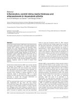
Measurement of diaphragm oxidative injury
Compared with control animals, protein oxidation, measured
by myofibrillar protein carbonyl levels, was significantly
increased after 18 hours of CMV (+63%, P < 0.001) and PSV
(+82%, P < 0.0005) (Figure 4). Myofibrillar protein oxidation
was not influenced by ventilator mode.
Discussion
The major finding of this study, which is the first to compare
PSV with control ventilation, is that, in contrast to CMV, PSV
did not increase diaphragmatic muscle proteolysis or
decrease protein synthesis. Both of these effects have been
shown to occur as a result of CMV-induced muscle atrophy
[2,11]. Finally, our results support the hypothesis that oxidative
injury, though indisputable, is probably not the trigger of CMV-
induced diaphragmatic proteolytic damage and thus of VIDD.
Before discussion of the results, some study limitations must
be pointed out.
Table 2
Body weight of control, pressure support ventilation, and controlled mechanical ventilation groups
Groups Initial body mass, grams Final body mass, grams
Control 253.5 ± 5.4 -
CMV at 6 hours 252.4 ± 4.5 253.5 ± 3.5
CMV at 18 hours 260.2 ± 3.2 258.6 ± 3.5
PSV at 6 hours 255.3 ± 3.8 255.4 ± 3.5
PSV at 18 hours 255.0 ± 3.0 258.3 ± 2.6
CMV, controlled mechanical ventilation; PSV, pressure support ventilation.
Figure 2
In vitro diaphragmatic proteolysisIn vitro diaphragmatic proteolysis. (a) Controlled mechanical ventilation
(CMV) increased total diaphragmatic proteolysis after 18 hours, but not
after 6 hours, of mechanical ventilation versus control (CON) and pres-
sure support ventilation (PSV). Units in (a) are nanomoles of tyrosine
per milligram of protein per hour. Both chymotrypsin-like activity (b) and
tripeptidylpeptidase II activity (c) were increased by 18 hours of CMV.
Units in (b) and (c) are relative fluorescence units (RFU) per microgram
per minute. Values are mean ± standard error. *P < 0.05 compared
with CON group.
†
P < 0.05 compared with PSV group at 6 and 18
hours.
‡
P < 0.05 compared with CMV group at 6 hours.
Critical Care Vol 12 No 5 Futier et al.
Page 6 of 9
(page number not for citation purposes)
Anesthetic protocol
The anesthetic agent, sodium pentobarbital, could have
affected the rate of muscle protein synthesis in the diaphragm.
However, both MV and spontaneously breathing animals were
anesthetized with sodium pentobarbital, so comparisons
between groups are valid. Moreover, a previous study has
reported that rats acutely anesthetized with sodium pentobar-
bital do not experience a significant decrease in protein syn-
thesis in skeletal muscle [36]. Additionally, general anesthesia
does not decrease protein synthesis in skeletal muscle in
healthy humans undergoing abdominal surgery [37]. Collec-
tively, these data indicate that protein synthesis is not altered
by anesthesia per se. The influence of continued exposure of
any given anesthetic agent (for example, 18 hours) would be
difficult to separate from the reduced use during that state.
However, the experiments reviewed above [36,37] report nor-
mal rates of protein synthesis in limb-locomotor skeletal mus-
cle during periods of time in which reduced use would not be
expected to have an effect on protein synthesis. These reports
[36,37] indicate that anesthesia does not affect protein syn-
thesis; therefore, the decreased rate of protein synthesis in the
diaphragm during MV is attributable to MV, not to the anes-
thetic as previously reported by several authors [2,4,6,11,38].
Diaphragmatic contraction
Prolonged MV results in diaphragmatic atrophy and contractile
dysfunction in animals. Evaluation of contractile diaphragmatic
properties in PSV and CMV will have been clinically relevant.
This study was not designed to respond to this question and
we discuss only MV-induced diaphragmatic protein altera-
tions. Further studies should focus on this point. Diaphrag-
matic contractions are avoided by CMV at a normal rate (80
cycles per minute). We have not tested this assessment but
several authors have done so previously [4] and used this pre-
viously reviewed paper for a recent study [2,11]. However, this
does not exclude the possibility that the animals were trigger-
ing the ventilator during CMV in the present study. This is a
real limitation of the manuscript.
Kinetics of controlled mechanical ventilation-induced
protein metabolism alteration
In the present study, we simultaneously analyze the effects of
MV on proteolysis, protein synthesis, and their kinetics. Con-
sistent with earlier findings [2], our results confirm the increase
in diaphragmatic proteolysis after 18 hours of CMV. Although
diaphragmatic proteolytic injury has been implicated in the
genesis of VIDD [7], less is known about modifications in dia-
phragmatic protein synthesis as a result of MV. Muscle atrophy
can result from increased proteolysis [39], decreased protein
synthesis [40], or both. Except for one recent study [11], none
had considered the possibility that diaphragm atrophy associ-
ated with CMV could also result from decreased protein syn-
thesis. We found both increased proteolysis and a time-
dependent decrease in protein synthesis. Moreover, our
results provide information about the probable kinetics of
CMV-induced protein metabolism modifications. Indeed, the
decrease in protein synthesis occurred extremely early (by the
sixth hour of CMV), was worsened by the duration of MV, and
preceded the increase in diaphragmatic proteolysis. It is inter-
esting to note that, in the study of Shanely and colleagues
[11], the results were obtained from the analysis of separate
studies of in vitro proteolysis and in vivo protein synthesis.
However, constant infusion of
13
C-leucine, which is used in
the analysis of in vivo protein synthesis, can modify an animal's
protein profile by altering insulin release, on both the tissue
and molecular levels [41], making interpretations between in
vivo and in vitro models difficult. In addition, the nutritional pro-
files of animals can limit the interpretation. Indeed, some
authors have compared the results obtained using fed [2] and
unfed animals, implying a negative protein assessment
[11,41]. On the other hand, in vivo protein synthesis should be
more relevant than in vitro proteolysis as used in our study.
These methodological differences could explain some differ-
ence in the results.
Figure 3
In vitro protein synthesis after 6 and 18 hours of controlled mechanical ventilation (CMV) and pressure support ventilation (PSV)In vitro protein synthesis after 6 and 18 hours of controlled mechanical
ventilation (CMV) and pressure support ventilation (PSV). Units are
nanomoles of phenylalanine (Phe) per milligram of protein per hour. Val-
ues are mean ± standard error. *P < 0.05 compared with control
(CON) group.
†
P < 0.05 compared with PSV group at 6 and 18 hours.
Figure 4
Protein-carbonyl content after 6 and 18 hours of controlled mechanical ventilation (CMV) and pressure support ventilation (PSV)Protein-carbonyl content after 6 and 18 hours of controlled mechanical
ventilation (CMV) and pressure support ventilation (PSV). Units are
nanomoles per milligram of protein. Values are mean ± standard error.
*P < 0.05 compared with control (CON) group.
Available online />Page 7 of 9
(page number not for citation purposes)
Pressure support ventilation-induced diaphragmatic
exercise
Our data showed that PSV limits MV-induced increases in pro-
teolysis and decreases in protein synthesis. Moreover, in con-
trast to CMV, modifications in protein metabolism were not
affected by PSV duration. Because of differences in proteoly-
sis/protein synthesis ratios, we hypothesized that PSV allows
the maintenance of protein turnover. In addition, because CMV
decreased protein synthesis, it is likely that CMV decreases or
completely inhibits protein turnover. These differences in mod-
ification of metabolism may be due to differences in the type of
diaphragmatic muscle damage caused by CMV and PSV.
Indeed, as for peripheral skeletal muscle models, during PSV
the diaphragm is subjected to exercise type activity through an
increase in respiratory activity (versus CMV) [42-44]. This
exercise would protect the diaphragm from modifications
related to muscular inactivity caused by CMV. During CMV,
there is a complete absence of neural activation and mechan-
ical activity in the diaphragm [4,45], which undergoes passive
shortening during mechanical expansion of the lungs [46,47].
This trauma has been implicated in the genesis of VIDD [2,11],
in particular during sarcomere injury [48,49] and during
decreased force-generating capacity of the diaphragm [7,50].
There has been little determination of the types of proteins
implicated in CMV-induced metabolic damage. CMV has been
shown to decrease the rate of mixed muscle protein synthesis
by 30% and to decrease the rate of myosin heavy chain pro-
tein synthesis by 65% [11]. Although our study was not
designed to analyze the type of proteins involved in the reduc-
tion of protein synthesis, it shed new light on the changes in
protein synthesis associated with the conservation of dia-
phragm activity. Further experiments are necessary to deter-
mine the specific proteins implicated in the increased protein
turnover observed with PSV. Our results also confirm that the
20S proteasome is involved in MV-induced proteolytic dam-
age [2,10]. CMV increases 20S proteasome activity in parallel
with the increase in diaphragmatic proteolysis. After 18 hours
of CMV, we observed an increase in the activity of extralyso-
somal TPPII, which degrades peptides generated by the pro-
teasome. Similarly, 72 hours of CMV increased the level of
MAF-box mRNA, which encodes an E3 ligase implicated in the
ubiquitination of proteins targeted for degradation via the pro-
teasome [38]. Together, these findings indicate the impor-
tance of the ubiquitin-proteasome pathway in CMV-induced
diaphragmatic muscle damage and in overall regulation of
muscle proteolysis [51] (as well as the importance of this enzy-
matic system within the skeletal muscle proteolytic machinery
[52,53]).
Is protein oxidation a real trigger?
Little is currently known concerning the triggers or molecular
signals of MV-induced protein metabolism modifications and
muscle atrophy [51,54]. Oxidative injury is induced by MV, and
increased protein oxidation and lipid peroxidation were found
to be associated with CMV [2,55]. Oxidative stress occurs
within a few hours after the start of CMV [9,56] and may play
a central role in the pathogenesis of CMV-induced diaphrag-
matic atrophy [7]. Oxidized proteins are associated with
increased proteolysis, which generates muscle atrophy and
dysfunction [57,58]. Because PSV does not increase proteol-
ysis (contrary to CMV) or decrease protein synthesis, it is likely
that PSV causes less oxidative injury. Our results confirm that
CMV is associated with diaphragmatic oxidative stress as indi-
cated by an increase in protein myofibrillar oxidation. The
increase in protein carbonyl levels parallels the increase in
20S proteasome activity, which specializes in degrading pro-
teins oxidized by reactive oxygen species [7,59]. Thus, oxi-
dized proteins may generate an increase in 20S proteasome
activity. Contrary to our hypothesis, we observed a similar oxi-
dation of myofibrillar protein with PSV. Thus, even if MV
causes oxidative stress, our findings support the hypothesis
that protein oxidation probably does not trigger the diaphrag-
matic proteolytic damage generated by CMV and its associ-
ated diaphragmatic dysfunction. Nevertheless, an
overproduction of free radicals may constitute the molecular
signal of CMV-increased proteolysis, either in mitochondria
(as suggested by an increase in manganese-superoxide dis-
mutase activity [9]) or via other metabolic pathways (such as
that involving xanthine oxidase [12]). There is also the possibil-
ity that other diaphragmatic regulating factors (such as apop-
tosis) might be involved [60].
Conclusion
We confirm that, within a few hours, CMV alters diaphragmatic
muscle protein metabolism. CMV first reduces protein synthe-
sis and then increases proteolysis. Compared with CMV, PSV
limits muscle wasting through a better protein balance despite
marked oxidative stress. If further study confirms our biochem-
ical findings with histological and electromyographical data,
PSV may be an alternative to CMV to limit muscle atrophy and
diaphragmatic dysfunction.
Competing interests
The authors declare that they have no competing interests.
Authors' contributions
EF and J-MC participated in the design of the study, carried
out the study, and helped to draft the manuscript. They con-
tributed equally to this work. LC, LM, LR, VS, and DA partici-
Key messages
• Controlled mechanical ventilation reduces protein syn-
thesis and secondly increases proteolysis.
• Pressure support ventilation limits muscle wasting
through a better protein balance.
• Pressure Support Ventilation may be an alternative to
Controlled mechanical Ventilation to limit diaphragmatic
atrophy.
Critical Care Vol 12 No 5 Futier et al.
Page 8 of 9
(page number not for citation purposes)
pated in the design of the study, performed biochemical
analysis, and helped to draft the manuscript. SJ, BJ and J-EB
participated in the design of the study and helped to draft the
manuscript. All authors read and approved the final
manuscript.
Acknowledgements
The authors thank Scott Butler for manuscript editing, Jean-Paul Mission
for statistical analysis, the members of the CICE-CENTI Unit, Faculty of
Medicine, Clermont-Ferrand, France, for their assistance, and the mem-
bers of the Human Nutrition Unit, Institut National de la Recherche
Agronomique, for their technical and scientific support. This work was
supported by the university hospital of Clermont-Ferrand.
References
1. Levine S, Nguyen T, Taylor N, Friscia ME, Budak MT, Rothenberg
P, Zhu J, Sachdeva R, Sonnad S, Kaiser LR, Rubinstein NA, Pow-
ers SK, Shrager JB: Rapid disuse atrophy of diaphragm fibers
in mechanically ventilated humans. N Engl J Med 2008,
358:1327-1335.
2. Shanely RA, Zergeroglu MA, Lennon SL, Sugiura T, Yimlamai T,
Enns D, Belcastro A, Powers SK: Mechanical ventilation-
induced diaphragmatic atrophy is associated with oxidative
injury and increased proteolytic activity. Am J Respir Crit Care
Med 2002, 166:1369-1374.
3. Sassoon CS: Ventilator-associated diaphragmatic dysfunction.
Am J Respir Crit Care Med 2002, 166:1017-1018.
4. Sassoon CS, Caiozzo VJ, Manka A, Sieck GC: Altered dia-
phragm contractile properties with controlled mechanical
ventilation. J Appl Physiol 2002, 92:2585-2595.
5. Vassilakopoulos T, Zakynthinos S, Roussos C: Bench-to-bedside
review: weaning failure – should we rest the respiratory mus-
cles with controlled mechanical ventilation? Crit Care 2006,
10:204.
6. Vassilakopoulos T: Ventilator-induced diaphragm dysfunction:
the clinical relevance of animal models. Intensive Care Med
2008, 34:7-16.
7. Vassilakopoulos T, Petrof BJ: Ventilator-induced diaphragmatic
dysfunction. Am J Respir Crit Care Med 2004, 169:336-341.
8. Lemaire F: Difficult weaning. Intensive Care Med 1993,
19(Suppl 2):S69-73.
9. Shanely RA, Coombes JS, Zergeroglu AM, Webb AI, Powers SK:
Short-duration mechanical ventilation enhances diaphrag-
matic fatigue resistance but impairs force production. Chest
2003, 123:195-201.
10. DeRuisseau KC, Shanely RA, Akunuri N, Hamilton MT, Van Gam-
meren D, Zergeroglu AM, McKenzie M, Powers SK: Diaphragm
unloading via controlled mechanical ventilation alters the
gene expression profile. Am J Respir Crit Care Med 2005,
172:1267-1275.
11. Shanely RA, Van Gammeren D, Deruisseau KC, Zergeroglu AM,
McKenzie MJ, Yarasheski KE, Powers SK: Mechanical ventilation
depresses protein synthesis in the rat diaphragm.
Am J Respir
Crit Care Med 2004, 170:994-999.
12. Kondo H, Nakagaki I, Sasaki S, Hori S, Itokawa Y: Mechanism of
oxidative stress in skeletal muscle atrophied by
immobilization. Am J Physiol 1993, 265:E839-844.
13. Li YP, Chen Y, Li AS, Reid MB: Hydrogen peroxide stimulates
ubiquitin-conjugating activity and expression of genes for spe-
cific E2 and E3 proteins in skeletal muscle myotubes. Am J
Physiol Cell Physiol 2003, 285:C806-812.
14. Betters JL, Criswell DS, Shanely RA, Van Gammeren D, Falk D,
Deruisseau KC, Deering M, Yimlamai T, Powers SK: Trolox atten-
uates mechanical ventilation-induced diaphragmatic dysfunc-
tion and proteolysis. Am J Respir Crit Care Med 2004,
170:1179-1184.
15. Maes K, Testelmans D, Powers S, Decramer M, Gayan-Ramirez G:
Leupeptin inhibits ventilator-induced diaphragm dysfunction
in rats. Am J Respir Crit Care Med 2007, 175:1134-1138.
16. Gayan-Ramirez G, Testelmans D, Maes K, Racz GZ, Cadot P,
Zador E, Wuytack F, Decramer M: Intermittent spontaneous
breathing protects the rat diaphragm from mechanical ventila-
tion effects. Crit Care Med 2005, 33:2804-2809.
17. Hering R, Bolten JC, Kreyer S, Berg A, Wrigge H, Zinserling J,
Putensen C: Spontaneous breathing during airway pressure
release ventilation in experimental lung injury: effects on
hepatic blood flow. Intensive Care Med 2008, 34:523-527.
18. Jolliet P, Tassaux D: Clinical review: patient-ventilator interac-
tion in chronic obstructive pulmonary disease. Crit Care 2006,
10:236.
19. Brander L, Slutsky AS: Assisted spontaneous breathing during
early acute lung injury. Crit Care 2006, 10:102.
20. Conti G, Arcangeli A, Antonelli M, Cavaliere F, Costa R, Simeoni F,
Proietti R: Sedation with sufentanil in patients receiving pres-
sure support ventilation has no effects on respiration: a pilot
study. Can J Anaesth 2004, 51:494-499.
21. Brochard L, Pluskwa F, Lemaire F: Improved efficacy of sponta-
neous breathing with inspiratory pressure support. Am Rev
Respir Dis 1987, 136:
411-415.
22. Brochard L, Harf A, Lorino H, Lemaire F: Inspiratory pressure
support prevents diaphragmatic fatigue during weaning from
mechanical ventilation. Am Rev Respir Dis 1989, 139:513-521.
23. National Research Council: Guide for the Care and Use of Labo-
ratory Animals Washington, DC: National Academies Press;
1996.
24. Le Bourdelles G, Viires N, Boczkowski J, Seta N, Pavlovic D, Aub-
ier M: Effects of mechanical ventilation on diaphragmatic con-
tractile properties in rats. Am J Respir Crit Care Med 1994,
149:1539-1544.
25. Schnader JY, Juan G, Howell S, Fitzgerald R, Roussos C: Arterial
CO
2
partial pressure affects diaphragmatic function. J Appl
Physiol 1985, 58:823-829.
26. Tischler ME, Desautels M, Goldberg AL: Does leucine, leucyl-
tRNA, or some metabolite of leucine regulate protein synthe-
sis and degradation in skeletal and cardiac muscle? J Biol
Chem 1982, 257:1613-1621.
27. Waalkes TP, Udenfriend S: A fluorometric method for the esti-
mation of tyrosine in plasma and tissues. J Lab Clin Med 1957,
50:733-736.
28. Temparis S, Asensi M, Taillandier D, Aurousseau E, Larbaud D,
Obled A, Bechet D, Ferrara M, Estrela JM, Attaix D: Increased
ATP-ubiquitin-dependent proteolysis in skeletal muscles of
tumor-bearing rats. Cancer Res 1994, 54:5568-5573.
29. Smith PK, Krohn RI, Hermanson GT, Mallia AK, Gartner FH,
Provenzano MD, Fujimoto EK, Goeke NM, Olson BJ, Klenk DC:
Measurement of protein using bicinchoninic acid. Anal
Biochem 1985, 150:76-85.
30. Combaret L, Tilignac T, Claustre A, Voisin L, Taillandier D, Obled
C, Tanaka K, Attaix D: Torbafylline (HWA 448) inhibits enhanced
skeletal muscle ubiquitin-proteasome-dependent proteolysis
in cancer and septic rats. Biochem J 2002, 361:185-192.
31. Hobler SC, Williams A, Fischer D, Wang JJ, Sun X, Fischer JE,
Monaco JJ, Hasselgren PO: Activity and expression of the 20S
proteasome are increased in skeletal muscle during sepsis.
Am J Physiol 1999, 277:R434-440.
32. Wray CJ, Tomkinson B, Robb BW, Hasselgren PO: Tripeptidyl-
peptidase II expression and activity are increased in skeletal
muscle during sepsis. Biochem Biophys Res Commun 2002,
296:41-47.
33. Fang CH, Li BG, Fischer DR, Wang JJ, Runnels HA, Monaco JJ,
Hasselgren PO: Burn injury upregulates the activity and gene
expression of the 20 S proteasome in rat skeletal muscle. Clin
Sci (Lond) 2000, 99:181-187.
34. Lowry OH, Rosebrough NJ, Farr AL, Randall RJ: Protein meas-
urement with the Folin phenol reagent. J Biol Chem 1951,
193:265-275.
35. Fagan JM, Sleczka BG, Sohar I: Quantitation of oxidative dam-
age to tissue proteins. Int J Biochem Cell Biol 1999,
31:751-757.
36. Heys SD, Norton AC, Dundas CR, Eremin O, Ferguson K, Garlick
PJ: Anaesthetic agents and their effect on tissue protein syn-
thesis in the rat. Clin Sci (Lond) 1989, 77:651-655.
37. Essen P, McNurlan MA, Wernerman J, Vinnars E, Garlick PJ:
Uncomplicated surgery, but not general anesthesia,
decreases muscle protein synthesis. Am J Physiol 1992,
262:E253-260.
Available online />Page 9 of 9
(page number not for citation purposes)
38. Sassoon CS, Zhu E, Caiozzo VJ: Assist-control mechanical ven-
tilation attenuates ventilator-induced diaphragmatic
dysfunction. Am J Respir Crit Care Med 2004, 170:626-632.
39. Bodine SC, Latres E, Baumhueter S, Lai VK, Nunez L, Clarke BA,
Poueymirou WT, Panaro FJ, Na E, Dharmarajan K, Pan ZQ, Valen-
zuela DM, DeChiara TM, Stitt TN, Yancopoulos GD, Glass DJ:
Identification of ubiquitin ligases required for skeletal muscle
atrophy. Science 2001, 294:1704-1708.
40. Ku Z, Yang J, Menon V, Thomason DB: Decreased polysomal
HSP-70 may slow polypeptide elongation during skeletal
muscle atrophy. Am J Physiol 1995, 268:C1369-1374.
41. Beaufrère B: [Amino acid metabolism in normal individuals].
Journ Annu Diabetol Hotel Dieu 2002:93-103.
42. Ji LL, Stratman FW, Lardy HA: Enzymatic down regulation with
exercise in rat skeletal muscle. Arch Biochem Biophys 1988,
263:137-149.
43. Wakshlag JJ, Kallfelz FA, Barr SC, Ordway G, Haley NJ, Flaherty
CE, Kelley RL, Altom EK, Lepine AJ, Davenport GM: Effects of
exercise on canine skeletal muscle proteolysis: an investiga-
tion of the ubiquitin-proteasome pathway and other metabolic
markers. Vet Ther 2002, 3:215-225.
44. Stupka N, Tarnopolsky MA, Yardley NJ, Phillips SM: Cellular
adaptation to repeated eccentric exercise-induced muscle
damage. J Appl Physiol 2001, 91:1669-1678.
45. Powers SK, Shanely RA: Exercise-induced changes in dia-
phragmatic bioenergetic and antioxidant capacity. Exerc Sport
Sci Rev 2002, 30:69-74.
46. Froese AB, Bryan AC: Effects of anesthesia and paralysis on
diaphragmatic mechanics in man. Anesthesiology 1974,
41:242-255.
47. Newman S, Road J, Bellemare F, Clozel JP, Lavigne CM, Grassino
A: Respiratory muscle length measured by sonomicrometry. J
Appl Physiol 1984, 56:753-764.
48. Williams PE, Goldspink G: The effect of denervation and dystro-
phy on the adaptation of sarcomere number to the functional
length of the muscle in young and adult mice. J Anat 1976,
122:455-465.
49. Farkas GA, Roussos C: Diaphragm in emphysematous ham-
sters: sarcomere adaptability.
J Appl Physiol 1983,
54:1635-1640.
50. Yang L, Luo J, Bourdon J, Lin MC, Gottfried SB, Petrof BJ: Con-
trolled mechanical ventilation leads to remodeling of the rat
diaphragm. Am J Respir Crit Care Med 2002, 166:1135-1140.
51. Attaix D, Combaret L, Pouch MN, Taillandier D: Regulation of
proteolysis. Curr Opin Clin Nutr Metab Care 2001, 4:45-49.
52. Attaix D, Combaret L, Kee AJ, Taillandier D: Mechanisms of ubiq-
uitination and proteasome-dependent proteolysis in skeletal
muscle. In Molecular Nutrition Edited by: Zempleni J, Daniel H.
Wallingford, Oxfordshire, UK: CABI Publishing; 2003:219-235.
53. Taillandier D, Combaret L, Pouch MN, Samuels SE, Bechet D,
Attaix D: The role of ubiquitin-proteasome-dependent proteol-
ysis in the remodelling of skeletal muscle. Proc Nutr Soc 2004,
63:357-361.
54. Jackman RW, Kandarian SC: The molecular basis of skeletal
muscle atrophy. Am J Physiol Cell Physiol 2004,
287:C834-843.
55. Jaber S, Sebbane M, Koechlin C, Hayot M, Capdevila X, Eledjam
JJ, Prefaut C, Ramonatxo M, Matecki S: Effects of short vs. pro-
longed mechanical ventilation on antioxidant systems in piglet
diaphragm. Intensive Care Med 2005, 31:1427-1433.
56. Zergeroglu MA, McKenzie MJ, Shanely RA, Van Gammeren D,
DeRuisseau KC, Powers SK: Mechanical ventilation-induced
oxidative stress in the diaphragm. J Appl Physiol 2003,
95:1116-1124.
57. Nagasawa T, Hatayama T, Watanabe Y, Tanaka M, Niisato Y, Kitts
DD: Free radical-mediated effects on skeletal muscle protein
in rats treated with Fe-nitrilotriacetate. Biochem Biophys Res
Commun 1997, 231:37-41.
58. Dean RT, Fu S, Stocker R, Davies MJ: Biochemistry and pathol-
ogy of radical-mediated protein oxidation. Biochem J 1997,
324(Pt 1):1-18.
59. Hussain SN, Vassilakopoulos T: Ventilator-induced cachexia.
Am J Respir Crit Care Med 2002, 166:1307-1308.
60. McClung JM, Kavazis AN, DeRuisseau KC, Falk DJ, Deering MA,
Lee Y, Sugiura T, Powers SK: Caspase-3 regulation of dia-
phragm myonuclear domain during mechanical ventilation-
induced atrophy.
Am J Respir Crit Care Med 2007,
175:150-159.