Báo cáo y học: "The effect of glutamine infusion on the inflammatory response and HSP70 during human experimental endotoxaemia" pptx
Bạn đang xem bản rút gọn của tài liệu. Xem và tải ngay bản đầy đủ của tài liệu tại đây (337.05 KB, 8 trang )
Open Access
Available online />Page 1 of 8
(page number not for citation purposes)
Vol 13 No 1
Research
The effect of glutamine infusion on the inflammatory response
and HSP70 during human experimental endotoxaemia
Anne Sofie Andreasen
1
*, Theis Pedersen-Skovsgaard
1
*, Ole Hartvig Mortensen
1
*, Gerrit van
Hall
1,2
, Pope Lloyd Moseley
1
and Bente Klarlund Pedersen
1
1
The Centre of Inflammation and Metabolism, Department of Infectious Diseases and Copenhagen Muscle Research Center, Rigshospitalet, Faculty
of Health Sciences, University of Copenhagen, Blegdamsvej, DK-2100 Copenhagen, Denmark
2
Department of Biomedical Sciences, University of Copenhagen, Tagensvej, DK-2200 Copenhagen, Denmark
* Contributed equally
Corresponding author: Anne Sofie Andreasen,
Received: 29 Oct 2008 Revisions requested: 6 Dec 2008 Revisions received: 19 Dec 2008 Accepted: 27 Jan 2009 Published: 27 Jan 2009
Critical Care 2009, 13:R7 (doi:10.1186/cc7696)
This article is online at: />© 2009 Andreasen et al.; licensee BioMed Central Ltd.
This is an open access article distributed under the terms of the Creative Commons Attribution License ( />),
which permits unrestricted use, distribution, and reproduction in any medium, provided the original work is properly cited.
Abstract
Introduction Glutamine supplementation has beneficial effects
on morbidity and mortality in critically ill patients, possibly in part
through an attenuation of the proinflammatory cytokine
response and a stimulation of heat shock protein (HSP)70. We
infused either alanine-glutamine or saline during endotoxin
challenge and measured plasma cytokines and HSP70 protein
expression.
Methods This crossover study, conducted in eight healthy
young men, was double-blind, randomized and placebo-
controlled. It was performed on 2 trial days, separated by a 4-
week washout period. The volunteers received an infusion of
alanine-glutamine at a rate of 0.025 g/(kg body weight × hour)
or saline for 10 hours. After 2 hours, an intravenous bolus of
Escherichia coli endotoxin (0.3 ng/kg) was administered. Blood
samples were collected hourly for the following 8 hours. HSP70
protein content in isolated blood mononuclear cells (BMNCs)
was measured by Western blotting.
Results Plasma glutamine increased during alanine-glutamine
infusion. Endotoxin reduced plasma glutamine during both trials,
but plasma glutamine levels remained above baseline with
alanine-glutamine supplementation. Endotoxin injection was
associated with alterations in white blood cell and differential
counts, tumour necrosis factor-α, IL-6, temperature and heart
rate, but glutamine affected neither the endotoxin-induced
change in these variables nor the expression of HSP70 in
BMNCs.
Conclusions Endotoxin reduced plasma glutamine
independently of alanine-glutamine infusion, but
supplementation allows plasma levels to be maintained above
baseline. Glutamine alters neither endotoxin-induced systemic
inflammation nor early expression of HSP70 in BMNCs.
Trial Registration ClinicalTrials.gov ID: NCT 00780520.
Introduction
The level of glutamine in plasma as well as in skeletal muscle
is depressed in critically ill patients compared with healthy
control individuals [1], and low glutamine levels in plasma and
muscles are associated with poor outcome [2,3]. In light of
these findings, glutamine has been classified as a 'condition-
ally essential amino acid', in that it is usually a nonessential
amino acid that must be supplemented during situations such
as critical illness [4], when endogenous glutamine production,
mainly in skeletal muscle, cannot keep up with the increased
demand. Several clinical trials of parental as well as enteral
glutamine supplementation to critically ill patients have shown
a beneficial effect both on infectious complications [5-7] and
mortality [8,9]; however, others were unable to detect
improvements in mortality or morbidity with parental supple-
mentation [10]. A meta-analysis of studies involving enterally
as well as parentally administered glutamine to seriously ill
patients identified a nonsignificant trend toward a positive
effect of high-dose parental glutamine on mortality and infec-
tious complications [11]. Because of heterogeneity in patient
populations included and the insufficient power of many of the
conducted trials, the clinical relevance of glutamine supple-
BMNC: blood mononuclear cell; CI: confidence interval; HSP: heat shock protein; IL: interleukin; TNF: tumour necrosis factor; WBC: white blood cell.
Critical Care Vol 13 No 1 Andreasen et al.
Page 2 of 8
(page number not for citation purposes)
mentation is still debated. On reviewing the literature, how-
ever, it appears that the beneficial effects of glutamine may
increase with higher doses or parenteral administration [12].
The association between low levels of glutamine, increased
mortality and morbidity, and the potential beneficial effect of
parental supplementation is still not fully understood. Animal
studies have revealed that glutamine supplementation attenu-
ates the release of the proinflammatory cytokines tumour
necrosis factor (TNF)-α and IL-6 in septic animals and
improves survival [13,14]; hence, a positive effect of parental
glutamine administration to critically ill patients may – at least
in part – be due to a modulation of excessive proinflammatory
cytokine release during the systemic inflammation associated
with critical illness in general, and with severe sepsis in partic-
ular.
Glutamine induces expression of the anti-inflammatory heat
shock protein (HSP)70 in cell cultures [15], animals [16-19]
and critically ill patients [20]. Rodents subjected to stimuli that
are known to induce a high intracellular level of HSPs and sub-
sequent induction of otherwise lethal sepsis exhibited attenu-
ated release of TNF-α and IL-6 and reduced mortality [21-23].
Based on these findings, the positive effect of glutamine in crit-
ical illness may be due either to a direct anti-inflammatory
effect of glutamine or to a glutamine-dependent increased
expression of HSP70 and the anti-inflammatory effects of this
protein.
In the present study we hypothesized that intravenous admin-
istration of glutamine attenuates the immune response during
acute inflammation. We thus established a human in vivo sep-
sis model (the human endotoxin model) in order to investigate
the effect of glutamine on the response of TNF-α, IL-6, cortisol
and white blood cell (WBC) subpopulations to a standardized
inflammatory stimulus (intravenous injection of E. coli endo-
toxin). Furthermore, we set out to determine the early effects
of glutamine and endotoxin on HSP70 content in immune
cells, in order to test the hypothesis that the HSP70 content
would increase during glutamine infusion but not be further
affected by endotoxin.
Materials and methods
Volunteers
Eight healthy young men (mean age 27.3 years, range 21 to
33 years) with body mass index 23.9 kg/m
2
(range 20.7 to
28.4 kg/m
2
), an unremarkable medical history and no regular
medication use were included in the study after they had given
informed oral and written consent. All participants underwent
a thorough clinical examination, including blood analyses (hae-
moglobin, WBC and differential counts, C-reactive protein,
blood glucose, electrolytes, and liver, kidney and thyroid func-
tion parameters) and electrocardiogram recording. All tests
were normal. The Ethical Committee of the Capital Region,
Denmark, approved the study ([H-KF] 01-144/98).
Materials
Glutamine was given as a dipeptide, consisting of alanine-
glutamine (Dipeptiven; Fresenius Kabi, Uppsala, Sweden) in a
stock concentration of 200 mg/ml of the dipeptide suspended
in saline. The solution used for infusion was a 20% (volume/
volume) solution of Dipeptiven and saline. Isotonic saline was
used as a placebo. The dipeptide solution could not visually be
distinguished from placebo, and the containers were covered
in a way that prevented both the volunteers and the investiga-
tors from distinguishing between them. The glutamine solution
and saline were prepared under sterile conditions on the day
before each trial.
Study design
The study was a randomized, double-blinded, placebo-control-
led, crossover trial. All volunteers participated on 2 trial days,
separated by 30 days. The volunteers were randomly assigned
to either infusion with alanine-glutamine on the first trial day
and placebo on the second (n = 5) or vice versa (n = 3). The
difference in the numbers of volunteers was due to one being
placed in the wrong group (human error).
The volunteers reported to the research unit at 06:30 a.m.
after an overnight fast. They were immediately placed in bed.
Catheters were placed bilaterally into an antecubital vein; one
catheter was used to draw blood samples and the other to
infuse glutamine or placebo. The volunteers were monitored
by continuous electrocardiography, noninvasive blood pres-
sure monitoring (every 15 minutes) and continuous measure-
ment of rectal temperature and venous oxygen saturation. At
07:00 a.m. (t = 0), infusion with glutamine or placebo was ini-
tiated and continued for 10 hours. The glutamine infusion rate
was set at 0.025 g/(kg body weight × hour). This rate has
been tested for safety [24] and is comparable to the doses
used in patient studies [5,6,8]. Placebo was infused at the
same rate. At 09:00 a.m., after 2 hours of alanine-glutamine or
placebo infusion, steady state was assumed to have been
achieved [24] and volunteers were given an intravenous bolus
injection with a standard reference E. coli endotoxin (Lot EC-
6; US Pharmacopeia Convention, Rockville, MD, USA) at a
dose of 0.3 ng/kg body weight. The vounteers were then mon-
itored for another 8 hours.
Blood sample and analysis of blood samples
Blood samples were drawn hourly from 0 hours and onward,
with additional samples at time 2.5 and 3.5 hrs (corresponding
to 0.5 and 1.5 hours, respectively, after endotoxin injection).
Whole blood was analyzed for WBC and differential counts
using standard biochemical procedures.
Samples for measuring plasma concentrations of glutamine,
cytokines and cortisol were drawn into tubes containing EDTA
and centrifuged; plasma was separated and immediately
stored at -80°C. Plasma glutamine concentration was deter-
mined enzymatically using an automated analyzer (Cobas
Available online />Page 3 of 8
(page number not for citation purposes)
Fara; F. Hoffmann-La Roche, Basel, Switzerland). Plasma
TNF-α, IL-6 and cortisol levels were determined using a com-
mercially available enzyme-linked immunosorbent assay
(Quantikine and Parameter; R&D systems, Minneapolis, MN,
USA). All measurements were taken in duplicate and means
were calculated for the subsequent statistical analyses. Blood
for isolation of blood mononuclear cells (BMNC) was drawn
into tubes containing heparin; BMNCs were isolated by den-
sity gradient centrifugation (Lymphoprep Nycomed Pharma
AS, Oslo, Norway) on LeucoSep tubes (Greiner, Fricken-
hausen, Germany). After separation, the BMNCs were imme-
diately suspended in a modified RIPA cell lysis buffer (50
mmol/l Tris-HCl [pH 7.4], 150 mmol/l NaCl, 1 mmol/l EGTA, 1
mmol/l EDTA, 0.25% NaDeoxycholate, 1% Triton X-100) con-
taining complete protease inhibitor cocktail (Roche, Basel,
Switzerland) and frozen at -80°C until measurement of HSP70
protein content using Western blotting, as described below.
HSP70 Western blotting
BMNCs resuspended in cell lysis buffer were thawed on ice,
pulled through a small-gauge syringe four to five times, and
centrifuged in a microcentrifuge at maximum speed at 4°C for
15 minutes. The supernatant was then transferred to a new
Eppendorf tube, and protein concentrations in this BMNC
lysate were determined using the BioRad DC kit (BioRad, Her-
cules, CA, USA) with bovine serum albumin as standard. All
measurements were conducted in triplicate.
Ten micrograms of BMNC protein lysate per lane were boiled
in Laemmli buffer and separated on 4% to 12% Bis-Tris gels
(Invitrogen, Taastrup, Denmark) and transferred to polyvinyli-
dene fluoride membranes (Hybond-P; GE Healthcare, Little
Chalfont, UK). Membranes were blocked for 1 hour at room
temperature in a blocking buffer (Tris-buffered saline [pH 7.6],
0.1% Tween-20, 5% skimmed milk). The membranes were
then incubated overnight at 4°C in a blocking buffer containing
a primary antibody against HSP70 (SPA-810; Stressgen, Vic-
toria, BC, Canada) at a 1:1,000 dilution. Subsequently, they
were washed three times for 5 minutes in a washing buffer
(Tris-buffered saline with 0.1% Tween-20) and incubated for
1 hour at room temperature with a secondary antibody (Rabbit
anti-mouse HRP, P0260; Dako, Glostrup, Denmark) at a
1:10,000 dilution in a blocking buffer, followed by three
washes of 5 minutes each in washing buffer. The protein
bands were detected using ECL (GE Healthcare) and quanti-
fied using CCD image sensor (ChemiDoc XRS; BioRad) and
software (Quantity One; BioRad).
Statistical analyses
All statistical analyses were done using SAS 9.1 (SAS Insti-
tute Inc., Cary, NC, USA). Log values were used when consid-
ered appropriate to approximate the normal distribution. P <
0.05 was considered statistically significant. A two-way analy-
sis of variance for longitudinal measures was performed on
each investigated variable using a means model (SAS PROC
MIXED) with the model TIME TREATMENT TIME*TREAT-
MENT, and with SUBJECT as a random factor. An autoregres-
sive covariate structure was assumed. Goodness-of-fit of the
mixed model was assessed by investigating the distribution of
the residuals. Significant changes in inflammatory markers
from the time of endotoxin administration to the following time
points were analyzed by paired t-tests using the Tukey-Kramer
method to adjust for multiple comparisons. Fractional changes
in glutamine levels from baseline to endotoxin injection and
from endotoxin injection to 4 hours after endotoxin injection
were calculated and evaluated by t-tests.
Results
Plasma glutamine
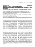
Plasma glutamine concentration increased by 55% (95% con-
fidence interval [CI] = 42% to 68%; P < 0.0001) above base-
line levels (from 500 μmol/l [95% CI = 430 to 570 μmol/l] to
764 μmol/l [95% CI = 708 to 821 μmol/l]) during the first 2
hours of glutamine infusion; in contrast, plasma glutamine did
not change during placebo infusion (Figure 1a). Two hours
after endotoxin administration, plasma glutamine decreased
with both treatments, reaching a nadir after 4 hours and then
gradually normalizing. When glutamine was infused, plasma
levels dropped by 21% (95% CI = 16% to 27%; P < 0.0001),
from 764 μmol/l (95% CI = 708 to 821 μmol/l) at the time of
endotoxin injection to 601 μmol/l (95% CI = 523 to 679 μmol/
l) 4 hours later; plasma levels thus remained above baseline
levels throughout the trial. During placebo infusion, plasma lev-
els also dropped significantly, from 484 μmol/l (95% CI = 427
to 541 μmol/l) to 369 μmol/l (95% CI = 333 to 406 μmol/l),
corresponding to a fractional decrease of 23% (95% CI =
15% to 31%; P = 0.0003). The endotoxin-induced drop in
plasma glutamine – absolute as well as fractional – was com-
parable between treatments.
Cytokines
TNF-α (P < 0.0001) and IL-6 (P < 0.05) plasma concentra-
tions both increased after endotoxin administration. However,
infusion with glutamine had no effect on the response of either
cytokine to endotoxin injection (Figure 1b,c).
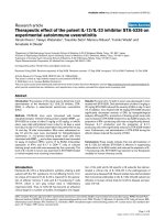
Heat shock protein-70
BMNCs were isolated at baseline and after 2 and 4 hours. No
significant changes were detected in BMNC HSP70 protein
concentrations over time or between trials (Figure 2).
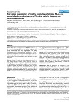
Other variables
Endotoxin injection induced characteristic changes (P < 0.05)
in the concentration of monocytes, lymphocytes and neu-
trophils, but these changes were unaffected by the infusion of
glutamine (Figure 3a–c). The plasma concentration of cortisol
decreased significantly during the first 2 hours of both
glutamine and placebo infusions (P < 0.0001). After endotoxin
administration, plasma cortisol increased (P < 0.0001), but
the time course was unaffected by glutamine (Figure 1d).
Critical Care Vol 13 No 1 Andreasen et al.
Page 4 of 8
(page number not for citation purposes)
Figure 1
Evolution of clinical and biochemical variables after endotoxin challenge and infusion with glutamine or placeboEvolution of clinical and biochemical variables after endotoxin challenge and infusion with glutamine or placebo. Shown is the time course of clinical
and biochemical variables after an intravenous bolus of endotoxin (vertical dotted line indicates time of endotoxin administration) and infusion with
glutamine (squares) or placebo (triangles) in young healthy volunteers: (a) plasma glutamine, (b) IL-6, (c) tumour necrosis factor (TNF)-α, (d) corti-
sol, (e) rectal temperature and (f) heart rate. Values are expressed as means ± standard error of the mean. The result of a mixed model analysis is
given below each graph. In panels e and f, hourly measurements are shown for clarity, although measurements performed every 15 minutes were
included in the analysis. *P < 0.05, post hoc t-test from the start of the trial to the time of endotoxin administration;
#
P < 0.05, post hoc t-test of the
difference from administration of endotoxin to time point during placebo infusion.
Available online />Page 5 of 8
(page number not for citation purposes)
Heart rate and temperature both increased (P < 0.05) after
endotoxin administration but were unaltered by glutamine infu-
sion (Figure 1e,f).
Discussion
This in vivo human study demonstrates a marked decrease in
plasma glutamine levels caused by intravenous injection of
endotoxin, the decrease being independent of the administra-
tion of glutamine. However, glutamine supplementation
allowed volunteers to maintain plasma levels above baseline in
spite of the endotoxin-induced drop. In contrast, plasma levels
dropped below baseline when placebo was infused. This find-
ing could be of clinical relevance, because patients with sep-
sis may experience several infectious bouts during their illness,
potentially depleting plasma glutamine levels successively, if
supplementation is not given. Clinical trials have shown that
both the plasma glutamine concentration and the muscle intra-
cellular glutamine content decline during severe sepsis and
critical illness [1,2], and the magnitude of decline in plasma
glutamine has been associated with increased mortality [3].
The potential mechanisms that underlie the decrease in
plasma glutamine during critical illness are yet to be unrav-
elled. The liver exhibits increased uptake of glutamine during
endotoxaemia in rats [25], possibly for the synthesis of acute
phase reactants; in addition, immune cells may utilize
glutamine at a higher rate during critical illness [26,27]. These
mechanisms may also account for the drop observed in our
study. Furthermore, the onset of the decline in plasma
glutamine in our study coincided with the peak concentrations
of plasma TNF-α and IL-6 at respectively 2 and 3 hours after
endotoxin administration. Our research group previously
showed that infusion of recombinant human IL-6 at high doses
to healthy volunteers lowers plasma glutamine substantially
[28]. These findings suggest that IL-6 may play a role in the
reduction in glutamine levels during inflammation.
The aim of the present study was to investigate whether
glutamine infusion had an effect on the cytokine and WBC
response to endotoxin. Refuting our hypothesis, we were una-
ble to detect any effect of glutamine on either cytokines or the
HSP70 content in BMNCs. Data from animal and cell line
studies have shown that glutamine may have anti-inflammatory
properties, and that this could be linked to increased intracel-
lular concentrations of HSP70 protein. Inflammation is
regarded to be a major contributor to the pathological conse-
quences of sepsis, and it has been suggested that treatments
able to attenuate the inflammatory response may improve sur-
vival [29,30]. We infused glutamine at clinically relevant rates
that have been tested for safety [24]. We achieved an increase
Figure 2
Evolution of HSP70 concentrations after endotoxin challenge and infusion with glutamine or placeboEvolution of HSP70 concentrations after endotoxin challenge and infusion with glutamine or placebo. Shown is the time course of heat shock pro-
tein (HSP)70 concentrations in blood mononuclear cells after endotoxin administration (vertical dotted line indicates time of injection) and infusion
with glutamine (squares) or placebo (triangles). Values are expressed as mean ± standard error of the mean. The result of a mixed model analysis is
given below the figure.
Critical Care Vol 13 No 1 Andreasen et al.
Page 6 of 8
(page number not for citation purposes)
in plasma glutamine of 55% above baseline, but we cannot
exclude the possibility that our inability to detect significant
attenuation of cytokine levels or induction of HSP70 protein
synthesis resulted from an insufficient dose or inadequate
duration of administration. Previous studies of HSP70 in
humans have identified substantial variation because of a vari-
ety of factors (for example, body temperature [31]), and the
endotoxin-mediated increases in cytokines are known to
exhibit large interindividual variations. Thus, the lack of positive
findings in the present study may also be ascribed to a small
number of volunteers, and the present findings should be inter-
preted with caution.
Conclusion
According to the results of the present study, endotoxin
decreased plasma glutamine levels. However, parenteral sup-
plementation of glutamine allowed the volunteers to maintain
their plasma glutamine above baseline levels, even though the
metabolism of glutamine in itself was unaffected by glutamine
supplementation, as demonstrated by a marked and compara-
ble decline in plasma glutamine after endotoxin injection in the
two groups. In contrast to our hypothesis, glutamine adminis-
tration had no effect on the cytokine or WBC response to
endotoxin and did not change the expression of HSP70 pro-
tein in BMNCs.
Figure 3
Evolution of leucocyte subpopulations after endotoxin challenge and infusion with glutamine or placeboEvolution of leucocyte subpopulations after endotoxin challenge and infusion with glutamine or placebo. Shown is the time course of leucocyte sub-
populations in plasma after endotoxin administration (vertical dotted line indicates time of injection) and infusion with glutamine (squares) or placebo
(triangles): (a) monocytes, (b) lymphocytes and (c) neutrophils. Values are expressed as mean ± standard error of the mean. The result of a mixed
model analysis is given below each graph.
#
P < 0.05, post hoc t-test of difference from administration of endotoxin to time point during placebo infu-
sion.
Available online />Page 7 of 8
(page number not for citation purposes)
Competing interests
The authors declare that they have no competing interests.
Authors' contributions
ASA and TPS conducted the study in the volunteers and
drafted the manuscript. OHM conducted the BMNC isolation
and HSP70 Western blotting, with the assistance of TPS and
PM. GvH conducted the analysis of plasma glutamine. PM and
BKP conceived the study, participated in its design and coor-
dination, and helped to draft the manuscript. All authors read
and approved the final manuscript.
Acknowledgements
Ruth Rousing, Hanne Villumsen and Lene Foged are acknowledged for
their technical assistance, and Kirsten Møller is acknowledged for read-
ing and commenting on the manuscript. The Centre of Inflammation and
Metabolism is supported by a grant from the Danish National Research
Foundation (# 02-512-55). This study was further supported by the
Danish Medical Research Council, the Commission of the European
Communities (contract no. LSHM-CT-2004-005272 EXGENESIS), the
Danish Food Industry Agency, and by grants from Thora and Viggo
Groves mindelegat. The Copenhagen Muscle Research Centre is sup-
ported by grants from the Capital Region of Denmark and the University
of Copenhagen.
References
1. Gamrin L, Essen P, Forsberg AM, Hultman E, Wernerman J: A
descriptive study of skeletal muscle metabolism in critically ill
patients: free amino acids, energy-rich phosphates, protein,
nucleic acids, fat, water, and electrolytes. Crit Care Med 1996,
24:575-583.
2. Roth E, Funovics J, Muhlbacher F, Schemper M, Mauritz W, Sporn
P, Fritsch A: Metabolic disorders in severe abdominal sepsis:
glutamine deficiency in skeletal muscle. Clin Nutr 1982,
1:25-41.
3. Oudemans-van Straaten HM, Bosman RJ, Treskes M, Spoel HJ
van der, Zandstra DF: Plasma glutamine depletion and patient
outcome in acute ICU admissions. Intensive Care Med 2001,
27:84-90.
4. Lacey JM, Wilmore DW: Is glutamine a conditionally essential
amino acid? Nutr Rev 1990, 48:297-309.
5. Dechelotte P, Hasselmann M, Cynober L, Allaouchiche B, Coeffier
M, Hecketsweiler B, Merle V, Mazerolles M, Samba D, Guillou YM,
Petit J, Mansoor O, Colas G, Cohendy R, Barnoud D, Czernichow
P, Bleichner G: L-alanyl-L-glutamine dipeptide-supplemented
total parenteral nutrition reduces infectious complications and
glucose intolerance in critically ill patients: the French control-
led, randomized, double-blind, multicenter study. Crit Care
Med 2006, 34:598-604.
6. Wischmeyer PE, Lynch J, Liedel J, Wolfson R, Riehm J, Gottlieb L,
Kahana M: Glutamine administration reduces Gram-negative
bacteremia in severely burned patients: a prospective, rand-
omized, double-blind trial versus isonitrogenous control. Crit
Care Med 2001, 29:2075-2080.
7. Houdijk AP, Rijnsburger ER, Jansen J, Wesdorp RI, Weiss JK,
McCamish MA, Teerlink T, Meuwissen SG, Haarman HJ, Thijs LG,
van Leeuwen PA: Randomised trial of glutamine-enriched
enteral nutrition on infectious morbidity in patients with multi-
ple trauma. Lancet 1998, 352:772-776.
8. Goeters C, Wenn A, Mertes N, Wempe C, Van AH, Stehle P, Bone
HG: Parenteral L-alanyl-L-glutamine improves 6-month out-
come in critically ill patients. Crit Care Med 2002,
30:2032-2037.
9. Griffiths RD, Jones C, Palmer TE: Six-month outcome of criti-
cally ill patients given glutamine-supplemented parenteral
nutrition. Nutrition 1997, 13:295-302.
10. Powell-Tuck J, Jamieson CP, Bettany GE, Obeid O, Fawcett HV,
Archer C, Murphy DL: A double blind, randomised, controlled
trial of glutamine supplementation in parenteral nutrition.
Gut
1999, 45:82-88.
11. Novak F, Heyland DK, Avenell A, Drover JW, Su X: Glutamine
supplementation in serious illness: a systematic review of the
evidence. Crit Care Med 2002, 30:2022-2029.
12. Bongers T, Griffiths RD, McArdle A: Exogenous glutamine: the
clinical evidence. Crit Care Med 2007, 35:S545-S552.
13. Singleton KD, Beckey VE, Wischmeyer PE: Glutamine prevents
activation of NF-kappaB and stress kinase pathways, attenu-
ates inflammatory cytokine release, and prevents acute respi-
ratory distress syndrome (ARDS) following sepsis. Shock
2005, 24:583-589.
14. Wischmeyer PE, Kahana M, Wolfson R, Ren H, Musch MM, Chang
EB: Glutamine reduces cytokine release, organ damage, and
mortality in a rat model of endotoxemia. Shock 2001,
16:398-402.
15. Wischmeyer PE, Riehm J, Singleton KD, Ren H, Musch MW,
Kahana M, Chang EB: Glutamine attenuates tumor necrosis
factor-alpha release and enhances heat shock protein 72 in
human peripheral blood mononuclear cells. Nutrition 2003,
19:1-6.
16. Wischmeyer PE, Kahana M, Wolfson R, Ren H, Musch MM, Chang
EB: Glutamine induces heat shock protein and protects
against endotoxin shock in the rat. J Appl Physiol 2001,
90:2403-2410.
17. Singleton KD, Serkova N, Banerjee A, Meng X, Gamboni-Robert-
son F, Wischmeyer PE: Glutamine attenuates endotoxin-
induced lung metabolic dysfunction: potential role of
enhanced heat shock protein 70. Nutrition 2005, 21:214-223.
18. Singleton KD, Wischmeyer PE: Oral glutamine enhances heat
shock protein expression and improves survival following
hyperthermia. Shock 2006, 25:295-299.
19. Singleton KD, Serkova N, Beckey VE, Wischmeyer PE: Glutamine
attenuates lung injury and improves survival after sepsis: role
of enhanced heat shock protein expression. Crit Care Med
2005, 33:1206-1213.
20. Ziegler TR, Ogden LG, Singleton KD, Luo M, Fernandez-Estivariz
C, Griffith DP, Galloway JR, Wischmeyer PE: Parenteral
glutamine increases serum heat shock protein 70 in critically
ill patients. Intensive Care Med 2005, 31:1079-1086.
21. Villar J, Ribeiro SP, Mullen JB, Kuliszewski M, Post M, Slutsky AS:
Induction of the heat shock response reduces mortality rate
and organ damage in a sepsis-induced acute lung injury
model. Crit Care Med 1994, 22:914-921.
22. Hotchkiss R, Nunnally I, Lindquist S, Taulien J, Perdrizet G, Karl I:
Hyperthermia protects mice against the lethal effects of endo-
toxin. Am J Physiol 1993, 265:R1447-R1457.
23. Chu EK, Ribeiro SP, Slutsky AS: Heat stress increases survival
rates in lipopolysaccharide-stimulated rats. Crit Care Med
1997, 25:1727-1732.
24. Ziegler TR, Benfell K, Smith RJ, Young LS, Brown E, Ferrari-Balivi-
era E, Lowe DK, Wilmore DW: Safety and metabolic effects of
L-glutamine administration in humans. JPEN J Parenter Enteral
Nutr 1990, 14:137S-146S.
25. Inoue Y, Pacitti AJ, Souba WW: Endotoxin increases hepatic
glutamine transport activity. J Surg Res 1993, 54:393-400.
26. Tjader I, Berg A, Wernerman J: Exogenous glutamine: compen-
sating a shortage? Crit Care Med 2007, 35:S553-S556.
Key messages
• An intravenous injection of E. coli endotoxin caused a
significant drop in plasma glutamine.
• Intravenous glutamine infusion allowed volunteers to
maintain plasma glutamine levels above baseline after
endotoxin injection.
• Glutamine infusion did not modify the early systemic
inflammatory response after endotoxin injection.
• Glutamine infusion did not change HSP70 expression
in BMNCs.
Critical Care Vol 13 No 1 Andreasen et al.
Page 8 of 8
(page number not for citation purposes)
27. Newsholme EA, Calder PC: The proposed role of glutamine in
some cells of the immune system and speculative conse-
quences for the whole animal. Nutrition 1997, 13:728-730.
28. van HG, Steensberg A, Fischer C, Keller C, Moller K, Moseley P,
Pedersen BK: Interleukin-6 markedly decreases skeletal mus-
cle protein turnover and increases nonmuscle amino acid uti-
lization in healthy individuals. J Clin Endocrinol Metab 2008,
93:2851-2858.
29. Cohen J: The immunopathogenesis of sepsis. Nature 2002,
420:885-891.
30. Hotchkiss RS, Karl IE: The pathophysiology and treatment of
sepsis. N Engl J Med 2003, 348:138-150.
31. Sandstrom ME, Madden LA, Taylor L, Siegler JC, Lovell RJ, Midg-
ley A, McNaughton L: Variation in basal heat shock protein 70
is correlated to core temperature in human subjects. Amino
Acids 2008 in press.