Báo cáo y học: "Pumpless extracorporeal interventional lung assist in patients with acute respiratory distress syndrome: a prospective pilot study" doc
Bạn đang xem bản rút gọn của tài liệu. Xem và tải ngay bản đầy đủ của tài liệu tại đây (167.61 KB, 7 trang )
Open Access
Available online />Page 1 of 7
(page number not for citation purposes)
Vol 13 No 1
Research
Pumpless extracorporeal interventional lung assist in patients
with acute respiratory distress syndrome: a prospective pilot study
Markus Zimmermann
1
, Thomas Bein
1
, Matthias Arlt
1
, Alois Philipp
2
, Leopold Rupprecht
2
,
Thomas Mueller
3
, Matthias Lubnow
3
, Bernhard M Graf
1
and Hans J Schlitt
4
1
Department of Anesthesiology, University of Regensburg Medical Center, Franz-Josef-Strauß-Allee 11, Regensburg, 93053, Germany
2
Department of Cardiothoracic and Vascular Surgery, University of Regensburg Medical Center, Franz-Josef-Strauß-Allee 11, Regensburg, 93053,
Germany
3
Department of Internal Medicine II, University of Regensburg Medical Center, Franz-Josef-Strauß-Allee 11, Regensburg, 93053, Germany
4
Department of General Surgery, University of Regensburg Medical Center, Franz-Josef-Strauß-Allee 11, Regensburg, 93053, Germany
Corresponding author: Markus Zimmermann,
Received: 2 Sep 2008 Revisions requested: 20 Oct 2008 Revisions received: 23 Dec 2008 Accepted: 30 Jan 2009 Published: 30 Jan 2009
Critical Care 2009, 13:R10 (doi:10.1186/cc7703)
This article is online at: />© 2009 Zimmermann et al.; licensee BioMed Central Ltd.
This is an open access article distributed under the terms of the Creative Commons Attribution License ( />),
which permits unrestricted use, distribution, and reproduction in any medium, provided the original work is properly cited.
Abstract
Introduction Pumpless interventional lung assist (iLA) is used in
patients with acute respiratory distress syndrome (ARDS) aimed
at improving extracorporeal gas exchange with a membrane
integrated in a passive arteriovenous shunt. In previous studies,
feasibility and safety of the iLA system was demonstrated, but
no survival benefit was observed. In the present pilot study we
tested the hypothesis that timely initiation of iLA using clear
algorithms and an improved cannulation technique will positively
influence complication rates and management of lung protective
ventilation.
Methods iLA was implemented in 51 patients from multiple
aetiologies meeting ARDS-criteria (American-European
Consensus) for more than 12 hours. Initiation of iLA followed an
algorithm for screening, careful evaluation and insertion
technique. Patients with cardiac insufficiency or severe
peripheral vascular disease were not considered suitable for
iLA. Arterial and venous cannulae were inserted using a new
strategy (ultrasound evaluation of vessels by an experienced
team, using cannulae of reduced diameter). The incidence of
complications and the effects on tidal volumes and inspiratory
plateau pressures were primary outcome parameters, while
oxygenation improvement and carbon dioxide removal
capabilities were secondary study parameters.
Results Initiation of iLA resulted in a marked removal in arterial
carbon dioxide allowing a rapid reduction in tidal volume (≤ 6 ml/
kg) and inspiratory plateau pressure. Adverse events occurred
in 6 patients (11.9%). The hospital mortality rate was 49%.
Conclusions The use of an indication algorithm for iLA in early
ARDS, combined with a refined application technique was
associated with efficient carbon dioxide removal and a reduced
incidence of adverse events. iLA could serve as an
extracorporeal assist to support mechanical ventilation by
enabling low tidal volume and a reduced inspiratory plateau
pressure.
Introduction
Pumpless extracorporeal interventional lung assist (iLA;
Novalung, Talheim, Germany) has been described in patients
with life-threatening forms of respiratory failure or acute respi-
ratory distress syndrome (ARDS) suffering from persistent
hypoxaemia and/or hypercapnia, unresponsive to conventional
therapy [1,2]. The iLA system is characterised by a novel mem-
brane gas exchange device with optimised blood flow inte-
grated into an arteriovenous heparin-coated bypass,
established by cannulation of the femoral artery and vein. A
passive shunt flow generated by the patient's blood pressure
gradient through the gas exchange device allows effective car-
bon dioxide extraction and moderate improvement in arterial
oxygenation [3].
ANOVA: analysis of variance; ARDS: acute respiratory distress syndrome; ECMO: extracorporeal membrane oxygenation; FIO
2
: fraction of inspired
oxygen; Fr: French; IBW: ideal body weight; ILA: interventional lung assist; PaO
2
: partial pressure of oxygen in arterial blood; PEEP: positive end
expiratory pressure; SBT: spontaneous breathing trial; SOFA: sequential organ failure assessment; V
T
: tidal volume.
Critical Care Vol 13 No 1 Zimmermann et al.
Page 2 of 7
(page number not for citation purposes)
We previously examined iLA implementation in a heterogene-
ous group of 90 severe critically ill patients at our institution
and reported a hospital mortality of 59% with a complication
rate of 24.4% [4]. Specifically, the patient groups with the
highest mortality were identified as patients with cancer, a high
demand for vasopressors, advanced age, morbid obesity or
those requiring long duration of mechanical ventilation. The
most frequent complications reported were related to the arte-
rial cannulation of the femoral vessels, resulting in distal limb
ischaemia, compartimental syndrome [4] or bleeding from the
cannulation sites requiring surgical intervention [1,5,6].
To date, solid evidence-based outcome data for iLA does not
exist, in part, because of inherent patient heterogeneity and
the rescue nature associated with the therapy, thereby leaving
most iLA protocols to be determined individually 'per case'. An
experience-based algorithm-guided approach to implement
arteriovenous iLA in the treatment of ARDS has recently been
published, setting new criteria for care [7]. The unmet need of
clinicians for systematically attained, well-structured studies to
reliably define indications for iLA sets a high value on publica-
tions of this nature and underscores the importance of estab-
lishing proven algorithms for ARDS therapy.
The present prospective pilot study defines an algorithm for
cannulation and implementation of iLA while refining inclusion
and exclusion criteria because we found evidence that arterial
insertion of cannulae with lower diameter might reduce the
rate of ischaemic complications. In contrast, the hypothesis
that timely initiation of iLA in an early phase of severe respira-
tory failure, and a consequent tidal volume reduction during
iLA therapy will positively influence outcome, will be deter-
mined in future randomised controlled trials.
Materials and methods
Patients
After a waiver by the Institutional Review Board at the Univer-
sity of Regensburg Medical Center was granted we report on
clinical results, mortality and complication rates in a 'second'
prospective cohort of ARDS patients treated by iLA-insertion
following an algorithm in comparison to our previously pub-
lished 'historical' retrospective study [4]. Obtainment of
informed consent was deemed by the Institutional Review
Board not to be required. Between October 2004 and March
2008, all 121 patients presenting with acute respiratory failure
were enrolled in the prospective study. iLA was implemented
in 51 patients suffering from ARDS mostly due to pneumonia,
trauma or sepsis. The algorithm for screening, evaluation and
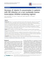
implementation of interventional lung assist is demonstrated in
Figure 1.
Before patients were considered potential candidates for iLA,
their clinical course was carefully evaluated. This included
optimisation of volume and vasoactive agent requirements,
ventilation strategy based on the concept of lung-protective
ventilation (moderate hypercapnia, increase in respiratory rate,
high positive end expiratory pressure (PEEP)), the use of
adjunctive therapeutic measures, for example, prone position
[8] or continuous lateral rotation therapy [9], as well as treat-
ment of the underlying disease in accordance to standard
intensive care procedures.
After a stabilisation period of 12 to 24 hours (Figure 1), a per-
sisting impairment in pulmonary gas exchange (partial pres-
sure of oxygen in arterial blood (PaO
2
)/fraction of inspired
oxygen (F
i
O
2
) 70 to 200 mmHg with PEEP of 10 cmH
2
O or
more and/or arterial pH less than 7.25 because of respiratory
acidosis [10]) was considered mandatory for the implementa-
tion of iLA. In more severe cases of hypoxaemia (PaO
2
/F
i
O
2
less than 70 mmHg), a pump-driven veno-venous extracorpor-
eal membrane oxygenation (ECMO) was preferably initiated.
Patients showing clinical signs of cardiac insufficiency, having
severe peripheral vascular disease, those in need of continu-
ously highly dosed vasoactive (noradrenaline greater than 0.4
μg/kg/minute) or inotropic agents were not considered suita-
ble for iLA, because the absence of shock or severe cardiovas-
cular instability is mandatory for the use of iLA.
Technique
Technical data of the iLA-system has been described in detail
previously [3,4]. In principle, iLA is a single use, ultracompact
extrapulmonary gas exchange system perfused by the heart.
Although carbon dioxide extraction mainly depends on a suffi-
cient sweep gas flow (about 10 L/minute) through the system,
patient oxygenation predominantly depends on arteriovenous
Figure 1
Algorithm for screening, evaluation and implementation of interventional lung assist (iLA)Algorithm for screening, evaluation and implementation of interventional
lung assist (iLA). **CLRT = continuous lateral rotation therapy. ARDS =
acute respiratory distress syndrome; ECMO = extracorporeal mem-
brane oxygenation; F
i
O
2
= fraction of inspired oxygen; HIT = heparin-
induced thrombocytopenia; PaO
2
= partial pressure of oxygen in arte-
rial blood; PEEP = positive end expiratory pressure.
Available online />Page 3 of 7
(page number not for citation purposes)
shunt volume, most strongly influenced by cannulae diameter,
and to a lesser extent, by the patients' mean arterial blood
pressure.
After ultrasonographic identification and assessment of the
diameters of the femoral artery as well as of the contralateral
femoral vein, cannulae were implanted by experienced physi-
cians using Seldinger's technique. The size of the arterial can-
nula was individually selected, based on vessel diameter to
ensure sufficient peripheral blood flow with a residual lumen of
30% after insertion. The diameter of the arterial cannula was
chosen to be 15 or 17 French (Fr), based conditionally on ade-
quate residual volume. The venous cannula was typically
selected two Fr sizes larger in order not to compromise flow
resistance. A platelet count or more than 60.000/μl and a par-
tial thromboplastin time less than 60 seconds were primary
coagulation needs for implementation of the cannulae.
Basic monitoring of the lower extremities included continuous
limb pulse oxymetry distal to the arterial cannulation site, deter-
mination of serum lactate and creatine kinase levels as well as
clinical inspection for any signs of restricted perfusion and/or
ischaemia. An overview of the implementation and monitoring
concept is given in Table 1.
Management
After insertion of the iLA-system, a de-escalation of invasive
ventilatory variables (tidal volume, plateau pressure, frequency,
F
i
O
2
) was performed, aimed at preventing the injured lung
from further (ventilator-induced) damage.
Changes in ventilatory parameters were applied as follows:
reduction of tidal volume (V
T
6 ml/kg ideal body weight (IBW)
or lower), of inspiratory plateau pressure (p
plat
≤ 30 cmH
2
O)
and of respiratory frequency (25 breaths/minute or less),
adoption of PEEP following the 'high alveoli'-concept of the
ARDS Clinical Trials Network [11]. Target arterial blood gases
during iLA-treatment were: PaO
2
70 mmHg or higher and pH
7.25 or higher.
After treatment of the underlying lung damage leading to a fur-
ther reduction in invasive mechanical ventilation (F
i
O
2
less
than 0.5, PEEP 12 cmH
2
O or less, assisted spontaneous
breathing) weaning from the iLA-system was initiated by start-
ing a 'cessation trial' (reduction of iLA sweep oxygen gas flow
to 1 L/minute) for a duration of two hours. If no major deterio-
ration of gas exchange variables was observed and no signifi-
cant increase in patients minute volume ventilation or
tachypnoea (40 breaths/minute or more) occurred, the cannu-
lae were manually removed, followed by sufficient and contin-
uous compression of the insertion sites for at least 30 minutes.
Thereafter, a pressure banding was applied for a period of 24
hours. Weaning from mechanical ventilation followed a clinical
ICU-guideline, in which a spontaneous breathing trial (SBT)
was implemented. In brief, SBT was daily screened and per-
formed with certain pulmonary (FiO
2
0.4 or less; PEEP 8
cmH
2
O or less, SaO
2
90% or higher), haemodynamic, neuro-
logical and metabolic conditions fulfilled. SBT was carried out
over one hour and the patient was extubated if no marked
deterioration in gas exchange, haemodynamic and stress-
associated parameters was observed.
Table 1
The concept of evaluation, insertion and clinical monitoring of the pumpless interventional lung assist (iLA) in patients with acute
respiratory distress syndrome (ARDS)
Evaluation and preparation Insertion Monitoring
Echocardiography:
exclusion of significant cardiac dysfunction
Preparation
of iLA system and introducer kit
System:
- continuous calculation of blood flow through
the device by transit time Doppler technology
Ultrasound:
assessment of femoral artery and vein diameter
Vascular cannula:
Artery: allowing a residual lumen ≥ 30% of the
vessel diameter maximum 17 Fr (adults)
Patient:
- continuous limb pulse oxymetry distal the
arterial cannulation site (toe)
Coagulation:
platelets > 60.000/μl aPTT < 60 seconds
haemoglobin ≥ 9 mg/dl access to blood bank
Vein:
+ 2 Fr. compared with arterial cannula
- clinical inspection for any signs of restricted
perfusion
Contraindication:
- coagulation disorder e.g. HIT
- cannulation by two experienced physicians - assessment of serum creatine kinase and
lactate regularly
- severe peripheral vascular disease -
continuously highly dosed vasoactive or
inotropic agents (Noradrenaline > 0.4 μg/kg/
minute)
- bolus application of 5000 IU heparin iv Arterial blood gases:
- early period (24 hours): frequently = every 4
hours
- connection of the system stepwise increase
of sweep gas flow to 10 l O
2
/minute
- late period (> 24 hours): every 8 hours
- continuous infusion of heparin (600 to 800
IU/hou via the arterial inflow cannula
APTT = activated partial thromboplastin time, HIT = heparin-induced thrombocytopenia, iv = intravenous.
Critical Care Vol 13 No 1 Zimmermann et al.
Page 4 of 7
(page number not for citation purposes)
The management of adverse events followed a consented
written algorithm. In cases of severe and persistent ischaemia
of a lower limb (no pulse oxymetry and doppler assessment of
leg arteries for more than two hours and/or visual impression
of impaired perfusion and/or acute elevation of ischaemia-indi-
cating laboratory values (creatine kinase, lactate)) an immedi-
ate removal of the cannulae was prescribed.
Statistical analysis
Statistical analysis was performed using SPSS Software, ver-
sion 16.0 (SPSS Inc., Chicago, IL, USA). As revealed by the
Kolmogorov-Smirnov method, most datasets significantly var-
ied from the pattern expected if they were drawn from a popu-
lation with a normal distribution. Non-parametric procedures
were therefore applied for intergroup (Mann-Whitney Rank
Sum Test, Kruskal-Wallis analysis of variance (ANOVA) on
Ranks), and intragroup analysis (Wilcoxon Signed Rank Test,
Friedman Repeated Measures ANOVA on Ranks). Results
were considered significant at p < 0.05. Data are presented
as median and interquartile range unless otherwise specified.
Results
Initiation of iLA resulted in a significant improvement in arterial
oxygenation and a marked removal in arterial carbon dioxide
within two hours allowing a rapid reduction in F
i
O
2
, minute
ventilation and inspiratory plateau pressure (Table 2). Further-
more, we were able to set lung protective tidal volume conse-
quently to a level below 6 ml/kg IBW without provoking severe
hypercapnia and/or acidosis. Following our PEEP/FiO
2
trial,
we found no changes in the PEEP level within 24 hours after
starting iLA. Insertion of iLA did not induce haemodynamic
instability although mean arterial pressure or the amount of
continuous infusion of noradrenaline remained unchanged, or
showed a tendency towards stabilisation. Sequential organ
failure assessment (SOFA)-score did not change significantly
after 24 hours following initiation of iLA.
Twenty-six of 51 patients (50.9%) survived ARDS, non-survi-
vors had a significant higher age, although the severity of dis-
ease (SOFA-score) and the severity of lung injury (Lung Injury
Score) were not different between survivors and non-survivors
(Table 3).
The frequency of complications is reviewed in Table 4. Tran-
sient episodes of lower limb ischaemia after arterial cannula-
tion occurred in three patients. This resulted in removal of the
cannula, resulting in normalisation of distal perfusion. Surgical
intervention was mandatory in one patient who developed a
compartment syndrome. From those four ischaemic complica-
tions, three patients had been cannulated with a 17 Fr cannula
for the arterial approach. Other complications were rarely seen
(cannula thrombosis, bleeding). In a total of six patients
(11.9%) adverse events were observed. No adverse event had
an effect on outcome.
Table 2
Changes in gas exchange, cardiovascular and respiratory variables before and during interventional lung assist (iLA) treatment
Pre-iLA 2 hours after insertion 24 hours after insertion
PaO
2
/FiO
2
75 (62 to 130) 102 (70 to 127) * 110 (86 to 160) *
PaCO
2
(mmHg) 73 (61 to 86) 44 (36 to 54) ** 41 (34 to 48) **
Arterial pH 7.23 (7.16 to 7.30) 7.38 (7.32 to 7.46) ** 7.44 (7.37 to 7.49) **§
MAP (mmHg) 73 (65 to 80) 83 (75 to 91) ** 81 (76 to 90)
Noradrenaline (μg/kg/minute) 0.16 (0.04 to 0.35) 0.11 (0.03 to 0.28) 0.09 (0.02 to 0.24) *
iLA-flow (L/minute) - 1.8 (1.6 to 2.0) 1.7 (1.5 to 2.0)
FiO
2
1 (0.8 to 1.0) 0.8 (0.7 to 1.0) ** 0.7 (0.6 to 0.9) **§S
MV (L/minute) 11.5 (9.3 to 12.5) 8.6 (6.4 to 10.5) ** 6.6 (5.5 to 8.3) **§S
V
T
ml/IBW 6.6 (5.3 to 7.2) 5.0 (4.0 to 6.4) ** 4.4 (3.4 to 5.4) **§S
RR (breaths/minute) 25 (22 to 27) 23 (20 to 30) 21 (18 to 26)
P
plat
(cmH
2
O) 35 (31 to 38) 34 (30 to 37) 30 (26 to 34) **
PEEP (cmH
2
O) 17 (14 to 20) 15 (11 to 19) * 17 (14 to 20)
Variables are presented as median values (interquartile ranges).
* p < 0.05 in comparison with pre-iLA
** p < 0.01 in comparison with pre-iLA
§p < 0.05 in comparison with two hours after insertion
§S p < 0.01 in comparison with two hours after insertion
FiO
2
= fraction of inspired oxygen; MAP = mean arterial pressure; MV = minute ventilation; PaCO
2
= partial pressure of carbon dioxide in arterial
blood; PaO
2
= partial pressure of oxygen in arterial blood; P
plat
= plateau pressure; PEEP = positive end expiratory pressure; RR = respiratory
rate; V
T
= tidal volume.
Available online />Page 5 of 7
(page number not for citation purposes)
Discussion
The main results of our prospective case series are: a change
in the indication spectrum resulted in a trend toward an
increased survival rate compared with the retrospective com-
parator study [4]; and a significant reduction in the incidence
of adverse events, especially ischaemic complications. In our
patients, iLA enabled a safe application of lung protective tidal
volume (V
T
= 6 ml/kg IBW) and in some patients even less
than 6 ml/kg without provoking severe acidosis. The combina-
tion of very low V
T
with high PEEP allowed the limitation of pla-
teau pressure at 30 cmH
2
O or lower and, thus the avoidance
of barotrauma. The use of smaller cannulae and an improved
cannulation technique (careful assessment of the vessels by
ultrasound, cowork-insertion by two experienced physicians)
may facilitate the reported improvements. With smaller cannu-
lae (arterial 17 Fr or lower), a sufficient blood flow of 1.0 to 1.5
L/minute allowed for adequate carbon dioxide-removal within
the circuit [12]. Although the support of a strict lung protective
ventilation [13] is our main goal for the use of iLA in ARDS
patients, the insertion of smaller cannulae is sufficient to reach
such a goal and minimises adverse ischaemic events. Improve-
ments in cannulae allowed the use of shorter (9 cm versus 14
cm) and thinner (13 versus 15 Fr) cannulae for arterial cannu-
lation. It is conceivable that further evolution of this technology
will contribute to greater improvements in the risk-benefit ratio
for the use of arteriovenous iLA.
Over the past three decades, an intensive scientific debate on
the effectiveness and possible harm of extracorporeal lung
assist systems was stimulated by clinical investigations study-
ing ECMO or extracorporeal carbon dioxide removal in rescue
situations (life-threatening hypoxaemia/hypercapnia) of
patients with ARDS [14-17]. In small prospective randomised
investigations, no clear survival benefit was demonstrated
using ECMO in comparison to 'conventional' treatment, and
the complication rate was shown to be high (more than 50%)
[14]. From today's point of view there were two major limita-
tions regarding the results and the interpretation of previous
studies. Specifically, patients were ventilated in the mode of
the 'Pre-ARDSNetwork-era' with high tidal volumes and rela-
tively low PEEP, thus a harmful potential of ventilator-induced
lung injury was postulated; and the 'historic' use of roller
pumps might have aggravated the high complication rate
(demand for elevated doses of heparin, induction of haemoly-
sis) and in the past, the technique of miniaturised centrifugal
pumps has been advocated [18-20].
A retrospective analysis of a new iLA system using an arterio-
venous shunt and a membrane lung, characterised by an
extremely-low flow resistance, demonstrated effective carbon
dioxide removal and a moderate oxygenation improvement in
severe ARDS, but no survival benefit in life-threatening hypox-
aemia/hypercapnia was observed [4]. This was potentially
Table 3
Patients characteristics and outcome
All Survivors Non-survivors
Patients 51 (100%) 26 (50.9%) 25 (49.1%)
Age (years) 52 (40 to 59) 44 (25 to 53) 58 (51 to 63) **
Female/male ratio 8/43 5/21 3/22
Body mass index 26.2 (23.7 to 31.1) 26.6 (23.8 to 31.1) 25.1 (23.9 to 28.4)
Days on ventilator (prior iLA) 4 (2 to 7) 3 (1 to 6) 5 (2 to 8)
Days on iLA 8 (6 to 11) 8 (6 to 10) 8 (4 to 16)
Lung injury score (Murray) 3.3 (3.25 to 3.7) 3.3 (3.3 to 3.7) 3.3 (3.1 to 3.7)
SOFA score (prior iLA) 10 (8 to 12) 9 (7.5 to 12) 10 (9 to 12)
SOFA score (24 hours after insertion) 10 (6 to 11) 9 (6 to 11) 10 (7 to 12)
Data are presented as median values (interquartile ranges), except female/male ratio.
** p < 0.01 in comparison with survivors.
ILA = interventional lung assist; SOFA = sequential organ failure assessment.
Table 4
Frequency of complications
Complication Number of patients (%)
Ischaemia of lower limb 3 (5.9)
Cannula thrombosis 1 (1.9)
Bleeding during cannulation 1 (1.9)
Compartmental syndrome (limb) 1 (1.9)
All 6 (11.8)
Critical Care Vol 13 No 1 Zimmermann et al.
Page 6 of 7
(page number not for citation purposes)
because of the lack of a clear algorithm and indication strat-
egy. Since 2004, we have worked to change the use of iLA in
terms of defining indications, refining insertion technique and
optimising the management of possible complications. In con-
trast to a previously described clinical concept aimed at 'res-
cue' in life-threatening gas exchange limitation, the new
algorithm introduced in this report stipulates the insertion of
iLA mainly for extracorporeal carbon dioxide-removal purposes
facilitating lung-protective ventilation. In our view, the most
important aspect of the presently described algorithm is the
indication strategy. In summary, we withdrew the iLA-system
from life-threatening 'rescue-situations' towards the support of
lung-protective ventilation in acute lung injury or early ARDS
on the threshold of becoming 'established' ARDS [10]. In this
concept, patients with a critically impaired pulmonary gas
exchange remain candidates for ECMO.
A comparison of cannula diameter, iLA effects on gas
exchange and complications between the present study and
our earlier work [4] demonstrated that the improved methodol-
ogy described here led to a significant reduction in the rate
and nature of complications. In the present prospective pilot
study, six patients (11.8%) had complications because of iLA
(transient ischaemia of a lower limb in three patients), while in
our recent retrospective analysis the incidence of complica-
tions was 24.4%, and serious ischaemic complications
occurred in nine patients (10%, p < 0.05). Furthermore, there
is a trend toward a decrease in mortality in our prospective
cohort (49.1%) compared with the retrospectively analysed
patients suffering from ARDS (58.9%).
Nevertheless, our present analysis has some limitations. Hav-
ing been conducted in a single-centre, the study might be sub-
ject to bias. Furthermore, the data stem from an expert team of
intensivists and perfusionists, having a long 'learning curve'
experience (> 150 applications) with the iLA system. Taking
this into consideration, we present – in contrast to our previ-
ous analysis – clinical data resulting from a strict algorithm
with defined indications (and contraindications).
Importantly. however, was also the identification of patient
groups that do not presently receive benefit from our iLA algo-
rithm. Specifically, in haemodynamically unstable patients
requiring high doses of vasopressors (noradrenaline 0.4 μg/
kg/minute or higher) or in patients with severe hypoxaemic
ARDS, a pump-driven ECMO is still the rescue measure of
choice.
Our new algorithm specifies indications for iLA differing from
ECMO that underscore the respective differences in therapy
concept. Although ECMO, characterised by high blood flow,
resembles an 'artificial lung' by producing significant exchange
of carbon dioxide and oxygen, iLA with its low blood flow pro-
vides impressive carbon dioxide elimination with a modest oxy-
genation improvement. Consequently iLA could be used as an
adjunct to mechanical ventilation affording optimised lung-pro-
tective ventilation strategies, with the objective of giving the
lungs time to heal [21].
Conclusion
Our data demonstrate that iLA can be an important tool ena-
bling advanced lung-protective ventilation in patients suffering
from ARDS. The use of an indication algorithm for iLA in early
ARDS, combined with a refined application technique was
associated with efficient carbon dioxide removal and a reduc-
tion in the incidence of adverse events, especially ischaemia
complications. With the ongoing technical evolution of smaller
cannulae, more efficient gas exchange membranes and easy
system handling, we hypothesise that iLA could serve as an
extracorporeal assist to support respirator ventilation by ena-
bling low tidal volume and reduced inspiratory plateau pres-
sure as an important tool in ARDS management. This
hypothesis is currently being tested by a prospective multicen-
tre randomised trial (ClinicalTrials NCT 00538928).
Competing interests
TB received lecture honorary from Novalung GmbH. The other
authors declare that they have no competing interests.
Authors' contributions
MZ made substantial contributions in data acquisition, patient
care and writing the manuscript. TB contributed to the study
design, statistical analysis and interpretation of data as well as
final approval of the manuscript. MA, AP, LR and TM equally
made substantial contributions in data acquisition and patient
care as well as reviewing the manuscript. HS and BG critically
revised the manuscript for important intellectual content.
References
1. Reng M, Philipp A, Kaiser M, Pfeifer M, Gruene S, Schoelmerich J:
Pumpless extracorporeal lung assist and adult respiratory dis-
tress syndrome. Lancet 2000, 356:219-220.
2. Liebold A, Reng CM, Philipp A, Pfeifer M, Birnbaum DE: Pumpless
extracorporeal lung assist – Experience with the first 20 cases.
Eur J Cardiothorac Surg 2000, 17:608-613.
Key messages
• The algorithm introduced in this report stipulates the
insertion of iLA mainly for extracorporeal carbon dioxide-
removal purposes enabling lung protective ventilation
strategies.
• iLA allowed a safe application of lung-protective tidal
volume (6 ml/kg or less) and a reduction in inspiratory
plateau pressure without provoking severe acidosis.
• The use of iLA in early ARDS, combined with a refined
application technique including the use of size-adapted
cannulae was associated with an efficient carbon diox-
ide removal and a low incidence of adverse events in
this prospective pilot study.
Available online />Page 7 of 7
(page number not for citation purposes)
3. Walles T: Clinical experience with the iLA Membrane Ventilator
pumpless extracorporeal lung-assist device. Expert Rev Med
Devices 2007, 4:297-305.
4. Bein T, Weber F, Philipp A, Prasser C, Pfeifer M, Schmid FX, Butz
B, Birnbaum D, Taeger K, Schlitt HJ: A new pumpless extracor-
poreal interventional lung assist in critical hypoxemia/hyper-
capnia. Crit Care Med 2006, 34:1372-1377.
5. Zimmermann M, Bein T, Philipp A, Ittner K, Foltan M, Drescher J,
Weber F, Schmid FX: Interhospital transportation of patients
with severe lung failure on pumpless extracorporeal lung
assist. Br J Anaesth 2006, 96:63-66.
6. Bein T, Prasser C, Philipp A, Müller T, Weber F, Schlitt HJ, Schmid
FX, Taeger K, Birnbaum D: Pumpless extracorporeal lung assist
using arterio-venous shunt in severe ARDS. Experience with
30 cases. Anaesthesist 2004, 53:813-819.
7. Deja M, Hommel M, Weber-Carstens S, Moss M, von Dossow V,
Sander M, Pille C, Spies C: Evidence-based therapy of severe
acute respiratory distress syndrome: an algorithm-guided
approach. J Int Med Res 2008, 36:211-221.
8. Guérin C: Ventilation in the prone position in patients with
acute lung injury/acute respiratory distress syndrome. Curr
Opin Crit Care 2006, 12:50-54.
9. Staudinger T, Kofler J, Müllner M, Locker GJ, Laczika K, Knapp S,
Losert H, Frass M: Comparison of prone positioning and con-
tinuous rotation of patients with adult respiratory distress syn-
drome: results of a pilot study. Crit Care Med 2001, 29:51-56.
10. Villar J, Pèrez-Mèndez L, Lopèz J, Belda J, Blanco J, Saralegui I,
Suàrez-Sipmann F, Lopèz J, Lubillo S, Kacmarek R, on behalf of the
HELP Network: An early PEEP/FiO
2
trial identifies different
degrees of lung injury in patients with acute respiratory dis-
tress syndrome. Am J Respir Crit Care Med 2007,
176:795-804.
11. Brower RG, Lanken PN, MacIntyre N, Matthay MA, Morris A,
Ancukiewicz M, Schoenfeld D, Thomson BT, National Heart, Lung,
and Blood Institute ARDS Clinical Trials Network: Higher versus
lower positive end-expiratory pressures in patients with the
acute respiratory distress syndrome. N Engl J Med 2004,
351:327-336.
12. Jayroe J, Wang D, Deyo DJ, Alpard SK, Bidani A, Zwischenberger
JB: The effect of augmented hemodynamics on blood flow
during arteriovenous carbon dioxide removal. ASAIO Journal
2003, 49:30-34.
13. Hager DN, Krishnan JA, Hayden DL, Brower RG, ARDS Clinical
Trials Network: Tidal volume reduction in patients with acute
lung injury when plateau pressures are not high. Am J Respir
Crit Care Med 2005, 172:1241-1245.
14. Zapol WM, Snider MT, Hill JD, Fallat RJ, Bartlett RH, Edmunds LH,
Morris AH, Peirce EC 2nd, Thomas AN, Proctor HJ, Drinker PA,
Pratt PC, Bagniewski A, Miller RG Jr: Extracorporeal membrane
oxygenation in severe acute respiratory failure. A randomized
prospective study. JAMA 1979, 242:2193-2196.
15. Bartlett RH, Roloff DW, Custer JR, Younger JG, Hirschl RB: Extra-
corporeal life support: the University of Michigan experience.
JAMA 2000, 283:904-908.
16. Brunston RL Jr, Zwischenberger JB, Tao W, Cardenas VJ Jr,
Traber DL, Bidani A: Total arteriovenous CO2 removal: Simpli-
fying extracorporeal support for respiratory failure. Ann Tho-
rac Surg 1997, 64:1599-1604.
17. Bindslev L, Eklund J, Norlander O, Swedenborg J, Olsson P, Nils-
son E, Larm O, Gouda I, Malmberg A, Scholander E: Treatment of
acute respiratory failure by extracorporeal carbon dioxide
elimination performed with a surface heparinized artificial
lung. Anesthesiology 1987, 67:117-121.
18. Christiansen S, Göbel C, Buhre W, Reul H, Autschbach R: Suc-
cessful use of a miniaturized bypass system with the DeltaS-
tream extracorporeal rotary blood pump. J Thorac Cardiovasc
Surg 2003, 125:43-44.
19. Arlt M, Philipp A, Zimmermann M, Voelkel S, Hilker M, Hobbhahn
J, Schmid C: First experiences with a new miniaturised life sup-
port system for mobile percutaneous cardiopulmonary
bypass. Resuscitation 2008, 77:345-350.
20. Kopp R, Dembinski R, Kuhlen R: Role of extracorporeal lung
assist in the treatment of acute respiratory failure. Minerva
Anestesiol 2006, 72:587-595.
21. Parsons P, Eisner M, Thompson B, Matthay MA, Ancukiewicz M,
Bernard GR, Wheeler AP, NHLBI Acute Respiratory Distress Syn-
drome Clinical Trials Network: Lower tidal volume ventilation
and plasma cytokine markers of inflammation in patients with
acute lung injury. Crit Care Med 2005, 33:1-6. Discussion 230–
232.