Antiarrhythmic Drugs A practical guide – Part 8 ppsx
Bạn đang xem bản rút gọn của tài liệu. Xem và tải ngay bản đầy đủ của tài liệu tại đây (155.59 KB, 19 trang )
128 Chapter 9
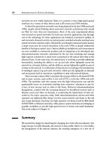
Table 9.3 Effectofantiarrhythmic drugsonpacing thresholds
Increase at normal drug levels Increase at toxic drug levels No increase
Flecainide Quinidine Lidocaine
Propafenone Procainamide Mexiletine
Amiodarone Disopyramide
Sotalol
several ways a nd is oftenclinically significant. Two major problems
caused by antiarrhythmic drugs are that they canchange the en-
ergy required for successful defibrillation and they canchange the
characteristics of the arrhythmiabeing treated.
The effectofantiarrhythmic drugsondefibr
illation energy re-
quirements isan important consideration because increasing the
defibrillation thresholdcan render an ICD ineffective. The effects of
various drugsondefibrillation energy requirements are summarized
in Table 9.4.I
ngeneral, drugs that block the sodium channel increase
defibrillation energy requirements (thus, Class IC drugs have the
most profound effect, and Class IA and Class IB drugstend to have
proportionally lesser effects), and drugs that block the potassium
channels (e.g., sotalol) decrease defibrillation energy require
ments.
Drugs that affect both the sodium and potassium channels (i.e., Class
IA drugsand amiodarone) have mixed effects—sometimes they in-
crease and sometimes they decrease defibrillation energy require-
ments. If one must prescribe a drug that has the potential of increas-
ing defibrillati
on energy requirements for a patient who has an ICD,
one shouldconsider retesting defibrillation thresholds after the drug
has been loaded to be sure that the ICD isstill capable of delivering
sufficientenergytoreliably defibrillate the patient.
Antiarrhythmic drugs
can also interact with ICDs by changing the
characteristicsofapatient’s ventricular tachycardia. By slowing the
Table 9.4 Effectofantiarrhythmic drugsondefibrillation thresholds
Increase Mixed effect Decrease
Flecainide Quinidine Sotalol
Propafenone Procainamide
Lidocaine Amiodarone
Mexiletine
Common adverse events with antiarrhythmic drugs 129
rate of ventricular tachycardia, a drug can render the arrhyth mia
more amenable to antitachycardia pacing, which potentially makes
the ICD more effective. On the other hand,byslowing the rate of
ventricular tachycardia below the recognition rate of the ICD, a drug
cancause the ICD to failtorecogniz
e(and therefore fail to treat) re-
current arrhythmias. Antiarrhythmic drugs can also cause reentrant
ventricular arrhythmias to recur more frequently or even to become
incessant, thus inducing frequent ICD therapy, which, in turn, can
cause excessive discomfort and premature battery de
pletion of the
ICD. Ingeneral, when one is compelled to add an antiarrhythmic
drug to the treatmentregimen of a patient with an ICD, one should
consider electrophysiologic testing to reexamine the characteristics
of the patient’s arrhythmias and to be sure that the ICD i
soptimally
programmed to treat the arrhythmias.
Reference
1Echt DS, Liebson PR, Mitchell B, et al. Mortality and morbidity in patients
receiving encainide, flecainideorplacebo. N EnglJMed 1991;324:781.
Part 3
Antiarrhythmic drugs in
the treatmentofcardiac
arrhythmias
CHAPTER 10
Basic principles of using
antiarrhythmic drugs
The first twosections of the book concerned the mechanismsofcar-
diac arrhythmias, the mechanism of action of antiarrhythmic drugs,
and the features of specific antiarrhythmic drugs. In thisfinal sec-
tion, that informationisapplied to the
use of antiarrhythmic drugs
in the treatmentofspecificcardiac arrhythmias. Chapter 10 reviews
some basic principles that should be kept in mind whenusing an-
tiarrhythmic drugs.
On the basisofthegenerally limited efficacyofantiarrhythmic
drugsaswell as their inherent propen
sity to cause serious problems,
the first principle should be completely self-evident;namely, one
should avoid using antiarrhythmic drugs whenever possible. Thus,
when one has decided to prescribe an antiarrhythmic drug, the final
step before actu
ally writing the order should be to ask, “Does this
patient really need this drug?” There are only two general conditions
in which using an antiarrhythmic drug isentirely appropriate: first,
when an arrhythmia needstobesuppressed because
it threatensto
cause death or permanent harm,and second, when an arrhythmia
needstobesuppressed because it produces significantsymptoms.
Before prescribing an antiarrhythmic drug, the physician should be
certain that the arrhythmia meets one of these t
wo conditions.
The second basic principle istokeep the goal of treatment clearly
in mind and to tailor the aggressiveness of one’s therapyaccordingly.
If one is treating an arrhythmiatoprevent death or permanent in-
jury, for instance, a relatively aggressive approach may be appropri
-
ate and necessary. In theory, if the object istospare life and limb,
one should err on the side of efficacy, perhaps willingly accepting
the risk of certain drug toxicities. Inpractice, however, as we will
see in Chapters 11 and 12, there are relatively fewinstances today
where oneought to rely p
rimarily on antiarrhythmic drugs to treat
arrhythmias that threaten life and limb.
133
134 Chapter 10
On the other hand, ifone is treating an arrhythmia to relieve
symptoms, a more circumspectapproach isappropriate. In these
cases, one generally shoulduse a stepwise strategy, beginning with
milder, less risky forms of treatment, and carefully reassessing the
risk-to-benefit ratio before each potential escalat
ion of therapy. All
too oftenphysicians pursue the treatment of relatively insignificant
arrhythmias with Ninja-like intensity, an error that can result in
unnecessary injury or death.
The final basic principle of using antiarrhythmic drugs is that, if
one feels compelled to expose a patie
nt to the risk of the drugs,
one should also feel compelled to take every reasonable precaution
to reduce the risks. For instance, given the almost universal risk
of proarrhythmia, one should oftenconsider placing patients on a
cardiacmonitor while antiarrhythmic drugs are being initi
ated be-
cause, although proarrhythmia can occuranytime during the course
of treatment, a significant proportion of these events occur during
the first 3 or 4days of drug usage. Most importantly, one must take
great care in deciding which drug to use. The choice must be indi-
v
idualized.
The accompanying tables summarize the factors that should be
consideredinchoosing antiarrhythmic drugs for patients with and
withoutsignificant underlying cardiacdisease.
Some drugs are plainly contraindicated for particular patients. Pro-
ca
inamide, for instance, shouldnot be usedinpatients with systemic
lupus erythematosus; quinidine shouldnot be usedinpatients with
chronic colitis;patients with severe lung disease (in whommild
drug-inducedpulmonary toxicity goes a long way) ideally shouldnot
receive ami
odarone;patients with a history of heart failure should
not receive drugs with negative inotropic effects.
Beyond these obvious individual considerations, the presenceor
absenceofunderlying heart disease is the most important variable in
choosing an antiarrhyth
mic drug,because heart disease predisposes
patients to reentrant circuits and, therefore, to proarrhythmia. As
shown in Table 10.1, beta blockers and Class IB drugs are the safest
choiceregardless of whether the patient has underlying heart dis-
ease. Class IC drugs are reasonably safe for patients with normal
hearts, butbecause they very frequently exacerbate ree
ntrantven-
tricular tachyarrhythmias, they are to be avoidedinpatients with
underlying cardiacdisease. Class IA drugs carry a moderate risk of
toxicity for patients without cardiacdisease because they cause both
torsades de pointes and end-organ toxicity;inpatients with cardia
c
Basic principles of using antiarrhythmic drugs 135
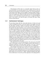
Table 10.1 Relative overall risk of serious toxicity from antiarrhythmic
drugs
∗
Increasing order of risk for patients Increasing order of risk for patients
with no underlying heart disease with underlying heart disease
†
Class II Class II
Class IB Class IB
Class IC Sotalol and dofetilide
Sotalol and dofetilide Amiodarone
Class IA Class IA
Amiodarone
‡
Class IC (should not use)
∗
Ranking of relative risks takes into account the risk of both proarrhythmia and
end-organ toxicity.
†
For patients with underlying heart disease, the ranking changes because these
patients have a much higher propensity for proarrhythmia. Amiodarone rises in
rank because of its relatively low risk of producing proarrhythmia. Class IC drugs
should virtually never be used in these patients.
‡
For patients without underlying heart disease, its impressive range of end-organ
toxicity makes amiodarone the riskiest drug.
disease, they also add a moderate risk of exacerbation of reentrant
arrhythmias. Sotalol and dofetilide carry a moderate risk of torsades
de pointes for all patients. Amiodarone carries a substantial risk of
significantend-organ toxicity for all patients, thoughonly a rela-
tively small risk of
proarrhythmia.
Table 10.2 ranks the efficacyofantiarrhythmic drugs for atrial
and ventricular tachyarrhythmias and for atrioventricular (AV)-
node-dependent arrhythmias. For atrial tachyarrhythmias, Class IA
drugs, sotalol, and dofetilide, are roughly equal in efficacy. Class
IC drugsand amiodarone are somewhat more effe
ctive than are
Class IA drugs, and Class IB drugs have virtually no efficacy for
these arrhythmias. Most antiarrhythmic agents have some degree
of efficacyagainst AV-node-dependent arrhythmias. For ventricu-
lar tachyarrhythmias, Class II and Class IB drugs are least effective;
amiodarone is m
ost effective.
Table 10.3 synthesizes the data from Tables 10.1 and 10.2 to gen-
eralize about the potential drugsofchoice for atrial and ventricular
tachyarrhythmias (keeping in mind that drug selectionmust be in-
dividualizedinevery case). The main considerationisalways to bal-
ance efficacy with safety.
136 Chapter 10
Table 10.2 Increasing order of relative efficacy for tachyarrhythmias
Atrial AV-node-dependent Ventricular
tachyarrhythmias
∗
tachyarrhythmias
†
tachyarrhythmias
Class IA Class IA Class II
Digoxin Class IB
Sotalol Class II Class IA
Dofetilide Verapamil
‡
Class IC
Class IC Sotalol Sotalol
Amiodarone Class IC Amiodarone
Amiodarone
Verapamil
§
Adenosine
∗
Atrial tachycardia, atrial fibrillation, and atrial flutter.
†
AV-nodal reentry and macroreentry (bypass-tract-mediated).
‡
When used orally for maintenance of sinus rhythm.
§
When used intravenously for acute termination of the arrhythmia.
The drug of choice in treating both atrial and ventricular tach-
yarrhythmias dependson the presence or absenceofunderlying
cardiacdisease. For instance, in the absence of heart disease, Class IC
drugs may offer the most favorable balance of efficacyand safety in
the treatment of atrial tachyarrhythmias. However, in the presence
of underlying heart di
sease, Class IC agents (because of their im-
pressive propensity to exacerbate reentrantventricular arrhythmias)
Table 10.3 Drugsofchoice for atrial and ventricular arrhythmias
∗
Underlying heart disease absent Underlying heart disease present
Atrial Ventricular Atrial Ventricular
arrhythmias
†
arrhythmias
‡
arrhythmias arrhythmias
Class IC
Sotalol
Class IA
Class II
Class IB
Sotalol
Class IC
Class IA
Amiodarone
Sotalol
Amiodarone
Class IA
Amiodarone
Sotalol
Class IA
∗
Drugs are listed in decreasing order of choice.
†
Atrial tachycardia, atrial fibrillation, and atrial flutter.
‡
Complex ventricular ectopy, ventricular tachycardia, and ventricular fibrillation.
Basic principles of using antiarrhythmic drugs 137
shouldnever be used. For ventricular arrhythmias, the primary con-
siderationinpatients without underlying heart disease (i.e., patients
in whom the risk for suddendeath is usually very low) istobesure
not to increase the risk of death by exposing the patients to the risk
of proarrhythmia. Thus,
in choosing drug therapy, one should err on
the side of safety; Class II and Class IB drugs should be considered
despite theirlimited effectiveness. As soon as one moves beyond
these two classes of drugs, onebeginsaccepting asubstantial risk of
proarrhythmia or other significant toxicity. On the other ha
nd, for
patients with underlying heart disease who require therapy for ven-
tricular arrhythmias, efficacy(which here includes avoiding proar-
rhythmia) is often the primary consideration.Thus, amiodarone
is often the first drug considereddespite its potential for causing
long-term end-organ toxicity. In the last column of Table 10.3, for
drugslisted as secondary choices after amiodarone, not only do the
oddsofefficacy decrease but the risk of proarrhythmia increases.
To summarize, whenit comes to using antiarrhythmic drugs, there
are no pretty choic
es. The best choice istoavoid them altogether.
If this is not possible, one must proceedwith the goals of treatment
clearly in mind and take every precaution to avoid producing more
problems than are caused by the arrhythmias being treated.
CHAPTER 11
Treatmentof
supraventricular
tachyarrhythmias
Traditionally, clinicians have tended to divide the supraventricu-
lar tachyarrhythmias into two broadcategories:paroxysmal atrial
tachycardia (PAT) and atrial flutter and atrial fibrillation. The term
PAT has falleninto disfavor of late (it isan artifact of the days before
the mechanismsofsupraven
tricular arrhythmias were understood),
butthisbimodal categorization of supraventricular arrhythmias still
lends itself nicely to a discussion of therapy.
Paroxysmal atrial tachycardia
PAT is a ter mused to describe regular supraventricular tachyarrhyth-
mias that occur with sudden onset and terminate equally suddenly.
Thus, PAT isacatchall phrase that incorporates virtually all reen-
trantsupraventricular arrhythmias except atrial fibrillation and atrial
flutter. More than 50% of PATs are c
aused by atrioventricular (AV)
nodal reentranttachycardia, and approximately 40% are caused by
macroreentranttachycardia mediated by an overt or concealed by-
pass tract. The remaining 10% or so of PATs are caused by reentrant
atrial tachycardiaorsinoatrial (SA) nod
al reentranttachycardia (see
Chapter 1 for a description of the mechanismsofsupraventricular
arrhythmias).
The acute and chronic therapies of PAT are listedinTable 11.1.
Acute therapy isaimed at terminating an episode of PAT. Ingen-
eral, this is easy to achieve. Since the AV node or the SA node isan
integral part of the reentrant circuit in 90–95% of PATs (the excep-
tionis reentrant atrial tachycardia, an arrhythmia that canusually
be recognized by the presenceofan unusual P-wave axis), maneu-
vers or drugs that produce transientSAnodal or AV nodal block are
138
Treatmentofsupraventricular tachyarrhythmias 139
Table 11.1 Acute and chronic treatmentofPAT
Acute treatment
Goal: Termination of the arrhythmia
Step 1: Vagal maneuvers, such as Valsalva (may be tried by the patient before
seeking medical attention)
Step 2: Intravenous administration of adenosine or verapamil
Termination by antitachycardia pacing or DC cardioversion (rarely necessary)
Chronic treatment
Goal: Prevention of recurrences
Infrequent or easy-to-terminate recurrences—no specific chronic therapy may
be necessary
Other types of recurrences
Treatment of choice—EP testing with RF ablation to abolish reentry
Drug therapy—one or more of several drugs may be tried empirically (see
Table 10.2)
EP, electrophysiologic; RF, radiofrequency.
highly effective in terminating supraventricular arrhythmias. Many
patients who have recurrent PAT can therefore terminate episodes
themselves by performing maneuvers that causeasudden increase
in vagal tone. Such maneuvers include Valsalva, carotid massage,
ocular massage, and dunking o
ne’s face in ice water. If pharmaco-
logic interventionis necessary, the treatmentofchoice is intravenous
adenosine, which isvirtually always effective—in fact, ifadenosine
fails to terminate the arrhythmia, the diagnosisofPATneedstobe
seriously rec
onsidered.Intravenous verapamil is also highly effec-
tive. Other AV nodal blocking drugs(digoxin and beta blockers) are
effective but have a muchlonger onset of action and,once loaded,
their effect persists. Unless these drugs are being administered for
chronic use, they are almost never gi
ven for acute treatmentofPAT.
Antitachycardia pacing techniques are also highly effective in termi-
nating supraventricular arrhythmias, butsincesomany less invasive
options are available, pacing is rarely usedunless an atrial pacemaker
is already in place.
The c
hronic therapy for PAT has undergone a revolutioninrecent
decades. Prior to the 1990s, pharmacologic therapy was the only
viable option for most patients. Although the choices of drug therapy
for the chronic treatment of PAT are broad and include all AV nodal
blocking agents (beta bloc
kers, calcium blockers, and digoxin)and
Class IA, Class IC, and Class III antiarrhythmic drugs, in earlier days
140 Chapter 11
many of these patients were aske d to take potentially toxic drugs
every day to prevent non-life-threatening arrhythmias that might
otherwise occuronly infrequently. Given that choice, many patients
quite reasonably opted for no therapy at all and accepted the fact that
they wou
ld have to make periodic pilgrimages to emergency rooms
to terminate acute episodes.
Fortunately, patients nolonger have to make suchachoice. Once
the mechanisms of the arrhythmias that cause PAT finally became
understood,and with parallel advances in technology, virtu
ally all
formsofPATbecame curable by the techniqueoftranscatheter abla-
tion.With thistechnique, critical components of the reentrant path-
ways responsible for a patient’s arrhythmia can be mappedinthe
electrophysiology catheterization laboratory and cau
terized (usually
with radiofrequencyenergy) directly through the electrophysiology
catheter. The success rate for curing AV nodal reentranttachycardias
and tachycardias mediated by bypass tracts (i.e, for the vast majority
of PATs) is well in excess of 95%. SA nodal reentry and intra-atr
ial
reentry can be curedwithasomewhat lower rate of success, but
these arrhythmias are rare. Today, patients with almost any form of
PAT should be referred for ablationif chronic drug therapyofany
type isbeing considered.
Atrial fibrillation and atrial flutter
Atrial fibrillation and atrial flutter are fundamentally different from
most of the arrhythmias that cause PAT because they arise in the
atrial myocardium itself, and therefore do not require either the
AV node or the SA node for their initiation or continuation. Atrial
fibrillation and atr
ial flutter canpersist in the presenceofanon-
functioning SA nodeorcomplete AV block. Therefore, the measures
commonly used to terminate PAT (i.e., producing transientAVnodal
block throughvagal maneuvers or by drug administration) do not
work with atri
al fibrillation and atrial flutter. Drugs that can termi-
nate these arrhythmias and preventrecurrence must necessarily act
on the atrial myocardium, namely, the Class IA, Class IC, and Class
III antiarrhythmic drugs. Therefore, treatmentaimed at maintain-
ing sinus rhythmis inherently difficult and relatively r
isky. Often,
it is more appropriate to accepta“lesser” therapeutic goal—that is,
to allow the underlying arrhythmiatopersist while controlling the
ventricular rate.
Treatmentofsupraventricular tachyarrhythmias 141
Table 11.2 Common underlying causes of atrial fibrillation and atrial flutter
Underlying heart disease
Valvular and congenital heart disease
Hypertensive heart disease
Acute ischemia or infarction
Cardiomyopathic diseases
Pericarditis
Systemic disorders
Hyperthyroidism
Acute pulmonary disease
Acute ethanol ingestion (“holiday heart”)
Stimulant administration or ingestion (e.g., caffeine, amphetamines,
and theophylline)
Unlike arrhythmias that cause PAT, atrial fibrillation and atrial
flutter often are related to an underlying disease process. The treat-
ment of these arrhythmias, therefore, should include a systematic
search for a primary cause. Table 11.2 lists the common underlying
causes of atrial fibrillation and atrial flutter.
Arrhythmias caused
by systemic processes (electrolyte distur-
bances, hyperthyroidism, pulmonary disease, and use of alcohol or
stimulant drugs) often improve or disappear once the systemic pro-
cess isaddressed. Arrhythmias associatedwith underlying heart dis-
ease, on the other hand, oftenpersist evenwhen therapy of heart
disease isoptimized.
Consequences
Atrial fibrillation and atrial flutter have three major consequences
that must be takeninto considerationwhenplanning therapy: loss
of the atrial kick, the rapid heart rate itself, and the risk of throm-
boembolism (Table 11.3).
Loss of atrial kick
The function of atrial contractionis to boost diastolic pressure within
the ventricles just before ventricular systole begins. End-diastolic
pressure (EDP) isofparamount importance in determining the force
of ventricular contraction and, therefore, of ventricular stroke vol-
ume. EDP issoimportant that, in general, ho
meostatic mechanisms
work to maintain itregardless of whether there isan atrial kick. The
importance of the atrial kick in maintaining adequate EDP directly
142 Chapter 11
Table 11.3 Major consequences of atrial fibrillation
Loss of atrial kick
Major hemodynamic compromise in patients with poor LV compliance
(i.e., patients with ventricular hypertrophy)
Mild-to-moderate hemodynamic compromise in patients with normal LV
compliance
Minimal-to-mild hemodynamic compromise in patients with increased LV
compliance (i.e., patients with dilated cardiomyopathies)
Tachycardia
Significant symptoms (palpitations and cardiac ischemia if CAD is present)
Tachycardiomyopathy (weakening of ventricular myocardium from chronic
tachycardia)
Thrombus formation
Stroke or other manifestations of thromboembolic disorder
CAD, coronary artery disease; LV, left ventricle.
dependson the relative compliance, or “stiffness,” of the ventri-
cle. The atrial kick isvitally important in patients whose ventri-
cles are noncompliant(i.e., stiff), a condition that occurs in the set-
ting of ventricular hypertrophy, whether the hypertrophy has bee
n
caused by aortic stenosis, hypertension,oridiopathic hypertrophic
cardiomyopathy. In these patients, a very high EDP is necessary to
maintain an adequate stroke volume, and the high EDP is provided,
at the last instantofdiastole, by the atrial kick. If the atrial kick is
lost (e.g
., because of the onset of atrial fibrillation), the only way
to achieve an adequate EDP istoraise the mean diastolic pressure,
that is, the pressure throughout diastole—and this is what exactly
happens. Because the heart’s compensatory mechanisms attemptto
maintain the EDP regardless of whether or
not there isan atrial kick,
the meandiastolic pressure suddenly rises and pulmonary conges-
tion ensues. Thus, patients with poor ventricular compliance de-
velop severe symptomsalmost immediately if atrial fibrillation oc-
curs; atrial k
ick isvital in these patients.
On the other hand, patients with dilatedcardiomyopathies have
enlarged, “baggy” ventricles that are significantly more compliant
thannormal. In these patients, the atrial kick contributes relatively
li
ttle to EDP because the relatively small volume of bloodprovided by
atrial contraction boosts pressure only slightly in ahighly compliant
ventricle. These patients tend to have relatively little change in their
baselinesymptoms with the onset of atrial fibrillation,and they often
Treatmentofsupraventricular tachyarrhythmias 143
are unable to perceive any difference, at least acutely, between sinus
rhythm and atrial fibrillation.
Patients with normal ventricular compliancetend to experience
intermediate symptoms with the onset of atrial fibrillation.With the
loss of the atrial kick, the ir EDP is m
aintained by a rise in m ean
diastolic pressure, but generally the elevations are not sufficientto
produce pulmonary edema. These patients canusually pinpoint the
timeofonset of atrial fibrillation,but in most cases, theirsymp-
toms are limited to palpitati
onsand a mild-to-moderate sensation of
breathlessness.
Tachycardia
Inpatients with normal AV conduction,tachycardiaensues immedi-
ately with the onset of atrial fibrillation or atrial flutter. The transient
decrease in stroke volume resulting from the loss of the atrial kick is
partially compensated by an increase in sympathetic tone, w
hich di-
rectly increases the heart rate and frequently also causes a sensation
of anxiety. The anxiety, in turn,further increases sympathetic tone.
Thus, it is not unusual for a patient with acute atrial fibrillation or
atrial flutter to present with very rapid heart rates and to experienc
e
extreme palpitations. Ingeneral, however, sympathetic tone drops
within afew hours, and the heart rate slowstomore reasonable
levels.
If heart rates remain elevatedchronically—for a period of weeks
or months—a tachycardiomyopathy may develop.Tachycardiomy-
opathy refers to the ventricular dysfunc
tion resulting from a per-
sistently elevated heart rate. Although relatively uncommon,this
conditionis indistinguishable from other formsofdilatedcardiomy-
opathy. Fortunately, tachycardiomyopathy is largely reversible if the
rapid heart rate is brought under control. In any case, the rapid heart
rates a
ccompanying atrial fibrillation and atrial flutter have signifi-
cance beyond merely producing palpitations.
Thromboembolism
Perhaps the major hemodynamic consequenceofatrial fibrillation
(and to a lesser extent, atrial flutter) is the risk of thromboembolism.
One-third of patients with chronic atrial fibrillation eventually expe-
rience stroke, and approximately 75% of those strokes are thought
to be embolic in natu
re. Both the incidence of atrial fibrillationit-
self and the yearly risk of stroke in patients with atrial fibrillation
increase with age. Atrial fibrillationis seeninapproximately 3% of
144 Chapter 11
patients who are of age 60, but in more than 10% of those 80 and
older. The yearly risk of stroke in 60-year-oldpatients with atrial
fibrillationisapproximately 2%, whereas that yearly risk increases
to more than 5% in patients 80 or older. Furthermore, for reasons
that are poorly understood, strokes that occur in patients with atrial
fibrillation are more li
kely to cause disability and mortality thando
strokes occurring in other patients. Antiembolic therapy with war-
farin, or to a lesser extent with aspirin, has been shown to signifi-
cantly reduce the risk of stroke in many patients with chronic atrial
fibrillation.Wewill discuss indicati
ons for anticoagulation below.
Treating atrial fibrillation and atrial flutter
When treating atrial fibrillation and atrial flutter, there are two basic
decisions that have to be made. First, should the patientreceive ther-
apyaimed at restoring and maintaining sinus rhythm (rhythmcon-
trol), or instead should the patient be allowed to remain in the tach-
yarrhythmia,
with therapeutic efforts being directed at controlling
the ventricular response (rate control)? And second, what should be
donetominimize the risk of stroke or other thromboembolic events?
Rhythm control versus rate control
Untilafew years ago, most cardiologists assumed that patients with
atrial fibrillationwould have improved outcomes if they could be
converted to and maintainedinnormal sinus rhythm.However, two
major randomizedclinical trials have now
shown that, at least using
currently available antiarrhythmic drug therapy, patients with atrial
fibrillation actually had better outcomes with rate control only.
Both the Atrial Fibrillation Follow-up Investigation of Rhythm
Management (AFFIRM)trial [1] and the Rate Control vers
us Elec-
trical Cardioversion (RACE) trial [2]randomizedpatients with atrial
fibrillation to therapy with either rhythmcontrol using antiarrhyth-
mic drugs or rate control only. Both studies showed a nearly signif-
icant trend towardworse outcomes with rhythmcontrol. Rhythm
co
ntrol with antiarrhythmic drugsyielded an increase in the pri-
mary end pointofdeath in the AFFIRM trial and an increasedin-
cidenceofaprimary composite end point(including death, heart
failure, thromboembolism, bleeding, requirement for a pacemaker,
and severe adverse d
rug reactions) in the RACE trial. Inneither study
was the quality of life improvedwith rhythmcontrol. Possibly more
Treatmentofsupraventricular tachyarrhythmias 145
importantly, the incidence of thromboembolismwas not reduced
with rhythmcontrol.
Experts and guidelines committees have concluded, from these
and other recenttrials, that for most patients with atrial fibrillation,
the rate-control approach is more appropriate. The use of antiar-
rhyth
mic drugs to try to maintain sinus rhythm shouldgenerally be
limited to patients who have persistentsymptoms of shortness of
breath, palpitations, heart failure, or angina despite adequate rate
control, or for those in whom adequate rate control cannot be at-
tained, or for patients who, after be
ing fully informed of the risks
and benefits, opt for rhythmcontrol themselves.
It has been speculated that the negative results reported by the AF-
FIRM and RACE trials regarding the strategy of rhythmcontrol have
mostly to do with the use of antiarrhythmic drugs, which are only
partially effective in maintaining sinus rhyth
m and which them-
selves cancause significant toxicity. Electrophysiologists, in partic-
ular, tend to subscribe to the theory that restoring sinus rhythm
by discovering and applying appropriate ablation techniques would
yielddifferent results from these twotrials. While there
is at least
a reasonable chance that these experts are correct, at this point no
study has shown that atrial fibrillation ablationprocedures lead to
better overall outcomes or reduce the risk of thromboembolism.
Catheter-based ablation techniques aimed at restoring and main-
taining sinus rhythminpatients with atr
ial fibrillation are still in the
developmental stages, and the efficacy for ablation for atrial fibril-
lationisstill relatively limited, while complications are nontrivial.
Incontrast, transcatheter ablation techniques are quite effective at
eliminating atrial fl
utter and are acceptably safe. For this reason,an-
tiarrhythmic drugs are used only rarely in the chronic management
of atrial flutter.
Cardioversion in atrial fibrillation and atrial flutter
There are at least two circumstances in which it is desirable to con-
vert patients from atrial fibrillation or atrial flutter backtonormal
sinus rhythm. The first is when a rhythm-control strategy has been
decidedupon,and the second is whenpatients present with p
arox-
ysmal atrial fibrillation or atrial flutter.
Paroxysmal atrial fibrillation and atrial flutter have beendefined
as arrhythmias that have beenpresent for less than 7 days (though
most paroxysmal atrial fibrillationpersists for less than24h). By
definition, then, patients who have paroxysmal episodes of atrial
146 Chapter 11
fibrillation or atrial flutter are usually in sinus rhythm. Therefore,
the primary goal of therapy in these patients ought to be to restore
normal sinus rhythm,and to dosowithin 24 hours of the onset
of the arrhythmia (to avoid the likelihood of formation of atrial
thrombi).
Inmost patients presenting with paroxys
mal atrial fibrillation and
atrial flutter, the arrhythmias will spontaneously revert to sinus
rhythmwithin afew hours of onset. Thus, in most instances, one
merely needstocontrol the heart rate and wait. However, if the
arrhythmia persists for 24 hours, elective cardioversion should be
performed
. If the patient has not presented for medical care until
the arrhythmia has persisted for more than48hours, cardioversion
should be postponeduntil 4weeks of anticoagulationwith war-
farin has been accomplished; warfarin should also be continue
d for
4weeks after cardioversion.
There are two methods for converting patients backtosinus
rhythm—direct-current (DC) cardioversion or pharmacologic car-
dioversion.DCcardioversion has a muchhigher efficacy rate and is
not proarrhythmic,and is the generally preferredmethod
.Ifdrug
therapy is chosen for cardioversion, propafenone, flecainide, ibu-
tilide, and dofetilide have been shown to be effective in restoring
sinus rhythminupto 60% of patients.
Rate control in atrial fibrillation
For patients who remain in chronic atrial fibrillation or atrial flutter,
controlling the ventricular response is important. Rapid ventricular
rates lead to symptomsofpalpitations, easy fatigue, breathlessness,
and poor exercise capacity. Persistenttachycardia can also lead to
car
diomyopathy.
Inmost patients, rate control can be achieved by the use of beta
blockers and verapamilordiltiazem.Especially in patients with heart
failure, digoxin may also be helpful in slowing the ventricular re-
sponse. Amiodarone is effective in slowing
the ventricular response
during chronic atrial fibrillation but is not usedcommonly for this
purpose because of its impressive toxicity. (The investigational drug
dronedarone, a “cousin” of amiodarone discussedinChapter 8, also
appears effective for th
is purpose and so far, appears to have much
less end-organ toxicity.)
Achieving adequate rate control means controlling the heart rate
both at rest and during exercise. The average resting heart rate
should be less than 80 beats/min,and during moderate ambulation,
Treatmentofsupraventricular tachyarrhythmias 147
it should be less than 110 beats/min. The overall average heart rate
during 24-hour Holter monitoring should be less than 100 beats/
min.
Titration of rate-control measures can be made by observing the
resting and exercise heart rates. Calcium blockers tend to slow the
heart rate at rest, while beta blockers tend to be more effective in
slowing the heart rate during exercise. Often,aco
mbination of drugs
is required, established by meansofatrial-and-error approach.
Adequate rate control can be achievedinthe large majority of
patients with pharmacologic therapy. However, occasional patients
cannot tolerate adequate
doses of beta blockers, calcium blockers,
or digoxin to achieve control. In these patients, strong consideration
ought to be given to transcatheter ablation of the AV junction to
achieve complete heart blockand the insertion of a permanent pace-
maker. Thistechniqu
e, while irreversible and seemingly somewhat
drastic, isactually relatively simple to perform and is very effective
and reasonably safe.
Rhythm control in atrial fibrillation
Whenever maintaining sinus rhythmis the goal of thera py, selecting
an appropriate antiarrhythmic drug requires consideration of the
available drugs themselves (i.e., theirlikely efficacyand toxicity), as
well as the clinical status of the patientbeing treated.Itshould be
kept in mind that, accor
ding to the best available data, a rhythm-
control strategy does not improve patients’ outcomes and further,
does not preclude the need for long-term anticoagulation.Thus, in
most cases, the aim of a rhythm-control strategy is merely to reduce
the incidenceand perhaps the severity of symptom
atic episodes of
atrial fibrillation.
Ingeneral, sixantiarrhythmic agents are primarily used today
in a rhythm-control strategy:disopyramide (Class IA), flecainide
and propafenone (Class IC), and sotalol, dofetilide, and amiodarone
(Class III). Table 11.4 lists the drugsofcho
iceaccording to the 2006
guidelines from the American College of Cardiology/American Heart
Association/European Society of Cardiology (ACC/AHA/ESC).
The general consensusofcardiologists, backedupby increasing
clinical evidence, is that amiodarone is the most effective a
ntiar-
rhythmic drug available for maintaining sinus rhythm after car-
dioversion from atrial fibrillation.Furthermore, it has a relatively
low incidenceofproarrhythmia, eveninpatients with underlying
heart disease. However, amiodarone carries the significantbaggage
148 Chapter 11
Table 11.4 Antiarrhythmic drugs for rhythmcontrol in atrial fibrillation
Patients with no structural heart disease (or with hypertension without LVH)
1st choice—propafenone, flecainide, and sotalol
2nd choice—amiodarone, dofetilide, and ablation
Patients with heart failure or significant LV dysfunction
1st choice—amiodarone and dofetilide
2nd choice—ablation
Patients with coronary artery disease
1st choice—sotalol and dofetilide
2nd choice—amiodarone or ablation
Patients with hypertension with LVH
1st choice—amiodarone
2nd choice—ablation
Special cases
Vagally mediated atrial fibrillation—disopyramide
Adrenergically mediated atrial fibrillation—beta blockers
LV, left ventricular; LVH, left ventricular hypertrophy.
of having atruly impressive array of potential end-organ toxicities—
not to mention the fact that its use for atrial fibrillationis not ap-
proved by the FDA—and itought to be reserved, in general, for pa-
tients whose hemodynamic compromise while in atrial fibrillation
is severe and in whom other antiarrhythmic drugs are not effective
or safe. The 2006 guidelines from the ACC/AHA/ESC recommended
that amiodaronebeused as first-line therapyonly in patients with
heart failure or significant left ventricular systolic dysfunction,orin
patients with hypertension a
nd left ventricular hypertrophy. (Left
ventricular hypertrophy, like systolic dysfunction, is often associ-
atedwith underlying electrophysiologic abnormalities that increase
the risk of proarrhythmia with other antiarrhythmic drugs.)
Flecainideand propafenone are reasonably effective in maintain
-
ing sinus rhythm,butbecause of their proarrhythmic potential they
should be used only in patients with no structural heart disease, and
who do not have coronary artery disease (or for that matter, a high
risk of developing coronary artery disease).
Sotalol and dofetilide are also moderately effective. These drugs
do not exa
cerbate reentrant arrhythmias (like the Class IC drugs do),
and as long as appropriate cautions are taken to reduce the incidence
of torsades de pointes, these are reasonably safe in patients with
underlying heart disease.