CARDIAC DRUG THERAPY - PART 6 docx
Bạn đang xem bản rút gọn của tài liệu. Xem và tải ngay bản đầy đủ của tài liệu tại đây (507.23 KB, 44 trang )
206 Cardiac Drug Therapy
• The Food and Drug Administration approval is for a 300-mg loading dose of clopidogrel,
but the guidelines for PCI released by the European Society of Cardiology (62) recommend
600 mg in patients with NSTEM and unstable angina slated for immediate (<6 h) PCI.
Virtually all NSTEMI patients with positive troponin levels should undergo coronary
angiograms, preferably 4–8 h after a 600-mg loading dose of clopidogrel and followed
if indicated by PCI.
• In many centers in the United States, high-risk patients are taken to the cath lab within 12
h of admission; following the results of angiograms, clopidogrel is given if bypass surgery
is not indicated.
• Surgery is indicated in <10% of this category of patients. Clopidogrel should be discontin-
ued 5 d prior to surgery to prevent major bleeding because clopidogrel irreversibly inhibits
ATP-induced platelet aggregation for the remaining platelets’ life.
An early aggressive strategy is advisable for virtually all high-risk patients because
adverse outcomes are reduced compared with conservative strategies that include PCI
delayed for several days. Delays also increases patient-hospital costs. Patients graded at
lower than high risk are catheterized within 48 h.
• Patients graded as low risk are discharged on a beta-blocker, aspirin, clopidogrel 75 mg, an
ACE inhibitor, and high-dose statin; stress testing including nuclear studies further assess
their risk and need for CT angiogram and probable PCI.
CHANGING STRATEGIES
Strategies are expected to change following the results of RCTs including the Acute
Catheterization and Urgent Intervention Triage Strategy (ACUITY) trial (63), which ran-
domized 13,800 patients with NSTEMI-ACS undergoing an invasive strategy randomly
to (1): UF heparin or enoxaparin with a glycoprotein (GP) IIb/IIIa blocker versus (2):
bivalirudin with a GP IIb/IIIa blocker versus (3): bivalirudin with provisional use of a GP
IIb/IIIa blocker (<7% received a platelet receptor blocker).
• Most important, bivalirudin administered alone without an added GP IIb/IIIa receptor blocker
was as effective in reducing ischemic outcomes as was UF heparin plus a GP IIb/IIIa blocker
or the study arm of enoxaparin plus a GP IIb/IIIa blocker but caused significantly (approx
50%) less major bleeding.
• Major bleeding caused by overdosing with UF heparin, LMWH, and a GP IIb/IIIa blocker
is a common problem worldwide.
• Bleeding is increased in centers in which clinicians have not taken adequate precautions
to lower doses in the face of age over 70 and/or renal dysfunction.
• The LMWHs, eptifibatide, and some GP IIb/IIIa blockers are eliminated by the kidneys,
and caution is required.
• Bivalirudin has a short half-life of 25 min following IV infusion; the once-daily dosing
without adjustment or monitoring is a remarkable feature. This agent is a major addition
to our armarmentarium. Fondaparinux has proved to be effective, as indicated by OASIS-
5, (26) and OASIS-6 (10). However, adjustment must be made to fondaparinux dosing if
the GFR is <40 mL/min (see Chapter 22).
Guidelines for PCI (64) and the use of heparins, antithrombins, and platelet GP IIb/IIIa
receptor blockers for the management of NSTEMI patients will continue to change as a
result of ongoing RCTs.
Chapter 11 / Management of Acute Myocardial Infarction 207
REFERENCES
1. Hamm CW, Bertrand M, Braunwald E. Acute coronary syndrome without ST elevation: Implementa-
tion of new guidelines. Lancet 2001;358:1533.
2. Thuresson M, Jarlov MB, Lindahl B, et al. Symptoms and type of symptom onset in acute coronary
syndrome in relation to ST elevation, sex, age, and a history of diabetes. Am Heart J 2005;150:234–242.
3. Topol EJ. The genomic basis of myocardial infarction. J Am Coll Cardiol 2005;46:1456–1465.
4. American Heart Association: Heart Facts 2005. All Americans. Available at: ricanheart.
org/downloadable/heart/1106668161495AllAmAfAmHeartFacts05.pdf. Accessed July 27, 2005.
5. Willett WC. Balancing life-style and genomics research for disease prevention Science 2002;296:
695–698.
6. Wang Q, Rao S, Shen G-Q, et al. Premature myocardial infarction novel susceptibility locus on chromo-
some 1p34-36 identified by genome-wide linkage analysis Am J Hum Genet 2004;74:262–271.
7. Topol EJ, McCarthy J, Gabriel S, et al. GeneQuest Investigators. Single nucleotide polymorphisms in
multiple novel thrombospondin genes may be associated with familial premature myocardial infarction
Circulation 2001;104:2641–2644.
8. DeWood MA, Spores J, Notske R, et al. Prevalence of total coronary occlusion during the early hours
of transmural myocardial infarction. N Engl J Med 1980;303:897.
9. ISIS-2 (Second International Study of Infarct Survival) Collaborative Group. Randomised trial of intra-
venous streptokinase, oral aspirin, both, or neither, among 17,187 cases of suspected acute myocardial
infarction: ISIS-2. Lancet 1988;2:350.
10. Randomized trial. The OASIS-6 Trial Group. JAMA 2006;295:1519–1530.
11. CAPRICORN Investigators. Effect of carvedilol on outcome after MI in patients with left ventricular
dysfunction; the CAPRICORN randomized trial. Lancet 2001;357:1385–1390.
12. Ambrosioni E, Borghi C, Magnani B, et al. for the Survival of Myocardial Infarction Long-Term Eval-
uation (SMILE) Study Investigators. The effect of the angiotensin-converting enzyme inhibitor zofen-
opril on mortality and morbidity after anterior myocardial infarction. N Engl J Med 1995;332:80.
13. Pfeffer MA, Braunwald E, Moye LA, et al. for the SAVE investigators. Effect of captopril on mortality
and morbidity in patients with left ventricular dysfunction after myocardial infarction: Results of the
Survival and Ventricular Enlargement trial. N Engl J Med 1992;327:669.
14. Vantrimpont P, Roleau JL, Chaun-Chaun W, et al. Additive beneficial effects of beta-blockers to angio-
tensin converting enzyme inhibitors in Survival and Ventricular Enlargement (SAVE) study. J Am Coll
Cardiol 1997;29:229.
15. Antman EM, Anbe DT, Armstrong PW, et al. ACC/AHA guidelines for the management of patients with
ST-elevation myocardial infarction—executive summary. J Am Coll Cardiol 2004;44:671–719.
16. GISSI: Italian Group. Effectiveness of intravenous thrombolytic treatment in acute myocardial infarction.
Lancet 1986;1:397.
17. ISIS Steering Committee. Intravenous streptokinase given within 0–4 h of onset of myocardial infarction
reduced mortality in ISIS-2. Lancet 1987;1:501.
18. Sheehan FH, Braunwald E, Canner P, et al. The effect of intravenous thrombolytic therapy on left ven-
tricular function: A report on tissue-type plasminogen activator and streptokinase from the thrombolysis
in myocardial infarction (TIMI phase I) trial. Circulation 1987;75:817.
19. GUSTO Investigators. An international randomized trial comparing four thrombolytic strategies for acute
myocardial infarction. N Engl J Med 1993;329:673.
20. ASSENT-2 Investigators. Assessment of the Safety and Efficacy of a New Thrombolytic: Single-bolus
tenecteplase compared with front-loaded alteplase in acute myocardial infarction: The ASSENT-2
double-blind randomized trial. Lancet 1999;354:716.
21. Collins R, Peto R, Baigent C, et al. Aspirin, heparin and fibrinolytic therapy in suspected acute myocar-
dial infarction. N Engl J Med 1997;336:847.
22. Thiemann DR, Coresh J, Schulman SP, et al. Lack of benefit for IV thrombolysis in patients with MI
who are older than 75 years. Circulation 2000;101:2239.
23. INJECT: International Joint Efficacy Comparison of Thrombolytics. Randomized, double blind compari-
son of reteplase-double bolus administration with streptokinase in acute myocardial infarction (INJECT):
Trial to investigate equivalence. Lancet 1995;46:329.
24. Global Use of Strategies to Open Occluded Coronary Arteries (GUSTO-III) Investigators. A compari-
son of reteplase with alteplase for acute myocardial infarction. N Engl J Med. 1997;337:1118.
25. Antman EM, Morrow DA, McCabe CH, et al. Enoxaparin versus unfractionated heparin with fibrinoly-
sis for ST-elevation myocardial infarction for the ExTRACT-TIMI 25 Investigators. N Engl J Med 2006;
354:1477–1488.
208 Cardiac Drug Therapy
26. The Fifth Organization to Assess Strategies in Acute Ischemic Syndromes Investigators. Comparison
of fondaparinux and enoxaparin in acute coronary syndromes. N Engl J Med 2006;354:1464–1476.
27. MIAMI Trial Research Group. Metoprolol in acute MI (MIAMI): A randomized placebo-controlled inter-
national trial. Eur Heart J 1985;6:199.
28. ISIS-1 Group. Randomized trial of intravenous atenolol among 16,027 cases of suspected acute myocar-
dial infarction: ISIS-1. Lancet 1986;2:57.
29. Khan M Gabriel. Which beta blocker to choose. In: Heart Disease Diagnosis and Therapy, 2nd ed.
Totowa, NJ, Humana Press, 2005, p. 55.
30. International Collaborative Study Group. Reduction of infarct size with the early use of timolol in acute
myocardial infarction. N Engl J Med 1984;310:9.
31. Norwegian Multicenter Study Group. Timolol-induced reduction in mortality and reinfarction in patients
surviving acute MI. N Engl J Med 1981;304:801.
32. Beta-blocker heart attack study group (BHAT). The Beta Blocker Heart Attack Trial. JAMA 1981;246:
2073.
33. Fourth International Study of Infarct Survival Collaborative Group. A randomized factorial trial assess-
ing early oral captopril, oral mononitrate, and intravenous magnesium sulfate in 58,050 patients with
suspected acute myocardial infarction. Lancet 1995;345:669.
34. Cannon CP, Braunwald E, McCabe CH, et al. Intensive versus moderate lipid lowering with statins after
acute coronary syndromes. N Engl J Med 2004;350:1495–1504.
35. Scirica BM, Morrow DA, Cannon CP, for the PROVE IT-TIMI 22 Investigators. Intensive statin therapy
and the risk of hospitalization for heart failure after an acute coronary syndrome in the PROVE IT-TIMI
22 Study. J Am Coll Cardiol 2006;47:2326–2331.
36. Woods KL, Fletcher S, Roffe C, et al. Intravenous magnesium sulphate in suspected acute myocardial
infarction: Results of the Second Leicester Intravenous Magnesium Intervention Trial (LIMIT-2). Lancet
1992;339:1553.
37. Woods KL, Fletcher S. Long-term outcome after intravenous magnesium sulphate in suspected acute myo-
cardial infarction: The Second Leicester Intravenous Magnesium Intervention Trial (LIMIT-2). Lancet
1994;343:816.
38. Bowli R. Mechanism of myocardial “stunning.” Circulation 1990;82:723.
39. MAGIC Trial Investigators. Early administration of intravenous magnesium to high-risk patients with
acute myocardial infarction in the Magnesium in Coronaries (MAGIC) Trial: A randomized controlled
trial. Lancet 2002;360:1189.
40. Lawrie DM, Higgins MR, Godman MJ, et al. Ventricular fibrillation complicating acute myocardial infarc-
tion. Lancet 1968;2:523.
41. Adgey AAJ, Geddes JS, Mulholland HC, et al. Incidence, significance, and management of early brady-
arrhythmia complicating acute myocardial infarction. Lancet 1968;2:1097.
42. Warren JV, Lewis RP. Beneficial effects of atropine in the pre-hospital phase of coronary care. Am J
Cardiol 1976;37:68.
43. Massumi RA, Mason DT, Amsterdam EA, et al. Ventricular fibrillation and tachycardia after intrave-
nous atropine for treatment of bradycardias. N Engl J Med 1972;287:336.
44. Wellens HJJ. Right ventricular infarction. N Engl J Med 1993;328:1036.
45. Zehender M, Casper W, Kauder E, et al. Right ventricular infarction as an independent predictor of prog-
nosis after acute inferior myocardial infarction. N Engl J Med 1993;328:981.
46. Hurst JW. Right ventricular infarction. N Engl J Med 1994;331:681.
47. Kinch JW, Ryan TJ. Right ventricular infarction. N Engl J Med 1994;17:1211.
48. Cotter G, Kaluski E,, Blatt A, et al. L-NMMA (a nitric oxide synthase inhibitor) is effective in the treat-
ment of cardiogenic shock. Circulation 2000;101:1358.
49. Hochman JS, Sleeper LA, Webb JG, et al. for the SHOCK investigators. Early revascularization in acute
MI complicated by cardiogenic shock: Should we emergently revascularize occluded coronaries for
cardiogenic shock? N Engl J Med 1999;341:625.
50. Hochman JS, Sleper LA, White HD. One year survival following early revascularization for cardiogenic
shock JAMA 2001;285:190–192.
51. Ray KK, Cannon CP, Cairns R, et al. for the PROVE IT-TIMI 22 Investigators. Early and late benefits
of high-dose atorvastatin in patients with acute coronary syndromes: Results from the PROVE IT-TIMI
22 trial. J Am Coll Cardiol 2005;46:1405–1410.
52. Topol EA, Moliterno DJ, Hermann HC, et al. for the TARGET investigators. Comparison of two platelet
glycoprotein IIb/IIIa inhibitors, tirofiban and abciximab for the prevention of ischemic events with per-
cutaneous coronary revascularization. N Engl J Med 2001;344:1888.
Chapter 11 / Management of Acute Myocardial Infarction 209
53. Cannon CP, Weintraub WS, Demopoulos LA, et al. Comparison of early invasive and conservative
strategies in patients with unstable coronary syndromes treated with the glycoprotein IIb/IIIa inhibitor
tirofiban. N Engl J Med 2001;344:1879.
54. Roffi M, Chew P, Mukherjee D, et al. Platelet glycoprotein IIb/IIIa inhibitors reduce mortality in
diabetic patients with non ST segment elevation acute coronary syndromes. Circulation 2001;104:2767.
55. Sabatine MS, Braunwald E. Will diabetes save the platelet blockers? Circulation 2001;104:2759.
56. The Clopidogrel in Unstable Angina to Prevent Recurrent Events trial investigators. Effects of clopido-
grel in addition to aspirin in patients with acute coronary syndromes without ST segment elevation. N Engl
J Med 2001;345:494.
57. Mehta S, Yusuf S, Peters R, et al. Effects of pre-treatment with clopidogrel and aspirin followed by long-
term therapy in patients undergoing percutaneous coronary intervention. The PCI-CURE study. Lancet
2001;358:527–533.
58. Steinhubl SR, Charnigo R, Topol EJ. Clopidogrel treatment prior to percutaneous coronary intervention:
When enough isn’t enough. JAMA 2006;295:1581–1582.
59. Giugliano RP, Braunwald E. The year in non-ST-segment elevation acute coronary syndromes. J Am Coll
Cardiol 2005;46:906–919.
60. Hochholzer W, Trenk D, Frundi D, et al. Time dependence of platelet inhibition after a 600-mg loading
dose of clopidogrel in a large, unselected cohort of candidates for percutaneous coronary intervention
Circulation 2005;111:2560–2564.
61. Patti G, Colonna G, Pasceri V, et al. Randomized trial of high loading dose of clopidogrel for reduction
of periprocedural myocardial infarction in patients undergoing coronary intervention. Results from the
ARMYDA-2 (Antiplatelet Therapy for Reduction of MYocardial Damage during Angioplasty) study.
Circulation 2005;111:2099–2106.
62. Silber S, Albertsson P, Aviles FF, et al. Guidelines for percutaneous coronary interventions: The task force
for percutaneous coronary interventions of the European Society of Cardiology. Eur Heart J 2005;26:
804–847.
63. Stone GW. Acute Catheterization and Urgent Intervention Triage Strategy Trial (ACUITY). Presented
at the 2006 ACC Annual Scientific Session, Mar 11–14, 2006, Atlanta, GA.
64. Smith SC Jr, Feldman TE, Hirshfeld JW Jr, et al. ACC/AHA/SCAI 2005 guideline update for percutane-
ous coronary intervention: A report of the American College of Cardiology/American Heart Association
Task Force on Practice Guidelines (ACC/AHA/SCAI Writing Committee to Update the 2001 Guidelines
for Percutaneous Coronary Intervention). J Am Coll Cardiol 2006;47:e1–121.
SUGGESTED READING
Antman EM, Morrow DA, McCabe CH, et al. Enoxaparin versus unfractionated heparin with fibrinolysis
for ST-elevation myocardial infarction for the ExTRACT-TIMI 25 Investigators. N Engl J Med 2006;
354:1477–1488.
Bavry AA, Lincoff AM. Is clopidogrel cardiovascular medicine’s double-edged sword? Circulation 2006;
113:1638–1640.
Beygui F, Collet J-P, Benoliel J-J, et al. High plasma aldosterone levels on admission are associated with death
in patients presenting with acute ST-elevation myocardial infarction. Circulation 2006;114:2604–2610.
Calhoun DA. Aldosterone and cardiovascular disease: Smoke and fire. Circulation 2006;114:2572–2574.
Cannon CP, Braunwald E, McCabe CH, et al. Intensive versus moderate lipid lowering with statins after acute
coronary syndromes N Engl J Med 2004;350:1495–1504.
Eisenstein EL, Anstrom KJ, Kong DF, et al. Clopidogrel use and long-term clinical outcomes after drug-eluting
stent implantation. JAMA 2007;297:159–168.
Giugliano RP, Braunwald E. The year in non-ST-segment elevation acute coronary syndromes. J Am Coll
Cardiol 2006;48:386–395.
Hirsch A, Windhausen F, Tijssen JGP for the Invasive versus Conservative Treatment in Unstable coronary
Syndromes (ICTUS) investigators. Long-term outcome after an early invasive versus selective invasive
treatment strategy in patients with non-ST-elevation acute coronary syndrome and elevated cardiac
troponin T (the ICTUS trial): a follow-up study. Lancet 2007;369:827–835.
Hockman JS, Lamas GA, Buller CE, et al. Coronary intervention for persistent occlusion after myocardial
infarction. J Engl J Med 2006;355:2395–2407.
Randomized Trial. The OASIS-6 Trial Group. JAMA 2006;295:1519–1530.
Remme WJ, Torp-Pedersen C, Cleland JGF, et al. Carvedilol protects better against vascular events than meto-
prolol in heart failure: results from COMET. J Am Coll Cardiol 2007; 49:963–971.
210 Cardiac Drug Therapy
Scirica BM, Morrow DA, Cannon CP, et al. for the PROVE IT-TIMI 22 Investigators. Intensive statin therapy
and the risk of hospitalization for heart failure after an acute coronary syndrome in the PROVE IT-TIMI
22 study. J Am Coll Cardiol 2006;47:2326–2331.
Scirica BM, Sabatine MS, Morrow DA, et al. The role of clopidogrel in early and sustained arterial patency
after fibrinolysis for ST-segment elevation myocardial infarction. The ECG CLARITY-TIMI 28 study.
J Am Coll Cardiol 2006;48:37–42.
Stevens LA, Coresh J, Greene T. Assessing kidney function—measured and estimated glomerular filtration
rate. N Engl J Med 2006;354:2473– 2483.
Stone GW, McLaurin BT, Cox DA, et al. Bivalirudin for patients with acute coronary syndromes. N Engl
J Med 2006;355:2203–2216.
Van Melle JP, Verbeek DEP, van den Berg MP, et al. Beta-blockers and depression after myocardial infarc-
tion: a multicenter prospective study. J Am Coll Cardiol 2006;48:2209–2214.
Von Känel, BS. Depression after myocardial infarction: Editorial. Unraveling the mystery of poor cardiovas-
cular prognosis and role of beta-blocker therapy. J Am Coll Cardiol 2006;48:2215–2217.
Chapter 12 / Management of Heart Failure 211
211
From: Contemporary Cardiology: Cardiac Drug Therapy, Seventh Edition
M. Gabriel Khan © Humana Press Inc., Totowa, NJ
12
Management of Heart Failure
THE SIZE OF THE PROBLEM
Heart failure (HF), unlike coronary heart disease (CHD), has no territorial boundaries.
• The world faces an epidemic of heart failure. The plague of HF is common in developed
and in developing countries.
• Although treatment strategies have improved considerably over the past decade, improve-
ment in outcomes remain modest and the incidence of HF is increasing. Some of this increase
is owing to an aging population in all countries.
• In the United States about 5 million individuals have HF. In addition, more than half a
million patients are diagnosed with a first episode of HF each year, and approximately 80%
of these are over age 65.
• In the United States, HF causes more than 300,000 deaths annually (1) over the past 10 yr
hospitalizations for HF have risen from approx 550,000 to approx 900,000 (2). The cost
worldwide is astronomic: in the United States more Medicare dollars are spent on the man-
agement of HF than for any other diagnosis (3) and this cost is estimated to be $28 billion
annually.
Prevention of HF is thus crucial, and physician education concerning the most appro-
priate drug cocktail to prescribe is vital.
This chapter gives relevant American College of Cardiology/American Heart Associ-
ation (ACC/AHA) guidelines (4) and class I recommendations. Class I comprises conditions
for which there is evidence and/or agreement that a given therapy is useful and effective.
CAUSES OF HEART FAILURE
• The many diseases causing HF must be sought (see Table 12-1) and treated aggressively
prior to symptomatic HF.
Basic Cause
Determine the basic cause of the heart disease. If the specific cause is present but is
not recognized (e.g., surgically correctable causes: significant mitral regurgitation may
be missed clinically; atrial-septal defect, arteriovenous fistula, constrictive pericarditis,
and cardiac tamponade are important considerations), the possibility of achieving a
complete cure, although rare, may be missed or the HF may become refractory. Cardiac
tamponade and constrictive pericarditis patients may deteriorate if routine measures for
treatment of HF are applied.
Note: Pulmonary edema and HF are not complete diagnoses; the basic cause and pre-
cipitating factors should be stated.
212 Cardiac Drug Therapy
The search for the etiology must be systematic, and the following routine check is
suggested:
1. Myocardial damage:
• Ischemic heart disease and its complications.
• Myocarditis.
• Cardiomyopathy.
2. Ventricular overload:
• Pressure overload.
a. Systemic hypertension.
b. Coarctation of the aorta.
c. Aortic stenosis.
d. Pulmonary stenosis.
• Volume overload.
a. Mitral regurgitation.
b. Aortic regurgitation.
c. Ventricular septal defect.
d. Atrial-septal defect.
e. Patent ductus arteriosus.
Table 12-1
Causes of Systolic Heart Failure and Diastolic Heart Failure
Systolic Heart Failure
Coronary heart disease ~40%
a
Hypertensive heart disease ~40%
Valvular heart disease ~15%
Other causes ~ 5%
Diabetes
Dilated cardiomyopathy
Myocarditis
Cardiotoxins
Diastolic Heart Failure: HFPEF
Left ventricular hypertrophy
Hypertensive heart disease (systolic and diastolic HF)
Chronic CHD
Diabetes
Myocardial fibrosis
Cardiomyopathy
Hypertrophic and restrictive
Amyloid heart disease
Sarcoidosis, hemochromatosis, metabolic storage disease
Hypertensive hypertrophic “cardiomyopathy” of the elderly:
aging heart (particularly women)
Arrhythmogenic right ventricular dysplasia
Constrictive pericarditis, pericardial effusion, and tamponade
Atrial myxoma
Systolic dysfunction is a principal cause of diastolic dysfunction.
a
CHD is approx 60% in the United States, but worldwide hypertension is more
common, particularly in blacks and Asians.
Chapter 12 / Management of Heart Failure 213
3. Restriction and obstruction to ventricular filling:
• Mitral stenosis.
• Cardiac tamponade.
• Constrictive pericarditis.
• Restrictive cardiomyopathies.
• Atrial myxoma.
4. Corpulmonale.
5. Others:
• Arteriovenous fistula.
• Thyrotoxicosis.
• Myxedema.
Factors precipitating heart failure:
1. Patient-physician problems:
• Reduction or discontinuation of medications.
• Salt binge.
• Increased physical or mental stress.
• Obesity.
2. Increased cardiac work precipitated by:
• Increasing hypertension (systemic or pulmonary).
• Arrhythmia; digoxin toxicity.
• Pulmonary embolism.
• Infection, e.g., bacterial endocarditis, chest, urinary, or others.
• Thyrotoxicosis or myxedema.
3. Progression or complications of the basic underlying heart disease:
• Ischemic heart disease—acute MI, left ventricular aneurysm, papillary muscle dysfunc-
tion causing mitral regurgitation.
• Valvular heart disease—increased stenosis or regurgitation.
4. Blood problems:
• Increased volume—transfusions of saline or blood.
• Decreased volume—overzealous use of diuretics.
• Anemia: hemoglobin < 5 g/100 mL (50 g/L), or in cardiacs < 9 g/100 mL (90 g/L).
• Electrolytes and acid-base problems (potassium, chloride, magnesium).
5. Drugs that affect cardiac performance and may precipitate HF:
• Nonsteroidal autoinflammatory drugs (NSAIDs): indomethacin, ibuprofen (Motrin;
Brufen), piroxicam (Feldene), and others.
• Beta-blockers.
• Corticosteroids.
• Disopyramide, procainamide.
• Calcium antagonists: verapamil, diltiazem, and, rarely, nifedipine.
• Digitalis toxicity.
• Vasodilators and antihypertensive agents that cause sodium and water retention. These
agents are further likely to precipitate HF if they cause an inhibition of increase in heart rate,
which is especially important in patients with severe bradycardia or sick sinus syndrome.
• Drugs that increase afterload and increase blood pressure.
• Adriamycin, daunorubicin, and mithramycin.
• Alcohol, acute excess (e.g., 8 oz of gin in a period of less than 2 h causes cardiac depres-
sion and a fall in the ejection fraction [EF]).
• Estrogens and androgens.
214 Cardiac Drug Therapy
• Antidepressants: tricyclic compounds.
• Ephedra can cause HF.
• Chlorpropamide enhances the activity of antidiuretic hormone secretion at the renal tubu-
lar site of action.
• In addition, HF may be classified as the commonly occurring systolic HF (approx 50%)
and diastolic HF (approx 25%), the managements of which have subtle and important
differences. A combination of systolic HF and diastolic HF, also termed HF with pre-
served ejection fraction (HFPEF) exists in approx 25% of patients (see Table 12-1).
Some would put the incidence of diastolic HF as approx 30–40%; if this is true, there
is less hope for outcome improvements because there are no satisfactory or proven
treatments for HFPEF. However, there are well-recognized difficulties in assessing left
ventricular (LV) diastolic function, and the diagnosis should be stated as probable, or
possible diastolic HF. Thus, the exact incidence awaits clarification (see Chapter 13,
Heart Failure Controversies).
DIAGNOSIS
1. Ensure that the diagnosis is correct by critically reviewing the history, physical exam, and
posteroanterior (PA) and lateral chest radiographs. Many patients are incorrectly treated
for HF on the basis of the presence of crepitations at the lung bases or peripheral edema.
Crepitations may be present in the absence of HF and may be absent with definite LV failure.
Edema is commonly owing to causes other than cardiac. The chest radiograph may be
positive before the appearance of crepitations. Edema or raised jugular venous pressure
(JVP) may be absent or incorrectly assessed.
2. Chest radiograph confirms the clinical diagnosis. It is most important to recognize the
radiologic findings of HF, listed as follows:
• Obvious constriction of the lower lobe vessels and dilation of the upper vessels related
to pulmonary venous hypertension are commonly seen in left HF, in mitral stenosis, and
occasionally with severe chronic obstructive pulmonary disease (COPD).
• Interstitial pulmonary edema: pulmonary clouding; perihilar haze; perivascular or peri-
bronchiolar cuffing; septal Kerley A lines and more commonly B lines.
• Effusions, subpleural or free pleural; blunting of the costophrenic angle, right greater
than left.
• Alveolar pulmonary edema (butterfly pattern).
• Interlobar fissure thickening related to accumulation of fluid (best seen in the lateral
film).
• Dilation of the central right and left pulmonary arteries. A right descending pulmonary
artery diameter > 17 mm (normal 9–16 mm) indicates an increase in pulmonary artery
pressure.
• Cardiac size: cardiomegaly is common; however, a normal heart size can be found in
several conditions causing definite HF:
a. Acute myocardial infarction (MI).
b. Mitral stenosis.
c. Aortic stenosis.
d. Acute aortic regurgitation.
e. Cor pulmonale.
• Cardiomegaly lends support to the diagnosis, severity, and etiology of HF but has been
overrated in the past. Such phrases as “no HF if the heart size on PA film is normal” are
to be discarded. The heart size may be normal in the presence of severe cardiac pathology,
that is, an LV aneurysm or repeated MIs that can cause hypokinetic, akinetic, or dys-
Chapter 12 / Management of Heart Failure 215
kinetic areas that may be observed on inspection of the chest wall but may not be
detectable on PA chest radiographs. Echocardiography is often necessary as it provides
the most useful information on the severity of valvular lesions, LV contractility, EF, and
verification of causes of HF.
It is necessary to exclude radiologic mimics of cardiogenic pulmonary edema:
a. Circulatory overload.
b. Lung infection—viral and other pneumonias.
c. Allergic pulmonary edema: heroin and nitrofurantoin.
d. Lymphangitic carcinomatosis.
e. Uremia.
f. Inhalation of toxic substances.
g. Increased cerebrospinal fluid (CSF) pressure.
h. Drowning.
i. High altitude.
j. Alveolar proteinosis.
3. BNP: Rapid testing of brain natriuretic peptide (BNP)in the emergency room helps dif-
ferentiate cardiac from pulmonary causes of acute dyspnea and has proved useful (5). The
popularity of BNP or amino-terminal pro-BNP as an aid to the diagnosis of HF continues
to increase (6; see Chapter 13).
4. Echocardiography is the single most useful diagnostic test to evaluate the causes of HF
and the heart function in patients confirmed to have HF clinically and radiologically (see
Table 12-2). The echocardiographic measurement of EF carries a substantial error but
should suffice for general patient management.
In patients in whom it is crucial to obtain an accurate EF, a gated radionuclide study
should be requested after the results of echocardiography. Echocardiography is the key
investigation because correctable causes of HF such as valvular disease, pericardial, and
other problems can be rapidly documented. Adequate information on LV function is pro-
vided, e.g., a poorly contractile ventricle, and fractional shortening should suffice. An EF
Table 12-2
Echocardiography, the Most Useful Test to Evaluate Patients with Proven Heart Failure
1. Assess left ventricular (LV) function and provide a sufficiently accurate ejection fraction
(EF)
a
for guidance of therapy
2. Screen for regional or global hypokinesis
3. Gives accurate cardiac dimensions; replaces radiology for cardiac chamber dilation
4. Assess regional LV wall motion abnormalities that indicate ischemia and significant
coronary heart disease
5. Assess hypertrophy, concentric or other
6. Left atrial enlargement common with valvular heart disease and an early sign of LV
hypertrophy
7. Assess valvular heart disease
8. Congenital heart disease
9. Diastolic dysfunction: assess after confirmation of normal systolic function and absence
of valvular disease
10. Pericardial disease, effusion, tamponade
11. Myocardial disease
a
Gated nuclear imaging is more accurate for EF in the absence of atrial fibrillation but does not assess
valves, hypertrophy, or items 3–11.
216 Cardiac Drug Therapy
< 40% is in keeping with decreased LV systolic function. Values of 20–30–35–40–45%
have some meaning to those who are used to these numbers. Also, the numbers assist with
reference to published articles that do not use fractional shortening. A radionuclide car-
diac scan is more accurate for the determination of EF but does not evaluate hypertrophy
or valvular, pericardial, and other diseases. The cost of a second test is not justifiable.
PATHOPHYSIOLOGY
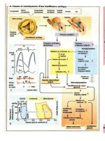
• It is most important to have a clear knowledge of the pathophysiology of HF, in particular
how LV work is dictated by systemic vascular resistance (SVR; see Fig. 12-1).
• Decrease neurohormonal activation; inhibition of the renin-angiotensin-aldosterone system.
• Inhibit LV remodeling.
• Improve myocardial hemodynamics.
• Increase cardiac output to deliver oxygenated blood to vital organs and to meet the meta-
bolic needs of the tissues, especially during normal activities and exercise.
• The cardiac output (CO) is reduced and filling pressure is increased. The low CO results
in a number of compensatory responses, as outlined in Fig. 12-1.
The following definitions are relevant:
• Cardiac output = stroke volume × heart rate (HR). Stroke volume is a reflection of pre-
load (filling pressure), myocardial contractility, and afterload (arterial impedance).
• LV work and myocardial oxygen consumption depend on
Fig. 12-1. Pathophysiology of heart failure.
Chapter 12 / Management of Heart Failure 217
a. HR × blood pressure (BP) (rate-pressure product).
b. BP = cardiac output × SVR.
The resistance or arterial impedance (afterload) against which the left ventricle must eject
is an important determinant of LV workload. A reduced SVR requires less energy and less
force of myocardial contraction to produce an increase in stroke volume.
SVR is automatically increased early in the development of HF and remains unchanged
or increases with increasing HF. This reaction is a necessity and is a normal compensatory
adjustment to maintain blood pressure and vascular homeostasis.
The compensatory adjustments are initiated by:
• Sympathetic stimulation that causes an increase in
a. Heart rate.
b. The force of myocardial contraction.
c. SVR.
• Activation of the renin-angiotensin-aldosterone system (RAAS), which causes
a. Intense arterial constriction and therefore an increase in SVR and blood pressure.
b. An increase in aldosterone, which produces distal sodium and water retention.
The important proximal tubular reabsorption of sodium is believed to be caused by a com-
bination of the preceding points and other as yet undetermined mechanisms (see Fig. 12-1).
• The renal response to a low CO in the normal subject is to maintain the blood pressure by
causing vasoconstriction and sodium and water reabsorption (saline autotransfusion). We
cannot expect the kidney to change its program when HF occurs. The kidney is behaving
appropriately in the wrong circumstances. Clearly, we can prevent the kidney from car-
rying out its program only if we switch off the initiating cause of the renal reflex, that is,
by increasing the cardiac output. Therefore, any drug that will increase CO will reduce the
renal response and lower SVR and further improve CO. An alternative strategy is to reset
the neurohormonal imbalance by the use of ACE inhibitors and aldosterone antagonists.
Note: Inotropic agents, digoxin or dobutamine, improve cardiac output and, therefore,
cause a fall in SVR.
MANAGEMENT GUIDE
Four golden rules dictate the efficient management of HF:
1. Ensure that the diagnosis of HF is correct, eliminating conditions that may mimic HF.
2. Determine and treat the basic cause of the heart disease. The rare surgical or medical cure
is worth the effort.
3. Search for the precipitating factors; remove or treat and prevent their recurrence to avoid
further episodes of HF. Withdraw drugs known to worsen HF: NSAIDs and notably cal-
cium antagonists (3,6) commonly administered to patients with hypertension and CHD.
4. The specific treatment of HF requires sound and up-to-date knowledge of the pathophysi-
ology of HF and the actions, indications, and side effects of the pharmacologic agents used
in its management.
Relieve symptoms and signs of HF by reducing raised filling pressures to near normal.
Therapeutic goals:
• Shift the cardiac function curve to the left and upward, decreasing the filling pressure yet
increasing stroke volume.
• Arrest or cause amelioration of the disease process.
218 Cardiac Drug Therapy
This can be achieved by the judicious use of
• Loop diuretics.
• Angiotensin-converting enzyme (ACE) inhibitors or angiotensin II receptor blockers (ARBs).
• Beta-blockers.
• Digoxin.
• Aldosterone antagonists: spironolactone or eplerenone.
• Statins for ischemic cardiomyopathy.
VASODILATORS
ACE Inhibitors/Angiotensin II Receptor Blockers
These agents are discussed in Chapter 3. ACE inhibitors and ARBs play a major role
in the management of HF.
Activation of the RAAS is an early manifestation of HF. The prime role of angiotensin
II is to support systemic blood pressure by:
• Causing systemic vasoconstriction, an increase in SVR.
• Stimulation of the central and peripheral effects of the sympathetic nervous system.
• Causing retention of sodium and water in the proximal nephron and directly by stimulation
of aldosterone production.
• Stimulating thirst and enhancing synthesis of vasopressin, thereby increasing total body
water.
In addition, angiotensin II preserves cerebral blood flow. Renal blood flow is pre-
served by selective vasoconstriction of the postglomerular (efferent) arterioles. Thus, the
influence of angiotensin II allows patients with severe HF to maintain blood pressure for
cerebral, renal, and coronary perfusion, and relatively normal values for serum creatinine
and blood urea nitrogen concentration also prevail. ACE inhibitors may cause a dramatic
decrease in glomerular filtration rate and increase azotemia in patients with HF and
hypotension. This deleterious effect can be minimized by reducing the patient’s depen-
dence on the renin-angiotensin system by reducing the dose of diuretic used. It is best to
choose an ACE inhibitor with a short action so as to allow brief restoration of the normal
homeostatic actions of the renin-angiotensin system (7). Long-acting agents may produce
prolonged hypotensive effects that may compromise cerebral and renal function and thus
may have disadvantages in such cases compared with short-acting agents (8). Initial low-
dose enalapril, 2.5 mg, caused a low 3.2% incidence of hypotension in a Scandinavian
study, proving the drug’s safe profile (9).
If ACE inhibitors or ARBs are not tolerated or are contraindicated, the combination
of hydralazine/isosorbide dinitrate (ISDN) should be tried; this combination is preferred
over ACE inhibitors in black patients as indicated in the Veterans Administration Heart
Failure Trial (A-HeFT) (10).
Data from the Veterans Administration Vasodilator Heart Failure Trial (V-HeFT)
suggested that patients with chronic HF could be considered for treatment with hydrala-
zine (25 mg three or four times daily) and ISDN (11), but use of captopril or enalapril is
preferred. In V-HeFT, the 2-yr reduction in mortality rate was 25%. Hydralazine and
ISDN were poorly tolerated and were withdrawn in 19% of patients. Only 55% of patients
were taking full doses of both drugs 6 mo after randomization (12). Improvement in sur-
vival was observed mainly in patients with New York Heart Association (NYHA) class
II HF. In this subset, 48 (24%) of 200 patients treated with enalapril and 66 (31%) of 210
patients treated with hydralazine/ISDN died (13).
Chapter 12 / Management of Heart Failure 219
The Cooperative North Scandinavian Enalapril Survival Study (CONSENSUS) (9)
showed that 6 mo of enalapril therapy produced a 40% reduction in mortality rate in
patients with NYHA class IV HF. The drug, when given as a 2.5-mg initial dose, is well
tolerated (9).
The Studies of Left Ventricular Dysfunction (SOLVD) investigators reported a mor-
tality rate of 35% in enalapril-treated patients with HF and an EF < 35% (14) and a mortal-
ity rate of 40% in the placebo group. Enalapril reduced the number of hospitalizations.
The prevention arm of SOLVD did not show a significant improvement in the survival
rate of patients with EF < 3 5% but without overt HF (14). Unfortunately, 65% of patients
in SOLVD were more than 4 wk post-MI. Thus, the study cannot be generalized to all
patients with HF.
Plasma renin levels are usually normal in patients with HF in NYHA class I and II, and
ACE inhibitors may not be logical therapy at this stage. When these patients are treated
with diuretics, however, plasma renin levels increase and ACE inhibitors may produce
salutary effects. In these situations not all patients benefit from ACE inhibitor therapy;
it is estimated that approx 50% of patients may improve in LV function, but survival data
are not available except in postinfarction patients. ACE inhibitors decrease LV hypertro-
phy, an important cause of diastolic dysfunction that predisposes to the late phase of the
failing ventricle. In the last phase of HF, both systolic and diastolic dysfunctions prevail.
Although ACE inhibitors have not proved useful in patients with mainly diastolic dys-
function, they can be used to prevent this condition. ACE inhibitors are not indicated as
monotherapy for patients with HF with CHD or hypertension.
D
OSAGE
Patients with very severe HF who are taking diuretics often have hyponatremia and
high plasma renin activity. These patients are likely to respond dramatically to ACE
inhibitors but with an associated profound fall in blood pressure. Thus, in this subset of
patients, it is necessary to discontinue diuretics and nitrates for 2–3 d and to initiate very-
low-dose captopril or enalapril therapy. The patient should be kept in bed for up to 3 h
following captopril administration. Captopril 6.25 mg is given twice daily for 1–2 d,
increasing the dose slowly to 6.25 mg three times daily and then 12.5 mg twice or three
times daily if systolic BP is >100 mmHg; at this stage, a low dose of diuretic is com-
menced. A captopril dose of 25 mg three times daily is often sufficient to provide benefit.
It may require 1–3 wk to achieve the dosage proved effective in clinical trials. Maximum
dose is 50 mg three times daily.
The physician must not be put off by mild hypotension (BP 90–100 mmHg) and must
be prepared to give ACE inhibitors a fair trial. Many weeks of treatment may be required
before clinical improvement becomes manifest. Pooled studies of a number of random-
ized placebo-controlled trials with other vasodilators compared with ACE inhibitor ther-
apy in patients with severe HF showed a significant improvement in survival in groups
treated with ACE inhibitors (15,16). Only captopril reduced wall stress and improved
functional class in 18 patients with dilated cardiomyopathy (17).
P
OST-MI HEART FAILURE WITH LEFT VENTRICULAR DYSFUNCTION
The following studies have tested the effect of ACE inhibitors:
• In the Survival and Ventricular Enlargement (SAVE) trial, 36,630 post-MI patients were
screened, but only 2231 of the 8938 patients with EF ≤ 40% were randomly assigned. Fol-
low-up at 3.5 yr showed a 37% reduction in the risk of developing HF and a 22% decrease
220 Cardiac Drug Therapy
in the risk of requiring hospitalization for HF (18). There was no significant reduction in
mortality rates.
• In the Acute Infarction Ramipril Efficacy (AIRE) study, ramipril was shown to improve
prognosis in post-MI patients with clinical evidence of HF (19).
Drug name: Captopril
Trade name: Capoten
Supplied: 12.5, 25, 50, 100 mg
Dosage: See text
DOSAGE
Withdraw diuretics and other antihypertensives for 24–48 h; then give a test dose of
3–6.5 mg and then the same dose twice daily, increasing to 12.5 mg two or three times
daily, preferably 1 h before meals (on an empty stomach).
The maximum suggested daily dose is 75–150 mg.
In renal failure, the dose interval is increased according to the creatinine clearance (see
Chapter 3 for a detailed account of adverse effects, cautions, interactions, and pharmaco-
kinetics).
Drug name: Enalapril
Trade names: Vasotec, Innovace (UK)
Supplied: 2.5, 5, 10, 20 mg
Dosage: 2.5-mg test dose; 8–12 h later start 2.5 mg twice daily, increasing over days
to weeks to 10–20 mg once or twice daily
Contraindications, side effects, and other considerations are discussed in Chapter 3
(see Table 3-1). Notably, the drug’s onset of action is delayed 2–4 h as opposed to capto-
pril (½–l h). Thus, an initial effect on hypotension is observed within 1 h after captopril
dosing and at about 2½ h with enalapril (17). Withdrawal of diuretics does not always pre-
vent marked hypotension or syncope (20), so caution is required with captopril and enala-
pril. In the Scandinavian study, a 2.5-mg initial dose caused a 3.2% withdrawal of patients
and a 31% reduction in the 1-yr mortality rate (9).
A RCT indicates that 20 mg of enalapril is as beneficial as 60 mg daily for HF treat-
ment (21).
Drug name: Lisinopril
Trade names: Prinivil, Zestril, Carace (UK)
Supplied: 5, 10, 20, 40 mg
Dosage: 2.5-mg test dose, then titrate dosage; 5–10 mg once daily, average 10–20 mg
daily. If no hypotension or adverse effects, the dose may be increased to
30–35 mg daily
The high dose was used in the Assessment of Treatment with Lisinopril and Survival
(ATLAS) study (22). Unfortunately, the ATLAS study compared 2.5–5 mg with 32.5–
35 mg lisinopril daily. It would make more clinical sense to have compared the dose
commonly used by cardiologists in clinical practice (i.e., 10–20 mg) as the low dose. The
Chapter 12 / Management of Heart Failure 221
results of the study showed a marginal difference; the high dose decreased modestly the
risk of hospitalization but not total mortality.
For other ACE inhibitors and ARBs, see Chapter 3. ARBs are advisable if ACE inhib-
itors are not tolerated (23,24).
Drug name: Hydralazine
Trade name: Apresoline
Supplied: 25, 50 mg
Dosage: 25 mg (average 50 mg) three times daily, max. 200 mg daily
Hydralazine is an effective vasodilator useful mainly when combined with oral nitrates,
as shown in V-HeFT I (12).
V-HeFT II indicated that hydralazine with ISDN is inferior to ACE inhibitor therapy
in achieving improved survival in patients with NYHA class II HF. Thirty-three percent
of patients cannot tolerate the drug because of headaches, dizziness, and other side
effects, and of the remaining 66% only half derive some benefit (25). Adverse effects were
similar in V-HeFT I and II. Hydralizine/ISDN may be used if an ACE inhibitor or ARB
is contraindicated. In A-HeFT, the combination significantly reduced mortality and
hospitalizations in patients of African origin (see Chapter 22).
Amlodipine
The Prospective Randomized Amlodipine Survival Evaluation (PRAISE)-2 study
showed that amlodipine caused neither benefit nor harm in patients with CHF. The result
of PRAISE-1 was the result of chance. In PRAISE-2, amlodipine increased the occur-
rence of pulmonary edema in patients with low EF (26). Calcium antagonists should not
be used in the treatment of HF or in patients with EF < 40%.
DIURETICS
Indications and Guidelines
Heart failure precipitated by acute MI: In this situation, the cautious use of titrated doses
of furosemide usually suffices.
Furosemide 20–40 mg intravenously (IV) followed by 40 mg, 30 min to 1 h later, is
given. If symptoms persist, diuresis is not established, and the BP is stable, 80 mg is given.
Ensure that the serum K
+
level remains normal; do not wait to see it fall to <3.5 mEq
(mmol)/L before adding potassium chloride (KC1).
In patients with moderate and severe HF who are predicted to have recurrent bouts of
HF and who are receiving digitalis, give furosemide 80–160 mg daily. Occasionally,
bumetanide produces a greater diuresis than furosemide.
The combination of furosemide and hydrochlorothiazide or metolazone(27) increases
diuresis and should be given a trial in patients refractory to furosemide or other loop
diuretics. In patients with refractory HF with severe renal failure, furosemide 160–
320 mg along with metolazone may be required to promote adequate diuresis.
Note that the diuretic and antihypertensive actions of furosemide and thiazides are
reduced by drugs that are prostaglandin inhibitors, in particular indomethacin and other
NSAIDs.
Torsemide (Demadex): 10–20 mg IV. Maximum single dose 100–200 mg
Bumetamide: 1.0 mg, maximum 4–8 mg.
222 Cardiac Drug Therapy
ALDOSTERONE ANTAGONISTS
Figure 12-1 indicates the role of increased aldosterone production in the pathophysi-
ology of HF. (See also Chapter 7, New Concepts.)
It is necessary to block aldosterone completely because it causes
• Na and water retention. This effect continues when the effects of short-acting, poorly ab-
sorbed loop diuretics, such as furosemide, have dissipated.
•K
+
and Mg loss.
• Myocardial and vascular fibrosis.
• Norepinephrine release and increased myocardial uptake of norepinephrine that can con-
tribute to sudden death; myocardial fibrosis that contributes to progressive HF.
• Although aldosterone antagonists have proved successful in reducing adverse outcomes
in patients with HF, their use in patients with impaired renal function is a risk factor for
hyperkalemia.
• Most important, elderly patients with a serum creatinine in the normal range often have
renal impairment, and the various formulas for assessing glomerular filtration rate (GFR)
have drawbacks, particularly in patients older than age 70.
Drug name: Spironolactone
Trade name: Aldactone
Supplied: 25 mg
Dosage: Initial 12.5 mg if serum assess K
+
5.0 or less and reassess in 3 d and at 1 wk;
if K
+
< 5.0 mEq/L, give 25 mg once daily
Reassess at 1 mo then every 3 mo: maintain K
+
4 to maximum 5.0 mEq/L
(mmol/L)
Use cautiously with close monitoring of serum potassium in patients with
serum creatinine 1.2–1.5 mg/dL (106–133 µmol/L) or estimated GFR
50–60 mL/min.
Avoid in patients with more severe renal dysfunction: estimated GFR or
creatinine clearance < 40 mL/min. The ACC/AHA advises < 30 mL/min
• A major breakthrough is the strong recommendation to add 25 mg spironolactone (Aldac-
tone) in patients with class III and IV HF because the drug caused a 30% reduction in the
risk of death among this class of patients with EF < 35% treated with loop diuretics, an
ACE inhibitor, and digoxin. Hospitalization for recurrent HF was significantly reduced
(28). Unfortunately, the dosage of ACE inhibitor used was smaller than that used in
modern clinical practice: mean dose captopril 63 mg, enalapril 15 mg, lisinopril 14.3 mg.
• Spironolactone causes gynecomastia and other androgenic effects, and eplerenone, which
does not have these effects, has proved effective in a RCT.
• The dose of spironolactone used in the Randomized Aldactone Evaluation (RALES) trial
was 25 mg (28).
• Caution is required in patients with abnormal renal function and in type II diabetes with
hyporeninemic hypoaldosteronism because severe hyperkalemia may ensue.
• If the serum K
+
reaches 5.0– 5.1 mEq/L, the dose of ACE inhibitor should be decreased
and loop diuretic increased before reducing the 25-mg dose of spironolactone.
• Serum K
+
should be evaluated at 3 d and 1–2 wk after starting treatment and then about
3 monthly. If the K
+
reaches 5.1 mEq/L, spironolactone should be discontinued. Caution:
Spironolactone or eplerenone should be used with close monitoring of serum potassium
in patients with serum creatinine 1.2–1.5 mg/dL (106–133 µmol/L) or estimated GFR 49–
59 mL/min (28a).
Chapter 12 / Management of Heart Failure 223
• Avoid in patients with more severe renal dysfunction: estimated GFR or creatinine clear-
ance < 40 mL/min. The ACC/AHA advises <30 mL/min.
• Elderly patients with a creatinine 1.2–1.4 mg/dL (102–123 µmol/L) within the normal range
may have a markedly reduced creatinine clearance (estimated GFR) of 49–59 mL/min.
In patients age > 75 yr, a normal serum creatinine does not indicate normal renal func-
tion. It is necessary to assess the GFR.
• However, caution is needed because the formula to determine estimated GFR gives inaccu-
rate results in patients older than age 70; in blacks a correction is required: multiply by 1.2.
• Avoid concomitant use of NSAIDS or cyclooxygenase-2 inhibitors.
Drug name: Eplerenone
Trade name: Inspra
Dosage: If baseline K
+
< 5.1 mEq/L, 12.5 mg once daily
Assess K
+
in 3 d and at 1 wk; if <5.0 mEq/L, increase to 25 mg once daily.
Reassess K
+
at 1 mo then every 3 mo: maintain K
+
4 to maximum 5.1 mEq/L
(mmol/L)
Max. 50 mg once daily
Use cautiously with close monitoring of serum potassium in patients with
serum creatinine 1.2–1.5 mg/dL (106–133 µmol/L) or estimated GFR
49–59 mL/min
Avoid in patients with more severe renal dysfunction: GFR or creatinine
clearance < 40 mL/min. The ACC/AHA advises < 30 mL/min; see above
cautions for spironolactone in the elderly
• Avoid concomitant use of NSAIDS or cyclooxygenase-2 inhibitors.
BETA-BLOCKERS
• Three beta-blocking drugs have been approved for the management of HF: carvedilol,
metoprolol extended release (Toprol XL), and bisoprolol.
• Beta-blockers play a major role in the management of patients with HF and are strongly rec-
ommended for the management of class I–III HF and also for compensated class IV patients.
• Beta-blocker therapy is initiated as soon as possible after the diagnosis of HF provided the
patient is free from fluid overload.
Some specialists have advocated the commencement of a beta-blocker prior to ACE
inhibition in the hypotensive patient because this may cause an improvement in LV func-
tion, thus allowing initiation (or an increase in dosage) of the ACE inhibitor (29).
• They are most effective in patients with ischemic heart disease and dilated cardiomyopa-
thy (30).
• Transmyocardial measurements have documented that the failing human heart is exposed
to increased adrenergic activity. Chronic adrenergic activation has adverse effects on the
natural course of heart muscle disease (31,32).
• These agents partially block RAAS and augment atrial and brain natriuretic peptide.
• It is often forgotten that beta-blockers significantly reduce renin secretion from the juxta-
glomerular cells of the kidney, which causes a decrease in angiotensin levels and reduced
aldosterone production; this action adds to their life-saving potential.
• Beta-blockers decrease renin and aldosterone production, which contributes to their life-
saving potential in patients with HF, but a mild increase in K
+
may occur and potentiate
that caused by spironolactone or eplerenone.
224 Cardiac Drug Therapy
Drug name: Carvedilol
Trade names: Coreg, Eucardic (UK)
Supplied: 12.5, 25 mg
Dosage: 3.125-mg test dose and then twice daily after food for 1–2 wk; increase to
6.25 mg twice daily wk 3–4; then 9.375 mg twice daily during weeks 5–8;
then if tolerated 12.5 mg twice daily. Increase slowly to the highest level
tolerated; max. 25 mg twice daily. See text for further advice
DOSAGE (FURTHER ADVICE)
• Carvedilol should be taken with food to slow the rate of absorption and reduce the inci-
dence of orthostatic effects. If dizziness, lightheadedness, or hypotension occur, the dose
of diuretic or ACE inhibitor should be reduced to allow up-titration of carvedilol or other
beta-blocker. If symptoms persist, the dose of carvedilol should be reduced.
• In an RCT (33), this drug resulted in a 67% reduction in mortality rate in patients with HF
treated with diuretics and an ACE inhibitor; almost all patients were taking digoxin. The Car-
vedilol Postinfarct Survival Controlled Evaluation (CAPRICORN) study (34) in patients
after MI with a mean EF of 33% found a 23% relative reduction in mortality, identical to
the result of a metaanalysis of 22 long-term RCTs in post-MI patients. Notably, a similar
benefit—a 2.3% absolute reduction in risk—was observed in SAVE, AIRE, and TRACE
with ACE inhibitors, i.e., 43 patients treated to save one life.
• The Carvedilol Prospective Randomized Cumulative Survival Study (COPERNICUS) trial
(35) studied 2289 patients with severe HF, EF 16–24%, but free from overt fluid retention
or recent treatment with IV diuretics or positive inotropic drugs. The results showed a
highly significant 35% reduction in all-cause mortality with carvedilol (see Chapter 22).
Beta-blocking drugs such as carvedilol have proved useful in improving survival and
decreasing the number of hospitalizations for worsening HF. The drug is as effective as
an ACE inhibitor in this setting. Carvedilol is indicated for the management of NYHA
class II–III HF; the drug has not been adequately tested in patients with class IV HF.
Compensated class IV HF patients free of fluid overload have shown benefit and should
be treated, judiciously, with carvedilol with up-titration of doses over 4–6 wk.
Drug name: Metoprolol succinate
Extended release
Metoprolol CR/XL
Trade names: Betaloc, Lopressor, Toprol XL
Supplied: 50, 100 mg
Dosage: 12.5 mg test dose and then once daily for 2 wk; then 25 mg; titrate over
4–8 wk to 100 mg usual maintenance dose; max. 200 mg once daily
In a randomized trial in 338 patients with HF from dilated cardiomyopathy, metoprolol
prevented clinical deterioration and improved symptoms and cardiac function (36). The
Metoprolol Extended-Release Randomized Intervention Trial in Heart Failure (MERIT-
HF) trial involving patients with class II and III HF, mean EF 28%, resulted in risk reduc-
tion of 33% for total mortality or worsening HF (37).
This agent was used in combination with diuretics, digoxin, and ACE inhibitors.
In a randomized trial in 50 patients with HF caused by ischemic heart disease, meto-
prolol 25–100 mg daily when added to standard HF therapy resulted in
Chapter 12 / Management of Heart Failure 225
• A decrease in the number of hospital admissions.
• Improved functional class.
• Increased EF.
• A greater increase in exercise duration compared with placebo (37). It is known that beta-
blocking agents cause a decrease in sudden cardiac deaths and increased survival in post-
MI patients, and this beneficial result may be obtained in patients with varying grades of HF.
Bisoprolol (Zebeta, Monocor)
In the Cardiac Insufficiency Bisoprolol Study II (CIBIS II) (38), bisoprolol resulted in
a significant decrease in mortality in patients with NYHA class III HF and EF < 35%. CIBIS
II involved 2647 patients aged 18–80 yr with class III or IV HF. Study patients received
ACE inhibitor and diuretic (digitalis was allowed) for at least 2 mo prior to bisoprolol or
placebo. Bisoprolol, initial dose 1.25 mg daily, was titrated at weekly intervals in 1.25-
mg increments for 4 wk and then up to a maximum of 10 mg daily. Bisoprolol therapy
reduced all-cause mortality by 32% (p = 0.00005) and sudden death by 45% (p = 0.001).
A 30% reduction in hospitalization occurred in the bisoprolol-treated group. Treatment
withdrawals in the bisoprolol-and placebo-treated patients were similar (approx 15%).
Drug name: Bisoprolol
Trade names: Zebeta, Monocor
Dosage: 1.25 mg test dose; then once daily; increase in 2–3 wk to 3.75 mg;
at 5–6 wk if tolerated 5-mg maintenance dose
At >12 wk, if needed max. dose 10 mg provided that the BP is >120 mmHg
systolic and heart rate > 55 beats/min
INOTROPIC AGENTS
Drug name: Digoxin
Trade name: Lanoxin
Supplied: 0.625, 0.125, 0.25 mg
Dosage: See text
Digoxin (Lanoxin) is the most reliable digitalis preparation and is used by the majority
of physicians. Remarks are confined to this drug.
Indications
• Atrial fibrillation with uncontrolled ventricular response is the most clear-cut indication.
• HF related to poor LV contractility. These patients usually have a third heart sound gallop
(S3), crepitations over the lung fields, and EF < 35%.
• A failure of diuretics and vasodilator therapy. Hypotension often limits the use of vasodila-
tors in patients with severe HF and a low EF. Digoxin has a role in this category of patients.
Digoxin is indicated for all patients with impaired systolic function and NYHA class
III, and IV HF (39). Patients with NYHA class II HF are often managed with diuretics and
ACE inhibitors, and recurrence of HF is an indication for the addition of digoxin.
• Ahmed and colleagues did a comprehensive post hoc analysis of the Digitalis Investigation
Group (DIG) trial (40). Digoxin showed a reduction in mortality and hospitalization for
HF (40).
226 Cardiac Drug Therapy
Digoxin is not usually recommended, or is of limited value, for the management of HF
resulting from or accompanied by:
• Acute MI, except from d 2 if HF is not easily controlled by furosemide, nitrates, ACE inhib-
itor, dobutamine, or nitroprusside.
• Advanced first-degree, second-degree, and complete atrioventricular (AV) block. (It is
preferable with second- and third-degree AV block to pace the patient and then use digitalis.)
• Patients with low EF, sinus rhythm, and no history of HF symptoms.
• The use of digoxin to minimize symptoms in patients with HF with preserved EF is not well
established.
• Mitral stenosis, normal sinus rhythm.
• Hypertrophic cardiomyopathy (HCM), except if HF is moderate or severe. (The drug is
potentially dangerous in HCM.)
• Sick sinus syndrome; it is advisable to put in a permanent pacemaker and then commence
beta-blocker therapy.
• Cor pulmonale, except for the management of atrial fibrillation with a fast ventricular re-
sponse or in patients with added severe LV failure exhibiting a low cardiac output and both
central and peripheral cyanosis.
A study by Arnold and colleagues (41) demonstrated that patients with proven HF show
improvement in hemodynamics during acute and long-term administration as well as dur-
ing exercise. Withdrawal of digoxin in that study produced a significant increase in pul-
monary capillary wedge pressure, heart rate, and SVR and a fall in stroke work index and
EF. After acute retreatment, all parameters improved, including exercise hemodynamics.
Gheorghiade and colleagues (42) have confirmed these hemodynamic effects of digoxin.
In a double-blind placebo-controlled study of patients with documented HF and no
reversible etiology, 16 of the 46 patients deteriorated between 4 d and 3 wk after stopping
digoxin (43).
Analysis of the Digoxin Study
The effect of digoxin on mortality and morbidity in patients with HF was reported in
1997 (39,40). The results of this study provided some answers to 200 yr of controversy
regarding the use of digitalis.
Digoxin was assigned to 3397 patients, and 3403 received diuretics and an ACE inhib-
itor. The mean EF was 28% ± 9%. The average follow-up was 37 mo. Fewer patients in the
digoxin group were hospitalized for worsening HF: 26.8% versus 34.7% in the placebo
group (p < 0.001; see Table 12-3).
• The study did not show a significant decrease in total mortality. Digoxin, however, did
not cause an increase in mortality. In fact, there was a trend toward a decrease in the risk
of death attributed to worsening HF (p = 0.06).
• Most important, the risk associated with the combined outcome of death related to HF
or hospitalization related to HF was significantly lower in the digoxin group (1041
versus 1291 patients, p < 0.01;see Table 12-3) and was similar to that observed in SAVE
and SOLVD attributed to the benefits of ACE inhibitor.
• Unfortunately, the study included only 2% class IV and 30% class III patients. Digoxin
is expected to benefit class III–IV patients; this subset was not well represented in the
study. Digoxin is strongly indicated in class III–IV HF and in patients with EF < 30%.
• Fortunately, the study showed that in patients with EF < 0.30, death or hospitalization related
to worsening HF occurred in 428 of 1127 in the digoxin group and in 556 of 1130 in the
placebo group, a 23.0% reduction. In patients with NYHA class III HF, death or hospitaliza-
Chapter 12 / Management of Heart Failure 227
tion occurred in 438 of 1118 in the digoxin group and in 552 of 1105 in the placebo group,
a 20.6% reduction (risk ratio 0.70 [95% confidence [CI] interval 0.61–0.79]). A 19% reduc-
tion was observed for the risk of death, CHF, or hospitalization (p = 0.001).
• A 22% decrease in death or hospitalization was observed in patients with cardiothoracic
ratio > 0.55. The study indicated that digoxin significantly decreases death or hospitaliza-
tion caused by worsening HF in patients with class II–III and IV HF with EF < 0.25 or with
cardiothoracic ratio > 0.55. The enalapril CONSENSUS study showed an increased survi-
val rate in class IV patients treated over 6 mo with enalapril added to diuretics and digoxin.
• Currently we strongly recommend this drug in patients with class III and IV HF, with EF
< 30%, and with increased LV volume and cardiothoracic ratio > 0.55. This is advisable
particularly if the systolic blood pressure is <100 mmHg caused by ACE inhibitors and
beta-blockers. Digoxin does not cause a decrease in BP. However, digoxin levels must
remain in the range 0.5–1 ng/mL.
Is the Combination of Digoxin and ACE Inhibitors Necessary?
The Randomized Assessment of Digoxin on Inhibitors of ACE (RADIANCE) study
included 178 patients with chronic HF and sinus rhythm who were clinically stable with
diuretics, an ACE inhibitor, and digoxin (44). Most patients (70%) were in NYHA class
II. In patients withdrawn from digoxin for 3 months, there was a sixfold worsening of HF.
Patients taking a placebo had a higher incidence of deterioration and worsening HF (23 ver-
sus 4 patients) and more deterioration in quality of life. The dose of digoxin in the RADI-
ANCE study was 0.38 mg daily, and serum digoxin levels ranged from 0.9 to 2.0 ng/mL.
• Digoxin favorably alters the neurohormonal imbalance that contributes to HF. It is, there-
fore, rational to use the triple combination of diuretics, ACE inhibitors, and digoxin to man-
age LV failure and improve symptoms, survival, and quality of life in virtually all patients
with class III–IV HF.
• A salutary interaction of digoxin and spironolactone: These two now have an important
role in the management of class III and IV HF. Na entry into Na channels in myofibroblasts
is enhanced by aldosterone and is the trigger for myocardial fibrosis.
Table 12-3
Effect of Digoxin on Mortality and Morbidity in Patients with Heart Failure
Digoxin Placebo %
(n = 3397) (n = 3403) reduction p Risk ratio
a
Worsening CHF 910 1180 22.9 <0.001
Death plus CHF 1041 1291 19.3 <0.001
Death owing to worsening CHF 394 449 0.06
Death or hospitalization 428/1127 556/1130 23.0 0.68
owing to CHF (0.60–0.70)
EF < 0.25
Class III or IV 438/1118 552/1105 20.6 0.70
Class IV only 2% (0.61–0.79)
of study group
Cardiothoracic 441/1176 567/1170 22.2 0.69
ratio > 0.55 (0.61–0.79)
a
Values in parentheses are 95% confidence intervals. CHF, congestive heart faillure; EF, ejection fraction.
Modified from the Digitalis Investigation Group. The effect of digoxin on mortality and morbidity in
patients with heart failure. N Engl J Med 1997;336:525.
228 Cardiac Drug Therapy
• When digoxin is added, there is a reduction in fibrosis because digoxin blocks sodium-potas-
sium adenosine triphosphatase (Na
+
,K
+
-ATPase) and thus decreases Na entry into fibro-
blasts and decreases fibrosis. In the spironolactone study, 72% of patients received digoxin.
Mechanism of Action
1. Digoxin increases the force and velocity of myocardial contraction in both the failing and
the nonfailing heart. It inhibits the function of the sodium pump, resulting in an increase
in intracellular sodium accompanied by an increase in cellular calcium. Digoxin causes the
Frank-Starling function curve to move upward and to the left, i.e., an improved ventricular
function curve. Improvement in cardiac output produces a favorable alteration of the com-
pensatory responses of HF including the neurohormonal response (see Fig. 12-1).
2. Electrophysiologic effects:
a. Decreases conduction velocity in the AV node, i.e., the drug sets up a “traffic jam” in the
AV node, producing an important reduction in ventricular response in atrial fibrillation.
b. Increases the slope of phase 4 diastolic depolarization and therefore increases automa-
ticity of ectopic pace makers.
3. Vasoconstrictor: The drug has a mild vasoconstrictor effect that increases total systemic
resistance. In the failing heart, however, the drug increases cardiac output, which counter-
acts the reflex stimulation of the sympathetic and angiotensin systems, resulting in vasodila-
tion and a fall in total systemic resistance, i.e., afterload reduction.
Dosage Considerations
Before writing an order for digoxin, review the indications and reassess renal function
and conditions that increase sensitivity (Table 12-4).
1. Loading Dose (Initial Dose)
For adults and children over 10 yr and in the absence of conditions that may increase sen-
sitivity—initial 0.5–1 mg orally, given as follows:
Table 12-4
Conditions in Which There is an Increased Sensitivity
to Digoxin and Conservative Dosing is Recommended
Elderly patients (age > 70 yr)
Renal dysfunction; creatinine > 1.2 mg/dL (106 µmol/L)
Estimated GFR < 50 mL/h
Thin patients, low skeletal mass
Hypokalemia
Hyperkalemia
Hypoxemia
Acidosis
Acute myocardial infarction
Hypomagnesemia
Hypercalcemia
Hypocalcemia
Myocarditis
Hypothyroidism
Amyloidosis
Chapter 12 / Management of Heart Failure 229
• Slow method: 0.5 mg immediately and 0.25 mg every 12 h for two doses (1 mg/24 h).
OR
• 0.25 mg twice daily for 2 d and then maintenance depending on age and renal function.
Note: A low skeletal mass means less binding to skeletal muscle receptors, and therefore
a smaller loading dose is required in the thin or elderly patient and in women. For a more
rapid effect (e.g., atrial fibrillation, heart rate 130–150/beats/min) give 0.5 mg immedi-
ately and then 0.25 mg every 6 h for two or three doses.
Intravenous: Mainly for atrial fibrillation, heart rate >150/beats/min in the absence of
digoxin therapy within the last 2 wk. Give either:
• 0.75 mg IV slowly over 5 min and then 0.25 mg every 2 h for two doses, under electro-
cardiography (ECG) cover, reassessing the patient before each dose is given. A total
dose of 1.25–1.5 mg is often necessary.
OR
• 0.75–1.25 mg as an infusion over 2 h or more, which is advised in the United Kingdom
when rapid control is needed.
2. Maintenance Dose
Digoxin is mainly excreted unchanged by the kidney, with an average half-life of 36 h.
In normal renal function, as a general rule give the following:
• Age < 70 yr: 0.25 mg daily, preferably at bedtime.
• Age > 70 yr: 0.125 mg daily, and reduced dose if renal impairment is present, to main-
tain a level of 0.5 to maximum 1 ng/mL.
Note: Atrial fibrillation with a fast ventricular response may require 0.125 mg daily or
twice weekly in addition to the preceding dose.
Caution:
• Serum digoxin levels > 1.0 ng/mL appear to increase mortality in women. The dose
should be 0.152 mg daily or less to achieve a level of 0.6–1 ng/mL.
• In renal failure, the dose interval is increased depending on the creatinine clearance.
The serum creatinine level is not an accurate measure of the creatinine clearance. It is
advisable to estimate the GFR. Despite such calculations, digoxin toxicity may develop
if renal dysfunction is present. Digoxin is best avoided in patients with moderate degree
of renal failure: estimated GFR < 30 mL/h.
• The use of digitalis in the presence of moderate or severe renal failure represents a
controversial area because the risk of toxicity is common.
The bioavailability of digitalis is reduced in malabsorption syndrome and by the following
drugs:
• Cholestyramine, colestipol.
• Neomycin.
• Antacids.
• Metoclopramide.
• Diphenylhydantoin.
• Phenobarbital.
• Phenylbutazone.
Serum Digoxin Levels and Interactions
Low therapeutic or even subtherapeutic digoxin levels < 1.0 ng/mL do not exclude toxic-
ity. Beneficial effects are observed in the low range 0.5–1.1 ng/mL; toxicity is rare if
levels are thus maintained. Determinations should be made no earlier than 6 h after the last
dose of digoxin. If digoxin is given at night, a serum level can be obtained at steady state
on the following morning, and the drug dosage can be modified when the patient is seen
during the day.
230 Cardiac Drug Therapy
Values of 2 ng/mL (2.6 nmol/L) may be associated with toxicity, and in some patients
levels of 2–3 ng/mL may not indicate toxicity. Patients must never be allowed to have levels
greater than 2 ng/mL µg/L. There are several limitations to these statements:
• The sensitivity of the myocardium and conducting system is important.
• Sensitivity of the radioimmunoassay is between 0.2 and 0.4 ng/mL digoxin. Values can dif-
fer (up to 30%).
• Spironolactone causes falsely raised values.
• Atorvastatin, quinidine, calcium antagonists, and amiodarone increase serum digoxin lev-
els. Decreased renal elimination of digoxin results from a quinidine-induced decrease in
tubular secretion of digoxin (46). Verapamil causes a significant increase in digoxin levels
that may result in severe bradycardia or asystole (47). Interaction with diltiazem is minimal
and with nifedipine insignificant. Amiodarone reduces clearance of digoxin and causes
a 25–75% increase in digoxin levels. A reduction of the dose of digoxin by 50% is recom-
mended when quinidine, quinine, verapamil, or amiodarone is given concurrently (48).
Clinical studies indicate that there is no clear-cut serum level that establishes the pres-
ence of toxicity. The serum digoxin level is useful when interpreted relative to the serum
potassium level and the clinical situation. The serum K
+
concentration increases with digi-
talis toxicity.
If digoxin toxicity is suspected, it is advisable to discontinue digoxin if:
• Serum digoxin level is >1.1 ng/mL with a serum K
+
level of 2.5–3.5 mEq (mmol)/L.
• Serum potassium level is <2.5 mEq (mmol)/L. In this case, digoxin should be withheld
regardless of the serum digoxin concentration.
Potassium depletion must be corrected before recommencing digoxin.
This seems a reasonable course of action because there are other treatments for the
management of HF.
Digitalis Toxicity
• The incidence of digoxin toxicity decreased during the 1990s, and toxicity occurs in about
one episode for every 20 yr of treatment. In large digoxin trials, adverse effects have been
no different than with placebo.
• The incidence of toxicity is rare with the lowered dose currently advised.
• The ECG may be nonspecific, but symptoms may provide enough clues, especially when
combined with knowledge of the results of the serum creatinine or creatinine clearance
and the maintenance dose of digoxin. This information should enable the physician to
reach a decision about whether or not the patient has digoxin toxicity. There is no need
to await notification of digoxin levels. If you suspect toxicity, withhold the drug for at least
48 h—there are other effective treatments for HF, and withholding a few doses of digoxin
will not be detrimental.
SYMPTOMS OF TOXICITY
• Gastrointestinal: anorexia, nausea, vomiting, diarrhea, and weight loss.
• Central nervous system: visual hallucinations, mental confusion, psychosis, restlessness,
insomnia, drowsiness, and extreme weakness; blue-green-yellow vision, blurring of vision,
and scotomas.
CARDIAC DYSRHYTHMIAS
1. Ventricular premature beats: bigeminal or multifocal.