Báo cáo y học: " HTLV-1 and -2 envelope SU subdomains and critical determinants in receptor binding" potx
Bạn đang xem bản rút gọn của tài liệu. Xem và tải ngay bản đầy đủ của tài liệu tại đây (610.38 KB, 14 trang )
BioMed Central
Page 1 of 14
(page number not for citation purposes)
Retrovirology
Open Access
Research
HTLV-1 and -2 envelope SU subdomains and critical determinants
in receptor binding
Felix J Kim
1,2
, Nicolas Manel
1
, Edith N Garrido
1
, Carine Valle
1
,
Marc Sitbon*
1
and Jean-Luc Battini*
1
Address:
1
Institut de Génétique Moléculaire de Montpellier (IGMM), CNRS-UMR5535, IFR122 1919 Rte de Mende, F-34293 Montpellier Cedex
5, France and
2
Current address: Memorial Sloan-Kettering Cancer Center 1275 York Ave, New York, NY, 10021, USA
Email: Felix J Kim - ; Nicolas Manel - ; Edith N Garrido - ;
Carine Valle - ; Marc Sitbon* - ; Jean-Luc Battini* -
* Corresponding authors
Abstract
Background: Human T-cell leukemia virus (HTLV) -1 and -2 are deltaretroviruses that infect a
wide range of cells. Glut1, the major vertebrate glucose transporter, has been shown to be the
HTLV Env receptor. While it is well established that the extracellular surface component (SU) of
the HTLV envelope glycoprotein (Env) harbors all of the determinants of interaction with the
receptor, identification of SU subdomains that are necessary and sufficient for interaction with the
receptor, as well as critical amino acids therein, remain to be precisely defined. Although highly
divergent in the rest of their genomes, HTLV and murine leukemia virus (MLV) Env appear to be
related and based on homologous motifs between the HTLV and MLV SU, we derived chimeric
HTLV/MLV Env and soluble HTLV-1 and -2 truncated amino terminal SU subdomains.
Results: Using these SU constructs, we found that the 183 and 178 amino terminal residues of the
HTLV-1 and -2 Env, respectively, were sufficient to efficiently bind target cells of different species.
Binding resulted from bona fide interaction with the HTLV receptor as isolated SU subdomains
specifically interfered with HTLV Env-mediated binding, cell fusion, and cell-free as well as cell-to-
cell infection. Therefore, the HTLV receptor-binding domain (RBD) lies in the amino terminus of
the SU, immediately upstream of a central immunodominant proline rich region (Env residues 180
to 205), that we show to be dispensible for receptor-binding and interference. Moreover, we
identified a highly conserved tyrosine residue at position 114 of HTLV-1 Env, Tyr
114
, as critical for
receptor-binding and subsequent interference to cell-to-cell fusion and infection. Finally, we
observed that residues in the vicinity of Tyr
114
have lesser impact on receptor binding and had
various efficiency in interference to post-binding events.
Conclusions: The first 160 residues of the HTLV-1 and -2 mature cleaved SU fold as autonomous
domains that contain all the determinants required for binding the HTLV receptor.
Published: 02 December 2004
Retrovirology 2004, 1:41 doi:10.1186/1742-4690-1-41
Received: 13 September 2004
Accepted: 02 December 2004
This article is available from: />© 2004 Kim et al; licensee BioMed Central Ltd.
This is an Open Access article distributed under the terms of the Creative Commons Attribution License ( />),
which permits unrestricted use, distribution, and reproduction in any medium, provided the original work is properly cited.
Retrovirology 2004, 1:41 />Page 2 of 14
(page number not for citation purposes)
Background
Human T-cell leukemia virus type 1 (HTLV-1) has been
found primarily in CD4+ and CD8+ T-lymphocytes in vivo
[1-3], whereas CD8+ T-lymphocytes are thought to be the
in vivo reservoir of HTLV-2 [4]. However, the in vitro tro-
pism of HTLV-1 and -2, as determined using HTLV enve-
lope-pseudotyped virions or envelope-induced cell fusion
assays, appears to be ubiquitous [5-7]. Indeed, we recently
showed that Glut1, the ubiquitous vertebrate glucose
transporter, serves as a receptor for HTLV-1 and -2 enve-
lope glycoprotein (Env) [8]. While the precise organiza-
tion and properties of the receptor-interacting Env
domains has not been reported, we found that the amino
terminal two-thirds of the HTLV-1 extracellular surface
component (SU) are sufficient to confer HTLV-1 tropism
to an ecotropic Friend murine leukemia virus (F-MLV)
Env [9]. A cell fusion interference assay performed with
this HTLV/F-MLV Env chimera and the parental Env con-
firmed that this 215 amino acid Env domain, harbors
HTLV-1 receptor-binding determinants [9].
The corresponding domain in MLV Env SU – located
upstream of a conserved K/R L L T/N L V Q motif in the
SU of the HTLV-1 and F-MLV Env [9,10] – is well charac-
terized and comprises two main functional regions: an
amino terminal sequence harboring the receptor-binding
determinants, VRA, VRB and VRC [11-13], and a proline-
rich region (PRR), starting at the first proline residue of
the GPRVPIGP sequence [11,14] and flanked by two
highly conserved GXDP [15] and CXXC [16] motifs (Fig-
ure 1). In the ecotropic and amphotropic (Ampho) MLV
Env, the PRR is a putative hinge region implicated in con-
formational changes, triggered after receptor binding, and
subsequent fusion [17,18]. In the central region of the
HTLV SU, a short sequence (Env residues 180 to 205) har-
bors high proline content and could be a homologue of
the MLV PRR.
Several studies using synthetic peptides and neutralizing
antibodies against the HTLV Env have shown that deter-
minants within this proline rich region homologue
(PRRH) are involved in interference to Env-mediated syn-
cytium formation [19-21]. The PRRH had been thought to
encode the receptor-binding domain, as based on cell-to-
cell fusion assays [19,22-24]. However, although PRRH
synthetic peptides can block HTLV Env-mediated syncytia
formation, they have no effect on HTLV SU binding [25]
and infection [26]. Indeed, we and others have shown
that Env receptor binding per se, as well as interference to
receptor-binding, cell-to-cell fusion, syncytium forma-
tion, and infection involve several distinct cell surface-
associated parameters [27-29]. In the present report, we
produced soluble forms of wild-type and mutant HTLV-1
and 2 SU amino terminal subdomains and tested their
receptor-binding abilities. We also tested their ability to
specifically interfere with HTLV Env cell surface binding,
Env-mediated cell-to-cell fusion, and retroviral infection.
By testing these essential parameters of Env-mediated dis-
semination, we delineated the Env receptor-binding
domain (RBD) to the first 160 residues of the mature
HTLV-1 and -2 SU, excluding the PRRH, and we identified
a conserved tyrosine residue at position 114 of HTLV-1
Env as a critical determinant for HTLV Env receptor
binding.
Results
Motif conservation and similar modular organization of
HTLV and MLV SU, and identification of a proline-rich
region homologue (PRRH) in the HTLV SU
As shown in Figure 1, our alignment of the MLV and HTLV
SU reveals several notable motif conservations outlining a
similar modular organization of the MLV SU and HTLV
SU. A (K/R)LL(T/N)LVQ motif, highly conserved between
the F-MLV and HTLV-1 SU, is located immediately down-
stream of the PRR and its PRRH counterpart, respectively.
Another highly conserved motif between MLV and HTLV,
GXDP, is found immediately upstream of the PRR/PRRH
(Figure 1). These two motifs compelled us to notice the
PRRH, between the PSQ and KLLTLVQ sequences in
HTLV-1, and between the PTQ and KILKFIQ sequences in
HTLV-2 (Figure 1). As counted from the first and last pro-
line in the delineated sequence, the PRRH has a proline
content of 30.8% and 30.4% for HTLV-1 and -2, respec-
tively. This is slightly lower than the 35.3%, 36%, 36%,
and 35.6% proline content for the ecotropic, polytropic,
xenotropic, and amphotropic MLV Env, respectively (Fig-
ure 1). The presence of a PRRH in the HTLV SU appeared
to be characteristic of their MLV-like modular organiza-
tion, since HTLV SU average proline content outside of the
PRRH does not exceed 11%.
Functional, soluble HTLV Env-receptor binding
determinants
MLV SU receptor binding determinants are all located
upstream of the PRR [11,30]. To test whether the HTLV
Env receptor binding determinants are also located
upstream of the potential PRRH, we constructed a chi-
meric Env and several soluble HTLV-1 and -2 SU amino
terminal subdomains. The chimeric HTLV/MLV Env,
H1
183
FEnv, comprises the 183 amino terminal residues of
the HTLV-1 SU ending with the PSQL residues fused to the
PIGP sequence of the F-MLV PRR (Figure 2A). In this Env
chimera the receptor-binding domain (first 269 residues)
of the F-MLV Env was replaced with the potentially corre-
sponding domain of the HTLV-1 Env SU (Figure 2A). The
chimeric H1
183
FEnv construct – which lacks the HTLV
PRRH but has the MLV PRR – was properly expressed in
transfected cells and was revealed on immunoblots with
an anti-MLV SU polyclonal antibody (Figure 3A). Accord-
ingly, an anti-HTLV-1 monoclonal antibody raised
Retrovirology 2004, 1:41 />Page 3 of 14
(page number not for citation purposes)
against a PRRH epitope did not bind this chimeric Env
(data not shown).
HTLV-1 and -2 SU amino terminal subdomains with or
without their respective PRRH were constructed as fusion
proteins with either an influenza hemagglutinin (HA) or
rabbit immunoglobulin Fc (rFc) carboxy terminal tag
(Figure 2B). The H1
215
SU and H2
211
SU subdomains com-
prise the first 215 and 211 residues, counting from the
first methionine in the signal peptide through the KLLT-
LVQ of HTLV-1 and KILKFIQ of HTLV-2 Env, respectively
(Figure 2B). The H1
179
SU and H2
178
SU, comprising the
amino terminal 179 and 178 amino acids of the HTLV-1
and -2 Env, respectively, exclude the PRRH sequence (Fig-
ure 2B).
Cell lysates and cell culture supernatants were analyzed to
evaluate intracellular expression and secretion of func-
tional SU amino terminal domains in transfected-cell cul-
tures, respectively. H1
215
SU and H2
211
SU, containing the
PRRH sequence, and H2
178
SU lacking this PRRH were all
efficiently expressed in transfected cells (Figure 3B). It is
noteworthy, however, that recovery of tagged H1
179
SU
molecules was largely inefficient because the vast majority
of this protein was cleaved (data not shown). In contrast,
no significant cleavage was observed with the other solu-
ble domains released in the medium (not shown) (Figure
3C). As expected for immunoadhesins, H1
215
SU,
H2
211
SU, and H2
178
SU rFc-tagged domains were detected
as dimers under non-reducing conditions (not shown).
Immunoblots of cell extracts revealed two forms of
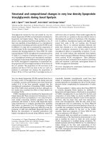
Homologous modular domains in HTLV and MLV envelopesFigure 1
Homologous modular domains in HTLV and MLV envelopes. Friend-MLV (F-MLV) Env and HTLV-1 Env are schemat-
ically represented as open and solid boxes, respectively. Boxes represent, from left to right, the signal peptide which comprises
the first 34 and 20 amino acid residues of F-MLV and HTLV Env, respectively, the extracellular surface component (SU) and the
transmembrane component (TM) including the carboxy terminal R peptide in F-MLV, which is cleaved in the mature Env glyco-
protein [64, 65]. Env landmark positions are indicated and the MLV proline-rich regions (PRR) and the HTLV SU PRR homo-
logue (PRRH) are delineated by vertical lines within the SU at the positions indicated by solid arrowheads. The PRR and PRRH
start at the first proline (P) residue downstream of the conserved GXDP motif. Env sequences represented in the figure are
obtained from F-MLV strain 57 (accession number CAA26561); P-MLV, F-MCF polytropic MLV (AAA46483); X-MLV, NZB
xenotropic MLV (AAA46531); A-MLV, amphotropic MLV strain 4070A (AAA46515); HTLV-2 (NP_041006); and HTLV-1, MT2
strain (VCLJMT). Residue numbering starts from the first methionine of the Env signal peptides. Proline residues and homolo-
gous motifs are noted in bold. Amino acid sequence alignments were performed using the Clustal program in the Megalign
alignment software package (DNAStar) with manual adjustments.
SU TM
215180
329
267
313 488
21
PSQL………
180
R
35
PRV…………………
675479267
HTLV-2
HTLV-1
F-MLV
X-MLV
A-MLV
P-MLV
MLV proline-rich region (PRR)
LLNLVQ
329
215
LLTLVQ
GYDPI-WF LNTE P SQLPPTAP - P LLP HSNLDHILEP SIP WKS KLLTLVQLTLQSTNYT CIVCI
GYDPL-WF ITSEP TQPPPTSP - P LVHDSDLEHVLTP STSWTT KILK FIQL TLQST NYS CMVCV
GRDPGLTFGIRLR YQNLGP RVP IG P N P VLADQLSLP R P N P L P K P AKS PPASNSTP TLISP S P T P TQPPPAGTGDRLLNLVQGAYQAL NLTNPD K TQECWLCL
GADPVTRFSLTRQ VLNVGP RVP IG P N P VITDQLPPSR P VQIM-LP R PPQ P PPP GAASIV-P ETAP - P SQQP GTGDRLLNLVDGAYQALNLTSPD K TQECWLCL
GADPVTRFSLTRQ VLNVGP RVP IG P N P VITDQLPPSQ P VQIM-LP R PPH P PPS GTVSMV-P GAPP- P SQQP GTGDRLLNLVEGAYQALNLTSPD K TQECWLCL
GTDPITMFSLTRQ VLNVGP RVP IG P N P VLP DQRLPSS P IEIVP A P Q PPS P LNTSYPPSTTSTP STS-P TSP SVP Q PPPGTGDRLLALVKGAYQALNLTNPD K TQECWLCL
-
F-MLV
HTLV-1
Retrovirology 2004, 1:41 />Page 4 of 14
(page number not for citation purposes)
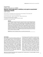
Schematic representation of HTLV/MLV Env chimeras and HTLV SU amino terminal subdomainsFigure 2
Schematic representation of HTLV/MLV Env chimeras and HTLV SU amino terminal subdomains. Env land-
mark positions are indicated and SU landmark sequences and positions are indicated by arrowheads. Open arrowheads indi-
cate the position of construct borders. (A) HTLV/MLV Env chimeras. The H1
215
FEnv and H1
183
FEnv HTLV/MLV Env chimeras
were obtained by replacing the 329 and 269 amino terminal residues of the F-MLV Env (open boxes) with the amino terminal
215 and 183 amino acid residues of the HTLV-1 Env (solid boxes), respectively. The H1
215
FEnv chimera, previously described
and formerly designated HHproFc [9], has been renamed here for sake of nomenclature homogeneity. (B) Soluble HTLV-1
(H1) and HTLV-2 (H2) SU amino terminal subdomains, H1
215
SU, H2
211
SU, H1
179
SU, and H2
178
SU were constructed as fusion
proteins with a carboxy terminal hemagglutinin (HA) or rabbit immunoglobulin Fc (rFc) tag. All amino acid residue numbering
starts from the first methionine of the HTLV-1 or -2 Env signal peptide, the amino terminal 20 and 21 aa residues, respectively.
180
21
313
488
CIVCI
215
229
HTLV-1
(H1)
229215180 183
GYDPIWFLNTEPSQ L PPTAPPLL PHSNLDHILEPSI PWK S KLLT LV QLTLQST NYT CIVCI
A
270
GPRVPIGP
675
479
CWLCL
NLVQ
350
329
F-MLV
(F)
35
183
H1 FEnv
183
PSQL/PIGP
589
393
CWLCLNLVQ
264
21
243
H1 FEnv
215
R
TLVQ
215
236
561365
CWLCL
21
HTLV-1
H2 SU
211
H1 SU
215
21
HTLV
229215
180
GYDPIWFLNTEPSQ LPPTAPPLLPHSNLDHILEP SIPWK S KLLTLVQLTLQST NYT CIVCI
B
225211176 178
H2 SU
178
HTLV-2 GYDPLWFITSEPTQ PPPTS PPLV HDS DL E H VL T PSTSWTT KILK FIQLTLQ ST NYS CM VCV
HTLV-1
229/225
21
PTQ
178
Tag
21
LLTLVQ
215
Tag
21
KILKFIQ
211
Tag
H1 SU
179
21
NTE
179
Tag
179
488
R
R
Retrovirology 2004, 1:41 />Page 5 of 14
(page number not for citation purposes)
intracellular H1
215
SU and H2
211
SU (Figure 3B); this was
likely due to variable glycosylation of these subdomains.
However, a single secreted, soluble form of each of these
amino terminal subdomains was detected in cell culture
supernatants (Figure 3C).
A truncated Ampho-MLV SU-rFc fusion protein that com-
prises the amino terminal 397 residues of the Ampho-
MLV Env fused to a carboxy terminal rFc tag was con-
structed (A
397
SU) and used as a heterologous control. A
single form of this truncated SU was efficiently expressed
in transfected cells (Figure 3B), and abundantly secreted
in cell culture medium (Figure 3C).
HTLV-1 and -2 SU subdomains with HTLV receptor binding
properties
The amino terminal subdomains were tested for their
ability to bind to HTLV receptor-presenting cells by flow
cytometry. Using this cell surface binding assay, all of the
soluble HTLV SU subdomains bound to the A23 hamster
fibroblast cell line (Figure 4) as well as to all other cell
lines tested, including 293T (human kidney fibroblasts),
NIH3T3 and NIH3T3TK
-
(murine fibroblasts) [29], HeLa
(human ovarian carcinoma cells), D17 (canine fibrob-
last), Jurkat (suspension human T cell line), activated pri-
mary human T cells, and numerous other cell lines and
primary cell types that are thought to express the HTLV
receptor. As expected from our previous work [31], none
of these soluble HTLV SU subdomains showed detectable
binding on resting T lymphocytes. Notably, binding of the
HTLV SU to these cells occurred whether they formed or
not syncytia in the presence of HTLV Env [29] and data
not shown). Binding by H2
178
SU was similar to H2
211
SU,
demonstrating that the first 158 residues of the mature
HTLV-2 SU, without the 20 amino acids of the amino ter-
minal signal peptide, are sufficient for cell surface bind-
ing, and therefore that the PRRH is not required for
receptor binding (Figure 4A).
To determine whether cell surface binding of these solu-
ble SU domains corresponded to bona fide binding to the
HTLV receptor, we performed an Env-specific binding
interference assay. In this assay, transfection of the above
described chimeric Env and SU subdomains into 293T
cells resulted in interference to cell surface binding by the
soluble HA-tagged H2
178
SU subdomain (Figure 4B).
Indeed, nearly complete interference was observed when
cells were transfected with the amino terminal subdomain
constructs, in the presence and absence of PRRH
sequences (H1
215
SU and H2
211
SU versus H1
183
FEnv and
H2
178
SU) (Figure 4B). This effect was specific as HTLV SU
binding was not inhibited by a heterologous A
397
SU
domain (Figure 4B). Therefore, we showed that the first
163 and 158 residues, with a cleaved signal peptide, of the
mature HTLV-1 and HTLV-2 SU, respectively, contained
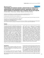
Intracellular expression of HTLV-1 Env chimeras and soluble SU subdomainsFigure 3
Intracellular expression of HTLV-1 Env chimeras and
soluble SU subdomains. Cell extracts (A, B) or culture
supernatants (C) were prepared from 293T cells transfected
with either full length Env (A) or soluble SU subdomains (B,
C) expression vectors as depicted in figure 2. Membranes
were probed with either (A) an anti-MLV SU antiserum to
detect F-MLV and H1
183
FEnv uncleaved Env precursor pro-
teins (F-MLV Prgp85 and H1
183
Fenv Pr, respectively) indi-
cated by arrowheads, and cleaved SU (F-MLV SUgp70 and
H1
183
FEnv SU, respectively) indicated by circles, or (B, C) an
anti-rabbit IgG antiserum to detect carboxy terminal rFc-
tagged soluble subdomains, including the Ampho-MLV SU
subdomain (A
397
SU).
B
C
A
H2 SU
178
H2 SU
211
Mock
WB # 65
H1 SU
215
A SU
397
Soluble SU subdomains in cell extracts
NM WB (Trnfxn #41)
H1 SU
215
H2 SU
211
H2 SU
178
Mock
A SU
397
Soluble SU subdomains in culture medium
Mock
H1
FEnv
183
F-MLV
F-MLV Pr gp85
F-MLV SU gp70
H1 FEnv SU
183
H1 FEnv Pr
183
Full length Env
Retrovirology 2004, 1:41 />Page 6 of 14
(page number not for citation purposes)
the entire HTLV Env RBD. These data also showed that
HTLV-1 and 2 cross-interfered, consistent with the fact
that they recognize the same cell surface receptor for infec-
tion [8,32].
Interference to HTLV Env-mediated cell-to-cell fusion by
HTLV SU amino terminal subdomains
Viral envelope interference occurs when cell surface recep-
tors are occupied by receptor-interacting Env components
[33-35]. Since interference to the different Env-mediated
functions involves distinct components [27-29], we also
tested the abilities of the H1
183
FEnv and the HTLV SU
amino terminal subdomains to interfere with HTLV Env-
mediated cell fusion. Interference to cell fusion was meas-
ured using a quantitative HTLV envelope cell fusion inter-
ference assay (CFIA), as previously described [9].
HTLV-1 Env-induced cell fusion was significantly dimin-
ished upon expression of the H1
215
SU subdomain in tar-
get cells, 12% ± 2% of control fusion (P < 0.001),
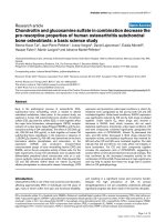
HTLV-1 and -2 SU subdomains interfere with HTLV Env SU cell surface bindingFigure 4
HTLV-1 and -2 SU subdomains interfere with HTLV Env SU cell surface binding. (A) Conditioned medium from
control 293T cells (open histograms) or from 293T cells expressing soluble rFc-tagged HTLV-1 H1
215
SU, HTLV-2 H2
211
SU and
H2
178
SU, or Ampho-MLV A
397
SU subdomains (filled histograms), were incubated with A23 hamster cells for 30' at 37°C and
binding was assessed by flow cytometry following addition of a secondary FITC-conjugated anti rabbit IgG antibody. Similar
results were obtained in binding assays performed using all cell lines described in the text. (B) To assess binding interference,
target 293T cells were transfected with the indicated Env construct and subsequently incubated with the HA-tagged H2
178
SU
domain (filled histograms). Binding was detected by FACS following incubation with an anti HA 12CA5 mouse mAb and a
FITC-conjugated anti mouse IgG antibody. Open histograms represent background levels of fluorescence. SU constructs are
schematically represented below each graph by solid (HTLV), open (F-MLV) or grey (Ampho-MLV) boxes.
H2
178
SU A
397
SU
Mock H1
215
SU H2
211
SU H2
178
SU A
397
SU
H1 FEnv
183
A
B
Interference to H2
178
SU binding
Cell surface binding
H2
211
SUH1
215
SU
Interfering Env or SU subdomain
Retrovirology 2004, 1:41 />Page 7 of 14
(page number not for citation purposes)
consistent with previous observations using the
H1
215
FEnv chimera [9]. Significant interference to cell
fusion was also observed with the H1
183
FEnv chimera,
which lacked a PRRH, down to 26% ± 4% of control
fusion (P < 0.001) (Figure 5). The corresponding HTLV-2
SU subdomains produced a nearly identical cell fusion
interference profile: interference by the H2
211
SU isolated
domain, in which the PRRH was maintained, resulted in
15% ± 3% of control cell fusion levels, while the H2
178
SU
subdomain, lacking the HTLV PRRH, inhibited HTLV-1
Env-induced cell fusion to 24% ± 6% of control levels (P
< 0.001) (Figure 5). It is noteworthy that similar data were
obtained when comparing cell fusion interference by
H1
215
FEnv and H1
183
FEnv. These effects were specific to
HTLV SU amino terminal domains as A
397
SU did not
interfere with HTLV-1 Env-mediated cell fusion (83% ±
11% of control fusion) (Figure 5). Furthermore, no inter-
ference was observed when these truncated HTLV SU frag-
ments and chimeric Env were tested against heterologous,
fusogenic control Env such as A∆R Env, F∆R, Xeno∆R and
VSVG (data not shown). Altogether, these results con-
firmed our findings that receptor-binding determinants
are present within the first 183 and 178 amino acids of the
HTLV-1 and -2 Env, respectively. They also indicated that
the PRRH (H1
215
SU and H2
211
SU), although unnecessary
for receptor binding, modulates the efficiency of interfer-
ence to HTLV Env-induced cell-to-cell fusion (P < 0.03).
Interference to HTLV Env-mediated infection by HTLV SU
amino terminal subdomains
Interference, as described above, was based on the inhibi-
tion of cell-to-cell fusion induced by fusogenic Env
expressed in the absence of other viral proteins. We fur-
ther evaluated the abilities of the Env chimeras and solu-
ble subdomains to specifically interfere with HTLV Env-
mediated infection. HTLV Env-pseudotyped MLV virions,
MLV(HTLV), were produced to infect 293T target cells.
Because these recombinant cell-free virions are not com-
petent for replication, this viral pseudotype infection
assay tests a single round of infection, and does not meas-
ure replication and subsequent exponential viral dissemi-
nation. Therefore, relative infection values are expressed
in linear rather than logarithmic scales.
Infection of mock-transfected target cells, devoid of inter-
fering Env domains, resulted in a mean infection value of
9905 ± 1117 infectious units per ml (iu/ml), and this was
taken as 100% control infection (Figure 6). Similar values,
8803 ± 1871 iu/ml or 89% ± 19% of control infection,
were obtained upon infection of target cells expressing a
heterologous SU subdomain, A
397
SU (Figure 6). Expres-
sion of the H1
183
FEnv and H1
215
FEnv chimeric Env in tar-
get cells significantly reduced MLV(HTLV) infection to
324 ± 98 iu/ml, 3.3% ± 1% of control infection, and to
307 ± 129 iu/ml, 3.1% ± 1.3% of control infection, respec-
tively (Figure 6 and data not shown). Similarly, the
H2
178
SU and H2
211
SU subdomains diminished
MLV(HTLV) infection to 191 ± 56 iu/ml and 215 ± 122 iu/
ml, 1.9% ± 0.6% and 2.2% ± 1.3% of control infection,
respectively (Figure 6). The specificity of interference to
infection by HTLV Env constructs was assessed by their
lack of interference abilities toward Ampho-MLV Env-
pseudotyped virions, MLV(Ampho) (data not shown).
Thus, for both HTLV-1 and -2, the amino terminal
domain upstream of the PRRH was sufficient for specific
interference to HTLV Env-mediated infection. Further-
more, in contrast to the cell fusion interference assays
described above, the PRRH did not detectably influence
MLV(HTLV) infection.
HTLV-1 and -2 SU subdomains interfere with HTLV Env-mediated cell fusionFigure 5
HTLV-1 and -2 SU subdomains interfere with HTLV
Env-mediated cell fusion. Cell-to-cell fusion assays were
performed by cocultivating fusogenic HTLV-1 Env-expressing
cells with target cells expressing the Env derivatives indicated
and schematically represented below each histogram. HTLV-
1 Env-mediated cell fusion in the presence of target cells
transfected with empty vector (Mock) yielded 200 to 1000
blue foci in 4 independent experiments and these levels were
defined as 100% cell fusion. Cell fusion levels in the presence
of HLTV SU mutants or the A
397
SU control Ampho-MLV SU
subdomain is shown as percent of control. Mean fusion per-
centages were determined from three to four independent
experiments. Error bars represent the standard error of the
mean.
Interfering Env or SU subdomain
Cell fusion (% control fusion)
Interference to HTLV Env-mediated cell fusion
Mock
H1 SU
215
H2 SU
211
H2 SU
178
100
26 ±4
0
20
40
60
80
100
120
12 ±2
15 ±3
24 ±6
83 ±11
A SU
397
H1
FEnv
183
Retrovirology 2004, 1:41 />Page 8 of 14
(page number not for citation purposes)
Because HTLV dissemination appears to occur mostly via
cell-to-cell contact, we also tested envelope interference to
infection by HTLV-1 SU amino terminal domains using a
cell-to-cell transmission interference assay. In this assay,
cells harboring interfering chimeric Env and soluble sub-
domains were cocultured with cells producing
MLV(HTLV) virions. Transfection of either chimeric Env
or soluble subdomains into HeLa target cells decreased
MLV(HTLV) infection to levels similar to those observed
in the cell fusion interference assay presented in figure 5
(data not shown).
Identification of residues within the HTLV SU amino
terminal domain that modulate receptor binding and
HTLV Env-mediated interference
Two key residues contained in the HTLV SU RBD and con-
served between HTLV-1 and -2, arginine 94 (Arg
94
) and
serine 101 (Ser
101
) for HTLV-1 Env which correspond to
Arg
90
and Ser
97
in HTLV-2 Env, have been shown to alter
cell-to-cell fusion and infection when mutated [36,37]. To
determine whether mutations of these residues had an
effect on receptor binding, we generated H1
215
SU sub-
domains with either Arg
94
or Ser
101
mutated to Ala, yield-
ing the mutant H1(R94A)SU and H1(S101A)SU
subdomains, respectively. We also evaluated mutations of
Asp
106
, mutant H1(D106A)SU, and Tyr
114
, mutant
H1(Y114A)SU, both residues found to be highly con-
served between all human and simian T cell leukemia
viruses (unpublished observations). Surprisingly, cell sur-
face binding profiles of H1(R94A)SU and H1(S101A)SU
mutants were not significantly altered when compared to
binding by the parental H1
215
SU, whereas the
H1(D106A)SU mutant presented reduced binding to
HTLV receptor-bearing cells and the H1(Y114A)SU
mutant showed a nearly complete abrogation of cell sur-
face binding (Figure 7A). Loss of binding observed with
the two latter mutants was not due to decreased soluble
SU fragment production, as assessed by immunoblotting
of transfected-cell culture media (Figure 7A). Moreover,
equivalent binding profiles were obtained when the same
mutations were introduced into the HTLV-2 soluble RBD
H2
178
SU (data not shown). Altogether, these experiments
demonstrated that Tyr
114
, and to a lesser extent Asp
106
, are
key residues involved in HTLV Env receptor binding.
We next tested the abilities of these mutants to interfere
with HTLV Env-mediated cell fusion and infection, using
the assays described above. As mentioned above, all wild-
type and mutant HTLV SU subdomains were produced
and secreted with a similar efficiency (Figure 7A).
Expression of the H1(D106A)SU and H1(Y114A)SU
mutants, with decreased capacities to bind the HTLV
receptor, correlated with decreased interference to HTLV
Env-mediated cell fusion and infection. Indeed,
H1(Y114A)SU, which had nearly undetectable level of
binding, showed the lowest levels of interference and thus
allowed the highest levels of HTLV Env-mediated cell
fusion and infection (56% ± 16% and 46% ± 10%, respec-
tively) (Figure 7). Nevertheless, levels of fusion and infec-
tion were lower than that observed when the heterologous
A
397
SU was used as a negative control of interference
(83% ± 11% and 89% ± 19% for cell fusion and infection,
respectively). Thus, overexpression of mutant HTLV SU
fragments with highly decreased receptor binding abilities
can still exert, albeit to a significantly lesser extent, inter-
ference to HTLV Env-mediated cell fusion and infection.
We found that similar levels of interference to HTLV Env-
mediated cell fusion and infection were observed when
either the parental H1
215
SU or the mutant H1(S101A)SU
were expressed in target cells (Figure 7B and 7C). This is
consistent with the capacity of this mutant to bind target
HTLV-1 and -2 SU subdomains interfere with infection by HTLV envelope-pseudotyped virionsFigure 6
HTLV-1 and -2 SU subdomains interfere with infec-
tion by HTLV envelope-pseudotyped virions. 293T
cells (5 × 10
5
) expressing the indicated interfering Env deriv-
atives were infected with cell-free HTLV-2 Env-pseudotyped
virions MLV(HTLV) carrying a LacZ reporter gene. Infected
cells were detected 2 days later by X-gal staining. Infection
values are represented as percent of control infection, i.e.,
relative to infection of mock (pCDNA3.1) transfected target
cells, calculated as infectious units per ml of virus containing
supernatant (i.u./ml). Data are representative of at least three
independent experiments performed in duplicate. Error bars
represent the standard error of the mean.
3.3 ±1
1.9 ±0.6 2.2 ±1.3
100
89 ±19
0
20
40
60
80
100
120
Interfering Env or SU subdomain
Relative infection (% control)
1.5 ±0.9
Mock
H1 SU
215
H2 SU
211
H2 SU
178
A SU
397
H1
FEnv
183
Retrovirology 2004, 1:41 />Page 9 of 14
(page number not for citation purposes)
cells at levels similar to that of wild type H1
215
SU. How-
ever, interference to HTLV Env-mediated cell fusion and
infection did not always correlate with cell surface bind-
ing profiles. While the H1(R94A)SU mutant inhibited cell
fusion and infection, its effects were significantly lower
than those of the wild-type H1
215
SU (56% ± 8% and 32%
± 2.3%, respectively) (Figure 7B,7C). Thus, although nei-
ther Arg
94
nor Ser
101
of the HTLV-1 SU appears to play a
direct role in binding, Arg
94
modulates HTLV Env-medi-
ated fusion and infection (Figure 7), likely via post-bind-
ing effects rather than binding per se. In conclusion,
Tyr114 appeared as the main determinant identified so far
for HTLV Env binding, whereas the effects previously
described with Arg
94
and Ser
101
are most likely associated
with post-binding events.
Discussion
Here, we report the generation of MLV Env with chimeric
HTLV/MLV SU and truncated HTLV-1 and -2 amino termi-
nal SU subdomains that can be expressed in and secreted
from eukaryotic cell lines in functional, soluble form.
Using these constructs, we demonstrated that the amino
terminal 163 and 158 residues (i.e., expunged of their Env
signal peptide) of the mature HTLV-1 and -2 Env SU,
respectively, were sufficient to exert both HTLV receptor
binding and efficient interference to diverse HTLV Env-
mediated functions, including binding, cell-to-cell fusion
and cell-free as well as cell-to-cell infection. Although the
PRRH sequence comprising amino acid residues 180 to
215 of the HTLV-1 Env and 176 to 211 of the HTLV-2 Env
was previously thought to be a receptor binding site, our
HTLV-1 SU amino terminal domain mutantsFigure 7
HTLV-1 SU amino terminal domain mutants. (A) H1
215
SU constructs were generated with the following SU amino ter-
minal point mutations; R94A, S101A, D106A and Y114A. The abilities of these soluble H1
215
SU constructs to bind 293T cells
were assessed by flow cytometry (gray histograms). The levels of expression of the various soluble SU subdomains are shown
under each histogram. The abilities of the H1
215
SU mutants to interfere with (B) HTLV Env-induced cell fusion and (C)
MLV(HTLV) pseudotype infection was assayed as described in Figs. 5 and 6. Data are representative of at least three independ-
ent experiments performed in duplicate. Error bars represent the standard error of the mean.
H1
215
SU H1(S101A)SUH1(R94A)SU H1(D106A)SU H1(Y114A)SU
100
0
20
40
60
80
100
120
Mock
H1(D106A)SU
H1(Y114A)SU
H1 SU
215
A SU
397
H1(R94A)SU
H1(S101A)SU
Interference to fusion
B
Mock
H1(D106A)SU
H1(Y114A)SU
H1 SU
215
A SU
397
H1(R94A)SU
H1(S101A)SU
0
20
40
60
80
100
120
Interference to infection
C
A
1.5 ±0.9
32 ±2.3
25 ±8
2 ±0.7
46 ±10
89 ±19
10083 ±11
38 ±0.4
56 ±16
12 ±2
56 ±8
16 ±0.3
Cell surface binding
Relative infection (% control)
Cell fusion (% control fusion)
Interfering Env or SU subdomain
HTLV-1 SU subdomains with a single amino acid mutation
Retrovirology 2004, 1:41 />Page 10 of 14
(page number not for citation purposes)
data preclude a major role for this region in the binding
properties described above. Indeed, whereas a synthetic
peptide composed of amino acids 197 to 216 and located
within the HTLV-1 PRRH, has been reported to interfere
with HTLV Env-induced syncytia formation [22], this pep-
tide was later shown to compete neither with receptor
binding of the entire HTLV-1 Env SU [38], nor with infec-
tion [26]. It is therefore likely that the effects reported for
PRRH-derived peptides, as measured by syncytia forma-
tion, are solely due to post-receptor binding events. How-
ever, we identified Tyr
114
of the HTLV-1 Env, which
corresponds to Tyr
110
of the HTLV-2 Env, as a key residue
in HTLV Env binding and for all the aforementioned
HTLV Env-mediated functional assays. We could not
detect binding of H1(Y114A)SU by flow cytometry, while
this mutant exerted residual, albeit significantly
decreased, interference to HTLV Env-mediated cell fusion
and infection. Altered folding outside of the binding
domain per se, rather than direct alteration of the receptor-
binding site, could also account for the lack of binding of
this mutant. However, we favor the latter hypothesis,
since the H1(Y114A)SU mutant was properly folded and
transported to the plasma membrane and secreted in the
medium as efficiently as wild type RBD, thus arguing
against gross misfolding of this mutant. Accordingly,
Tyr
114
appears to be conserved in all known human and
simian T cell leukemia viruses strains, which share the
same receptor.
The receptor-binding site in MLV RBD is composed of a
combination of several cysteine loops located upstream of
the PRR [11,39] which is linked to a conserved anti-paral-
lel β core [13]. The isolation of an F-MLV SU amino termi-
nal subdomain allowed crystallization of MLV RBD and
the modeling of the RBD cysteine loop arrangement [13].
The precise organization of cysteine loops, likely to har-
bor the receptor binding determinants, within the HTLV
SU amino terminus remains to be established. Neverthe-
less, the identification of Tyr
114
as a key HTLV-1 RBD resi-
due points at this determinant as a very likely receptor-
binding core. This, together with previous works relying
on syncytia formation and cell-to-cell transmission
[36,37], will help to distinguish between bona fide recep-
tor binding determinants and determinants involved at a
post-binding level.
Another recently identified determinant, the Pro-His-Gln
SU motif conserved among gammaretroviruses such as
MLV and feline leukemia viruses (FeLV), has been deter-
mined to play a major role in viral entry during post-bind-
ing events [40]. The mechanism of this effect involves a
direct interaction of MLV SU soluble forms with Env
attached SU carboxy terminus [41-46]. This interaction
between the SU amino and carboxy termini leads to the T
cell-restricted tropism of a natural isolate of FeLV, FeLV T,
in which the SU Pro-His-Gln motif is mutated. Indeed,
FeLV T is restricted in cat to T cells because they naturally
express an endogenous soluble FeLV RBD-related factor
called FeLIX that trans-complements the lack of the SU
Pro-His-Gln motif in the FeLV T Env and restores its post-
binding defect [47]. Despite the HTLV-1 and F-MLV SU
homologous modular organization and the assignment of
several common motifs between the two latter SU, no
obvious Pro-His-Gln motif homologue is present in the
HTLV SU amino terminus. Whether a FeLIX-like molecule
that interacts with HTLV Env exists in human T cells
remains to be addressed. Furthermore, the fact that the
Pro-His-Gln has been shown to play a major role in trans-
activation of viral infection in several gammaretroviruses
which are efficiently infectious as cell-free virions
[42,44,48], raises the question whether the apparent lack
of such a motif in the HTLV simple oncovirus-like SU is
linked to the relative inefficiency of HTLV Env-mediated
infection by cell-free virions. The HTLV SU subdomains
described here should prove to be valuable in addressing
such questions.
The recent identification of Glut1, the ubiquitous glucose
transporter of vertebrates [49], as a receptor for HTLV Env
[8] adds an additional similarity between the Env of
HTLV, a deltaretrovirus, and that of gammaretroviruses.
All these virus Env recognize multimembrane-spanning
metabolite transporters [50,51]. This and the common
modular organization of the HTLV and MLV SU raise
questions regarding the origin of the HTLV Env. It has pre-
viously been reported that envelopes of invertebrate retro-
viruses may have been "captured" from other viruses [52-
54]. As HTLV and MLV have strongly divergent overall
genomic organizations, "envelope capture" from related
ancestor genes might account for the close relationship
between the Env of these phylogenetically distant viruses
[10].
Conclusions
We have generated truncated domains of the HTLV Env
amino terminus, upstream of residues 183 and 178 of the
HTLV-1 and -2 Env, respectively, that were sufficient to
bind target cells of different species through interaction
with the HTLV Env receptor. We also identified a tyrosine
at position 114 and 110 in HTLV-1 and -2 Env, respec-
tively, as a key determinant for this binding. In addition
to their use for further exploration of the mechanisms
involved in HTLV entry, the tagged HTLV-1 and -2 RBD
subdomains described here are novel tools for the detec-
tion of Glut1 cell surface expression and intracellular traf-
ficking. Indeed, we tracked intracellular expression of
EGFP-tagged HTLV SU subdomains by time-lapse micros-
copy, and found that they are preferentially routed toward
cell-cell contact areas (unpublished observations), where
Glut1 is particularly abundant [55] and our unpublished
Retrovirology 2004, 1:41 />Page 11 of 14
(page number not for citation purposes)
observations). Furthermore, those HTLV SU derivatives
could be of particular importance in view of the key roles
played by Glut1 in various biological processes, including
T cell survival and activation [31,56], tumor genesis
[57,58], and neuronal activity [59]. Interestingly, soluble
HTLV SU subdomains inhibit Glut1-mediated glucose
transport, and accordingly, expression of mutants with
diminished receptor binding ability resulted in less pro-
nounced inhibition [8] and data not shown). Thus, these
HTLV SU derivatives could also be used as glucose trans-
port inhibitors. These data demonstrate the potential for
the novel and broad utility of these reagents in the study
of HTLV infection as well as biological processes involving
glucose transport and metabolism.
Materials and methods
Construction of chimeric Env and HTLV-1 and -2 SU
subdomains
To exchange the PRR and PRRH regions, we introduced an
allelic MfeI restriction site in the HTLV-1 and F-MLV Env.
Introduction of this site in F-MLV resulted in the substitu-
tion of a glutamine and leucine (QL) dipeptide for the
parental arginine and valine (RV) residues of the GPRV-
PIGP motif, at the start of the MLV Env PRR. Introduction
of the MfeI site in the PSQL motif of the HTLV-1 SU main-
tained the parental QL residues, at the start of the HTLV
Env PRRH. By exchanging domains at the MfeI sites, we
derived the H1
183
FEnv chimera containing the amino ter-
minal 183 residues of the HTLV Env followed by the F-
MLV PRR. In this chimera, the PSQL/PIGP hybrid
sequence is generated at the exchange border, and the
PRRH of HTLV is replaced by the F-MLV PRR (Figure 2A).
In contrast, the entire PRRH of HTLV-1 is present in the
H1
215
FEnv chimera – this Env chimera has been previ-
ously described and designated HHproFc [9]. The
H1
183
FEnv and H1
215
FEnv chimeras, as well as the paren-
tal HTLV-1 and F-MLV Env, were inserted in an allelic
fashion into the previously described pCEL retroviral Env
expression vector [60]. The HTLV-2 Env expression vector,
pCSIX/H2, was constructed by inserting the HindIII –
EcoRI fragment from pHTE-2 (a gift from M-C Dokhelar)
encompassing the HTLV-2 env gene, the pX region and the
3' LTR into pCSI (CMV promoter, SV-40 intron) [61] at
the HindIII and EcoRI restriction sites.
The H1
215
SU, H2
211
SU, H1
179
SU, and H2
178
SU sub-
domains, corresponding to the HTLV-1 and -2 SU amino
terminus with and without their respective PRRH, were
generated by PCR and subcloned into the pCSI expression
vector as fusion proteins harboring a carboxy terminal rFc
or HA tag (Figure 2B). The H1(R94A)SU, H1(S101A)SU,
H1(D106A)SU, and H1(Y114A)SU substitution mutants
were generated by oligonucleotide-directed PCR muta-
genesis on the H1
215
SU vector and subcloned into the
pCSI expression vector. All PCR-generated DNA fragments
were sequenced using an ABI Prism 310 sequencer. Clon-
ing details are available upon request.
Protein expression and immunoblots
Approximately 5 × 10
5
293T cells per 35 mm well were
transfected with 5 µg of vectors using a calcium-phos-
phate-Hepes buffered saline (HBS) transfection protocol.
Transfection medium was replaced with 3 ml of fresh cul-
ture medium twenty hours post-transfection. Forty-eight
hours post-transfection cell culture medium (superna-
tant) was recovered and filtered through a 0.45 µm pore-
size membrane to remove cell debris. Twenty µl were
directly analyzed by SDS-PAGE (15% polyacrylamide
gel), and the rest was aliquoted and stored at -20°C for
later use in binding assays (see below). Cell extracts were
collected 48 h post-transfection in 1 ml of cell lysis buffer
(50 mM Tris-HCl [pH 8.0], 150 mM NaCl, 0.1% sodium
dodecyl sulfate [SDS], 1% Nonidet P-40, 0.5% deoxycho-
late, and a cocktail of mammalian protease inhibitors
[Sigma]) and clarified by two successive centrifugations at
13,000 rpm for 10 min at 4°C in a microcentrifuge.
Approximately 20 µl of each extract, adjusted after nor-
malization for protein concentration using the Bradford
assay (Sigma), were subjected to electrophoresis on SDS-
15% acrylamide gels, followed by transfer onto nitrocellu-
lose (Protran; Schleicher & Schuell). Membranes were
blocked in phosphate-buffered saline (PBS) containing
5% powdered milk and 0.5% Tween 20, probed with a
1:1000 dilution of a goat anti-RLV gp70 polyclonal
antibody (Viromed) followed by a horseradish peroxi-
dase-conjugated anti-goat immunoglobulin (for detec-
tion of chimeric Env), or goat anti-rabbit-IgG-horseradish
peroxidase-conjugated immunoglobulins (for detection
of rFc-tagged SU subdomains). Immunoblots were subse-
quently washed three times with PBS-0.1% Tween 20 and
revealed by chemiluminescence (ECL+, Amersham).
Binding and binding interference assays
Binding assays were performed as previously described
[31]. Briefly, 5 × 10
5
target cells were detached with a PBS-
EDTA solution, collected by centrifugation, incubated for
30' at 37°C with 300 µl of rabbit Fc-tagged soluble HTLV-
1, HTLV-2, or Ampho-MLV truncated SU, washed, labeled
with an anti-rabbit-IgG FITC-conjugated antibody, and
analyzed on a FACSCalibur (Becton Dickinson). Data
analysis was performed using the CellQuest software
(Becton Dickinson). For interference studies, 293T cells
were transfected with 4 µg of Env or Env SU subdomain
expression vectors (carboxy terminal rFc-tagged forms)
using the calcium-phosphate-HBS method. Under these
conditions, transfection efficiencies ranged from approxi-
mately 80 to 90% of the target cells. Twenty-four and 48
hours post-transfection, cells were collected and trans-
fected 293T cells expressing the different interfering HTLV
or Ampho-MLV domains were incubated with a
Retrovirology 2004, 1:41 />Page 12 of 14
(page number not for citation purposes)
challenging HA-tagged soluble HTLV-2 SU amino termi-
nal subdomain (H2
178
SU-HA). Cells were stained using a
primary 12CA5 anti HA antibody followed by an anti-
mouse-IgG FITC-conjugated antibody before detection by
flow cytometry.
Envelope interference to cell fusion assay
Briefly, the HTLV/MLV Env chimera, H1
183
FEnv, was used
to interfere with challenging HTLV Env. The interfering
non-fusogenic H1
183
FEnv and truncated HTLV SU sub-
domains were transiently transfected into
HeLaCD4LTRLacZ, a cell line highly susceptible to HTLV
Env-induced fusion that contains a stably integrated Tat-
dependent LacZ expression vector [62]. These transfect-
ants were cocultured with Tat-expressing NIH3T3(TK-)
cells (NIH3T3(TK-)Tat) that were transiently transfected
with the challenging HTLV Env. The NIH3T3(TK-)Tat cell
line is resistant to HTLV-Env-induced syncytia formation,
despite its ability to express the HTLV receptor and to bind
HTLV Env, and thus can be used to precisely monitor
fusion of the HeLaCD4LTRLacZ target cells [9,29].
H1
183
FEnv Env and truncated HTLV SU subdomains
plasmid DNA (2 to 3 µg) was transfected into
HeLaCD4LTRLacZ cells, while challenging, fusogenic
HTLV-1 Env plasmid (1 µg) was transfected into
NIH3T3(TK-)Tat. The interfering Env or SU subdomain-
presenting cells were detached 24 hours post-transfection
and 1–2 × 10
5
cells were cocultured for 24 hours with 1–
2 × 10
5
challenging HTLV-1 Env-presenting NIH3T3(TK-
)Tat cells. Subsequently, the cocultured cells were fixed
and stained for β-galactosidase expression as described
previously [60]. Transfection efficiencies of the
HeLaCD4LTRLacZ target cells were approximately 50%.
Mock transfections were performed with similar amounts
of control plasmid DNAs. Env interference was measured
by the decreased number of blue foci and was expressed as
percent blue foci of control fusion (mock-transfected
target cells). Data are represented as mean interference (±
standard deviation), and statistical significance of interfer-
ence levels was determined using a pairwise Student's t
test.
Envelope interference to infection assay
MLV(Ampho) and MLV(HTLV) pseudotyped virions were
produced after transfection of 10
6
293T cells with 5 µg
pCSI/Ampho or pCSIX/H2, respectively, 5 µg pCL/Gag-
Pol [29] and 10 µg of pCLMFG-LacZ [63], using a calcium-
phosphate-HBS transfection protocol. Supernatants were
recovered 48 hours post transfection and filtered through
0.45 µm pore-size membrane to remove cell debris, and
stored at -80°C. The pCLMFG-LacZ plasmid is a retroviral
expression vector that provides a packageable RNA coding
for the LacZ gene marker. pCSI/Ampho is an expression
vector encoding the Ampho-MLV Env, and the HTLV-2
Env expression vector, pCSIX/H2, is described above.
Virion-containing supernatants were used to infect target
293T cells expressing the chimeric Env or HTLV RBD
subdomains. Transfection efficiencies of target 293T cells
were >80% in all experiments. Infections were performed
36–48 hours post-transfection on cultures grown in 12
well plates (Costar) at 37°C, medium was changed 24
hours later, and confluent cell monolayers were fixed,
stained for β-galactosidase activity before counting blue
foci. Interference to infection was determined by infecting
transfected target cells with approximately 100 and 1000
iu. Infection was evaluated as described above, and the
number of LacZ-positive blue colonies counted was nor-
malized by multiplying by the appropriate dilution factor.
The resulting infection values were analyzed as iu/ml of
virus containing supernatant. Subsequently the relative
infection levels in cells expressing the HTLV SU domains
were compared to those of mock transfected cells and
were expressed as percentages of control infection (%
control).
List of abbreviations used
HTLV Human T-cell leukemia virus
SU envelope extracellular surface component
Env envelope glycoprotein
MLV murine leukemia virus
F-MLV Friend-MLV
RBD receptor-binding domain
PRR proline-rich region
PRRH proline rich region homologue
Ampho amphotropic
HA influenza hemagglutinin
rFc rabbit immunoglobulin constant fragment
A
397
SU Ampho-MLV Env fused to a carboxy terminal rFc
tag
CFIA cell fusion interference assay
iu/ml infectious units per ml
Arg
94
arginine 94
Ser
101
serine 101
Tyr
114
tyrosine 114
Retrovirology 2004, 1:41 />Page 13 of 14
(page number not for citation purposes)
FeLV feline leukemia viruses
HBS Hepes buffered saline
PBS phosphate-buffered saline
SDS sodium dodecyl sulfate
Competing interests
The authors declare that they have no competing interests.
Authors' contributions
FJK designed and realized or supervised most of the exper-
iments and co-wrote the manuscript. NM participated to
some molecular constructions, set up, realized and ana-
lyzed most binding assays and FACS analyses and partici-
pated to the redaction of the manuscript. ENG set up and
performed the cell-to-cell transmission assay and per-
formed the corresponding experiments, CV constructed
some of the RBD point mutants and tested them, MS ini-
tiated the project, co-participated in the design of the
study, co-coordinated its realization and co-wrote the
manuscript, and JLB realized some of the molecular con-
structs, performed some of the experiments, co-partici-
pated in the design of the study, co-coordinated its
realization and co-wrote the manuscript. All authors read
and approved the final manuscript.
Acknowledgements
We thank N. Taylor for helpful discussion and critical reading of the man-
uscript, G. Labesse for his help in protein sequence analyses, R.K. Naviaux
for the gift of pCL-Eco and pMFG-LacZ plasmids, J.A. Young for the rabbit
Fc plasmid, J C. Dantonel for the anti-HA antibody, F. Carbonell for tech-
nical assistance, and all the members of our laboratory for insightful discus-
sion. FJK was supported by an award from the Philippe Foundation and
successive fellowships from the Agence Nationale pour la Recherche con-
tre le SIDA (ANRS), the Association pour la Recherche contre le Cancer
(ARC), and the Fondation de France. NM is supported by a graduate stu-
dent fellowship from the MRT. JLB and MS are supported by the Institut
National de la Santé et de la Recherche Médicale (INSERM). This work was
supported by grants from ARC (ARC Nos. 5989 and 3424), Fondation de
France (Nos. 2291 and 2138) and Association Française contre les Myopa-
thies (AFM No.7706) to MS.
References
1. Richardson JH, Edwards AJ, Cruickshank JK, Rudge P, Dalgleish AG:
In vivo cellular tropism of human T-cell leukemia virus type
1. J Virol 1990, 64:5682-5687.
2. Hanon E, Stinchcombe JC, Saito M, Asquith BE, Taylor GP, Tanaka Y,
Weber JN, Griffiths GM, Bangham CR: Fratricide among CD8(+)
T lymphocytes naturally infected with human T cell lympho-
tropic virus type I. Immunity 2000, 13:657-664.
3. Nagai M, Brennan MB, Sakai JA, Mora CA, Jacobson S: CD8(+) T
cells are an in vivo reservoir for human T-cell lymphotropic
virus type I. Blood 2001, 98:1858-1861.
4. Wang TG, Ye J, Lairmore MD, Green PL: In vitro cellular tropism
of human T cell leukemia virus type 2. AIDS Res Hum Retroviruses
2000, 16:1661-1668.
5. Sutton RE, Littman DR: Broad host range of human T-cell leuke-
mia virus type 1 demonstrated with an improved pseudotyp-
ing system. J Virol 1996, 70:7322-7326.
6. Okuma K, Nakamura M, Nakano S, Niho Y, Matsuura Y: Host range
of human T-cell leukemia virus type I analyzed by a cell
fusion-dependent reporter gene activation assay. Virology
1999, 254:235-244.
7. Trejo SR, Ratner L: The HTLV receptor is a widely expressed
protein. Virology 2000, 268:41-48.
8. Manel N, Kim FJ, Kinet S, Taylor N, Sitbon M, Battini JL: The Ubiq-
uitous Glucose Transporter GLUT-1 Is a Receptor for
HTLV. Cell 2003, 115:449-459.
9. Kim FJ, Seiliez I, Denesvre C, Lavillette D, Cosset FL, Sitbon M: Def-
inition of an amino-terminal domain of the human T-cell
leukemia virus type 1 envelope surface unit that extends the
fusogenic range of an ecotropic murine leukemia virus. J Biol
Chem 2000, 275:23417-23420.
10. Kim FJ, Manel N, Battini JL, Sitbon M: Emergence of vertebrate
retroviruses and envelope capture. Virology 2004, 318:183-191.
11. Battini JL, Heard JM, Danos O: Receptor choice determinants in
the envelope glycoproteins of amphotropic, xenotropic, and
polytropic murine leukemia viruses. J Virol 1992, 66:1468-1475.
12. Battini JL, Danos O, Heard JM: Receptor-binding domain of
murine leukemia virus envelope glycoproteins. J Virol 1995,
69:713-719.
13. Fass D, Davey RA, Hamson CA, Kim PS, Cunningham JM, Berger JM:
Structure of a murine leukemia virus receptor-binding glyc-
oprotein at 2.0 angstrom resolution. Science 1997,
277:1662-1666.
14. Koch W, Hunsmann G, Friedrich R: Nucleotide sequence of the
envelope gene of Friend murine leukemia virus. J Virol 1983,
45:1-9.
15. Gallaher WR, Ball JM, Garry RF, Martin-Amedee AM, Montelaro RC:
A general model for the surface glycoproteins of HIV and
other retroviruses. AIDS Res Hum Retroviruses 1995, 11:191-202.
16. Sitbon M, d'Auriol L, Ellerbrok H, André C, Nishio J, Perryman S,
Pozo F, Hayes SF, Wehrly K, Tambourin P, Galibert F, Chesebro B:
Substitution of leucine for isoleucine in a sequence highly
conserved among retroviral envelope surface glycoproteins
attenuates the lytic effect of the Friend murine leukemia
virus. Proc Natl Acad Sci USA 1991, 88:5932-5936.
17. Andersen KB: A domain of murine retrovirus surface protein
gp70 mediates cell fusion, as shown in a novel SC-1 cell fusion
system. J Virol 1994, 68:3175-3182.
18. Lavillette D, Maurice M, Roche C, Russell SJ, Sitbon M, Cosset FL: A
proline-rich motif downstream of the receptor binding
domain modulates conformation and fusogenicity of murine
retroviral envelopes. J Virol 1998, 72:9955-9965.
19. Tanaka Y, Zeng L, Shiraki H, Shida H, Tozawa H: Identification of a
neutralization epitope on the envelope gp46 antigen of
human T cell leukemia virus type I and induction of neutral-
izing antibody by peptide immunization. J Immunol 1991,
147:354-360.
20. Tanaka Y, Tanaka R, Terada E, Koyanagi Y, Miyano-Kurosaki N,
Yamamoto N, Baba E, Nakamura M, Shida H: Induction of antibody
responses that neutralize human T-cell leukemia virus type
I infection in vitro and in vivo by peptide immunization. J Virol
1994, 68:6323-6331.
21. Londos-Gagliardi D, Jauvin V, Armengaut MH, Astier-Gin T, Goetz M,
Huet S, Guillemain BJ: Influence of amino acid substitutions on
antigenicity of immunodominant regions of the HTLV type I
envelope surface gylcoprotein: a study using monoclonal
antibodies raised against relevant peptides. AIDS Res Hum
Retroviruses 1999, 15:909-920.
22. Sagara Y, Inoue Y, Shiraki H, Jinno A, Hoshino H, Maeda Y: Identifi-
cation and mapping of functional domains on human T-cell
lymphotropic virus type 1 envelope proteins by using syn-
thetic peptides. J Virol 1996, 70:1564-1569.
23. Delamarre L, Pique C, Pham D, Tursz T, Dokhelar MC: Identifica-
tion of functional regions in the human T-cell leukemia virus
type I SU glycoprotein. J Virol 1994, 68:3544-3549.
24. Delamarre L, Rosenberg AR, Pique C, Pham D, Callebaut I, Dokhelar
MC: The HTLV-I envelope glycoproteins: structure and
functions. J Acquir Immune Defic Syndr Hum Retrovirol 1996,
13:S85-91.
25. Jassal SR, Pohler RG, Brighty DW: Human T-cell leukemia virus
type 1 receptor expression among syncytium-resistant cell
lines revealed by a novel surface glycoprotein-immunoad-
hesin. J Virol 2001, 75:8317-8328.
Retrovirology 2004, 1:41 />Page 14 of 14
(page number not for citation purposes)
26. Jinno A, Haraguchi Y, Shiraki H, Hoshino H: Inhibition of cell-free
human T-cell leukemia virus type 1 infection at a postbinding
step by the synthetic peptide derived from an ectodomain of
the gp21 transmembrane glycoprotein. J Virol 1999,
73:9683-9689.
27. Chung M, Kizhatil K, Albritton LM, Gaulton GN: Induction of syn-
cytia by neuropathogenic murine leukemia viruses depends
on receptor density, host cell determinants, and the intrinsic
fusion potential of envelope protein. J Virol 1999, 73:9377-9385.
28. Siess DC, Kozak SL, Kabat D: Exceptional fusogenicity of Chi-
nese hamster ovary cells with murine retroviruses suggests
roles for cellular factor(s) and receptor clusters in the mem-
brane fusion process. J Virol 1996, 70:3432-3439.
29. Kim FJ, Manel N, Boublik Y, Battini JL, Sitbon M: Human T-cell
leukemia virus type 1 envelope-mediated syncytium forma-
tion can be activated in resistant Mammalian cell lines by a
carboxy-terminal truncation of the envelope cytoplasmic
domain. J Virol 2003, 77:963-969.
30. Ott D, Rein A: Basis for receptor specificity of nonecotropic
murine leukemia virus surface glycoprotein gp70SU. J Virol
1992, 66:4632-4638.
31. Manel N, Kinet S, Battini JL, Kim FJ, Taylor N, Sitbon M: The HTLV
receptor is an early T-cell activation marker whose expres-
sion requires de novo protein synthesis. Blood 2003,
101:1913-1918.
32. Sommerfelt MA, Weiss RA: Receptor interference groups of 20
retroviruses plating on human cells. Virology 1990, 176:58-69.
33. Rein A: Interference grouping of murine leukemia viruses: a
distinct receptor for the MCF-recombinant viruses in mouse
cells. Virology 1982, 120:251-257.
34. Rein A, Schultz A: Different recombinant murine leukemia
viruses use different cell surface receptors. Virology 1984,
136:144-152.
35. Chesebro B, Wehrly K: Different murine cell lines manifest
unique patterns of interference to superinfection by murine
leukemia viruses. Virology 1985, 141:119-129.
36. Rosenberg AR, Delamarre L, Preira A, Dokhelar MC: Analysis of
functional conservation in the surface and transmembrane
glycoprotein subunits of human T-cell leukemia virus type 1
(HTLV-1) and HTLV-2. J Virol 1998, 72:7609-7614.
37. Delamarre L, Rosenberg AR, Pique C, Pham D, Dokhelar MC: A
novel human T-leukemia virus type 1 cell-to-cell transmis-
sion assay permits definition of SU glycoprotein amino acids
important for infectivity. J Virol 1997, 71:259-266.
38. Brighty DW, Jassal SR: The synthetic peptide P-197 inhibits
human T-cell leukemia virus type 1 envelope-mediated syn-
cytium formation by a mechanism that is independent of
Hsc70. J Virol 2001, 75:10472-10478.
39. Battini JL, Danos O, Heard JM: Definition of a 14-amino-acid pep-
tide essential for the interaction between the murine leuke-
mia virus amphotropic envelope glycoprotein and its
receptor. J Virol 1998, 72:428-435.
40. Bae Y, Kingsman SM, Kingsman AJ: Functional dissection of the
Moloney murine leukemia virus envelope protein gp70. J Virol
1997, 71:2092-2099.
41. Barnett AL, Cunningham JM: Receptor binding transforms the
surface subunit of the mammalian C-type retrovirus enve-
lope protein from an inhibitor to an activator of fusion. J Virol
2001, 75:9096-9105.
42. Barnett AL, Davey RA, Cunningham JM: Modular organization of
the Friend murine leukemia virus envelope protein underlies
the mechanism of infection. Proc Natl Acad Sci U S A 2001,
98:4113-4118.
43. Barnett AL, Wensel DL, Li W, Fass D, Cunningham JM: Structure
and mechanism of a coreceptor for infection by a pathogenic
feline retrovirus. J Virol 2003, 77:2717-2729.
44. Lavillette D, Ruggieri A, Russell SJ, Cosset FL: Activation of a cell
entry pathway common to type C mammalian retroviruses
by soluble envelope fragments. J Virol 2000, 74:295-304.
45. Lavillette D, Boson B, Russell SJ, Cosset FL: Activation of Mem-
brane Fusion by Murine Leukemia Viruses Is Controlled in
cis or in trans by Interactions between the Receptor-Binding
Domain and a Conserved Disulfide Loop of the Carboxy Ter-
minus of the Surface Glycoprotein. J Virol 2001, 75:3685-3695.
46. Lavillette D, Ruggieri A, Boson B, Maurice M, Cosset FL: Relation-
ship between SU subdomains that regulate the receptor-
mediated transition from the native (fusion-inhibited) to the
fusion-active conformation of the murine leukemia virus
glycoprotein. J Virol 2002, 76:9673-9685.
47. Anderson MM, Lauring AS, Burns CC, Overbaugh J: Identification
of a cellular cofactor required for infection by feline leuke-
mia virus. Science 2000, 287:1828-1830.
48. Farrell KB, Ting YT, Eiden MV: Fusion-defective gibbon ape
leukemia virus vectors can be rescued by homologous but
not heterologous soluble envelope proteins. J Virol 2002,
76:4267-4274.
49. Mueckler M, Hresko RC, Sato M: Structure, function and biosyn-
thesis of GLUT1. Biochem Soc Trans 1997, 25:951-954.
50. Overbaugh J, Miller AD, Eiden MV: Receptors and entry cofactors
for retroviruses include single and multiple transmembrane-
spanning proteins as well as newly described glycophosphati-
dylinositol-anchored and secreted proteins. Microbiol Mol Biol
Rev 2001, 65:371-389.
51. Tailor CS, Lavillette D, Marin M, Kabat D: Cell surface receptors
for gammaretroviruses. Curr Top Microbiol Immunol 2003,
281:29-106.
52. Malik HS, Henikoff S, Eickbush TH: Poised for contagion: evolu-
tionary origins of the infectious abilities of invertebrate
retroviruses. Genome Res 2000, 10:1307-1318.
53. Rohrmann GF, Karplus PA: Relatedness of baculovirus and gypsy
retrotransposon envelope proteins. BMC Evol Biol 2001, 1:1.
54. Pearson MN, Rohrmann GF: Transfer, Incorporation, and Sub-
stitution of Envelope Fusion Proteins among Members of the
Baculoviridae, Orthomyxoviridae, and Metaviridae (Insect
Retrovirus) Families. J Virol 2002, 76:5301-5304.
55. Virgintino D, Robertson D, Benagiano V, Errede M, Bertossi M,
Ambrosi G, Roncali L: Immunogold cytochemistry of the blood-
brain barrier glucose transporter GLUT1 and endogenous
albumin in the developing human brain. Brain Res Dev Brain Res
2000, 123:95-101.
56. Rathmell JC, Vander Heiden MG, Harris MH, Frauwirth KA, Thomp-
son CB: In the absence of extrinsic signals, nutrient utilization
by lymphocytes is insufficient to maintain either cell size or
viability. Mol Cell 2000, 6:683-692.
57. Smith TA: Facilitative glucose transporter expression in
human cancer tissue. Br J Biomed Sci 1999, 56:285-292.
58. Younes M, Lechago LV, Somoano JR, Mosharaf M, Lechago J: Wide
expression of the human erythrocyte glucose transporter
Glut1 in human cancers. Cancer Res 1996, 56:1164-1167.
59. Magistretti PJ, Pellerin L: [Functional brain imaging: role meta-
bolic coupling between astrocytes and neurons]. Rev Med
Suisse Romande 2000, 120:739-742.
60. Denesvre C, Sonigo P, Corbin A, Ellerbrok H, Sitbon M: Influence
of transmembrane domains on the fusogenic abilities of
human and murine leukemia retrovirus envelopes. J Virol
1995, 69:4149-4157.
61. Battini JL, Rasko JE, Miller AD: A human cell-surface receptor for
xenotropic and polytropic murine leukemia viruses: possible
role in G protein-coupled signal transduction. Proc Natl Acad Sci
U S A 1999, 96:1385-1390.
62. Dragic T, Charneau P, Clavel F, Alizon M: Complementation of
murine cells for human immunodeficiency virus envelope/
CD4-mediated fusion in human/murine heterokaryons. J Virol
1992, 66:4794-4802.
63. Naviaux RK, Costanzi E, Haas M, Verma IM: The pCL vector sys-
tem: rapid production of helper-free, high-titer, recom-
binant retroviruses. J Virol 1996, 70:5701-5705.
64. Ragheb JA, Anderson WF: pH-independent murine leukemia
virus ecotropic envelope-mediated cell fusion: implications
for the role of the R peptide and p12E TM in viral entry. J Virol
1994, 68:3220-3231.
65. Rein A, Mirro J, Haynes JG, Ernst SM, Nagashima K: Function of the
cytoplasmic domain of a retroviral transmembrane protein:
p15E-p2E cleavage activates the membrane fusion capability
of the murine leukemia virus Env protein. J Virol 1994,
68:1773-1781.