Báo cáo y học: "HIV-1 Tat, apoptosis and the mitochondria: a tubulin link?" ppt
Bạn đang xem bản rút gọn của tài liệu. Xem và tải ngay bản đầy đủ của tài liệu tại đây (377.5 KB, 4 trang )
BioMed Central
Page 1 of 4
(page number not for citation purposes)
Retrovirology
Open Access
Commentary
HIV-1 Tat, apoptosis and the mitochondria: a tubulin link?
Mauro Giacca*
Address: Molecular Medicine Laboratory, International Centre for Genetic Engineering and Biotechnology (ICGEB), Trieste, Italy
Email: Mauro Giacca* -
* Corresponding author
Abstract
The Tat protein of HIV-1 is a powerful activator of viral gene expression. Besides this essential
function at the HIV-1 promoter, the protein also exerts a remarkable number of other biological
activities, among which the induction of cellular apoptosis. Two papers now published in
Retrovirology provide possible molecular mechanisms for the pro-apoptotic effect of Tat, which
involve the cell's microtubular network and the mitochondrial pathway of apoptosis.
Although more than 20 years have passed since the iden-
tification of HIV as the cause of AIDS, several essential
questions about its pathogenicity remain as yet unan-
swered. In particular, a central, still unresolved issue is the
mechanism underlying the progressive development of
immunodeficiency. It is now well established that HIV
infection determines a rapid turnover of infected CD4
cells [1,2]; consistent with this finding, multiple molecu-
lar pathways triggered by different HIV proteins are
known to lead to cell apoptosis [3,4]. However, the capac-
ity of the immune system to regenerate its cells by far
exceeds the number of dying HIV infected cells. Thus, the
extension of the apoptotic message to neighboring,
bystander cells has long been recognized as a potential
mechanism sustaining the immunodeficiency that accom-
panies HIV disease progression [5].
In this context, the finding that the virus-encoded Tat pro-
tein is released by the infected cells and can be taken up
by neighboring, uninfected cells via an endocytic mecha-
nism [6,7] has long suggested the possibility that some of
the bystander apoptotic effects exerted by HIV might be
mediated by this protein. Over ten years ago different
investigators did indeed show that extracellular Tat can
trigger apoptosis in T-cell lines and primary T-cells [8,9].
The classical apoptotic pathway, involving the cell's mito-
chondria, is regulated by the Bcl-2 family of proteins. This
family contains both anti-apoptotic (Bcl-2, Bcl-XL) and
pro-apotpotic (Bax, Bid, Bim) members that exert their
function primarily at the mitochondrion by either pre-
venting or inducing mitochondrial dysfunction. Upon
receiving a death signal, the pro-apoptotic proteins trans-
locate from the cytoplasm to the outer mitochondrial
membrane, where they interact with their pro-apoptotic
partners. This occurrence is followed by mitochondrial
dysfunction, release of pro-apoptotic proteins out of the
mitochondrion (among which, a prominent role can be
ascribed to cytochrome c), and subsequent caspase activa-
tion [10]. One of the cellular events that trigger the mito-
chondrial pathway of apoptosis is the disturbance of the
dynamic formation of microtubules in the cell. This event
can be triggered by a variety of microtubule-targeted,
tubulin-polymerizing agents (MTPAs), which include
paclitaxel (Taxol) and several other anticancer drugs [11].
Following intracellular uptake, MPTAs bind β-tubulin and
promote tubulin polymerization, which interferes with
the function of the mitotic spindle resulting in mitotic
arrest at the metaphase-anaphase transition and subse-
quent induction of the mitochondrial pathway of
apoptosis.
Published: 07 February 2005
Retrovirology 2005, 2:7 doi:10.1186/1742-4690-2-7
Received: 03 February 2005
Accepted: 07 February 2005
This article is available from: />© 2005 Giacca; licensee BioMed Central Ltd.
This is an Open Access article distributed under the terms of the Creative Commons Attribution License ( />),
which permits unrestricted use, distribution, and reproduction in any medium, provided the original work is properly cited.
Retrovirology 2005, 2:7 />Page 2 of 4
(page number not for citation purposes)
A link between microtubule polymerization and the pro-
apoptotic effect of Tat has first been suggested a few years
ago in the observation that Tat directly interacts with the
αβ-tubulin dimers and polymerized microtubules in the
cytoplasm of the cell [12]. The functional consequence of
this interaction, which requires the integrity of four amino
acids in the conserved Tat core domain, is the stabilization
of microtubules and the consequent prevention of micro-
tubule depolymerization. This disturbance in the micro-
tubular network is a powerful inducer of the
mitochondrial pathway of cellular apoptosis, an event
that is transduced by the pro-apoptotic Bcl-2 relative Bim.
These findings supported previous observations that had
already shown that Tat causes changes in mitochondrial
membrane permeability [13,14] and that it interferes with
the polymerization of microtubules [15].
Two papers now published in Retrovirology extend the link
between the microtubule network, the mitochondrial
pathway of apoptosis, and Tat. De Mareuil and coworkers
show that Tat enhances tubulin polymerization into
microtubules, an effect similar to that exerted by the
MTPAs, and physically associates with the polymerized
microtubuli [16]. As opposed to paclitaxel, however, Tat
only increases the rate of tubulin polymerization while it
does not permanently affect the organization of the
microtubule network, nor does it blocks cell cycle progres-
sion. Most notably, the ability of different Tat variants to
induce tubulin polymerization correlates with their capac-
ity to induce apoptosis. Similar to paclitaxel and other
microtubuli damaging agents, the pro-apoptotic effect of
Tat parallels the induction of cyctochrome c release from
the mitochondria, a critical event triggering apoptosis.
The accompanying manuscript by Epie and coworkers
describes the identification of a microtubule-associated
protein, LIS1, which specifically binds Tat [17]. In the
course of a biochemical project entailing the fractionation
of T-cell extracts searching for Tat-associated kinases that
phosphorylate the C-terminal domain of RNA polymer-
ase II – a known biochemical activity associated to Tat -,
these authors found that LIS1 co-purifies with a complex
of proteins including one of the CTD kinases, CDK7, its
cyclin partner, cyclin H and the MAT1 co-factor. Of note,
out of the four purified proteins, only LIS1 directly bound
Tat, as shown by GST-pulldown and co-immunoprecipi-
tation experiments, and by the yeast two hybrid assay.
LIS1 is known to regulate microtubule dynamics by inter-
acting with dynein and additional components of the
dynein motor [18].
What might be the relevance of these findings in the con-
text of HIV-1 infection? They clearly provide a mechanism
for CD4 T-cell apoptosis and for the extension of the
apoptotic effect to bystander, uninfected cells in the
lymph node. Moreover, the interaction of Tat with the
microtubular network might explain the occurrence of
neuropathogenesis accompanying the progression of HIV
disease, since many human neurodegenerative conditions
are elicited by a reorganization of the neuronal cytoskele-
ton [19]. Thus, the disturbance of the microtubular net-
work induced by Tat adds to other potentially pro-
apoptotic mechanisms induced by the protein, such as the
upregulation of FasL [9], TRAIL [20], Bax [21] and caspase
8 [22] and the downregulation of Bcl2 [21].
As commonly happens in biology, the findings reported
in these manuscripts raise more questions than answers.
First, the Tat domains involved in the described interac-
tions are different, a surprising finding given the very
small size of Tat. This observation might possibly suggest
that Tat is part of a large multi-molecular complex associ-
ated with the tubular network, making multiple contacts
with different proteins. This issue can be experimentally
addressed biochemically, or even within the cell, by tak-
ing advantage of the biophysical techniques available to
investigate protein-protein interactions in vivo [23]. Sec-
ondly, the role of LIS1, if any, in the Tat-triggered mito-
chondrial pathway of apoptosis or in the functions of
CDK7 and its partners, with which it unexpectedly co-
purifies is unclear. Third, and most importantly, it
remains to be seen whether the concentration at which Tat
binds tubulin and exerts its pro-apoptotic effects is com-
patible with the concentration at which the protein is
expressed in the infected cells and diffuses to neighboring
cells. As a matter of fact, the measurement of the extracel-
lular concentration of Tat still remains a holy grail in the
HIV research field [24], partly due to the weak avidity of
the currently available anti-Tat antibodies, partly because
of the biological property of extracellular Tat that is
sequestered by extracellular matrix proteoglycans [25].
Until more reliable methods are developed to determine
the levels of extracellular Tat in vivo, the full biological
implications of Tat-induced apoptosis cannot be entirely
appreciated.
Abbreviations
MTPAs: microtubule-targeted, tubulin-polymerizing
agents
CTD: carboxy-terminal domain
Competing interests
The author(s) declare that they have no competing
interests.
Retrovirology 2005, 2:7 />Page 3 of 4
(page number not for citation purposes)
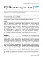
To form microtubules, α- and β-tubulin molecules join to form a heterodimerFigure 1
To form microtubules, α- and β-tubulin molecules join to form a heterodimer. These dimers then attach to other dimers form-
ing oligomers that elongate into protofilaments; eventually, the oligomers will join to give rise to a ringed microtubule. Micro-
tubules or unpolymerized tubulin bind microtubule-associated proteins (MAPs), which regulate polymerization, facilitate
assembly, stabilize the microtubules and regulate microtubular transport of macromolecules and vesicles. The HIV-1 Tat pro-
tein binds to αβ-tubulin dimers and microtubules thus enhancing microtubule polymerization, and to the microtubule-associ-
ated protein LIS1, which is also known to facilitate assembly of microtubules. Disturbance of the dynamics of microtubular
network formation activates the intrinsic mitochondrial apoptotic pathway. Pro-apoptotic Bcl2 family members – in particular,
Bim – are recruited to the mitochondrion; as a consequence, the mitochondrial membrane potential collapses, and pro-apop-
totic factors are released into the cytoplasm. These include reactive oxygen intermediates (ROIs), apoptosis-inducing factor
(AIF), and cytochrome c, among others. Release of cytochrome c is a point of no return as it leads to autoactivation of caspase
9, which in turn proceeds to cleave the downstream effector caspases (caspase 3, 6, etc.).
Publish with BioMed Central and every
scientist can read your work free of charge
"BioMed Central will be the most significant development for
disseminating the results of biomedical research in our lifetime."
Sir Paul Nurse, Cancer Research UK
Your research papers will be:
available free of charge to the entire biomedical community
peer reviewed and published immediately upon acceptance
cited in PubMed and archived on PubMed Central
yours — you keep the copyright
Submit your manuscript here:
/>BioMedcentral
Retrovirology 2005, 2:7 />Page 4 of 4
(page number not for citation purposes)
References
1. Wei X, Ghosh SK, Taylor ME, Johnson VA, Emini EA, Deutsch P, Lif-
son JD, Bonhoeffer S, Nowak MA, Hahn BH, Saag MS, Shaw GM:
Viral dynamics in human immunodeficiency virus type 1
infection. Nature 1995, 373:117-122.
2. Ho DD, Neumann AU, Perelson AS, Chen W, Leonard JM, Markowitz
M: Rapid turnover of plasma virions and CD4 lymphocytes in
HIV-1 infection. Nature 1995, 373:123-126.
3. Roshal M, Zhu Y, Planelles V: Apoptosis in AIDS. Apoptosis 2001,
6:103-116.
4. Gougeon ML: Apoptosis as an HIV strategy to escape immune
attack. Nat Rev Immunol 2003, 3:392-404.
5. Finkel TH, Tudor-Williams G, Banda NK, Cotton MF, Curiel T,
Monks C, Baba TW, Ruprecht RM, Kupfer A: Apoptosis occurs
predominantly in bystander cells and not in productively
infected cells of HIV- and SIV-infected lymph nodes. Nat Med
1995, 1:129-134.
6. Mann DA, Frankel AD: Endocytosis and targeting of exogenous
HIV-1 Tat protein. EMBO J 1991, 10:1733-1739.
7. Fittipaldi A, Ferrari A, Zoppe M, Arcangeli C, Pellegrini V, Beltram F,
Giacca M: Cell membrane lipid rafts mediate caveolar endo-
cytosis of HIV-1 Tat fusion proteins. J Biol Chem 2003,
278:34141-34149.
8. Li CJ, Friedman DJ, Wang C, Metelev V, Pardee AB: Induction of
apoptosis in uninfected lymphocytes by HIV-1 Tat protein.
Science 1995, 268:429-431.
9. Westendorp MO, Frank R, Ochsenbauer C, Stricker K, Dhein J, Wal-
czak H, Debatin KM, Krammer PH: Sensitization of T cells to
CD95-mediated apoptosis by HIV-1 Tat and gp120. Nature
1995, 375:497-500.
10. Alimonti JB, Ball TB, Fowke KR: Mechanisms of CD4+ T lym-
phocyte cell death in human immunodeficiency virus infec-
tion and AIDS. J Gen Virol 2003, 84:1649-1661.
11. Bhalla KN: Microtubule-targeted anticancer agents and
apoptosis. Oncogene 2003, 22:9075-9086.
12. Chen D, Wang M, Zhou S, Zhou Q: HIV-1 Tat targets microtu-
bules to induce apoptosis, a process promoted by the pro-
apoptotic Bcl-2 relative Bim. Embo J 2002, 21:6801-6810.
13. Macho A, Calzado MA, Jimenez-Reina L, Ceballos E, Leon J, Munoz E:
Susceptibility of HIV-1-TAT transfected cells to undergo
apoptosis. Biochemical mechanisms. Oncogene 1999,
18:7543-7551.
14. Ferri KF, Jacotot E, Blanco J, Este JA, Kroemer G: Mitochondrial
control of cell death induced by HIV-1-encoded proteins. Ann
N Y Acad Sci 2000, 926:149-164.
15. Battaglia PA, Zito S, Macchini A, Gigliani F: A Drosophila model of
HIV-Tat-related pathogenicity. J Cell Sci 2001, 114:2787-2794.
16. de Mareuil J, Carre M, Barbier P, Campbell GR, Lancelot S, Opi S,
Esquieu D, Watkins J, Prevot C, Braguer D, Briand C, Peyrot V, Loret
EP: HIV-1 Tat protein enhances microtubule polymerization.
Retrovirology 2005 in press.
17. Epie N, Ammosova T, Sapir T, Voloshin Y, Lane WS, Turner W,
Reiner O, Nekhai S: HIV-1 Tat interacts with LIS1 protein. Ret-
rovirology 2005 in press.
18. Xiang X: LIS1 at the microtubule plus end and its role in
dynein-mediated nuclear migration. J Cell Biol 2003,
160:289-290.
19. Drewes G, Ebneth A, Mandelkow EM: MAPs, MARKs and micro-
tubule dynamics. Trends Biochem Sci 1998, 23:307-311.
20. Zhang M, Li X, Pang X, Ding L, Wood O, Clouse K, Hewlett I, Dayton
AI: Identification of a potential HIV-induced source of
bystander-mediated apoptosis in T cells: upregulation of
trail in primary human macrophages by HIV-1 tat. J Biomed Sci
2001, 8:290-296.
21. Sastry KJ, Marin MC, Nehete PN, McConnell K, el-Naggar AK,
McDonnell TJ: Expression of human immunodeficiency virus
type I tat results in down-regulation of bcl-2 and induction of
apoptosis in hematopoietic cells. Oncogene 1996, 13:487-493.
22. Bartz SR, Emerman M: Human immunodeficiency virus type 1
Tat induces apoptosis and increases sensitivity to apoptotic
signals by up-regulating FLICE/caspase-8. J Virol 1999,
73:1956-1963.
23. Marcello A, Ferrari A, Pellegrini V, Pegoraro G, Lusic M, Beltram F,
Giacca M: Recruitment of human cyclin T1 to nuclear bodies
through direct interaction with the PML protein. EMBO J
2003, 22:2156-2166.
24. Xiao H, Neuveut C, Tiffany HL, Benkirane M, Rich ER, Murphy PM,
Jeang KT: Selective CXCR4 antagonism by Tat: implications
for in vivo expansion of coreceptor use by HIV-1. Proc Natl
Acad Sci USA 2000, 97:11466-11471.
25. Tyagi M, Rusnati M, Presta M, Giacca M: Internalization of HIV-1
Tat requires cell surface heparan sulfate proteoglycans. J Biol
Chem 2001, 276:3254-3261.