Báo cáo y học: "Heat shock protein 70 expression, keratin phosphorylation and Mallory body formation in hepatocytes from griseofulvin-intoxicated mice" pptx
Bạn đang xem bản rút gọn của tài liệu. Xem và tải ngay bản đầy đủ của tài liệu tại đây (3.19 MB, 15 trang )
BioMed Central
Page 1 of 15
(page number not for citation purposes)
Comparative Hepatology
Open Access
Research
Heat shock protein 70 expression, keratin phosphorylation and
Mallory body formation in hepatocytes from
griseofulvin-intoxicated mice
Michel Fausther, Louis Villeneuve and Monique Cadrin*
Address: Département de chimie-biologie, Université du Québec à Trois-Rivières, 3351 boulevard des Forges, C.P. 500, Québec, Trois-Rivières,
Canada G9A 5H7
Email: Michel Fausther - ; Louis Villeneuve - ; Monique Cadrin* -
* Corresponding author
Abstract
Background: Keratins are members of the intermediate filaments (IFs) proteins, which constitute
one of the three major cytoskeletal protein families. In hepatocytes, keratin 8 and 18 (K8/18) are
believed to play a protective role against mechanical and toxic stress. Post-translational
modifications such as phosphorylation and glycosylation are thought to modulate K8/18 functions.
Treatment of mouse with a diet containing griseofulvin (GF) induces, in hepatocytes, modifications
in organization, expression and phosphorylation of K8/18 IFs and leads, on the long term, to the
formation of K8/18 containing aggregates morphologically and biochemically identical to Mallory
bodies present in a number of human liver diseases. The aim of the present study was to investigate
the relationship between the level and localization of the stress inducible heat shock protein 70
kDa (HSP70i) and the level and localization of K8/18 phosphorylation in the liver of GF-intoxicated
mice. The role of these processes in Mallory body formation was studied, too. The experiment was
carried out parallely on two different mouse strains, C3H and FVB/n.
Results: GF-treatment induced an increase in HSP70i expression and K8 phosphorylation on
serines 79 (K8 S79), 436 (K8 S436), and K18 phosphorylation on serine 33 (K18 S33) as determined
by Western blotting. Using immunofluorescence staining, we showed that after treatment, HSP70i
was present in all hepatocytes. However, phosphorylated K8 S79 (K8 pS79) and K8 S436 (K8
pS436) were observed only in groups of hepatocytes or in isolated hepatocytes. K18 pS33 was
increased in all hepatocytes. HSP70i colocalized with MBs containing phosphorylated K8/18.
Phophorylation of K8 S79 was observed in C3H mice MBs but was not present in FVB/n MBs.
Conclusions: Our results indicate that GF intoxication represents a stress condition affecting all
hepatocytes, whereas induction of K8/18 phosphorylation is not occurring in every hepatocyte. We
conclude that, in vivo, there is no direct relationship between GF-induced stress and K8/18
phosphorylation on the studied sites. The K8/18 phosphorylation pattern indicates that different
cell signaling pathways are activated in subpopulations of hepatocytes. Moreover, our results
demonstrate that, in distinct genetic backgrounds, the induction of K8/18 phosphorylation can be
different.
Published: 12 August 2004
Comparative Hepatology 2004, 3:5 doi:10.1186/1476-5926-3-5
Received: 26 February 2004
Accepted: 12 August 2004
This article is available from: />© 2004 Fausther et al; licensee BioMed Central Ltd.
This is an open-access article distributed under the terms of the Creative Commons Attribution License ( />),
which permits unrestricted use, distribution, and reproduction in any medium, provided the original work is properly cited.
Comparative Hepatology 2004, 3:5 />Page 2 of 15
(page number not for citation purposes)
Background
Intermediate filaments (IFs) with microtubules and actin
microfilaments are the major cytoskeletal components of
most vertebrate cells [1-4]. IF proteins constitute a large
family of proteins that is divided into five types [1,2]. The
expression of the different IF proteins is differentiation
and tissue specific [1,5]. Keratins expressed in epithelial
cells, represent the largest and most complex subtype of IF
proteins (more than 20 proteins)[2]. They are classified
into two groups, the type I (acidic K9 to K20) and the type
II (neutral-basic, K1 to K8), which form obligate heter-
opolymers composed of equimolar amounts of type I and
type II keratins [2,6].
It is now generally accepted that, in multilayered epithe-
lia, one of the function for keratins IFs is to protect the tis-
sue from mechanical stress [7-9]. The first evidences for
this function came from studies on epidermis, which
showed that transgenic mice lacking epidermal keratins,
or expressing mutated keratins, displayed blistering skin
disease phenotypes, similar to human skin diseases such
as epidermolysis bullosa simplex or epidermolytic hyper-
keratosis [7,10,11].
As for epidermal keratins, the production of transgenic
mice targeting K8 or K18 has been necessary to unravel the
role of IFs in simple epithelium such as in the liver. In
hepatocytes, K8/18 is the only keratin pair and thus both
keratins are necessary to form an IF network. Transgenic
mice expressing K8 or K18 carrying mutations that affect
filament formation, develop mild hepatitis and display
greater liver sensitivity to mechanical and toxic stress than
wild type animals [12,13].
Recent studies from Ku et al. [14-16] have shown that
mutations on K8/18 predispose to the development of
liver disease in humans. Moreover, modifications in IF
organization and the formation of keratin containing
aggregates, named Mallory bodies (MBs), are observed in
different liver diseases such as alcoholic hepatitis, Wil-
son's disease, Indian childhood cirrhosis and liver steato-
sis in obesity [17-21]. Other proteins, such as ubiquitin
and the heat shock protein 70 kDa (HSP70), are also
present in MBs and could play a role in their formation
[22-24]. Taken together, these results support the hypoth-
esis that keratins are necessary to preserve the hepatocytes
integrity upon stressful conditions. It is still unclear how
keratins accomplish these protective roles. Previous stud-
ies have shown that modifications in keratin phosphor-
ylation are associated with various conditions such as
mitosis, apoptosis and stress, suggesting a role for this
post-translational modification in the modulation of ker-
atin-related functions [25-27].
Long-term treatment of mice with a diet containing grise-
ofulvin (GF) induces the development of an hepatitis
associated with the formation of MBs, which are biochem-
ically and morphologically similar to those found in
humans [19,28]. This animal model constitutes a useful
tool to investigate the keratin dynamics in the response of
hepatocytes to the presence of a hepatotoxic agent.
In the present study, we investigated the effect of chronic
GF intoxication on hepatocytes from C3H and FVB/n
mouse strains. We monitored the expression of the stress
inducible form of the heat shock protein 70 kDa (HSP70i)
and the induction of K8/18 phosphorylation at specific
sites: K8 on serine 79 (K8 S79), K8 on serine 436 (K8
S436), K18 on serine 52 (K18 S52) and K18 on serine 33
(K18 S33) (reviewed in [26,29]). We also examined the
possible relationship between HSP70i expression and K8/
18 phosphorylation, during the development of hepatitis
and in MB formation.
Results
Induction of HSP70i and K8/18 phosphorylation upon GF-
treatment in C3H and FVB/n mouse livers
The modifications in the amount of HSP70i, K8/18, and
phosphorylated keratins (K8 pS79, K8 pS436 and K18
pS33) were analyzed by Western Blotting of total proteins
from control and GF-treated C3H and FVB/n mouse livers
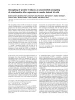
(2 weeks, 6 weeks and 5 months). GF intoxication
induced an increase in keratin levels in livers from both
mouse strains (Fig. 1A,1B). HSP70i, which was present in
control livers of both mouse strains, was also increased by
the treatment (Fig. 1A).
Total proteins from C3H and FVB/n livers were probed
with antibodies against K8 pS79, K8 pS436 and K18 pS33
(Fig. 1C,1D,1E,1F,1G). Significant changes in K8 and K18
phosphorylation occurred after GF-treatment in both
mouse strains (Fig. 1C,1D,1E,1F,1G). Small amounts of
K8 pS79 and K18 pS33 were found in control livers (Fig.
1C,1D and 1E,1F), whereas K8 pS436 was not detected
(Fig. 1G). After 2 weeks of treatment, an increase in the
amount of all phosphokeratin species studied was
observed. The phosphorylation levels of K8 S436 and K18
S33 remained higher than control values in both mouse
strains for the entire treatment (Fig. 1E,1F,1G). However,
when compared with 2 week treatment, a decrease in K8
pS436 and K18 pS33 was noted after 5 months of treat-
ment (Fig. 1E,1F,1G). Similarly, a decrease in K8 S79
phosphorylation was observed after 5 months of treat-
ment, in C3H mice (Fig. 1C). However in FVB/n mice, K8
pS79 was not detected after the same period of treatment
(Fig. 1D).
Comparative Hepatology 2004, 3:5 />Page 3 of 15
(page number not for citation purposes)
Localization of HSP70i and K8/18 during GF intoxication
We analyzed at the cellular level, by double immunofluo-
rescence staining, the distribution of HSP70i and IFs on
liver sections of control and GF-treated C3H and FVB/n
mouse livers.
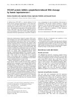
In control hepatocytes, IFs formed a complex cytoplasmic
network that was denser at the cell membrane and partic-
ularly around the bile canaliculi (Fig. 2A). Our biochemi-
cal analysis showed that HSP70i was present in control
hepatocytes. However, by immunofluorescence, we did
not detect the presence of HSP70i in the cells (Fig. 2B).
After 2 weeks of treatment, most of the hepatocytes were
enlarged and the bile canaliculi were dilated. IF network
was denser around dilated bile canaliculi (Fig. 2C). All
hepatocytes contained a very dense cytoplasmic IF net-
work. These modifications were accompanied by an
increase in the amount of HSP70i in hepatocytes and a
granular staining was detectable at the membrane and in
the nuclei (Fig. 2D). A few cells showed a high level of
HSP70i. After 5 months of treatment, there was a mosaic
pattern of cells with and without IF staining (Fig. 2E).
HSP70i showed a granular staining pattern in many hepa-
tocytes and was also present in MBs (Fig. 2F).
Phosphorylation of K8 S79, K8 S436 and K18 S33 during
GF intoxication
Cryosections of control and GF-treated C3H and FVB/n
mouse livers were fixed with 4% paraformaldehyde and
processed for double immunofluorescence staining. As
mentioned above, control mice showed hepatocytes with
a cytoplasmic IF network, which was denser at the cell
periphery (Fig. 3,4,5A). K8 pS79 and K8 pS436 were not
generally detected in the IF network of control hepato-
cytes. Only, occasionally some doublet cells, most likely
representing cells in mitosis, were stained (data not
shown). A basal level of phosphorylation for K18 S33 was
detected at the periphery of all hepatocytes (Fig. 5B).
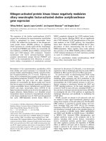
After 2 weeks of GF-treatment, hepatocytes were enlarged
and an increase in cytoplasmic IF network was observed
(Fig. 3,4,5C). This treatment induced the phosphoryla-
tion of K8 on S79 and S436 in some hepatocytes. K8 pS79
and K8 pS436 were present in clusters of cells scattered
over the whole liver (Fig. 3,4,7D). The groups of cells
stained with the anti-K8 pS79 or anti-K8 pS436 usually
surrounded damaged cells (Fig. 3,7D). In the case of K8
pS79, IFs located in the cytoplasm and at the periphery of
the cells were highly stained (Fig. 3D,3E). For K8 pS436,
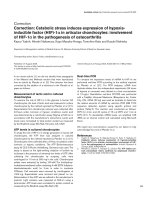
Biochemical analysis of livers from C3H and FVB/n miceFigure 1
Biochemical analysis of livers from C3H and FVB/n mice. Western blots from C3H mouse livers; A, K8 and HSP70i; C,
K8 pS79; E, K18 pS33; G, K8 pS436. Western blots from FVB/n mouse livers; B, K8; D, K8 pS79; F, K18 pS33.
Comparative Hepatology 2004, 3:5 />Page 4 of 15
(page number not for citation purposes)
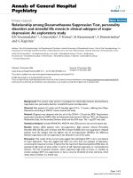
the staining was stronger at the cell periphery and around
the dilated bile canaliculi (Fig. 4D). In addition to their
presence in clusters of cells, K8 pS79 and K8 pS436 dis-
played an intense cytoplasmic staining in some isolated
cells or cell doublets (Fig. 3E). Since both epitopes
showed similarities in their patterns of distribution, we
asked whether they were present in the same hepatocytes.
Immunostaining for the detection of K8 pS79 and K8
pS436 were performed on serial liver sections of GF-
treated mouse liver. Our results showed that the groups of
hepatocytes positives for K8 pS79 were also positive for
K8 pS436 (Fig. 6).
In the case of K18 S33, its phosphorylation was increased
in most (if not all) hepatocytes. Most of the staining was
Distribution of keratin IFs and HSP70i in hepatocytes from control and GF-fed C3H miceFigure 2
Distribution of keratin IFs and HSP70i in hepatocytes from control and GF-fed C3H mice. A, C, E keratin IFs; B,
D, F HSP70i; A, B) control; C, D) 2 week treatment; E, F) 5 month treatment. Arrows in E and F indicate reactive MBs with
Troma 1 (anti-K8) and anti-HSP70i, respectively. Scale bar = 20 µm.
Comparative Hepatology 2004, 3:5 />Page 5 of 15
(page number not for citation purposes)
observed at the periphery of the cells, delimitating clearly
the bile canaliculi. A few hepatocytes showed high levels
of cytoplasmic K18 pS33 (Fig. 5D).
After 6 weeks of GF-treatment, the distribution of hepato-
cytes containing K8 pS79 and K8 pS436 was different
from the one observed after 2 weeks of treatment. Clusters
of labeled cells were smaller, whereas labeled isolated
cells became more prominent (Fig. 3G,4F). Singlet and
doublet cell(s) highly labeled with K8 pS79 and K8 pS436
were also present (Fig. 3H). K18 pS33 was present in most
hepatocytes and showed a similar pattern as the one
observed after staining with Troma1 (Fig. 5F).
After 5 months of GF-treatment, MBs were present in
some hepatocytes in both mouse strains (Fig. 3I,4G,5G).
MBs had variable size and different positions depending
on the cell and were observed in cells with or without a
visible intracytoplasmic IF network, as detected with
Troma 1. In both mouse strains, K8 pS436 and K18 pS33
were present in MBs (Fig. 4,5H), whereas K8 pS79 seemed
to be absent (Fig. 3J). Experiments described above were
also performed using cold acetone instead of 4%
Distribution of keratin IFs and K8 pS79 in hepatocytes from control and GF-fed C3H miceFigure 3
Distribution of keratin IFs and K8 pS79 in hepatocytes from control and GF-fed C3H mice. A, C, F, I keratin IFs;
B, D, E, G, H, J K8 pS79; A, B) control; C, D, E) 2 week treatment; F, G, H 6 week treatment; I, J 5 month treatment. Arrow in
D indicates clusters of cells containing K8 pS79. Empty arrowheads in I and J indicate MBs reactive with Troma 1 but not with
LJ4 (anti-K8 pS79), respectively. Scale bar = 20 µm.
Comparative Hepatology 2004, 3:5 />Page 6 of 15
(page number not for citation purposes)
Distribution of keratin IFs and K8 pS436 in hepatocytes from control and GF-fed C3H miceFigure 4
Distribution of keratin IFs and K8 pS436 in hepatocytes from control and GF-fed C3H mice. A, C, E, G keratin
IFs; B, D, F, H K8 pS436; A, B) control; C, D) 2 week treatment; E, F) 6 week treatment; G, H); 5 month treatment. Arrows in
D indicate clusters of cells containing K8 pS436. Filled arrowheads in G and H indicate reactive MBs with Troma 1 and 5B3
(anti-K8 pS436), respectively. Scale bar = 20 µm.
Comparative Hepatology 2004, 3:5 />Page 7 of 15
(page number not for citation purposes)
Distribution of keratin IFs and K18 pS33 in hepatocytes from control and GF-fed C3H miceFigure 5
Distribution of keratin IFs and K18 pS33 in hepatocytes from control and GF-fed C3H mice. A, C, E, G keratin
IFs; B, D, F, H K18 pS33; A, B) control; C, D) 2 week treatment; E, F) 6 week treatment; G, H) 5 month treatment. Asterisk in
D shows an hepatocyte containing a high level of K18 pS33; arrow indicates a dilated bile canaliculi. Filled arrowheads in G and
H indicate reactive MBs with Troma 1 and Ab8250 (anti-K18 pS33), respectively. Scale bar = 20 µm.
Comparative Hepatology 2004, 3:5 />Page 8 of 15
(page number not for citation purposes)
paraformaldehyde. After acetone fixation, no difference in
the staining pattern was observed for GF-treatment of 2
and 6 weeks in both mouse strains (Fig. 7). However, dif-
ferences were observed for MB staining in the 5 months
GF-treated mouse liver. In C3H mouse strain livers, K8
pS79 was present in many MBs although some of them
showed no staining (Fig. 7H). In FVB/n mouse strain liv-
ers, K8 pS79 was not present in most MBs (Fig. 8). No dif-
ference in staining of MBs was observed for K8 pS436 and
K18 pS33.
Localization of phosphorylated K8 species and HSP70i
during GF intoxication
Double immunostaining with anti-HSP70i and anti-
phosphorylated keratins (K8 pS79 or K8 pS436) was per-
formed for studying the localization of HSP70i in relation
to keratin phosphorylation. The results showed that
HSP70i and phosphorylated K8 species colocalized in
some cells (Fig. 9). However, in most of the cells, the colo-
calization was not observed.
Discussion
The functional significance of K8/18 in simple epithelium
has been the subject of numerous studies over the last dec-
ade [29-35]. Although most of these reports lead towards
roles for K8/18 in the resistance of cells to mechanical and
toxic stress, the molecular mechanisms underlying these
phenomena remain to be elucidated. To date, most of our
understanding of the pathways involving keratins in the
response of hepatocytes to toxic stress comes from the
Colocalization of K8 pS79 and K8 pS436 in hepatocytes from GF-fed C3H dmiceFigure 6
Colocalization of K8 pS79 and K8 pS436 in hepatocytes from GF-fed C3H dmice. A, C K8 pS79; B, D K8 pS436; A,
B, C, D 2 week treatment. Arrows in A and B indicate clusters of hepatocytes containing both K8 pS79 and K8 pS436. Note:
reactivity in nuclei observed in A, B, C and D represents non-specific staining due to the secondary antibody. Scale bar = 20
µm.
Comparative Hepatology 2004, 3:5 />Page 9 of 15
(page number not for citation purposes)
Distribution of keratin IFs and K8 pS79 in hepatocytes from control and GF-fed C3H miceFigure 7
Distribution of keratin IFs and K8 pS79 in hepatocytes from control and GF-fed C3H mice. A, C, E, G keratin IFs;
B, D, F, H K8 pS79; A, B) control; C, D) 2 week treatment; E, F) 6 week treatment; G, H) 5 month treatment. Arrow in D indi-
cates clusters of cells containing K8 pS79; asterisk shows a damaged hepatocyte. Filled arrowheads in G and H indicate MBs
reactive with Troma 1 and LJ4 (anti-K8 pS79), respectively; empty arrowheads indicate MBs reactive with Troma 1 but not
with LJ4 (anti-K8 pS79), respectively. Scale bar = 20 µm.
Comparative Hepatology 2004, 3:5 />Page 10 of 15
(page number not for citation purposes)
analyses of various cell lines [36-38]. K8/18 phosphoryla-
tion at specific sites has been proposed to be a key factor
in the regulation of those keratin functions. In this regard,
K8 pS79, K8 pS436, K18 pS52 and K18 pS33 are the most
studied phosphorylation sites [39].
In vivo, K8/18 are also subjected to phosphorylation and,
as suggested by in vitro studies, it is proposed to help hepa-
tocytes to cope with toxic stress [26,29,35]. For instance,
transgenic mice expressing human K18 S52 mutated in
alanine mutant are more susceptible to drug-induced liver
injury than transgenic mice over expressing wild type
human K18 [40].
In the present study, we showed that the chronic intoxica-
tion of mice with GF, which is known to induce modifica-
tions in keratin organization and formation of MBs [33],
was associated with increased expression of the stress pro-
tein HSP70i. GF-treatment resulted in a rapid increase in
the expression of HSP70i. This modification was already
perceptible after 2 weeks of treatment and was maintained
for the whole period of treatment. This result provides
direct evidence that GF-treatment, which has been pro-
posed to constitute an oxidative stress for hepatocytes
[41], triggers signaling pathways involved in cellular pro-
tection [33]. This interpretation of our biochemical data is
in agreement with our immunofluorescence study, which
showed that HSP70i partly relocalized to the nucleus dur-
ing the treatment. This distribution pattern is typical of
the distribution of HSP70i in stressed cells [42,43].
We have previously shown that GF intoxication induced
an overall increase in K8/18 phosphorylation [44,45].
Here, we show that GF-treatment is associated with mod-
ifications in K8/K18 phosphorylation at specific sites such
as: K8 S79, K8 S436 and K18 S33. Among the studied
phosphorylation sites, and because it was present and was
increased in all treated hepatocytes, K18 pS33 was the
Distribution of keratin IFs and K8 pS79 in hepatocytes from GF-fed FVB/n miceFigure 8
Distribution of keratin IFs and K8 pS79 in hepatocytes from GF-fed FVB/n mice. A, C keratin IFs; B, D K8 pS79; A,
B, C, D 5 month treatment. Asterisks in A and B indicate MBs reactive with Troma 1 but not with LJ4; arrows in C and D indi-
cate MBs reactive with Troma 1 and LJ4, respectively. Scale bar = 20 µm.
Comparative Hepatology 2004, 3:5 />Page 11 of 15
(page number not for citation purposes)
keratin phosphoepitope the tissue distribution of which
resembled that of HSP70i. The phosphorylation of K18
S33 has been shown to play a role in keratin reorganiza-
tion during mitosis and by linking 14-3-3 proteins, to
modulate their function [46,47]. Hence, we propose that
K18 S33 phosphorylation could be linked to IF reorgani-
zation during GF intoxication. Moreover, because K18
pS33 is increased in all hepatocytes, it could be implicated
in the stress response by participating in the relocalization
and/or the recruitment of molecules or factors implicated
in stress-induced cell signaling.
In contrast to K18 pS33, phosphorylated K8 species, K8
pS79 and K8 pS436 were not present in control mice
hepatocytes. After 2 and 6 weeks of treatment, we
observed an increase in the level of phosphorylation on
these sites. However, contrary to HSP70i and K18 pS33,
these phosphorylation sites were only present in isolated
cells (singlet or doublets) or clusters of cells. Labeled sin-
glet or doublet cells were more numerous after staining
with the anti-K8 pS79 than after staining with the anti-K8
pS436. These cells could correspond to cells that are
undergoing mitosis. This is supported by previous studies
which showed that the phosphorylation of K8 on S79 and
S436 occurs during mitosis [25,48]. This interpretation is
also in agreement with the work of Stumptner et al. [49],
which showed the presence of cell doublets reactive with
the anti-K8 pS79 after a short treatment with DDC that
induces on the long term MB formation. The discrepancy
in the number of cells stained for K8 pS79 and K8 pS436,
Distribution of phosphorylated keratin IFs and HSP70i in hepatocytes from GF-fed C3H miceFigure 9
Distribution of phosphorylated keratin IFs and HSP70i in hepatocytes from GF-fed C3H mice. A K8 pS79; B, D
HSP70i; C K8 pS436; A, B, C, D 2 week treatment. Arrows in A and B indicate cells in which HSP70i and K8 pS79 colocalized.
Arrows in C and D indicate cells in which HSP70i and K8 pS436 colocalized. Scale bar = 20 µm.
Comparative Hepatology 2004, 3:5 />Page 12 of 15
(page number not for citation purposes)
both in the singlet and doublet cells, suggests that differ-
ent kinases are involved in the phosphorylation of those
sites.
The presence of K8 pS79 and K8 pS436 was also detected
in islets of cells. Interestingly, both antigens were present
in the same clusters of cells surrounding unstained cells
that were most likely undergoing apoptosis. These
unstained cells are evocative of detached cells during
anoikis, an apoptotic process that can be induced by loss
of cell-cell anchorage. Stress and apoptosis has been
shown to modulate K8 S79 and K8 S436 phosphorylation
[25,48]. The observed phosphorylation could indicate
that these hepatocytes are stressed hepatocytes intended
to apoptosis. However, analysis of the livers for the pres-
ence of apoptosis showed that only a few hepatocytes are
going through programmed cell death and groups of cells
in apoptosis were never observed (data not shown). We
propose that the apoptotic cell could represent the starting
point of a signal transduction pathway to neighboring
cells. The activation of specific kinases that would phos-
phorylate keratins could provide those cells a resistance to
apoptosis. This latter interpretation is in agreement with
the notion that K8/18 intermediate filaments play a key
role in the protection of cells against apoptosis [26,35].
Liao et al. [50] have shown that HSP70 associates with K8/
18 via K8. Our study show that colocalization of HSP70i
and IFs occurs only in a few hepatocytes. Since the hepa-
tocytes, in which colocalization was observed, contained
K8 pS79 or K8 pS436, HSP70i binding to IFs in these cells
may be related to the presence of keratin phosphorylation
and participates to cellular pathways involving
phosphorylated K8/18 on specific sites. Ku et al. [51] have
shown that phosphorylation could modulate K8/18 ubiq-
uitination and ensuing turnover. Knowing that binding of
HSP70 to a protein can affect its targeting by kinases or
phosphatases [52], HSP70i could bind to phosphorylated
K8 species, prevent dephosphorylation by specific phos-
phatases, and thereby enhance phosphorylation-medi-
ated K8/18 protection from degradation by the ubiquitin
pathway [51,53]. However, since HSP70i and phosphor-
ylated K8 species colocalized only in a few cells over the
whole tissue, the relevance of this phenomenon in the
response to the presence of the hepatotoxin needs to be
addressed and further investigations will be necessary to
confirm that hypothesis.
Chronic intoxication of mice with GF induces the forma-
tion of MBs. Numerous studies have demonstrated the
presence of different phosphorylated K8/18 species
within MBs, suggesting that K8/18 phosphorylation could
participate in the MB formation processes [49,54]. In our
experiments, we showed that K8 pS436 and K18 pS33
were present in all observed MBs, whereas K8 pS79 was
present in MBs in C3H mice hepatocytes but not in FVB/
n mice. The difference in the presence of K8 pS79 phos-
phoepitope within MBs suggests that phosphorylation at
that specific site is not essential for MB formation. How-
ever, as suggested by Stumptner et al. [49], because K8
pS436 and K18 pS33 are always detected in MBs, phos-
phorylation on these sites could be implicated in the proc-
esses of MB formation. Taken together, those results
indicate that in the context of MB formation, K8/18 phos-
phorylation should not be considered as a general phe-
nomenon but as specific events that affect precise sites on
K8 or K18. The difference observed between keratin phos-
phorylation in C3H and FVB/n mice indicates that the
genetic background influences the response of hepato-
cytes to toxic stress. This interpretation is in agreement
with the results obtained with K8-null mice which dis-
played variable phenotypes depending on the genetic
background [30,31].
The treatment with GF that represents a toxic stress, most
likely, involves the activation of stress activated protein
kinases (SAPKs) in some hepatocytes. SAPKs p38 and JNK
are physiologic kinases for K8 S79 and K8 S436 [37,55].
We postulate that p38 kinase and/or JNK are activated by
GF-treatment in some hepatocytes and are responsible for
the modifications in K8 phosphorylation we observed. K8
and K18 give different patterns of phosphorylated cells
indicating that, under the same conditions, K8 and K18
phosphorylation is regulated differently.
Conclusions
Our results show that increases in HSP70i, K8/18 expres-
sion and K8/18 phosphorylation constitute early events in
the response of hepatocytes to the presence of GF. These
observations support a role for keratins in preserving cel-
lular integrity during stress conditions induced by the
presence of a chemical agent [33,35]. HSP70i expression
in hepatocytes after GF-treatment is not directly related to
K8/18 phosphorylation at the studied sites: K8 S79, K8
S436 and K18 S33. With regard to MB formation, it
appears that both HSP70i and K8/18 phosphorylation
might contribute to the IF aggregation processes. The
involvement of K8/18 phosphorylation in MB formation
seems to be related only to specific sites and dependent on
mouse genetic inheritance.
Methods
Experimental design
Experiments were performed with adult C3H mice
(Charles River Canada, St-Constant, QC) and FVB/n mice
(Baribault et al. 1994) weighing 25 to 30 g. Two mouse
strains were used to minimize the potential effect of dif-
ferent genetic background on the response of hepatocytes
and to facilitate the interpretation of the data. All animals
were housed with a 12-hour light-dark cycle and allowed
Comparative Hepatology 2004, 3:5 />Page 13 of 15
(page number not for citation purposes)
the consumption of water and of a standard mouse semi-
synthetic diet (Texlad Test Diet, Madison, WI), both ad
libitum. GF-treated mice were fed a diet containing 2.5%
(w/w) GF (Schering Corp., Kenilworth, NJ) for different
periods of time: 2 weeks, 6 weeks and 5 months according
to the method of Denk et al. [28]. Control mice were fed
the same diet without GF. For control and each period of
GF-treatment, experimental groups included 3 animals.
Mice were sacrificed by cervical dislocation and livers were
snap frozen in methylbutane precooled with liquid nitro-
gen and stored at -70°C before use. All experiments were
conducted according to the requirements of Canadian
Council Animal Care and the "Université du Québec à
Trois-Rivières" Animal Welfare Committee. For micro-
scopical studies, paraformaldehyde and cold acetone were
routinely used, as fixatives, to ensure that the staining pat-
terns were not a consequence of the fixative used.
Reagents
The antibodies used were as following: Troma 1, a rat
monoclonal antibody (rAb) that recognizes K8 [56]; LJ4,
a mouse monoclonal antibody (mAb) that recognizes
human K8 pS73 equivalent to mouse K8 pS79 [25]; mAb
5B3 that recognizes K8 pS431 equivalent to mouse K8
pS436 [48]; 8250, a rabbit polyclonal antibody (pAb) that
recognizes K18 pS33 [46] and a pAb that recognizes the
stress inducible form of HSP70, HSP70i (Stressgen, Victo-
ria, BC). The secondary antibodies for fluorescence micro-
scopy were as follows: tetramethylisothiocyanate (TRITC)
or fluorescein isothiocyanate (FITC) conjugated goat anti-
rat IgG, FITC-conjugated donkey anti-rabbit IgG (Jackson
Immunoresearch, Bio/Can Scientific, Mississauga, ON).
The M.O.M. kit and Avidin/Biotin blocking kit (Vector
®
Laboratories Canada, Burlington, ON) were used to per-
form immunolabelling with mAbs LJ4 and 5B3. The sec-
ondary antibodies used for Western blotting were as
follows: biotinylated goat anti-rat IgG, biotinylated don-
key anti-mouse IgG and peroxydase donkey anti-rabbit
IgG (Jackson Immunoresearch, Bio/Can Scientific, Missis-
sauga, ON). Other reagents used were: Horseradish
Streptavidin Peroxydase-conjugated (SPC) (Jackson
Immunoresearch, Bio/Can Scientific, Mississauga, ON),
Bovine Serum Albumin (BSA) (Jackson Immunoresearch,
Bio/Can Scientific, Mississauga, ON), Leupeptin (Sigma-
Aldrich Canada, Oakville, ON), Pepstatin (Sigma-Aldrich
Canada, Oakville, ON), Aprotinin (Sigma-Aldrich
Canada, Oakville, ON), Normal Horse Serum (NHS)
(Vector
®
Laboratories Canada, Burlington, ON), Luminol
(Amersham Pharmacia Biotech, Oakville, ON).
Gel electrophoresis and immunoblotting
Livers were homogenized in 62.5 mM Tris-HCl, pH 6.8
containing 2.3 % (w/v) SDS, 50 mM sodium fluoride, 10
mM EDTA, 1 mM sodium pyrophosphate, 1 mM DTT, 1
mM PMSF, 1 µM leupeptin (Sigma-Aldrich Canada,
Oakville, ON), 1 µM pepstatin (Sigma-Aldrich Canada,
Oakville, ON), 2.5 µg/ml aprotinin (Sigma-Aldrich Can-
ada, Oakville, ON). Proteins were separated by electro-
phoresis on 10% SDS-polyacrylamide gels [57]. Protein
concentration was determined by the Lowry method,
modified for the presence of SDS [58], and equal amounts
of proteins (5 to 12.5 µg) were loaded on each well. Gels
were stained with 0.1% Coomassie Blue, or transferred
onto nitrocellulose membranes (Biorad laboratories Can-
ada, Mississauga, ON), and processed for immunodetec-
tion. Membranes were blocked overnight with 5% (w/v)
non-fat dry milk (Carnation, Nestlé
®
) in PBS (Phosphate
Buffer Saline, 0.137 M NaCl, 2.7 mM KCl, 4.3 mM
Na
2
HPO
4
, 14.7 mM KH
2
PO
4,
pH 7.2), incubated with the
primary antibodies for 45 min, at room temperature,
washed in PBS containing 0.2% (v/v) Tween 20 and incu-
bated for 45 min with the appropriate secondary anti-
body: biotinylated goat anti-rat IgG (Jackson
Immunoresearch, Bio/Can Scientific, Mississauga, ON),
biotinylated donkey anti-mouse IgG (Jackson Immunore-
search, Bio/Can Scientific, Mississauga, ON) and horse-
radish peroxydase donkey anti-rabbit IgG (Jackson
Immunoresearch, Bio/Can Scientific, Mississauga, ON).
When biotinylated secondary antibodies were used, mem-
branes were washed with PBS-Tween 20 and incubated
with streptavidin conjugated with horseradish peroxydase
(Jackson Immunoresearch, Bio/Can Scientific, Missis-
sauga, ON) for 30 min and washed with PBS-Tween 20.
The chemiluminescent horseradish peroxydase substrate
Luminol (Amersham Pharmacia Biotech, Oakville, ON)
was added to the membranes according to recommenda-
tions of the company, and membranes were exposed to
Blue X-Omat X-ray film sheets (Mandel Scientific Com-
pany, Guelph, ON) to localize antibody binding.
Fluorescence microscopy
Cryosections (4 µm) of fresh liver were fixed with 4% (w/
v) paraformaldehyde in PBS pH 7.2 for 20 min, at room
temperature, and rinsed in PBS or TBS (Tris Buffer Saline,
10 mM Tris-HCl, 0.138 M NaCl, 2.7 mM KCl, pH 7.4)
upon staining protocols. Since fixation can affect anti-
body-binding capacity, cryosections were also fixed with
cold acetone (-20°C) for 10 min. For the detection of K8,
sections were incubated with rAb Troma 1 at room tem-
perature, washed in PBS and incubated with a FITC or a
TRITC conjugated goat anti-rat IgG (Jackson Immunore-
search, Bio/Can Scientific, Mississauga, ON) for 45 min,
at room temperature. For immunostaining of K18 pS33,
sections were incubated for 1 hour, at room temperature
with anti-K18 pS33 (8250) diluted in PBS containing
10% (w/v) BSA, washed in PBS and incubated for 45 min
with a FITC conjugated donkey anti-rabbit IgG in PBS
containing 10% BSA (Jackson Immunoresearch, Bio/Can
Scientific, Mississauga, ON). Immunostaining with anti-
K8 pS79 (LJ4) and anti-K8 pS436 (5B3) mAbs, was done
Comparative Hepatology 2004, 3:5 />Page 14 of 15
(page number not for citation purposes)
using the M.O.M. (mouse on mouse) detection kit (Vec-
tor
®
Laboratories Canada, Burlington, ON) and an Avidin/
Biotin blocking kit (Vector
®
Laboratories Canada, Burling-
ton, ON) according to recommendations of the company.
Normal horse serum (Vector
®
Laboratories Canada, Burl-
ington, ON) was added to solution during incubation
step with secondary antibody. For heat shock proteins
staining, liver sections were incubated with anti-HSP 70i
diluted in TBS containing 10% BSA for 45 min at 37°C,
washed in TBS and incubated for 45 min at 37°C with a
FITC conjugated donkey anti-rabbit IgG (Jackson Immu-
noresearch, Bio/Can Scientific, Mississauga, ON) diluted
in TBS containing 10% BSA. For detection of HSP70i, K8
pS79 and K8 pS436, the sections were treated with 1% (v/
v) Nonidet P-40 (Sigma-Aldrich Canada, Oakville, ON)
following fixing step with 4% paraformaldehyde.
The tissues were mounted in P-phenylene diamine diluted
in 50% (v/v) glycerol. The slides were kept at -20°C and
photomicrographs were collected using an Olympus
®
BX60 photomicroscope.
List of abbreviations
HSP70i – inducible form of 70 kDa Heat shock protein.
GF – griseofulvin. IFs – intermediate filaments. K8 – kera-
tin 8. K8/18 – keratin 8 and keratin 18. K8 S79 – serine 79
on keratin 8. K8 pS79 – phosphorylated serine 79 on ker-
atin 8. MBs – Mallory bodies.
Authors' contributions
MF carried out all western blotting analyses, performed
the immunofluorescence studies and participated in draft-
ing the manuscript. LV participated in the design of the
study. MC participated in the design of study, its coordi-
nation and drafting the manuscript. All authors read and
approved the final manuscript.
Acknowledgements
We thank Dr B. Omary from the Department of Medicine, Palo Alto VA
Medical Center and Stanford University, Palo Alto, California, for providing
antibodies directed against phosphorylated keratins. We also thank Dr N.
Marceau from the "Centre de recherche de L'Hôtel-Dieu de Québec
(CHUQ)", Québec, for providing Troma 1 and FVB/n mice. This work was
supported by a grant from NSERC.
References
1. Lazarides E: Intermediate filaments as mechanical integrators
of cellular space. Nature 1980, 283:249-256.
2. Fuchs E, Weber K: Intermediate filaments: structure, dynam-
ics, function, and disease. Annu Rev Biochem 1994, 63:345-382.
3. Schliwa M: The cytoskeleton: An introductory survey. In : Alfert
M, Beerman W, Goldstein L, Porter K R, eds Cell Biology Monograhs Volume
13. Edited by: Springer-Verlag. New York, Wien; 1986.
4. Herrmann H, Hesse M, Reichenzeller M, Aebi U, Magin TM: Func-
tional complexity of intermediate filament cytoskeletons:
from structure to assembly to gene ablation. Int Rev Cytol 2003,
223:83-175.
5. Steinert PM, Roop DR: Molecular and cellular biology of inter-
mediate filaments. Annu Rev Biochem 1988, 57:593-625.
6. Moll R, Franke WW, Schiller DL, Geiger B, Krepler R: The catalog
of human cytokeratins: patterns of expression in normal epi-
thelia, tumors and cultured cells. Cell 1982, 31:11-24.
7. Vassar R, Coulombe PA, Degenstein L, Albers K, Fuchs E: Mutant
keratin expression in transgenic mice causes marked abnor-
malities resembling a human genetic skin disease. Cell 1991,
64:365-380.
8. Fuchs E, Cleveland DW: A structural scaffolding of intermedi-
ate filaments in health and disease. Science 1998, 279:514-519.
9. Coulombe PA, Omary MB: 'Hard' and 'soft' principles defining
the structure, function and regulation of keratin intermedi-
ate filaments. Curr Opin Cell Biol 2002, 14:110-122.
10. Coulombe PA, Hutton ME, Letai A, Hebert A, Paller AS, Fuchs E:
Point mutations in human keratin 14 genes of epidermolysis
bullosa simplex patients: genetic and functional analyses. Cell
1991, 66:1301-1311.
11. Porter RM, Lane EB: Phenotypes, genotypes and their contribu-
tion to understanding keratin function. Trends Genet 2003,
19:278-285.
12. Loranger A, Duclos S, Grenier A, Price J, Wilson-Heiner M, Baribault
H, Marceau N: Simple epithelium keratins are required for
maintenance of hepatocyte integrity. Am J Pathol 1997,
151:1673-1683.
13. Ku NO, Michie S, Oshima RG, Omary MB: Chronic hepatitis,
hepatocyte fragility, and increased soluble phosphoglycok-
eratins in transgenic mice expressing a keratin 18 conserved
arginine mutant. J Cell Biol 1995, 131:1303-1314.
14. Ku NO, Darling JM, Krams SM, Esquivel CO, Keeffe EB, Sibley RK, Lee
YM, Wright TL, Omary MB: Keratin 8 and 18 mutations are risk
factors for developing liver disease of multiple etiologies. Proc
Natl Acad Sci U S A 2003, 100:6063-6068.
15. Ku NO, Gish R, Wright TL, Omary MB: Keratin 8 mutations in
patients with cryptogenic liver disease. N Engl J Med 2001,
344:1580-1587.
16. Ku NO, Wright TL, Terrault NA, Gish R, Omary MB: Mutation of
human keratin 18 in association with cryptogenic cirrhosis. J
Clin Invest 1997, 99:19-23.
17. Denk H, Krepler R, Lackinger E, Artlieb U, Franke WW: Immuno-
logical and biochemical characterization of the keratin-
related component of Mallory bodies: a pathological pattern
of hepatocytic cytokeratins. Liver 1982, 2:165-175.
18. Hazan R, Denk H, Franke WW, Lackinger E, Schiller DL: Change of
cytokeratin organization during development of Mallory
bodies as revealed by a monoclonal antibody. Lab Invest 1986,
54:543-553.
19. Jensen K, Gluud C: The Mallory body: theories on development
and pathological significance (Part 2 of a literature survey).
Hepatology 1994, 20:1330-1342.
20. Jensen K, Gluud C: The Mallory body: morphological, clinical
and experimental studies (Part 1 of a literature survey).
Hepatology 1994, 20:1061-1077.
21. Denk H, Stumptner C, Zatloukal K: Mallory bodies revisited. J
Hepatol 2000, 32:689-702.
22. Mayer R, Lowe J, Landon M, McDermott H, Tuckwell J, Doherty F,
Laszlo L: Ubiquitin and the lysosome system: molecular path-
ological and experimental findings. Eds Maresca, B and Lindquist,
S Edited by: Springer-Verlag. New York, Heidelberg; 1991:pp.
299-314.
23. Yuan QX, Marceau N, French BA, Fu P, French SW: Heat shock in
vivo induces Mallory body formation in drug primed mouse
liver. Exp Mol Pathol 1995, 63:63-76.
24. Riley NE, Li J, McPhaul LW, Bardag-Gorce F, Lue YH, French SW:
Heat shock proteins are present in mallory bodies (cytoker-
atin aggresomes) in human liver biopsy specimens. Exp Mol
Pathol 2003, 74:168-172.
25. Liao J, Ku NO, Omary MB: Stress, apoptosis, and mitosis induce
phosphorylation of human keratin 8 at Ser-73 in tissues and
cultured cells. J Biol Chem 1997, 272:17565-17573.
26. Omary MB, Ku NO, Liao J, Price D: Keratin modifications and
solubility properties in epithelial cells and in vitro. Subcell
Biochem 1998, 31:105-140.
27. Toivola DM, Zhou Q, English LS, Omary MB: Type II keratins are
phosphorylated on a unique motif during stress and mitosis
in tissues and cultured cells. Mol Biol Cell 2002, 13:1857-1870.
28. Denk H, Gschnait F, Wolff K: Hepatocellar hyalin (Mallory bod-
ies) in long term griseofulvin-treated mice: a new experi-
Publish with BioMed Central and every
scientist can read your work free of charge
"BioMed Central will be the most significant development for
disseminating the results of biomedical research in our lifetime."
Sir Paul Nurse, Cancer Research UK
Your research papers will be:
available free of charge to the entire biomedical community
peer reviewed and published immediately upon acceptance
cited in PubMed and archived on PubMed Central
yours — you keep the copyright
Submit your manuscript here:
/>BioMedcentral
Comparative Hepatology 2004, 3:5 />Page 15 of 15
(page number not for citation purposes)
mental model for the study of hyalin formation. Lab Invest
1975, 32:773-776.
29. Ku NO, Zhou X, Toivola DM, Omary MB: The cytoskeleton of
digestive epithelia in health and disease. Am J Physiol 1999,
277:G1108-37.
30. Baribault H, Penner J, Iozzo RV, Wilson-Heiner M: Colorectal
hyperplasia and inflammation in keratin 8-deficient FVB/N
mice. Genes Dev 1994, 8:2964-2973.
31. Baribault H, Price J, Miyai K, Oshima RG: Mid-gestational lethality
in mice lacking keratin 8. Genes Dev 1993, 7:1191-1202.
32. Gilbert S, Loranger A, Daigle N, Marceau N: Simple epithelium
keratins 8 and 18 provide resistance to Fas-mediated apop-
tosis. The protection occurs through a receptor-targeting
modulation. J Cell Biol 2001, 154:763-773.
33. Cadrin M, Hovington H, Marceau N, McFarlane-Anderson N: Early
perturbations in keratin and actin gene expression and fibril-
lar organisation in griseofulvin-fed mouse liver. J Hepatol 2000,
33:199-207.
34. Marceau N, Loranger A, Gilbert S, Daigle N, Champetier S: Keratin-
mediated resistance to stress and apoptosis in simple epithe-
lial cells in relation to health and disease. Biochem Cell Biol 2001,
79:543-555.
35. Omary MB, Ku NO, Toivola DM: Keratins: guardians of the liver.
Hepatology 2002, 35:251-257.
36. Ku NO, Omary MB: Expression, glycosylation, and phosphor-
ylation of human keratins 8 and 18 in insect cells. Exp Cell Res
1994, 211:24-35.
37. Ku NO, Azhar S, Omary MB: Keratin 8 phosphorylation by p38
kinase regulates cellular keratin filament reorganization:
modulation by a keratin 1-like disease causing mutation. J Biol
Chem 2002, 277:10775-10782.
38. Liao J, Lowthert LA, Omary MB: Heat stress or rotavirus infec-
tion of human epithelial cells generates a distinct hyperphos-
phorylated form of keratin 8. Exp Cell Res 1995, 219:348-357.
39. Ku NO, Liao J, Chou CF, Omary MB: Implications of intermedi-
ate filament protein phosphorylation. Cancer Metastasis Rev
1996, 15:429-444.
40. Ku NO, Michie SA, Soetikno RM, Resurreccion EZ, Broome RL,
Omary MB: Mutation of a major keratin phosphorylation site
predisposes to hepatotoxic injury in transgenic mice. J Cell Biol
1998, 143:2023-2032.
41. Knasmuller S, Parzefall W, Helma C, Kassie F, Ecker S, Schulte-Her-
mann R: Toxic effects of griseofulvin: disease models, mecha-
nisms, and risk assessment. Crit Rev Toxicol 1997, 27:495-537.
42. Stege GJ, Li L, Kampinga HH, Konings AW, Li GC: Importance of
the ATP-binding domain and nucleolar localization domain
of HSP72 in the protection of nuclear proteins against heat-
induced aggregation. Exp Cell Res 1994, 214:279-284.
43. Welch WJ, Feramisco JR: Nuclear and nucleolar localization of
the 72,000-dalton heat shock protein in heat-shocked mam-
malian cells. J Biol Chem 1984, 259:4501-4513.
44. Kawahara H, Cadrin M, French SW: Ethanol-induced phosphor-
ylation of cytokeratin in cultured hepatocytes. Life Sci 1990,
47:859-863.
45. Cadrin M, Anderson NM, Aasheim LH, Kawahara H, Franks DJ,
French SW: Modifications in cytokeratin and actin in cultured
liver cells derived from griseofulvin-fed mice. Lab Invest 1995,
72:453-460.
46. Ku NO, Liao J, Omary MB: Phosphorylation of human keratin 18
serine 33 regulates binding to 14-3-3 proteins. Embo J 1998,
17:1892-1906.
47. Ku NO, Michie S, Resurreccion EZ, Broome RL, Omary MB: Keratin
binding to 14-3-3 proteins modulates keratin filaments and
hepatocyte mitotic progression. Proc Natl Acad Sci U S A 2002,
99:4373-4378.
48. Ku NO, Omary MB: Phosphorylation of human keratin 8 in
vivo at conserved head domain serine 23 and at epidermal
growth factor-stimulated tail domain serine 431. J Biol Chem
1997, 272:7556-7564.
49. Stumptner C, Omary MB, Fickert P, Denk H, Zatloukal K: Hepato-
cyte cytokeratins are hyperphosphorylated at multiple sites
in human alcoholic hepatitis and in a mallory body mouse
model. Am J Pathol 2000, 156:77-90.
50. Liao J, Lowthert LA, Ghori N, Omary MB: The 70-kDa heat shock
proteins associate with glandular intermediate filaments in
an ATP-dependent manner. J Biol Chem 1995, 270:915-922.
51. Ku NO, Omary MB: Keratins turn over by ubiquitination in a
phosphorylation-modulated fashion. J Cell Biol 2000,
149:547-552.
52. Gabai VL, Meriin AB, Mosser DD, Caron AW, Rits S, Shifrin VI, Sher-
man MY: Hsp70 prevents activation of stress kinases. A novel
pathway of cellular thermotolerance. J Biol Chem 1997,
272:18033-18037.
53. Bardag-Gorce F, van Leeuwen FW, Nguyen V, French BA, Li J, Riley
N, McPhaul LW, Lue YH, French SW: The role of the ubiquitin-
proteasome pathway in the formation of mallory bodies. Exp
Mol Pathol 2002, 73:75-83.
54. Stumptner C, Fuchsbichler A, Lehner M, Zatloukal K, Denk H:
Sequence of events in the assembly of Mallory body compo-
nents in mouse liver: clues to the pathogenesis and signifi-
cance of Mallory body formation. J Hepatol 2001, 34:665-675.
55. He T, Stepulak A, Holmstrom TH, Omary MB, Eriksson JE: The
intermediate filament protein keratin 8 is a novel cytoplas-
mic substrate for c-Jun N-terminal kinase. J Biol Chem 2002,
277:10767-10774.
56. Boller K, Kemler R, Baribault H, Doetschman T: Differential distri-
bution of cytokeratins after microinjection of anti-cytokera-
tin monoclonal antibodies. Eur J Cell Biol 1987, 43:459-468.
57. Laemmli UK: Cleavage of structural proteins during the
assembly of the head of bacteriophage T4. Nature 1970,
227:680-685.
58. Lowry OH, Rosebrough NJ, Farr AL, Randall RJ: Protein measure-
ment with the Phenol folin reagent. J Biol Chem 1951,
193:265-275.