Báo cáo y học: "Endothelin-1 enhances fibrogenic gene expression, but does not promote DNA synthesis or apoptosis in hepatic stellate cell" doc
Bạn đang xem bản rút gọn của tài liệu. Xem và tải ngay bản đầy đủ của tài liệu tại đây (332.18 KB, 12 trang )
BioMed Central
Page 1 of 12
(page number not for citation purposes)
Comparative Hepatology
Open Access
Research
Endothelin-1 enhances fibrogenic gene expression, but does not
promote DNA synthesis or apoptosis in hepatic stellate cells
Masahiko Koda*
1,2
, Michael Bauer
1
, Anja Krebs
1
, Eckhart G Hahn
1
,
Detlef Schuppan
1,3
and Yoshikazu Murawaki
2
Address:
1
First Department of Medicine, University of Erlangen-Nuernberg, Erlangen, Germany,
2
Second Department of Internal Medicine, Faculty
of Medicine, Tottori University, Yonago 683-8504, Japan and
3
Division of Gastroenterology, Beth Israel Deaconess Medical Center, Harvard
Medical School, Boston, MA, USA
Email: Masahiko Koda* - ; Michael Bauer - ;
Anja Krebs - ; Eckhart G Hahn - ;
Detlef Schuppan - ; Yoshikazu Murawaki -
* Corresponding author
Abstract
Background: In liver injury, the pool of hepatic stellate cell (HSC) increases and produces
extracellular matrix proteins, decreasing during the resolution of fibrosis. The profibrogenic role
of endothelin-1 (ET-1) in liver fibrosis remains disputed. We therefore studied the effect of ET-1
on proliferation, apoptosis and profibrogenic gene expression of HSCs.
Results: First passage HSC predominantly expressed endothelin A receptor (ETAR) mRNA and
4th passage HSC predominantly expressed the endothelin B receptor (ETBR) mRNA. ET-1 had no
effect on DNA synthesis in 1st passage HSC, but reduced DNA synthesis in 4th passage HSC by
more than 50%. Inhibition of proliferation by endothelin-1 was abrogated by ETBR specific
antagonist BQ788, indicating a prominent role of ETBR in growth inhibition. ET-1 did not prevent
apoptosis induced by serum deprivation or Fas ligand in 1st or 4th passage HSC. However, ET-1
increased procollagen α1(I), transforming growth factor β-1 and matrix metalloproteinase (MMP)-
2 mRNA transcripts in a concentration-dependent manner in 1st, but not in 4th passage HSC.
Profibrogenic gene expression was abrogated by ETAR antagonist BQ123. Both BQ123 and BQ788
attenuated the increase of MMP-2 expression by ET-1.
Conclusion: We show that ET-1 stimulates fibrogenic gene expression for 1st passage HSC and
it inhibits HSC proliferation for 4th passage HSC. These data indicate the profibrogenic and
antifibrogenic action of ET-1 for HSC are involved in the process of liver fibrosis.
Background
Hepatic stellate cells (HSC) are responsible for the storage
of retinoid and the control of sinusoidal blood flow in
normal liver. In liver injury, HSC number is markedly
increased and transformed into myofibroblast-like cells,
termed activated HSC. Activated HSC produce extracellu-
lar matrix components, matrix metalloproteinases and
their inhibitors [1-3]. All of them decreasing during the
resolution of the fibrotic tissue.
Endothelin (ET)-1, a 21 amino acid peptide, plays multi-
functional roles in a variety of tissues and cells [4,5]. In
Published: 24 October 2006
Comparative Hepatology 2006, 5:5 doi:10.1186/1476-5926-5-5
Received: 01 March 2006
Accepted: 24 October 2006
This article is available from: />© 2006 Koda et al; licensee BioMed Central Ltd.
This is an Open Access article distributed under the terms of the Creative Commons Attribution License ( />),
which permits unrestricted use, distribution, and reproduction in any medium, provided the original work is properly cited.
Comparative Hepatology 2006, 5:5 />Page 2 of 12
(page number not for citation purposes)
the liver, ET-1 induces vascular constriction and stimu-
lates glycogenolysis and the synthesis of lipid mediators
[6,7]. ET-1 is secreted by sinusoidal endothelial cells and
by activated HSC [8], and activated HSC that express high
numbers of ET receptors [1] respond to ET-1 with spread-
ing and expression of α-smooth muscle actin [8,9]. The
cellular receptors for ET-1 are the endothelin A receptor
(ETAR) and the endothelin B receptor (ETBR) [10,11]. The
expression of ETAR and ETBR are different between quies-
cent and activated HSC or between early- and late-acti-
vated states in HSC.
ET-1 is involved in the evolution of tissue fibrosis and ET-
1 overexpressing transgenic mice develop renal fibrosis
[12]. ET-1 can increase collagen synthesis in cardiac
fibroblasts and vascular smooth muscle cells [13,14]. In
the liver ET-1 contributes to HSC activation and fibrogen-
esis by upregulation of type I collagen gene expression
[15]. We previously showed that in a rat model of second-
ary biliary fibrosis a selective ETAR antagonist reduced
collagen accumulation even in an advanced stage of fibro-
sis [16]. However, the exact role of ET-1 as a modulator of
HSC proliferation, apoptosis and extracellular matrix
metabolism remains unclear. Therefore, in the present
study we investigated the effects of ET-1, as well as the
ETAR and the ETBR on the proliferation, apoptosis and
extracellular matrix production of HSC in states of early
and late activation, corresponding to different expressions
of ETAR and ETBR.
Results
The gene expression of ETAR and ETBR in 1st or 4th
passage HSC
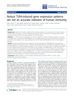
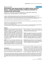
In 1st passage HSC, the ETAR mRNA expression was sig-
nificantly higher than the ETBR mRNA expression (Fig. 1).
However, the ETAR mRNA dramatically decreased in 4th
passage HSC. On the other hand, the ETBR mRNA expres-
sion significantly increased 1.9-fold in 4th passage HSC.
The relative expression ratio of ETAR to ETBR was higher
in 1st passage HSC than in 4th passage HSC.
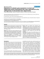
Effect of ET-1 on HSC proliferation
ET-1 in 1st passage HSC did not affect DNA synthesis in
the presence of 0.125%, 5% or 10% fetal calf serum (FCS)
(Fig. 2A), or in the presence of 10
-6
M of BQ123, a selec-
tive ETAR antagonist, or BQ788, a selective ETBR antago-
nist (data not shown). In contrast, ET-1 (10
-10
, 10
-8
, 10
-6
M) dose-dependently reduced DNA synthesis of 4th pas-
sage HSC only in 10% FCS, with maximal inhibition
(49.3%) at 10
-7
M ET-1 (Fig. 2B). This effect was mediated
by the ETBR, since Sarafotoxin (S6c), a selective ETBR ago-
nist, dose-dependently inhibited DNA synthesis (40%
inhibition at 10
-6
M), even in the absence of ET-1 (Fig.
2B). The involvement of the ETBR was confirmed when
ET-1 (10
-6
M) in the presence of the ETAR antagonist
BQ123 (10
-6
M) still reduced DNA synthesis, while the
combination of ET-1 and the ETBR antagonist BQ788 (10
-
6
M) abrogated the inhibitory effect of ET-1 on serum-
stimulated DNA synthesis.
Effect of ET-1 on HSC apoptosis
Spontaneous apoptosis rate was 0.99 ± 0.08% (mean ±
SD, n = 6) in 1st and 2.86 ± 0.52% in 4th passage HSC (n
= 6) when cultured in 10% FCS for 24 h. Addition of ET-
1 (10
-10
, 10
-8
, 10
-6
M) did not alter the basal level of apop-
tosis (1.03%, 1.32% and 1.19%, respectively, in 1st pas-
sage HSC, and 1.46%, 1.53% and 1.25%, respectively in
4th passage HSC). To induce significant apoptosis, cells
were either serum-deprived or treated with Fas-ligand
(Table 1, Fig. 3A). ET-1 (10
-8
M or 10
-6
M) had no effect on
apoptosis induced by serum deprivation in early and late
passage HSC, and did not rescue the cells from apoptosis
when added one h before addition of Fas-ligand (Fig. 3B).
Simultaneous addition of ET-1 and the ETAR and ETBR
antagonists also did not alter Fas-ligand induced apopto-
sis both in 1st and 4th passage HSC.
Effect of ET-1 on HSC matrix-related gene expression
ET-1 at concentrations of 10
-8
M and 10
-6
M increased pro-
collagen α1(I) mRNA expression 1.4- and 1.8-fold,
respectively, in 1st passage HSC, while no effect was found
in 4th passage HSC (Fig. 4). Tissue inhibitor of metallo-
proteinase-1 (TIMP-1) transcript levels remained
unchanged both in 1st and 4th passage HSC (Fig. 4). Only
the ETAR antagonist, BQ123, completely blocked ET-1
enhanced procollagen α1(I) mRNA expression, while the
ETBR antagonist, BQ788, had no effect (Fig. 5). ET-1 (10
-
8
M and 10
-6
M) increased transforming growth factor β-1
(TGFβ-1) mRNA expression 1.2–1.3-fold in 1st passage
HSC which was blocked by the ETAR antagonist. In addi-
tion, ET-1 (10
-8
M and 10
-6
M) upregulated matrix metal-
loproteinase-2 (MMP-2) mRNA transcripts 4- and 6-fold,
respectively, in 1st passage HSC, and both the ETAR and
the ETBR antagonist inhibited this induction completely
(Fig. 6). In 4th passage HSC no effect of ET-1 on TGFβ-1
and MMP-2 mRNA expression was found (data not
shown). [These findings clearly show that ET-1 stimulated
profibrogenic gene expression, i.e. procollagen α1(I),
TGFβ-1 and MMP-2 mRNA, only in 1st passage HSC and
via the ETAR.]
Discussion
Several studies have implicated ET-1 in fibrogenesis of the
kidneys, the cardiovascular system and liver fibrosis.
However, the role of ET-1 in hepatic fibrogenesis and in
particular in HSC matrix production and apoptosis
remains controversial. Therefore we examined cell prolif-
eration, apoptosis and extracellular matrix metabolism of
ET-treated HSC in an early and a late state of activation.
We used 1st passage and 4th passage HSC as an early and
Comparative Hepatology 2006, 5:5 />Page 3 of 12
(page number not for citation purposes)
late state of activation. Our study has shown that ETAR is
dominant in 1st passage HSC and ETBR is dominant in
4th passage HSC. Our results agreed with those reported
by other investigators [11,20]. The progressive activation
in HSC in culture is associated with progressive shift from
a relative predominance of ETAR to relative predomi-
nance of ETBR. A predominance of ETBR was observed
when the cells had undergone complete transition to
myofibroblastic-like phenotype. In vivo study, ETBR is
predominantly expressed in both normal liver and cir-
rhotic liver and overexpressed especially in cirrhotic liver
[21,22]. Taken together, HSC predominantly express
ETAR in early state, activated by several cytokine or liver
damage, and predominantly express ETBR on late acti-
vated state.
Previous studies reported the mitogenic potential of ET-1
in coronary smooth muscle cells and alveolar fibroblasts
[23,24], and Rockey et al. [8] and Pinzani et al. [11] dem-
onstrated that ET-1 stimulates DNA synthesis in early cul-
tured HSC in the presence of low concentrations of FCS.
We were unable to demonstrate any mitogenic effect of
The gene expression of ETAR and ETBR in early- and late-passage HSCFigure 1
The gene expression of ETAR and ETBR in early- and late-passage HSC. A: The gene expression of ETAR and ETBR
in 1st and 4th passage HSC. B: the relative expression ratio of ETAR to ETBR in 1st and 4th passage HSC. HSC were plated on
25 cm
2
dishes at a density of 1.0 × 10
5
cells/dish in DMEM containing 10% FCS. After confluence, cells were washed with PBS
and placed in DMEM with 0.125% FCS for 24 hours. RNA isolation and real time PCR using SYBR Green were performed
according to Material and Methods. Data were normalized to GAPDH mRNA levels. Results are given as mean ± SD (n = 5). *:
p < 0.05.
ETAR
ETAR
ETBR
ETBR
1st passage HSC
4th passage HSC
1st passage
HSC
4th passage
HSC
A
B
*
*
*
*
*
ETAR/ETBR expression ratio
Normalized mRNA levels
Comparative Hepatology 2006, 5:5 />Page 4 of 12
(page number not for citation purposes)
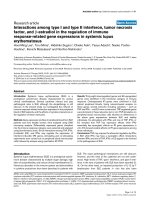
Effect of ET-1 on DNA synthesis and proliferation in HSCFigure 2
Effect of ET-1 on DNA synthesis and proliferation in HSC. A: Effect of ET-1 on DNA synthesis of 1st passage HSC.
BrdU incorporation into DNA was measured in 8 × 10
3
HSC during 48 h and at different concentrations of FCS. Absorbance
values of controls were set as 100% for each concentration of FCS and values normalized to the respective control culture lev-
els. Results were means ± SD (n = 8). There was no effect of ET-1 on the proliferation of 1st passage HSC. B: Inhibitory effect
of ET-1 on the proliferation of 4th passage HSC. BrdU incorporation into DNA was measured in 8x10
3
HSC during 48 hours
at different concentrations of ET-1 or the ETBR agonist S6c with 10% FCS. The ETAR antagonist, BQ123, or the ETBR antago-
nists, BQ788, were added at 10
-6
M in the presence of 10
-6
M of ET-1. Results are given as mean ± SD (n = 8). Absorbance val-
ues of controls were set as 100% and values for cultures under the influence of each concentration of ET-1 or S6c are shown.
*p < 0.05, **p < 0.01 vs control, ## p < 0.01 vs ET -1 10
-6
M.
0
20
40
60
80
100
120
0
20
40
60
80
100
120
control -10
-8 -7 -6
ET-1
+
BQ123
control -10
-8 -7 -6
S6c log(Mol)
(%)
BrdU incorporation
**
**
*
****
*
**
##
ET-1 log(Mol)
ET-1
+
BQ788
0
20
40
60
80
100
120
140
0
20
40
60
80
100
120
140
control
-10 -8 -7 -6
control
-10 -8 -7 -6control
-10 -8 -7 -6
ET-1 log(Mol)
0.125% fetal calf serum
5% fetal calf serum 10% fetal calf serum
(%)
BrdU incorporation
ET-1 log(Mol) ET-1 log(Mol)
A
B
Comparative Hepatology 2006, 5:5 />Page 5 of 12
(page number not for citation purposes)
Table 1: The effects of endothelin-1 on apoptosis induced serum deprivation in 1st and 4th passage HSC.
1
st
passage HSC 4
th
passage HSC
Apoptosis after serum deprivation Apoptosis after serum deprivation
72 hr (%) 120 hr (%) 168 hr (%) 72 hr (%) 120 hr (%) 168 hr (%)
Control 38.8 ± 7.5 40.2 ± 6.6 55.6 ± 10.0 40.2 ± 3.2 46.9 ± 5.2 42.3 ± 5.6
ET-1(10
-8
M) 37.3 ± 9.3 42.4 ± 4.6 56.3 ± 8.0 35.3 ± 4.4 46.9 ± 3.4 36.7 ± 3.3
ET-1(10
-6
M) 31.9 ± 9.9 40.1 ± 5.5 53.3 ± 11.3 36.7 ± 5.8 55.4 ± 14.3 46.2 ± 11.9
Data are given as mean ± standard deviation. ET-1: endothelin-1.
ET-1 in 1st and 4th passage HSC. Moreover, we found
inhibition of cell proliferation by ET-1 in 4th passage
HSC. This discrepancy is likely explained by primary cul-
ture and low FCS concentrations (2–5%) which those
authors used, whereas we used activated HSC after one or
4th passages that appear to resemble the myofibroblasts
obtained by outgrowth from explants of normal liver by
Mallat et al. [20]. In these myofibroblastic HSC use of the
selective ETBR agonist sarafotoxin (6Sc) and the selective
ETBR antagonist (BQ788) demonstrated that this growth
inhibitory effect was mediated by the ETBR. We showed
that passaging of HSC induced a predominance of ETBR
over the ETAR. Taken together, we conclude that ET-1
induces inhibition of cell proliferation in long-term acti-
vated but not early HSC, and that ET-1 does not contrib-
ute to liver fibrosis due to stimulation of HSC
proliferation.
Although ET-1 has been described as a survival factor for
various kinds of cells [25,26], its effect on HSC apoptosis
had not been studied. Using serum deprivation we were
able to induce a reproducible apoptosis rate of 40% both
in 1st and 4th passage HSC. In addition, we induced HSC
apoptosis via the Fas signaling cascade. Fas has been dem-
onstrated to be expressed in liver and to be overexpressed
in acute or chronic liver diseases [27,28]. Furthermore,
activated HSC are more susceptible to Fas-ligand induced
apoptosis than quiescent HSC [19,29-31]. Using both
proapoptotic stimuli, ET-1 did not rescue 1st or 4th pas-
sage HSC from apoptosis.
We could show that ET-1 dose-dependently stimulated
the expression of procollagen α1(I) mRNA in 1st, but not
in 4th passage HSC. Similarly, ET-1 upregulated the
expression of TGFβ-1, the strongest profibrogenic
cytokine. These fibrogenic functions of ET-1 were inhib-
ited by the ETAR antagonist. Our findings are in accord
with data showing that early passage HSC predominantly
express the ETAR, whereas 4th passage HSC and myofi-
broblasts obtained by outgrowth mainly express the ETBR
[11,20]. Contrary to our results, Gandhi et al. [32]
reported that ET-1 stimulates collagen synthesis in HSC
via the ETBR. Although the cause of this difference is
unknown, many reports have shown that the stimulatory
effect of ET-1 for procollagen synthesis in fibroblasts and
vascular smooth muscle cell is mediated by the ETAR
[13,14], in agreement with our present findings in HSC.
Furthermore, we could demonstrate [16] that only an
ETAR in contrast to a mixed (ETAR and ETBR) antagonist
[33] inhibits hepatic fibrosis in rats with secondary biliary
fibrosis due to bile duct ligation and scission in vivo.
Excess extracellular matrix proteins are degraded by
matrix metalloproteinases (MMPs), which are regulated
by specific inhibitors, in particular tissue inhibitor of
MMPs 1 (TIMP-1), which appears to play an important
profibrogenic role in hepatic fibrogenesis [34]. We found
no effect of ET-1 on TIMP-1 expression in 1st and 4th pas-
sage HSC. However, ET-1 stimulated the expression of
MMP-2 mRNA in 1st passage HSC. The upregulation of
MMP-2 favours degradation of the normal subendothelial
matrix, with subsequent replacement by a nonfunctional
interstitial extracellular matrix, including procollagen I. It
also accelerates HSC activation and invasiveness [35,36].
Therefore, ET-1 further likely promotes unfavourable
matrix turnover through the stimulation of collagen 1,
TGFβ-1 and MMP-2.
Although procollagen α1(I) or TGFβ-1 expression were
suppressed only by the ETAR antagonist, MMP-2 expres-
sion induced by ET-1 was inhibited both by the ETAR and
the ETBR antagonist. The reason for this is yet unclear. It
can be speculated that the regulation of MMP-2 expres-
sion may involve other promoter elements than those
stimulated by TGFβ-1. Thus the NFκB family of transcrip-
tion factors induces expression and activation of MMP-2
[37]. Furthermore, ET-1 enhances the DNA-binding activ-
ity of NFκB via ETBR [38]. Therefore, MMP-2 may be
upregulated by both ET-receptors via NFκB.
While it is still not possible to examine to which stages of
liver fibrosis progression early and late passage HSC cor-
respond, hepatic concentrations of ET-1 and densities of
ET-receptors are increased in human and experimental
Comparative Hepatology 2006, 5:5 />Page 6 of 12
(page number not for citation purposes)
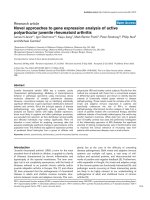
HSC apoptosisFigure 3
HSC apoptosis. A :Apoptotic rate of 1st and 4th passage HSC induced by different concentrations of Fas-ligand. Apoptotic
rate of HSC was measured after HSC were incubated in DMEM with 0.125% FCS containing Fas-ligand at different concentra-
tions for 24 hours. Results are given as mean ± SD (n = 4). **: p < 0.01 vs control. B :Effect of ET-1 and ET receptor antago-
nists on Fas-induced apoptosis of 1st and 4th passage HSC. HSC in DMEM containing 0.125% FCS were incubated with ET -1
alone at 10
-8
M or 10
-6
M with or without Fas-ligand (50 ng/ml), or with ET-1 (10
-6
M), FasL and the ETAR antagonist BQ123 or
the ETBR antagonist BQ788 (both at 10
-6
M). Results are given as mean ± SD (n = 8).
0
10
20
30
40
50
60
70
0
10
20
30
40
50
60
70
control
10 50
Fas ligand(ng/ml)
control
10 50
(%)
1st passage HSC 4th passage HSC
**
**
**
**
Apoptotic rate
Fas ligand(ng/ml)
0
10
20
30
40
50
60
70
0
10
20
30
40
50
60
70
control
10 50
Fas ligand(ng/ml)
control
10 50
(%)
1st passage HSC 4th passage HSC
**
**
**
**
Apoptotic rate
Fas ligand(ng/ml)
A
0
10
20
30
40
50
60
70
80
0
10
20
30
40
50
60
70
80
0
10
20
30
40
50
60
70
0
10
20
30
40
50
60
70
control
Fas L
+
ET-1
(-8)
+
ET-1
(-6)
+
BQ123
(-6)
+
BQ788
(-6)
control
+
ET-1
(-8)
+
ET-1
(-6)
+
BQ123
(-6)
+
BQ788
(-6)
(%) (%)
Apoptotic rate
Fas L
1st passage HSC
4th passage HSC
0
10
20
30
40
50
60
70
80
0
10
20
30
40
50
60
70
80
0
10
20
30
40
50
60
70
0
10
20
30
40
50
60
70
control
Fas L
+
ET-1
(-8)
+
ET-1
(-6)
+
BQ123
(-6)
+
BQ788
(-6)
control
+
ET-1
(-8)
+
ET-1
(-6)
+
BQ123
(-6)
+
BQ788
(-6)
(%) (%)
Apoptotic rate
Fas L
1st passage HSC
4th passage HSC
B
Comparative Hepatology 2006, 5:5 />Page 7 of 12
(page number not for citation purposes)
liver cirrhosis [8,11,32], part of which are contributed by
sinusoidal endothelial cells [39]. Interestingly, a recent
report has shown that TGFβ-1 reduces ET receptor density
in HSC, especially that of the ETBR [40]. Our observation
that 4th passage HSC express more functional ETBR than
ETAR are in line with findings that 4th passage HSC
become less sensitive to auto- and paracrine TGFβ-1 stim-
ulation [41].
Conclusion
ET-1 stimulates the expression of procollagen α1(I) and
TGFβ-1 (through the ETAR) and MMP-2 (through both
ETAR and ETBR) in 1st passage HSC, whereas it inhibits
HSC proliferation in late stages of HSC activation. This
suggests that ET-1 is profibrogenic in early and possibly
antifibrogenic in late stages of hepatic fibrogenesis.
Materials and methods
Materials
Cell culture materials were purchased from Biochem AG
(Berlin, Germany) or Life Technology (Karlsruhe, Ger-
many). ET-1, the ETBR agonist sarafotoxin S6c, the ETAR
antagonist BQ123, the ETBR antagonist BQ788. Fas lig-
and were purchased from Alexis Biochemicals (Gruen-
burg, Germany). BrdU colorimetric cell proliferation
ELISA was from Roche (Mannheim, Germany). Primers
and probes for real time PCR were synthesized at MWG-
Biotech AG (Ebersberg, Germany) and Superscript II
RNase H
-
reverse transcriptase was from Life Technologies
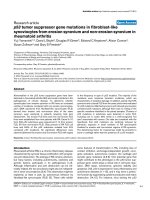
Effect of ET-1 on procollagen type I and TIMP-1 transcript levels of 1st and 4th passage HSCFigure 4
Effect of ET-1 on procollagen type I and TIMP-1 transcript levels of 1st and 4th passage HSC. Cells were incu-
bated without or with 10
-8
M or 10
-6
M ET-1 for 48 hours. Total RNA from HSC was reverse transcribed and transcript levels
of procollagen α1(I) and TIMP-1 were determined by real time quantitative PCR based on the Taqman technology. Data were
normalized to GAPDH mRNA levels. Results are given as mean ± SD (n = 4). **: p < 0.01 vs controls.
0
20
40
60
80
100
120
140
160
180
200
0
20
40
60
80
100
120
140
160
180
200
0
20
40
60
80
100
120
140
160
180
200
0
20
40
60
80
100
120
140
160
180
200
control
ET
(-8)
ET
(-6)
control
ET-1
(-8)
ET-1
(-6)
control
ET-1
(-8)
ET-1
(-6)
control
ET-1
(-8)
ET-1
(-6)
procollagen procollagen
TIMP-1
TIMP-1
**
Normalized m-RNA levels
(%) (%)
1st passage HSC
4th passage HSC
0
20
40
60
80
100
120
140
160
180
200
0
20
40
60
80
100
120
140
160
180
200
0
20
40
60
80
100
120
140
160
180
200
0
20
40
60
80
100
120
140
160
180
200
control
ET
(-8)
ET
(-6)
control
ET-1
(-8)
ET-1
(-6)
control
ET-1
(-8)
ET-1
(-6)
control
ET-1
(-8)
ET-1
(-6)
procollagen procollagen
TIMP-1
TIMP-1
**
Normalized m-RNA levels
(%) (%)
1st passage HSC
4th passage HSC
Comparative Hepatology 2006, 5:5 />Page 8 of 12
(page number not for citation purposes)
(Karlsruhe, Germany). Random hexamers and oligo(dT)
primer were from Promega (Mannheim, Germany). Other
reagents were purchased from Sigma (Seele, Germany).
Cell preparation
HSC were isolated from male Wistar rats (400–500 g,
from Schoenwalde, Germany) fed ad libitum using the
collagenase-perfusion method and purified on a
Nycodenz gradient as described (17). In brief, the liver
was perfused through the portal vein and using an inferior
vena cava outflow using calcium free Hank's balanced salt
solution (HBSS) (Life Technology, Karlsruhe, Germany)
maintained at 37°C at a rate of 10 ml/min for 10 min. The
perfusion was continued with HBSS containing 1.3 mM
CaCl
2
, 0.08% protease E, 0.05% collagenase type IV and
0.001% DNase 1 at a rate of 10 ml/min for 30 min. The
cell suspension was subjected to density gradient centrifu-
gation. The HSC-enriched fraction was suspended in
DMEM containing penicillin (250 U/ml), streptomycin
(250 μg/ml) and 10% FCS, and seeded at a density of 1 ×
10
6
cells/ml. Cell viability was greater than 91% as deter-
mined by Trypan Blue exclusion. HSC purity, as assessed
by phase-contrast microscopy and vitamin A autofluores-
cence immediately after plating, and by immunoreactivity
for desmin one week after plating, was greater than 95%,
with a yield ranging from 1.2 × 10
7
to 1.5 × 10
7
HSC/rat.
Effect of ET receptor antagonists on procollagen α1(I) mRNA expression of 1st passage HSCFigure 5
Effect of ET receptor antagonists on procollagen α1(I) mRNA expression of 1st passage HSC. Cells were stimu-
lated with 10
-6
M ET-1 in the absence or presence of the ETAR antagonist BQ123 (10
-6
M) or the ETBR antagonist BQ788 (10
-
6
M) for 48 hours. The mRNA levels were determined by real time quantitative PCR. Results are given as mean ± SD (n = 4).
**p < 0.01 vs control,
##
p < 0.01 vs ET-1 (10
-6
M).
0
20
40
60
80
100
120
140
160
180
200
0
20
40
60
80
100
120
140
160
180
200
control ET-1
ET-1
+
BQ123
(%)
**
Normalized procollagen α 1(I) mRNA levels
ET-1
+
BQ788
# #
0
20
40
60
80
100
120
140
160
180
200
0
20
40
60
80
100
120
140
160
180
200
control ET-1
ET-1
+
BQ123
(%)
**
Normalized procollagen α 1(I) mRNA levels
ET-1
+
BQ788
# #
Comparative Hepatology 2006, 5:5 />Page 9 of 12
(page number not for citation purposes)
The cells were subcultured (split ratio 1:3) in DMEM with
10% FCS, penicillin (100 IU/ml), streptomycin (100 μg/
ml) and amphotericin B.
We used 1st passage HSC as an early activated state and
4th passage HSC as a late activated state.
DNA synthesis
Cells were plated in 96-well dishes at a density 8 × 10
3
cells/well in complete culture medium. After 24 hours the
cells were washed with PBS and placed in DMEM with
0.125% FCS for 48 hours. This medium was removed and
the cells were placed in fresh DMEM with 0.125%, 2% or
10% FCS containing ET-1 at different concentrations.
After 48 hours incubation with BrdU at 37°C, BrdU incor-
porated into DNA was measured by ELISA according to
the manufacture's protocol.
Induction of apoptosis
Either serum deprivation [18] or Fas-ligand [19] were
used to induce apoptosis in HSC. In serum deprivation
apoptosis, control was made with 10% serum. In Fas-lig-
and induced apoptosis, control was run without Fas-lig-
and. There was no vehicle control. For serum deprivation,
the cells were plated in 6-well dishes at a density of 2 ×
10
4
/well in complete culture medium. After 24 h the cells
were washed with PBS and placed in DMEM with 0.125%
FCS containing ET-1 at increasing concentrations for 72,
Effect of ET-1 on TGFβ-1 and MMP-2 transcript levels of 1st passage HSC and influence of the ET receptor antagonistsFigure 6
Effect of ET-1 on TGFβ-1 and MMP-2 transcript levels of 1st passage HSC and influence of the ET receptor
antagonists. Cells were stimulated with 10
-6
M ET-1 in absence or presence of the ETAR antagonist BQ123 (10
-6
M) or the
ETBR antagonist BQ788 (10
-6
M). TGFβ-1 and MMP-2 transcript levels were determined by real time quantitative PCR, using
Taqman technology, and data were normalized to GAPDH mRNA. Results are given as mean ± SD (n = 4). **: p < 0.01, *: p <
0.05 vs controls,
##
p < 0.01 vs ET-1 (10
-6
M).
0
20
40
60
80
100
120
140
160
control
ET-1
(-8)
ET-1
+
BQ123
Normalized TGFß-1 mRNA levels
ET-1
+
BQ788
0
100
200
300
400
500
600
700
800
Normalized MMP-2 mRNA levels
ET-1
(-6)
control
ET-1
(-8)
ET-1
+
BQ123
ET-1
+
BQ788
ET-1
(-6)
*
**
**
# #
# #
# #
(%)
(%)
0
20
40
60
80
100
120
140
160
control
ET-1
(-8)
ET-1
+
BQ123
Normalized TGFß-1 mRNA levels
ET-1
+
BQ788
0
100
200
300
400
500
600
700
800
Normalized MMP-2 mRNA levels
ET-1
(-6)
control
ET-1
(-8)
ET-1
+
BQ123
ET-1
+
BQ788
ET-1
(-6)
*
**
**
# #
# #
# ## #
(%)
(%)
Comparative Hepatology 2006, 5:5 />Page 10 of 12
(page number not for citation purposes)
120 or 168 h. The medium was exchanged after 72 or 120
h and the apoptotic rate measured by flow cytometry. For
Fas-ligand induced apoptosis, the cells were seeded in 6-
well dishes in complete culture medium and placed in
DMEM with 0.125% FCS for 24 h as before. After 24 h the
medium was replaced by fresh DMEM with 0.125% FCS
containing 1 μg/ml of Fas enhancer (mouse IgG) and 10
to 50 μg/ml Fas-ligand for 24 h. To investigate the influ-
ence of ET-1 on Fas-ligand induced apoptosis, HSC were
cultured with increasing concentrations of ET-1 described
above.
Flow cytometric quantification of apoptotic HSC
HSC were trypsinized and centrifuged for 10 minutes at
500 g. Cells were fixed in 3 ml of 75% ethanol/25% PBS,
diluted in 10 ml PBS and centrifuged for 10 minutes at
500 g. After resuspension in PBS, digestion of RNA with
RNase A (500 μl of 500 μg/ml) at 37°C for 30 minutes
and staining with propidium Iodide at a final concentra-
tion of 100 μg/ml, cell cycle stages were determined by
flow cytometry (Coulter, Epics X/XL Flow cytometry Sys-
tem, Krefeld, Germany). SubG1 events were quantified as
correlate for the rate of apoptosis. At least 12000 events
were collected for each analyzed sample.
Inhibition of ETA and ETB receptors
HSC were plated on 25 cm
2
dishes at a density of 1.0 × 10
5
cells/dish in DMEM containing 10% FCS. After conflu-
ence, cells were washed with PBS and placed in DMEM
with 0.125% FCS for 48 hours. Thereafter cells were incu-
bated with ET-1 for 48 hours in the presence of the ETAR
antagonist BQ123 (10
-6
M) or ETBR antagonist BQ788
(10
-6
M).
RNA isolation and reverse transcription
Total RNA of HSC was extracted by using the acid-phenol
guanidium method. The RNA concentration was deter-
mined by absorbance at 260 nm and the RNA quality ver-
ified by electrophoresis on an ethidium bromide stained
1% agarose gel. Total RNA was reverse transcribed in a
final volume of 20 μl containing 1 × RT buffer (500 μM
each dNTP, 3 mM MgCl2 75 mM KCl, 50 mM Tris-HCl pH
8.3), 10 units of Superscript II RNase H
-
reverse tran-
scriptase (Gibco BRL, Life Technologies, Karlsruhe, Ger-
many), 1 μl of 50 ng/μl random hexamers (Promega,
Mannheim, Germany), 0.5 μl of 100 pmol/ml oligo(dT)
primer and 1~5 μg of total RNA. The samples were incu-
bated at 20°C for 10 minutes, 42°C for 30 min and
reverse transcriptase was inactivated by heating to 99°C
for 5 min and cooling to 5°C for 5 min.
Real time quantitative PCR
We used a Light Cycler System (Roche, Tokyo) and a Light
Cycler-FastStart DNA Master SYBR Green I kit to quantify
mRNA of ETAR and ETBR. Nucleotide sequences of ETAR
and ETBR for the primers were as follows; ETAR (accession
no. NM012550) sense: -ACCAGTCCAAAAGCCTCA-,
antisense: -TCTGCACAGGGTTAGTTCA-; ETBR (accession
no. NM017333) sense: -AACTTCCGCTCCAGCAAT-, anti-
sense: -TCCCGAGGCTTCATTCAT Conditions for real-
time PCR were as follows: 10 min denaturing at 95°C, 10
s annealing at 64 or 62°C, and 5–9 s amplification at
72°C. Forty cycles were performed and then followed by
melting curve analysis to verify the correctness of the
amplification. Analysis of the data was performed accord-
ing to the manufacturer's instructions, using Light Cycler
software version 3.5.3.
The Taqman technology was used to quantify procollagen
I, TIMP-1, TGFβ-1, and MMP-2 mRNA. This method relies
on the correlation between the abundance of mRNA and
the number of PCR cycles necessary to reach a threshold
of detection of a fluorescent probe released during each
successive replication. A standard curve performed with a
serial dilution of a sample showed a constant slope when
amplification occurred between 10 and 40 cycles. Real
time quantitative PCR analysis was performed with a PE
applied Biosystems 7700 sequence Detector (Perkin-
Elmer Applied Biosystems, Faster City, CA), which is a
combined thermal cycler and fluorescence detector. Spe-
cific primers and probes for real time PCR were chosen
with the assistance of the software Primer Express (Perkin-
Elmer Applied Biosystems, Faster City, CA). Rat nucle-
otide sequences for the primers and hybridization probes
were as follows; glyceraldehyde 3-phosphate dehydroge-
nase (GAPDH)(accession no. M17701) sense: -CCT GCC
AAG TAT GAT GAC ATC AAG A-, antisense: -GTA GGC
CAG GAT GCC CTT TAG T-, probe: -CTC GGC CGC CTG
CTT CAC CA-; procollagen I (α1) (accession no. Z78279)
sense: -TTC GGC TCC TGC TCC TCT TA-, antisense: -GTA
TGC AGC TGA CTT CAG GGA TGT-, probe: -TTC TTG
GCC ATG CGT CAG GAG GG-; TIMP-1 (accession no.
U06179) sense: -TCC TCT TGT TGC TAT CAT TGA TAG
CTT-, antisense: -CGC TGG TAT AAG GTG GTC TCG AT-,
probe: -TTC TGC AAC TCG GAC CTG GTT ATA AGG-;
TGFβ-1 (accession no. X52498) sense: -AGAAGTCAC-
CCGCGTGCTAA-, antisense: -TCCCGAATGCTCGACG-
TATTGA-, probe: -
ACCGCAACAACGCAATCTATGACAAAACCA-; MMP-2
(accession no. X71466) sense: -CCGAGGACTATGACCG-
GGATAA-, antisense: CTTGTTGCCCAGGAAAGTGAAG-,
probe: -TCTGCCCCGAGACCGCTATGTCCA
Ten microliters of the RT samples was used for quantita-
tive two step PCR with a 5 minute denaturation step at
95°C, followed by 40 cycles of 15 seconds at 95°C and 1
min at 65°C in the presence of 200 nM specific forward
and reverse primers, 100 mM specific fluorogenic probe,
5 mM MgCl2, 50 mM KCl, 10 mM Tris buffer (PH 8.3),
200 μM each dNTP, and 1.25 units of DNA polymerase.
Comparative Hepatology 2006, 5:5 />Page 11 of 12
(page number not for citation purposes)
Each sample was analyzed in duplicate and a calibration
curve constructed using a 2-fold serial dilution of a stand-
ard cDNA preparation obtained from total RNA of
untreated HSC run in parallel with each analysis. For each
sample, the amounts of procollagen α1(I), TIMP-1, TGFβ-
1 and MMP-2 were divided by the amount of GAPDH to
obtain normalized procollagen α1(I), TIMP-1, TGFβ-1 or
MMP-2 values.
Statistical analysis
Statistical analyses were performed using Statview Version
5 (SAS Institute Inc. NC). Significance of the differences
was studied using the Mann-Whitney U test for non-para-
metric variables. Statistical significance was regarded
when p < 0.05.
Competing interests
The author(s) declare that they have no competing inter-
ests.
Authors' contributions
MS performed most experiments and wrote the manu-
script. MB and AK helped perform experiments. DS, EH
and YM participated in the study design and helped to
draft the manuscript. All authors read and approved the
final manuscript.
Acknowledgements
Supported by the Interdisciplinary Center of Clinical Research (IZKF), grant
B21, of the University of Erlangen-Nuernberg and by the German Network
for Hepatitis (Hepnet).
References
1. Housset CN, Rockey DC, Friedman SL, Bissell M: Hepatic
lipocytes: a major target for endothelin-1. J Hepatol 1995,
22:55-60.
2. Friedman SL: Molecular regulation of hepatic fibrosis, an inte-
grated cellular response to tissue injury. J Biol Chem 2000,
275:2247-2250.
3. Pinzani M, Rombouts K: Liver fibrosis: from the bench to clinical
targets. Dig Liv Dis 2004, 36:231-242.
4. Yanagisawa M, Kurihara H, Kimura S: A novel potent vasocon-
strictor peptide produced by vascular endothelial cells.
Nature 1988, 332:411-415.
5. Clozel M, Gray GA, Breu V, Loffler B, Osterwalder R: The endothe-
lin ET-B receptor mediates both vasodilation and vasocon-
striction. Biochem Biophys Res Comm 1992, 186:867-873.
6. Gandhi CR, Stephenson K, Olson MS: Endothelin, a potent pep-
tide agonist in the liver. J Biol Chem 1990, 265:17432-17435.
7. Rockey DC, Weisiger RA: Endothelin induced contractility of
stellate cells from normal and cirrhotic rat liver: Implica-
tions for regulation of portal pressure and resistance. Hepa-
tology 1996, 24:233-240.
8. Rockey DC, Fouassier L, Chung JJ, Carayon A, Vallee P, Rey C, Hous-
set C: Cellular localization of endothelin-1 and increased pro-
duction in liver injury in the rat: Potential for autocrine and
paracrine effects on stellate cells. Hepatology 1998, 27:472-480.
9. Reinehr RM, Kubitz R, Peters-Regehr T, Bode JG, Haeussinger D:
Activation of rat stellate cells in culture is associated with
increased sensitivity to endothelin 1. Hepatology 1998,
28:1566-1577.
10. Housset C, Rockey DC, Bissell DM: Endothelin receptors in rat
liver: Lipocytes as a contractile target for endothelin 1. Proc
Natl Acad Sci USA 1993, 90:9266-9270.
11. Pinzani M, Milani S, De Franco R, Grappone C, Caligiuri A, Gentilini
A, Tosti-Guerra C, Maggi M, Failli P, Ruocco C, Gentilini P: Endothe-
lin 1 is overexpressed in human cirrhotic liver and exerts
multiple effects on activated hepatic stellate cells. Gasteroen-
terology 1996, 110:534-548.
12. Hocher B, Thone-Reineke C, Rohmeiss P, Schmager F, Slowinski T,
Burst V, Siegmund F, Quertermous T, Bauer C, Neumayer HH, Sch-
leuning WD, Theuring F: Endothelin-1 transgenic mice develop
glomerulosclerosis, interstitial fibrosis, and renal cysts but
not hypertension. J Clin Invest 1997, 99:1380-1389.
13. Guarda E, Katwa LC, Myers PR, Tyagi SC, Weber KT: Effects of
endothelin on collagen turnover in cardiac fibroblasts. Cardi-
ovasc Res 1993, 27:2130-2134.
14. Rizvi MA, Katwa L, Spadone DP, Myers PR: The effects of endothe-
lin-1 on collagen type I and type III synthesis in cultured por-
cine artery vascular smooth muscle cells. J Mol Cell Cardiol
1996, 28:243-252.
15. Rockey DC, Chung JJ: Endothelin antagonism in experimental
hepatic fibrosis. Implications for endothelin in the pathogen-
esis of wound healing. J Clin Invest 1996, 98:1381-1388.
16. Cho JJ, Hocher B, Herbst H, Jia J-D, Ruehl M, Hahn EG, Riecken EO,
Schuppan D: An oral endothelin A receptor antagonist blocks
collagen synthesis and deposition in advanced rat liver fibro-
sis. Gastroenterology 2000, 118:1169-1178.
17. Ramm GA: Isolation and culture of rat hepatic stellate cells. J
Gastroenterol Hepatol 1998, 13:846-851.
18. Shichiri M, Kato H, Marumo F, Hirata Y: Endothelin-1 as an auto-
crine/paracrine apoptosis survival factor for endothelial
cells. Hypertension 1997, 30:1198-1203.
19. Nagata S: Fas-induced apoptosis. Int Med 1998, 37:179-181.
20. Mallat A, Fouassier L, Preaux AM, Gal CS, Raufaste D, Rosenbaum J,
Dhumeaux D, Jouneaux C, Mavier P, Lotersztajn S: Growth inhibi-
tory properties of endothelin-1 in human hepatic myofibrob-
lastic Ito cells. An endothelin B receptor-mediated pathway.
J Clin Invest 1995, 96:42-49.
21. Yokomori H, Oda M, Yasogawa Y, Nishi Y, Ogi M, Takahashi M, Ishii
H: Enhanced expression of endothelin B receptor at protein
and gene levels in human cirrhotic liver. Am J Pathol 2001,
159:1353-1362.
22. Ikura Y, Ohsawa M, Naruko T, Muraguchi T, Hirayama M, Suekane T,
Fukushima H, Sugama Y, Shirai N, Kayo S, Yoshimi N, Ehara S, Tan-
zawa K, Ueda M: Expression of the hepatic endothelin system
in human cirrhotic livers. J Pathol 2004, 204:304-310.
23. Hafizi S, Allen SP, Goodwin AT, Chester AH, Yacoub MH: Endothel-
lin-1 stimulates proliferation of human coronary smooth
muscle via the ETA receptor and is co-mitogenic with
growth factors. Atherosclerosis 1999, 146:351-359.
24. Shahar I, Fireman E, Topilsky M, Grief J, Schwarz Y, Kivity S, Ben-
Efraim S, Spirer Z: Effect of endothelin-1 on α-smooth muscle
actin expression and on alveolar fibroblasts proliferation in
interstitial lung diseases. Int J Immunopharmacol 1999,
21:759-775.
25. Wu-Wong JR, Chiou WJ, Dickinson R, Opgenorth TJ: Endothelin
attenuates apoptosis in human smooth muscle cells. Biochem
J 1997, 328:733-737.
26. Shichiri M, Yokokura M, Marumo F, Hirata Y: Endothelin-1 inhibits
apoptosis of vascular smooth muscle cells induced by nitric
oxide and serum deprivation via MAP kinase pathway. Arteri-
oscler Thromb Vasc Biol 2000, 20:989-997.
27. Seishima M, Takemura M, Saito K, Ando K, Noma A: Increased
serum soluble Fas (sFas) concentrations in HCV-positive
patients with liver cirrhosis. J Hepatol 1997, 27:424-425.
28. Taieb J, Mathurin P, Poynard T, Gougerot-Pocidalo MA, Chollet-Mar-
tin S: Raised plasma soluble Fas and Fas-ligand in alcoholic
liver disease. Lancet 1998, 351:1930-1931.
29. Saile B, Knittel T, Matthes N, Schott P, Ramadori G: CD95/CD95L-
mediated apoptosis of the hepatic stellate cell. A mechanism
terminating uncontrolled hepatic stellate cell proliferation
during hepatic tissue repair. Am J Pathol 1997, 151:1265-1272.
30. Gong WR, Pecci A, Roth S, Lahme B, Beato M, Gressner AM: Trans-
formation-dependent susceptibility of rat hepatic stellate
cells to apoptosis induced by soluble Fas ligand. Hepatology
1998, 28:492-502.
31. Saile B, Matthes N, Knittel T, Ramadori G: Transforming growth
factor beta and tumor necrosis factor alpha inhibit both
Publish with BioMed Central and every
scientist can read your work free of charge
"BioMed Central will be the most significant development for
disseminating the results of biomedical research in our lifetime."
Sir Paul Nurse, Cancer Research UK
Your research papers will be:
available free of charge to the entire biomedical community
peer reviewed and published immediately upon acceptance
cited in PubMed and archived on PubMed Central
yours — you keep the copyright
Submit your manuscript here:
/>BioMedcentral
Comparative Hepatology 2006, 5:5 />Page 12 of 12
(page number not for citation purposes)
apoptosis and proliferation of activated rat hepatic stellate
cells. Hepatology 1999, 30:196-202.
32. Gandhi CR, Kuddus RH, Uemura T, Rao AS: Endothelin stimulates
transforming growth factor-beta1 and collagen synthesis in
stellate cells from control but not cirrhotic rat liver. Eur J
Pharmacol 2000, 406:311-318.
33. Poo JL, Jimenez W, Maria Munoz R, Bosch-Marce M, Bordas N,
Morales-Ruiz M, Perez M, Deulofeu R, Sole M, Arroyo V, Rodes J:
Chronic blockade of endothelin receptors in cirrhotic rats:
hepatic and hemodynamic effects. Gastroenterology 1999,
116:161-167.
34. Schuppan D, Krebs A, Bauer M, Hahn EG: Hepatitis C and liver
fibrosis. Cell Death Differ 2003, 10:S59-S67.
35. Olaso E, Ikeda K, Eng FJ, Xu L, Wang L-H, Lin HC, Friedman SL:
DDR2 receptor promotes MMP-2-mediated proliferation
and invasion by hepatic stellate cells. J Clin Invest 2001,
108:1369-1378.
36. Gaca MD, Zhou X, Issa R, Kiriella K, Iredale JP, Benyon RC: Base-
ment membrane-like matrix inhibits proliferation and colla-
gen synthesis by activated rat hepatic stellate cells: evidence
for matrix-dependent deactivation of stellate cells. Matrix Biol
2003, 22:229-239.
37. Philip S, Bulbule A, Kundu GC: Matrix metalloproteinase-2:
Mechanism and regulation of NF-κB-mediated activation
and its role in cell motility and ECM-invasion. Glycoconjugate J
2004, 21:429-441.
38. Wilson SH, Simari RD, Lerman A: The effect of endothelin-1 on
nuclear factor Kappa B in macrophages. Biochem Biophy Res
Commun 2001, 286:968-972.
39. Kamegaya Y, Oda M, Yokomori H, Ishii H: Role of endothelin
receptors in endothelin-1-induced morphological changes of
hepatic sinusoidal endothelial fenestrae: morphometric
evaluation with scanning electron microscopy. Hepatol Res
2002, 22:89-101.
40. Gabriel A, Kuddus RH, Rao AS, Gandhi CR: Down-regulation of
endothelin receptors by transforming growth factor beta1 in
hepatic stellate cells. J Hepatol 1999, 30:
440-450.
41. Dooley S, Delvoux B, Lahme B, Mangasser-Stephan K, Gressner AM:
Modulation of transforming growth factor beta response and
signaling during transdifferentiation of rat hepatic stellate
cells to myofibroblasts. Hepatology 2000, 31:1094-1106.