Báo cáo y học: "TGF-β dependent regulation of oxygen radicals during transdifferentiation of activated hepatic stellate cells to myofibroblastoid cells" potx
Bạn đang xem bản rút gọn của tài liệu. Xem và tải ngay bản đầy đủ của tài liệu tại đây (1.13 MB, 12 trang )
BioMed Central
Page 1 of 12
(page number not for citation purposes)
Comparative Hepatology
Open Access
Research
TGF-β dependent regulation of oxygen radicals during
transdifferentiation of activated hepatic stellate cells to
myofibroblastoid cells
Verena Proell
1
, Irene Carmona-Cuenca
2
, Miguel M Murillo
2,3
,
Heidemarie Huber
1
, Isabel Fabregat
3
and Wolfgang Mikulits*
1
Address:
1
Department of Medicine I, Division: Institute of Cancer Research, Medical University of Vienna, Borschke-Gasse 8a, A-1090 Vienna,
Austria,
2
Departamento de Bioquímica y Biología Molecular, Facultad de Farmacia, Universidad Complutense de Madrid, Madrid 28040, Spain
and
3
IDIBELL-Institut de Recerca Oncològica, Gran Via s/n, Km 2.7, L'Hospitalet, Barcelona, Spain
Email: Verena Proell - ; Irene Carmona-Cuenca - ;
Miguel M Murillo - ; Heidemarie Huber - ; Isabel Fabregat - ;
Wolfgang Mikulits* -
* Corresponding author
Abstract
Background: The activation of hepatic stellate cells (HSCs) plays a pivotal role during liver injury
because the resulting myofibroblasts (MFBs) are mainly responsible for connective tissue re-
assembly. MFBs represent therefore cellular targets for anti-fibrotic therapy. In this study, we
employed activated HSCs, termed M1-4HSCs, whose transdifferentiation to myofibroblastoid cells
(named M-HTs) depends on transforming growth factor (TGF)-β. We analyzed the oxidative stress
induced by TGF-β and examined cellular defense mechanisms upon transdifferentiation of HSCs to
M-HTs.
Results: We found reactive oxygen species (ROS) significantly upregulated in M1-4HSCs within
72 hours of TGF-β administration. In contrast, M-HTs harbored lower intracellular ROS content
than M1-4HSCs, despite of elevated NADPH oxidase activity. These observations indicated an
upregulation of cellular defense mechanisms in order to protect cells from harmful consequences
caused by oxidative stress. In line with this hypothesis, superoxide dismutase activation provided
the resistance to augmented radical production in M-HTs, and glutathione rather than catalase was
responsible for intracellular hydrogen peroxide removal. Finally, the TGF-β/NADPH oxidase
mediated ROS production correlated with the upregulation of AP-1 as well as platelet-derived
growth factor receptor subunits, which points to important contributions in establishing
antioxidant defense.
Conclusion: The data provide evidence that TGF-β induces NADPH oxidase activity which causes
radical production upon the transdifferentiation of activated HSCs to M-HTs. Myofibroblastoid
cells are equipped with high levels of superoxide dismutase activity as well as glutathione to
counterbalance NADPH oxidase dependent oxidative stress and to avoid cellular damage.
Published: 20 February 2007
Comparative Hepatology 2007, 6:1 doi:10.1186/1476-5926-6-1
Received: 29 May 2006
Accepted: 20 February 2007
This article is available from: />© 2007 Proell et al; licensee BioMed Central Ltd.
This is an Open Access article distributed under the terms of the Creative Commons Attribution License ( />),
which permits unrestricted use, distribution, and reproduction in any medium, provided the original work is properly cited.
Comparative Hepatology 2007, 6:1 />Page 2 of 12
(page number not for citation purposes)
Background
Antioxidant defense mechanisms evolved as a conse-
quence of the aerobic lifestyle caused by the photosyn-
thetic activity of herbal organisms, which in turn depends
on the capability of oxygen reduction occurring during
respiration. Reactive oxygen species (ROS) are essential
for a couple of processes within the cell and play a critical
role in several diseases including liver damage [1]. ROS
are produced (i) by the interaction of ionizing radiation
with biological molecules, (ii) during cellular respiration
and (iii) by myeloperoxidase and nicotinamide-adenine
dinucleotide phosphate (NADPH) oxidase of phagocytic
cells such as neutrophils and macrophages. In addition,
several non-phagocytotic cell types such as hepatocytes
[2] and hepatic stellate cells (HSCs) [3] have also been
shown to express a NADPH oxidase-like enzyme playing
an important role in the generation of ROS [4].
Strong oxidants like ROS can damage proteins, lipids (lip-
idperoxidation) as well as DNA, and therefore have been
suggested to have a critical implication in carcinogenesis
[5]. As a consequence, each cell type harbors several
defense mechanisms against the noxious effects of oxida-
tive stress. Two enzymes play a major protective role,
namely the superoxide dismutase (SOD), which converts
two superoxide anions (O
2
-
) into hydrogen peroxide
(H
2
O
2
) and oxygen, and catalase, which promotes the
conversion of hydrogen peroxide to water and molecular
oxygen. Antioxidants such as ascorbic acid, β-carotene
and α-tocopherol also reduce danger from accidentally
produced ROS. Another defense mechanism is based on
glutathione (γ-glutamyl-cysteinyl-glycine, GSH), which
participates in many different cellular actions including
nutrient metabolism and regulation of cellular events
such as signal transduction, cytokine production, cell pro-
liferation, apoptosis and immune response [6]. However,
GSH is mainly known as an intracellular redox system
exhibiting two conformations, the antioxidant "reduced
glutathione" tripeptide conventionally termed as the
above mentioned GSH, and the oxidized form, a sulfur-
sulfur linked compound known as glutathione disulfide
(GSSG).
Apart from putative harmful consequences caused by
ROS, recent reports demonstrate that free radicals are also
implicated in cell signaling, especially in tumor cells and
cells determined to undergo apoptosis. There exist strong
evidence particularly for liver diseases that increased pro-
duction of free radicals and/or impaired antioxidant
defense mechanisms are involved. As a consequence,
numerous studies have been focused on the pathological
significance of ROS in liver injury as well as on therapeutic
intervention with antioxidants [1,7-10].
Hepatic stellate cells play a pivotal role during liver injury.
In the adult healthy liver, HSCs are considered as the prin-
cipal storage site of retinoids, whereas HSCs get activated
to myofibroblasts (MFBs) upon liver damage. This
transdifferentiation is accompanied by drastic morpho-
logical changes including loss of cytoplasmic lipid drop-
lets and alterations in protein synthesis patterns, which
comprises de novo synthesis of α-smooth muscle actin [11-
14]. Furthermore, HSC-derived MFBs are mainly respon-
sible for extracellular matrix (ECM) remodeling in the
fibrotic liver, which represents a hallmark of fibrogenesis.
In particular, MFBs secrete high levels of the interstitial
collagens I and III [15] as well as several matrix metallo-
proteinases (MMPs) [14,16] and tissue inhibitors of
MMPs [16-18], resulting in a dense and rigid network of
matrix constituents which exerts physical stress on sur-
rounding cells.
Whether ROS are implicated in HSC activation and which
molecular mechanisms are the basis for the transdifferen-
tiation of HSCs to MFBs is still a matter of debate. Lee and
colleagues demonstrated that ROS are indispensable for
HSCs activation and that c-myc and NF-κB act as molecu-
lar mediators of oxidative stress [19]. In addition, co-cul-
ture experiments have shown that extracellular ROS,
produced by stable cytochrome P450 2E1 (CYP2E1) over-
expression in HepG2 cells, facilitate activation of quies-
cent HSCs in vitro, resulting in increased expression of
collagen I and α-SMA [20]. Moreover, treatment of hepa-
tocytes with nitrilotriacetate complex results in oxidative
stress response. It has been shown that transfer of condi-
tioned medium on HSCs stimulated their proliferation as
well as collagen I accumulation within these cells [21].
Similar results were obtained in Kupffer and other inflam-
matory cells, which have been shown to produce H
2
O
2
[19,22,23].
One of the most extensively studied antagonistic player of
ROS is GSH, which has been reported to be significantly
upregulated in cultured primary HSCs at day seven com-
pared to freshly isolated HSCs [24]. In addition, long-
term cultured HSCs exhibit a higher synthesis rate of GSH
compared to cells in short-term culture. In contrast, no
increased GSH or γ-glutamyl-cysteine synthetase (GCS)
level has been observed in isolated HSCs from fibrotic rat
livers after 8 weeks of bile duct ligation or 4 weeks of CCl
4
treatment [24].
Recently, we published a hepatic stellate cell line referred
to as M1-4HSC [25], which has been isolated from p19
ARF
null mice and represents activated HSCs displaying an
amazing plasticity concerning their morphology. Since
they have undergone spontaneous activation in vitro, M1-
4HSCs have already lost fat-storing droplets and express
high amounts of α-SMA. Due to TGF-β administration,
Comparative Hepatology 2007, 6:1 />Page 3 of 12
(page number not for citation purposes)
these cells are provoked to undergo a further activation
process to myofibroblastoid cells, termed M-HTs [25,26].
Hence, this cellular model provides the unique ability to
study late stage events of HSCs activation, i.e. the transdif-
ferentiation of activated HSCs to MFBs. Indeed, most
studies investigating HSC activation have employed
freshly isolated, quiescent HSCs and monitored spontane-
ous activation which takes place as soon as cells are cul-
tured in vitro, whereby TGF-β is suggested to accelerate
transdifferentiation even though it is not required [27].
We addressed the question whether oxidative stress is
implicated in late stage activation of M1-4HSCs to myofi-
broblastoid M-HTs. In order to elucidate whether ROS
plays a role in TGF-β driven transdifferentiation, we mon-
itored ROS levels during the first 72 hours of TGF-β treat-
ment, which is referred to as induction phase. We show
that ROS are upregulated during TGF-β driven HSCs acti-
vation, whereas M-HTs displayed a very effective counter-
regulation to TGF-β induced oxidative stress by
upregulation of SOD enzymatic activity rather than cata-
lase. In addition, genes implicated in the response to oxi-
dative stress such as c-fos and c-jun as well as platelet-
derived growth factor (PDGF) receptors α and β are
shown to be regulated which points to their regulatory
functions in establishing resistance to oxidative stress.
Results and discussion
Increase of ROS levels during the induction phase of TGF-
β
driven M1-4HSCs activation to MFBs
The cell line M1-4HSC represents activated HSCs, which
undergo further activation to myofibroblast-like cells in
response to TGF-β [25]. The induction phase refers to 72
hours of TGF-β treatment, which is characterized by the
change to a myofibroblastoid morphology (Fig. 1A). After
20 days of TGF-β administration, the cells represent acti-
vated MFBs with a stable phenotype, termed M-HTs.
Transdifferentiation of M1-4HSCs to M-HTs shows
increased nuclear accumulation of Smad2/3 (Fig. 1B),
indicating a further activation of TGF-β signaling. In addi-
tion, M-HTs exhibit decreased expression of desmin (Fig.
1C), as reported recently [25]. This cellular model pro-
vides the unique ability to monitor late stage events dur-
ing fibrogenesis, since spontaneous activation has already
occurred. In order to examine whether ROS are implicated
in this transdifferentiation from activated HSCs to MFBs,
we analyzed intracellular hydrogen peroxide during the
induction phase compared to untreated M1-4HSCs and
M-HTs. Hydrogen peroxide was used as a general marker
of oxidative stress since all forms of oxygen radicals that
occur intracellularly are finally converted into H
2
O
2
. We
observed a significant increase in ROS levels after 48 and
72 hours of TGF-β treatment (Fig. 2A), whereas no eleva-
tion of hydrogen peroxide levels was determined after 24
hours. Since basal levels of ROS are already induced in
M1-4HSCs compared to quiescent HSCs, as reported by
several investigators [20,28-30], TGF-β is obviously able
to provide accumulation of hydrogen peroxide in M1-
4HSCs. In contrast, M-HTs showed about 40% reduced
intracellular hydrogen peroxide content compared to
untreated M1-4HSCs (Fig. 2B). Hence, these data raised
the question whether the lowered ROS levels in M-HTs
were caused by reduced ROS production or by the upreg-
ulation of cellular antioxidant defense mechanisms. To
properly tackle this issue we asked for the major source of
ROS in M1-4HSCs caused by TGF-β administration.
TGF-
β
treatment of M1-4HSCs results in induction of
NADPH oxidase activity
In most cell types, mitochondria-anchored enzymes pro-
vide the majority of ROS such as NADPH-ubiquinone oxi-
doreductase and ubiquinol cytochrome oxidoreductase
[31]. Another important source for ROS is NADPH oxi-
dase, which has been shown to be active in several non-
phagocytotic cell types including HSCs [32]. This NADPH
oxidase-like enzyme is a multi-protein complex consisting
of the transmembrane proteins p22
phox
and the p91
phox
-
related enzymes of the NADPH oxidase (Nox) family, the
cytosolic proteins p47
phox
and p67
phox
as well as the small
GTP binding protein Rac. NADPH oxidase activity
depends amongst others on the co-enzyme flavin and can
be therefore inhibited by diphenyleneiodonium chloride
(DPI). The involvement of NADPH oxidase in the TGF-β-
dependent increase of oxidative stress in M1-4HSC was
obtained by measurements of ROS content in cells that
have been treated with TGF-β 1 in the presence of DPI.
M1-4HSC starved for 6 hours and administrated with
TGF-β 1 for 3 hours resulted in an increase of ROS levels
to 50% (Fig. 2C). This accumulation of oxidative stress
was impaired by simultaneous co-incubation with TGF-β
and DPI, as ROS levels under these conditions were com-
parable to those measured in control cells.
Hence, we analyzed whether NADPH oxidase activity was
affected in response to TGF-β1. The analysis revealed a
strong elevation of NADPH oxidase activity after 48 hours
which decreased again after 72 hours (Fig. 2D). In agree-
ment with ROS levels observed at 24 hours, no NADPH
oxidase activity could be detected. In contrast, myofibrob-
lastoid M-HTs exhibited a highly elevated activity of the
ROS producing enzyme compared to untreated M1-
4HSCs. These results excluded that the intracellular
hydrogen peroxide levels in these cells were caused by a
low production of free radicals. In line with these data, the
components of NADPH oxidase were found to be differ-
entially transcribed. Control M1-4HSC and those treated
with TGF-β as well as M-HTs were analyzed for NADPH
oxidase components via linear, semi-quantitative RT-PCR.
RhoA was used as control for RT-PCR because no varia-
tions in expression levels have been found upon transdif-
Comparative Hepatology 2007, 6:1 />Page 4 of 12
(page number not for citation purposes)
ferentiation of M1-4HSCs, as described recently [25].
Both Nox4 and p47
phox
were significantly upregulated
during the induction phase of M-1HSCs activation to M-
HTs (Fig. 3). Nox4 and p47
phox
mRNA levels were already
augmented after 24 hours of TGF-β1 administration
although NADPH oxidase activity was increased 48 hours
post TGF-β1 treatment. This discrepancy might be
explained by the fact that transcription precedes transla-
tion and functional activation of the enzyme. Nox4 tran-
script levels were also found to be elevated in M-HTs (Fig.
3), which was in line with the high NADPH oxidase activ-
ity (Fig. 2D). Notably, Nox1, gp91
phox
(Nox2) and Nox3
showed comparably enhanced mRNA levels in M-HTs
(data not shown).
Taken together, these data point to a direct influence of
TGF-β on NADPH oxidase activity and subsequent ROS
accumulation during the transdifferentiation of M1-
4HSCs to MFBs. According to the literature, upregulation
of ROS due to TGF-β has also been shown in various cell
types such as vascular smooth muscle cells [33], hepato-
cytes [34], fetal lung fibroblasts [35], cardiac fibroblasts
[36] and also HSCs [28], most frequently by upregulation
of NADPH oxidase activity [33,35-37]. However, the data
available for HSCs refer to (i) the activation of quiescent
HSCs and (ii) to proportionally short incubation times in
comparison to the fibrosis model employed in this study.
We focused on the induction phase within 72 hours com-
pared to untreated, but already spontaneously activated
parental M1-4HSCs and M-HTs, the latter grown for long-
term in TGF-β supplemented medium and comparable to
HSC-derived MFBs in vivo. These cells display very high
NADPH oxidase activity as well as increased p47
phox
and
Nox mRNA despite of diminished levels of free radicals.
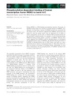
Cellular model of hepatic fibrosisFigure 1
Cellular model of hepatic fibrosis. (A) Morphological changes of M1-4HSCs treated with TGF-β1 either for 72 hours or
for long-term (myofibroblastoid M-HT) as analyzed by phase contrast microscopy. (B) Nuclear translocation of Smad2/3 as vis-
ualized by confocal immunofluorescence analysis. (C) Confocal immunofluorescence images after staining of cells with anti-
desmin antibody.
A
B
C
40 µm
M1-4HSC
M1-4HSC + 72h TGF-β M-HT
40 µm
Phase
Smad2/3
Desmin
40 µm
Comparative Hepatology 2007, 6:1 />Page 5 of 12
(page number not for citation purposes)
TGF-β mediated accumulation of ROS associates with increased NADPH oxidase activityFigure 2
TGF-β mediated accumulation of ROS associates with increased NADPH oxidase activity. (A) During the TGF-β
dependent transdifferentiation of M1-4HSCs, ROS levels increase after 48 and 72 hours. (B) M-HTs show a reduction of ROS
levels to about 50% as compared to untreated M1-4HSCs. (C) DPI inhibits TGF-β caused ROS accumulation in M1-4HSCs. (D)
TGF-β treatment of M1-4HSCs induces NADPH oxidase activity after 48 hours. M-HTs display vast NADPH oxidase activity.
For all situations, n = 3. * p < 0.05.
D
0
0.2
0.4
0.6
0.8
1.0
1.2
1.4
pmol NADPH / min · mg
*
+
2
4
h
TG
F
-
β
+
4
8
h
TG
F
-
β
+
7
2
h
TG
F
-
β
M
-
H
T
M
1
-
4
H
S
C
0
50
100
150
+
2
4
h
TG
F
-
β
+
4
8
h
TG
F
-
β
+
7
2
h
TG
F
-
β
*
*
M
1
-
4
H
S
C
A
Percent (ROS)
0
50
100
150
200
Percent (ROS)
+
TG
F
-
β
+
D
P
I
+
TG
F
-
β
M
1
-
4
H
S
C
C
*
Percent (ROS)
50
100
150
M
-
H
T
*
M
1
-
4
H
S
C
B
0
Comparative Hepatology 2007, 6:1 />Page 6 of 12
(page number not for citation purposes)
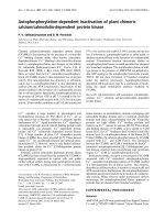
Expression profiling of oxidative stress components by semiquantitative RT-PCRFigure 3
Expression profiling of oxidative stress components by semiquantitative RT-PCR. GCS, γ-glutamylcysteine syn-
thetase; GSHPx, glutathione peroxidase; GSSG-R, glutathione reductase; SOD 1, Cu/Zn superoxide dismutase; SOD 2, mito-
chondrial superoxide dismutase. The constitutive expression of rhoA is shown as loading control.
GCS
GSHPx
GSSG-R
Nox4
p47
M
1
-
4
H
S
C
+
2
4
h
T
G
F
-
β
+
4
8
h
T
G
F-
β
+
7
2
h
T
G
F-
β
M
-
H
T
rhoA
Catalase
SOD 1
SOD 2
Comparative Hepatology 2007, 6:1 />Page 7 of 12
(page number not for citation purposes)
Accordingly, Bataller et al. have recently shown the tran-
scriptional upregulation of p47
phox
in quiescent HSCs and
activated HSCs isolated from healthy and cirrhotic rat liv-
ers, respectively [32]. In order to clarify the contradiction
of reduced oxidative stress in M-HTs with a concomitant
high activity of NADPH oxidase, we addressed the ques-
tion for the regulation of counteracting mechanisms.
Enzymatic defense mechanisms reduce TGF-
β
induced
oxidative stress in M-HTs
To examine whether enzymatic defense strategies partici-
pate in the protection against intracellular ROS accumula-
tion, we analyzed alterations in the enzyme activity of
SOD. The superoxide anion O
2
-
is produced by NADPH
oxidase and arises as free radical through leaking away
from respiratory chain. In mammals, three SOD isoforms
have been identified such as cytosolic Cu/Zn-SOD (SOD
1), mitochondrial Mn-SOD (SOD 2), and extracellular
Cu/Zn SOD (SOD 3), which are responsible for the
destruction of O
2
-
to hydrogen peroxide and oxygen. Sub-
sequently, catalase and/or GSH redox cycle are responsi-
ble for removing hydrogen peroxide from the cell with
water and oxygen as products. No significant alteration in
SOD activity was observed during initiation of TGF-β
driven MFB activation, whereas SOD activity in M-HTs
was upregulated to 50% as compared to M1-4HSC (Fig.
4A). RT-PCR revealed a slight upregulation of SOD 1
mRNA during induction phase and showed highest
expression in M-HTs (Fig. 3). The expression of SOD 2
mRNA also slightly increased after TGF-β treatment of
M1-4HSCs. The high level expression of SOD 2 transcripts
maintained upon kinetics of TGF-β administration and
was even observed in M-HTs. An increase in SOD1 expres-
sion might produce a gain in the cytosolic SOD activity,
which counteracts ROS production at the plasma mem-
brane level. These results are in line with data obtained by
the analysis of NADPH oxidase, which showed strongly
enhanced activity in M-HTs, indicating huge amounts of
superoxide anion that has to be removed mainly by the
involvement of SOD.
Taken together these results point to an essential role for
SOD 1 in M-HTs, facing an augmented superoxide anion
content that has to be removed in order to protect cells
from unfavorable consequences. This is essential even
though oxidative stress supports the establishment of
HSC activation and fibrosis, which has also been shown
for stellate cells in the pancreas (PSC). For instance, Emori
et al. reported an important role of SOD in PSC activation,
as blocking by diethyldithiocarbamate resulted in a signif-
icant induction of α-SMA positive cells [38]. Therefore, we
consider that TGF-β induced elevation of ROS is crucial
for the transdifferentiation of M1-4HSCs to M-HTs. How-
ever, MFBs also depend on the reduction of free radical
accumulation in order to survive.
Regulation of defense mechanisms against oxidative stress during the TGF-β driven transdifferentiation of M1-4HSCs to M-HTsFigure 4
Regulation of defense mechanisms against oxidative
stress during the TGF-β driven transdifferentiation of
M1-4HSCs to M-HTs. (A) SOD activity (n = 2). (B) Cata-
lase activity (n = 4). (C) Glutathione levels (n = 3). * p < 0.05.
Percent (Catalase)
0
20
40
60
80
100
120
*
*
+
2
4
h
T
G
F
-
β
+
4
8
h
T
G
F
-
β
+
7
2
h
T
G
F
-
β
M
-
H
T
M
1
-
4
H
S
C
B
0
Percent (SOD activity)
50
100
150
200
*
+
2
4
h
T
G
F
-
β
+
4
8
h
T
G
F
-
β
+
7
2
h
T
G
F
-
β
M
-
H
T
M
1
-
4
H
S
C
A
C
0
50
100
150
200
250
300
Percent (GSH+GSSG)
*
*
+
2
4
h
T
G
F
-
β
+
4
8
h
T
G
F
-
β
+
7
2
h
T
G
F
-
β
M
-
H
T
M
1
-
4
H
S
C
Comparative Hepatology 2007, 6:1 />Page 8 of 12
(page number not for citation purposes)
Catalase fails to resist elevated hydrogen peroxide levels
In order to reduce oxidative stress, intracellular H
2
O
2
is
dismutated to water and oxygen either by catalase or GSH
redox cycle. Interestingly, we observed a slight downregu-
lation of catalase activity during induction phase and a
moderate upregulation in M-HTs as compared to parental
M1-4HSCs (Fig. 4B). Corresponding mRNA levels were
not regulated at all (Fig. 3) which leads to the conclusion
that catalase is not crucially involved in oxidative stress
defense during M1-4HSCs activation to M-HTs. These
data are contrary to Bleser et al. who demonstrated that
catalase mRNA levels where strongly induced in activated
HSCs in vivo as well as in vitro. Therefore, we suggest that
catalase induction represents an early event in HSCs acti-
vation, which does not participate in the counterregula-
tion of oxidative stress in M-HTs. Previous data indicate a
discriminating role between low and high concentration
of H
2
O
2
, determining whether catalase or GSH redox cycle
is more likely to clear free radicals. In general, it is pro-
posed that GSH is more efficient at low intracellular H
2
O
2
concentrations whereas high amounts of H
2
O
2
are prefer-
entially removed by catalase [29,39-41]. This points to
rather moderate H
2
O
2
levels in M1-4HSCs, which might
be important in signal transduction supporting the late
stage activation to M-HTs. Therefore, we hypothesized
that the GSH redox cycle must have considerable implica-
tions in developing resistance to ROS in M-HTs.
Glutathione upregulation refers resistance to TGF-
β
induced oxidative stress in activated HSCs
Among various other functions, GSH is mainly involved
in the maintenance of the intracellular redox homeostasis
including removal of hydrogen peroxide. We found that
total glutathione levels were upregulated after 24 and 48
hours, and were even more than doubled after 72 hours of
TGF-β treatment (Fig. 4C). This elevation in intracellular
glutathione content was further detected in M-HTs, which
exhibited a 2.5 fold higher level than untreated M1-
4HSCs. Moreover, we analyzed whether the expression of
redox cycle components are affected. Noteworthy, the
production of glutathione is achieved by de novo synthesis
through synthetases such as γ-glutamyl-cysteine syn-
thetase (GCS). Interestingly, RT-PCR analyses of the corre-
sponding transcript showed that GCS was slightly induced
after 24 hours TGF-β treatment and maintained elevated
in M-HTs (Fig. 3). In addition, we examined the mRNA
level of glutathione peroxidase (GSHPx) and glutathione
reductase (GSSG-R), which are suggested to be involved in
removing peroxides (using GSH as substrate) and reduc-
ing GSSG, respectively. However, no modulation of tran-
script levels was found.
Taken together, these results suggest a direct regulation of
NADPH oxidase by TGF-β and increased ROS levels as
well as a particular contribution of GSH in the resistance
to augmented oxidative stress. Interestingly, Bleser et al.
proposed catalase to be more effective to remove high
local concentrations of ROS, which are represented by
intracellular produced H
2
O
2
. Contrary, extracellular
H
2
O
2
results in consumption of GSH. This might be true
for the early activation phase of quiescent HSCs but not
for the completion of transdifferentiation to MFBs. Since
catalase activity was slightly downregulated during 72
hours of TGF-β treatment and reached a moderate activity
in M-HTs, catalase might not be effective in removing
H
2
O
2
. In conclusion, these data indicate that SOD activity
is responsible for reduction of oxidative stress in M-HTs in
cooperation with GSH. In order to gain insight into how
these pathways might be regulated, we analyzed target
genes that are involved in the response to oxidative stress.
Transcriptional upregulation of AP-1 transcription factors
and PDGF receptor subunits during HSCs activation to
myofibroblastoid M-HTs
Since AP-1 transcription factor is involved in stress
response, we examined the regulation of its subunits c-fos
and c-jun by RT-PCR during the transdifferentiation of
M1-4HSCs to M-HTs. The upregulation of both mRNAs
was maintained in M-HTs (Fig. 5), which points to a reg-
ulatory function of AP-1 involved in establishing resist-
ance to oxidative stress. Since it has been shown that
PDGF is regulated upon oxidative stress [3], we deter-
mined mRNA levels of PDGF receptors α and β in M1-
4HSCs. Indeed, PDGF receptor transcripts were increased
within the induction phase, since upregulation of PDGF-
Rα mRNA was detected after 48 hours and even further
increased after 72 hours as well as in M-HTs. Unlike
PDGF-Rα, whose mRNA levels were not affected after 24
hours TGF-β administration, PDGF-Rβ mRNA abundance
already peaked at 24 hours with a more than 10-fold
induction. Besides its well known function as potent
mitogen, PDGF is implicated in numerous other processes
including wound healing and the formation of connective
tissue by stimulating the production of several matrix
molecules such as collagens and fibronectin [42]. This is
in accordance with our data since HSC-derived M-HTs
secrete vast amounts of these ECM components (unpub-
lished data), which mimics the in vivo situation during
liver fibrogenesis. In addition, it has been shown by
Adachi et al. that PDGF-BB ligand induces NADPH oxi-
dase to produce ROS, which in turn stimulates prolifera-
tion of LI-90 cells [3]. Thus, the upregulation of PDGF-Rβ
expression might contribute to the increase of NADPH
oxidase activity in M-HTs.
In summary, we show that even though M-HTs harbor
hyperactive NADPH oxidase, these myofibroblastoid
derivatives of M1-4HSCs have reduced ROS levels com-
pared to the untreated cell line. The cellular antioxidant
defense mechanism depends on the increased activity of
Comparative Hepatology 2007, 6:1 />Page 9 of 12
(page number not for citation purposes)
SOD, which converts the free radical O
2
-
to hydrogen per-
oxide that is subsequently reduced either by the GSH
redox cycle or by catalase. Since catalase does not seem to
be affected during this process of HSC activation, we sug-
gest that the resistance to oxidative stress in M-HTs hinges
on the significantly increased availability of GSH.
Conclusion
The current investigation demonstrates the TGF-β
dependent production of reactive oxygen species upon
transdifferentiation of derivatives of hepatic stellate cells
(M1-4HSC line) to M-HTs. The data provide evidence that
(i) the increase of oxidative stress correlates with a gain in
NADPH oxidase activity, and (ii) superoxide dismutase
activation in cooperation with glutathione reduces radical
accumulation in myofibroblastoid cells. These defense
mechanisms are suggested to be particularly relevant in
order to protect myofibroblastoid cells from harmful con-
sequences caused by oxidative stress.
Materials and methods
Cell lines
M1-4HSC and derivative M-HT lines were grown in
DMEM plus 10% fetal calf serum (FCS) as described pre-
viously [25]. M-HTs were additionally supplemented with
1 ng/ml TGF-β1 (R&D Systems, Minneapolis, USA). All
cells were kept at 37°C and 5% CO
2
, and routinely
screened for the absence of mycoplasma.
Confocal immunofluorescence microscopy
Cells were fixed and permeabilized as described recently
[25]. Primary antibodies were used at following dilutions:
anti-Smad2/3 (Transduction Laboratories, Lexington,
UK), 1:100; anti-desmin (DAKO Corp., Carpinteria, CA,
USA), 1:100. After application of cye-dye conjugated sec-
ondary antibodies (Jackson Laboratories, West-Grove,
USA), imaging of cells was performed with a TCS-SP con-
focal microscope (Leica, Heidelberg, Germany). Nuclei
were visualized using To-PRO3 at a dilution of 1:10,000
(Invitrogen, Carlsbad, USA).
Measurement of intracellular ROS
Intracellular ROS was measured as previously described
[43] with minor modifications. Briefly, cells were plated
in 12 well plates and treated with TGF-β1 for the indicated
time. For measurement, cells were incubated for 1 hour
with 2.5 µM of the oxidation-sensitive probe 2'7'-dichlo-
rodihydrofluorescein diacetate (DCFH-DH) (Invitrogen,
Carlsbad, USA) in DMEM plus 10% FCS. Cellular fluores-
cence intensity was measured at 485/20 and 530/25 nm
with Fluorimeter (Wallace) and depicted in percentage
with respect to control, as represented by untreated M1-
4HSCs.
Diphenyleneiodonium chloride (DPI; Sigma) was used at
a final concentration of 20 µM. M1-4HSCs have been
treated with TGF-β1 for 3 hours or co-incubated with DPI
(4 hours) and TGF-β (3 hours) during overall starvation of
6 hours. Cellular fluorescence intensity was again
depicted in percentage with respect to control, repre-
sented by 6 hours starved M1-4HSCs.
Analysis of NADPH oxidase activity
Cells were harvested by trypsinization, pelleted by centrif-
ugation at 2,500 g for 5 min at 4°C, and resuspended in
PBS, followed by incubation with 250 µmol/l NADPH.
NAD(P)H oxidase activity was analyzed as previously
described [31]. NADPH consumption was monitored by
the decrease in absorbance at λ = 340 nm for 5 min. For
analysis of specific NADPH oxidase activity, the rate of
consumption of NADPH inhibited by DPI was measured
by adding 10 µmol/l DPI 30 min prior to measurement.
For normalization, protein concentration was determined
by lysis of an aliquot of cells by adding SDS and protein
measurement by Lowry solution. The absorption extinc-
tion coefficient used to calculate the amount of NADPH
consumed was 6.22 mM
-1
cm
-1
. Results were expressed as
pmol/l of substrate per minute per milligram of protein.
Glutathione determination
Cells were washed twice, scraped in PBS at 90% density
and centrifuged at 950 g for 5 min at 4°C. Cellular glu-
tathione was extracted in a buffer containing 0.2% Triton
X-100, 2.5% sulfosalicylic acid, and then centrifuged at
Steady state transcript levels of PDGF receptors and AP-1 components as analyzed by semiquantitative RT-PCRFigure 5
Steady state transcript levels of PDGF receptors and
AP-1 components as analyzed by semiquantitative
RT-PCR. The constitutive expression of rhoA is shown as
loading control.
PDGF-Rα
PDGF-Rβ
c-fos
c-jun
rhoA
M
1
-
4
H
S
C
+
2
4
h
T
G
F
-
β
+
4
8
h
T
G
F
-
β
+
7
2
h
T
G
F
-
β
M
-
H
T
Comparative Hepatology 2007, 6:1 />Page 10 of 12
(page number not for citation purposes)
10,000 g for 10 min at 4°C. The supernatant was used for
determination of total (GSH and GSSG) glutathione by
the Griffith's method, modified as described previously
[44,45]. Using glutathione as standard, glutathione con-
tent is expressed as pmol/µg protein and represented as
percentage with respect to untreated M1-4HSCs (control).
Analysis of superoxide dismutase activity
Enzyme activity was determined as previously described
[31]. Briefly, cells were harvested as described for glutath-
ione determination. Pellets were lysed in 150 µl 50 mM
di-sodiumphosphate buffer containing 0.5% Triton X-
100, 1 mM PMSF and 5 µg/ml Leupeptin and sonicated.
Lysates were purified by centrifugation at 13,000 g for 10
min at 4°C. SOD activity was measured by monitoring the
autooxidation of 6-hydroxy-dopamine. Autooxidation is
inhibited by 6-hydroxy-dopamine consuming superoxide
generated during this process, as described previously
[31,46]. Briefly, the kinetics of autooxidation of 6-
hydroxy-dopamine were monitored by λ = 490 nm for 60
sec under conditions that resulted in linear kinetics.
Assays of protein extracts (20–30 µg protein in 20 µl pro-
tein extract) were carried out under conditions that
resulted in 40% – 60% inhibition of the autooxidation of
6-hydroxy-dopamine. Measurements were repeated three
times. Data were calculated as percentage of inhibition of
the autooxidation of 6-hydroxy-dopamine that was
obtained with 10 µg protein. The values are depicted as
percentage with respect to untreated M1-4HSCs (control).
Analysis of catalase activity
Cell harvest and protein extract preparation was per-
formed as described for SOD activity measurement. Cata-
lase activity was measured by monitoring the
disappearance of hydrogen peroxide at λ = 240 nm [46].
The reaction mixture contained 40 – 80 µg protein, 50
mmol/l potassium phosphate buffer, pH 7.0, and 10
mmol/l H
2
O
2
. Changes in absorbance were measured for
100 sec. The specific activity was calculated as previously
described [31] and depicted as percentage with respect to
untreated M1-4HSCs (control).
Reverse transcription polymerase chain reaction (RT-PCR)
The extraction of poly(A)+ mRNA, reverse transcription to
cDNA and PCR were performed as described previously
[47]. The conditions for the linear PCR reaction were opti-
mized for each primer pair. The oligonucleotide forward
and reverse primers correspond to mouse catalase (5'-CAA
CGC TGA GAA GCC TAA-3' and 5'-CGC ACA GCA CAG
GAA TAA-3'), c-fos (5'-GCT GAC AGA TAC ACT CCA AGC
GG-3'and 5'-AGG AAG ACG TGT AAG TAG TGC AG-3'),
γ-glutamylcysteine synthetase (5'-CCT CAT TCC GCT GTC
CAA-3' and 5'-CTG CAC ACG CCA TCC TAA-3'), GSPH-1
(5'-TTC GGA CAC CAG GAG AAT-3' and 5'-GCA GCC
AGT AAT CAC CAA-3'), GSSG reductase (5'-GCG TGG
AGG TGT TGA AGT and 5'-TTC ACC GCT ACA GCG AAG-
3'), c-jun (5'-AGA GTT GCA CTC ACT GTG GCT GAA-3'
and 5'-AGA ACA GTC CGT CAC TTC AC-3'), Nox4 (5'-
TTGCTACTGCCTCCATCAAG-3' and Nox4 5'
ATCAACAGCGTGCGTCTAAC-3'), p47
phox
(5'-CCG AGG
CTC ACA TCT GTA-3' and 5'-CAC CAG CTC GTG TCA
AGT-3'), PDGF-Rα (5'-CAG ACT TCG GAA GAG AGT
GCC ATC-3' and 5'-CAG TAC AAG TTG GCG CGT GTG G-
3'), PDGF-Rβ (5'-CCT GAA CGT GGT CAA CCT GCT-3'
and 5'-GGC ATT GTA GAA CTG GTC GT-3'), RhoA (5'-
GTG GAA TTC GCC TTG CAT CTG AGA AGT-3' and 5'-
CAC GAA TTC AAT TAA CCG CAT GAG GCT-3'), SOD 1
(5'-AGC GGT GAA CCA GTT GTG-3' and 5'-CGG CCA
ATG ATG GAA TGC-3') and SOD 2 (5'-ACA ACT CAG
GTC GCT CTT-3' and 5'-AGC AGG CAG CAA TCT GTA-
3'). The specific amplicons were analyzed by agarose gel
electrophoresis and visualized with ethidium bromide.
Statistics
All results are expressed as mean ± standard error of the
median (S.E.M.). Comparisons to control, as represented
by untreated M1-4HSCs, were performed using Student's
t-test in case of Figure 2B. With regard to all other data, sta-
tistical analyses were performed using ANOVA followed
by the post-hoc Duncan test. All data showed normal dis-
tribution as analyzed by the Kolmogorov-Smirnov test.
Competing interests
The author(s) declare that they have no competing inter-
ests.
Authors' contributions
VP performed most of the experiments and also drafted
the manuscript. ICC and MMM carried out measurements
on NADPH oxidase activity and supported VP by prepar-
ing cellular extracts and statistical analyses. HH performed
immunofluorescence analyses. IF participated in the
design of the study and was involved with the particular
expertise on oxidative stress. WM coordinated the study
and finally edited the manuscript. All authors have read
and approved the content of the manuscript.
Acknowledgements
The authors wish to thank Dr. Mario Mikula and Dr. Alexandra Fischer for
critical reading of the manuscript, and Dr. Margarita Fernández for helpful
comments. This work was supported by grants from the "Hochschuljubi-
läumsstiftung der Stadt Wien" (W.M.), from the "Jubiläumsfonds der Oes-
terreichischen Nationalbank", OENB 10171 (W.M.), from the
Herzfelder'schen Familienstiftung (W.M.), from Acciones Integradas Öster-
reich – Spanien (I.F. and W.M.) and from the Ministerio de Educación y
Ciencia (BMC03-524, IF), Spain. M.M. is recipient of a fellowship from the
Ministerio de Educación y Ciencia, Spain. I.C. is recipient of a fellowship of
Comunidad de Madrid, Spain.
References
1. Loguercio C, Federico A: Oxidative stress in viral and alcoholic
hepatitis. Free Radic Biol Med 2003, 34:1-10.
Comparative Hepatology 2007, 6:1 />Page 11 of 12
(page number not for citation purposes)
2. Caraceni P, Ryu HS, van Thiel DH, Borle AB: Source of oxygen free
radicals produced by rat hepatocytes during postanoxic
reoxygenation. Biochim Biophys Acta 1995, 1268:249-254.
3. Adachi T, Togashi H, Suzuki A, Kasai S, Ito J, Sugahara K, Kawata S:
NAD(P)H oxidase plays a crucial role in PDGF-induced pro-
liferation of hepatic stellate cells. Hepatology 2005,
41:1272-1281.
4. Babior BM: NADPH oxidase: an update. Blood 1999,
93:1464-1476.
5. Loft S, Poulsen HE: Cancer risk and oxidative DNA damage in
man. J Mol Med 1996, 74:297-312.
6. Wu G, Fang YZ, Yang S, Lupton JR, Turner ND: Glutathione
metabolism and its implications for health. J Nutr 2004,
134:489-492.
7. Jones BE, Czaja MJ: III. Intracellular signaling in response to
toxic liver injury. Am J Physiol 1998, 275:G874-878.
8. Kaplowitz N: Mechanisms of liver cell injury. J Hepatol 2000,
32:39-47.
9. Jakus V: The role of free radicals, oxidative stress and antioxi-
dant systems in diabetic vascular disease. Bratisl Lek Listy 2000,
101:541-551.
10. Schlenker T, Feranchak AP, Schwake L, Stremmel W, Roman RM, Fitz
JG: Functional interactions between oxidative stress, mem-
brane Na(+) permeability, and cell volume in rat hepatoma
cells. Gastroenterology 2000, 118:395-403.
11. Friedman SL, Roll FJ, Boyles J, Bissell DM: Hepatic lipocytes: the
principal collagen-producing cells of normal rat liver. Proc
Natl Acad Sci U S A 1985, 82:8681-8685.
12. Mak KM, Lieber CS: Lipocytes and transitional cells in alcoholic
liver disease: a morphometric study. Hepatology 1988,
8:1027-1033.
13. Rockey DC, Boyles JK, Gabbiani G, Friedman SL: Rat hepatic
lipocytes express smooth muscle actin upon activation in
vivo and in culture. J Submicrosc Cytol Pathol 1992, 24:193-203.
14. Arthur MJ, Friedman SL, Roll FJ, Bissell DM: Lipocytes from nor-
mal rat liver release a neutral metalloproteinase that
degrades basement membrane (type IV) collagen. J Clin Invest
1989, 84:1076-1085.
15. Knittel T, Schuppan D, Meyer zum Buschenfelde KH, Ramadori G:
Differential expression of collagen types I, III, and IV by fat-
storing (Ito) cells in vitro. Gastroenterology 1992, 102:1724-1735.
16. Knittel T, Kobold D, Piscaglia F, Saile B, Neubauer K, Mehde M, Timpl
R, Ramadori G: Localization of liver myofibroblasts and
hepatic stellate cells in normal and diseased rat livers: dis-
tinct roles of (myo-)fibroblast subpopulations in hepatic tis-
sue repair. Histochem Cell Biol 1999, 112:387-401.
17. Iredale JP, Murphy G, Hembry RM, Friedman SL, Arthur MJ: Human
hepatic lipocytes synthesize tissue inhibitor of metallopro-
teinases-1. Implications for regulation of matrix degradation
in liver. J Clin Invest 1992, 90:282-287.
18. Vyas SK, Leyland H, Gentry J, Arthur MJ: Rat hepatic lipocytes
synthesize and secrete transin (stromelysin) in early primary
culture. Gastroenterology 1995, 109:889-898.
19. Lee KS, Buck M, Houglum K, Chojkier M: Activation of hepatic
stellate cells by TGF alpha and collagen type I is mediated by
oxidative stress through c-myb expression. J Clin Invest 1995,
96:2461-2468.
20. Nieto N, Friedman SL, Cederbaum AI: Stimulation and prolifera-
tion of primary rat hepatic stellate cells by cytochrome P450
2E1-derived reactive oxygen species. Hepatology 2002,
35:62-73.
21. Svegliati Baroni G, D'Ambrosio L, Ferretti G, Casini A, Di Sario A, Sal-
zano R, Ridolfi F, Saccomanno S, Jezequel AM, Benedetti A: Fibro-
genic effect of oxidative stress on rat hepatic stellate cells.
Hepatology 1998, 27:720-726.
22. Casini A, Ceni E, Salzano R, Biondi P, Parola M, Galli A, Foschi M, Cali-
giuri A, Pinzani M, Surrenti C: Neutrophil-derived superoxide
anion induces lipid peroxidation and stimulates collagen syn-
thesis in human hepatic stellate cells: role of nitric oxide.
Hepatology 1997, 25:361-367.
23. Murrell GA, Francis MJ, Bromley L: Modulation of fibroblast pro-
liferation by oxygen free radicals. Biochem J 1990, 265:659-665.
24. Maher JJ, Saito JM, Neuschwander-Tetri BA: Glutathione regula-
tion in rat hepatic stellate cells. Comparative studies in pri-
mary culture and in liver injury in vivo. Biochem Pharmacol 1997,
53:637-641.
25. Proell V, Mikula M, Fuchs E, Mikulits W: The plasticity of p19 ARF
null hepatic stellate cells and the dynamics of activation. Bio-
chim Biophys Acta 2005, 1744:76-87.
26. Fischer AN, Herrera B, Mikula M, Proell V, Fuchs E, Gotzmann J,
Schulte-Hermann R, Beug H, Mikulits W: Integration of Ras sube-
ffector signaling in TGF-beta mediated late stage hepatocar-
cinogenesis. Carcinogenesis 2005, 26:931-942.
27. Hellerbrand C, Stefanovic B, Giordano F, Burchardt ER, Brenner DA:
The role of TGFbeta1 in initiating hepatic stellate cell activa-
tion in vivo. J Hepatol 1999, 30:77-87.
28. Segawa M, Kayano K, Sakaguchi E, Okamoto M, Sakaida I, Okita K:
Antioxidant, N-acetyl-L-cysteine inhibits the expression of
the collagen alpha2 (I) promoter in the activated human
hepatic stellate cell line in the absence as well as the pres-
ence of transforming growth factor-beta. Hepatol Res 2002,
24:305-315.
29. De Bleser PJ, Xu G, Rombouts K, Rogiers V, Geerts A: Glutathione
levels discriminate between oxidative stress and transform-
ing growth factor-beta signaling in activated rat hepatic stel-
late cells. J Biol Chem 1999, 274:33881-33887.
30. Garcia-Trevijano ER, Iraburu MJ, Fontana L, Dominguez-Rosales JA,
Auster A, Covarrubias-Pinedo A, Rojkind M: Transforming growth
factor beta1 induces the expression of alpha1(I) procollagen
mRNA by a hydrogen peroxide-C/EBPbeta-dependent
mechanism in rat hepatic stellate cells. Hepatology 1999,
29:960-970.
31. Herrera B, Murillo MM, Alvarez-Barrientos A, Beltran J, Fernandez M,
Fabregat I: Source of early reactive oxygen species in the
apoptosis induced by transforming growth factor-beta in
fetal rat hepatocytes. Free Radic Biol Med 2004, 36:16-26.
32. Bataller R, Schwabe RF, Choi YH, Yang L, Paik YH, Lindquist J, Qian
T, Schoonhoven R, Hagedorn CH, Lemasters JJ, Brenner DA:
NADPH oxidase signal transduces angiotensin II in hepatic
stellate cells and is critical in hepatic fibrosis. J Clin Invest 2003,
112:1383-1394.
33. Gorlach A, Brandes RP, Bassus S, Kronemann N, Kirchmaier CM,
Busse R, Schini-Kerth VB: Oxidative stress and expression of
p22phox are involved in the up-regulation of tissue factor in
vascular smooth muscle cells in response to activated plate-
lets. Faseb J 2000, 14:1518-1528.
34. Kayanoki Y, Fujii J, Suzuki K, Kawata S, Matsuzawa Y, Taniguchi N:
Suppression of antioxidative enzyme expression by trans-
forming growth factor-beta 1 in rat hepatocytes. J Biol Chem
1994, 269:15488-15492.
35. Thannickal VJ, Day RM, Klinz SG, Bastien MC, Larios JM, Fanburg BL:
Ras-dependent and -independent regulation of reactive oxy-
gen species by mitogenic growth factors and TGF-beta1.
Faseb J 2000, 14:1741-1748.
36. Cucoranu I, Clempus R, Dikalova A, Phelan PJ, Ariyan S, Dikalov S,
Sorescu D: NAD(P)H oxidase 4 mediates transforming
growth factor-beta1-induced differentiation of cardiac
fibroblasts into myofibroblasts. Circ Res 2005, 97:900-907.
37. Carmona-Cuenca I, Herrera B, Ventura JJ, Roncero C, Fernandez M,
Fabregat I: EGF blocks NADPH oxidase activation by TGF-
beta in fetal rat hepatocytes, impairing oxidative stress, and
cell death. J Cell Physiol 2006, 207:322-330.
38. Emori Y, Mizushima T, Matsumura N, Ochi K, Tanioka H, Shirahige A,
Ichimura M, Shinji T, Koide N, Tanimoto M: Camostat, an oral
trypsin inhibitor, reduces pancreatic fibrosis induced by
repeated administration of a superoxide dismutase inhibitor
in rats. J Gastroenterol Hepatol 2005, 20:895-899.
39. Jones DP, Eklow L, Thor H, Orrenius S: Metabolism of hydrogen
peroxide in isolated hepatocytes: relative contributions of
catalase and glutathione peroxidase in decomposition of
endogenously generated H2O2. Arch Biochem Biophys 1981,
210:505-516.
40. Makino N, Mochizuki Y, Bannai S, Sugita Y: Kinetic studies on the
removal of extracellular hydrogen peroxide by cultured
fibroblasts. J Biol Chem 1994, 269:1020-1025.
41. Cohen G, Hochstein P: Glutathione Peroxidase: The Primary
Agent for the Elimination of Hydrogen Peroxide in Erythro-
cytes. Biochemistry 1963, 2:1420-1428.
42. Heldin CH, Westermark B: Mechanism of action and in vivo role
of platelet-derived growth factor. Physiol Rev 1999,
79:1283-1316.
Publish with BioMed Central and every
scientist can read your work free of charge
"BioMed Central will be the most significant development for
disseminating the results of biomedical research in our lifetime."
Sir Paul Nurse, Cancer Research UK
Your research papers will be:
available free of charge to the entire biomedical community
peer reviewed and published immediately upon acceptance
cited in PubMed and archived on PubMed Central
yours — you keep the copyright
Submit your manuscript here:
/>BioMedcentral
Comparative Hepatology 2007, 6:1 />Page 12 of 12
(page number not for citation purposes)
43. Valdes F, Murillo MM, Valverde AM, Herrera B, Sanchez A, Benito M,
Fernandez M, Fabregat I: Transforming growth factor-beta acti-
vates both pro-apoptotic and survival signals in fetal rat
hepatocytes. Exp Cell Res 2004, 292:209-218.
44. Sanchez A, Alvarez AM, Benito M, Fabregat I: Apoptosis induced
by transforming growth factor-beta in fetal hepatocyte pri-
mary cultures: involvement of reactive oxygen intermedi-
ates. J Biol Chem 1996, 271:7416-7422.
45. Sanchez A, Alvarez AM, Benito M, Fabregat I: Cycloheximide pre-
vents apoptosis, reactive oxygen species production, and glu-
tathione depletion induced by transforming growth factor
beta in fetal rat hepatocytes in primary culture. Hepatology
1997, 26:935-943.
46. Ellerby LM, Bredesen DE: Measurement of cellular oxidation,
reactive oxygen species, and antioxidant enzymes during
apoptosis. Methods Enzymol 2000, 322:413-421.
47. Gotzmann J, Huber H, Wolschek M, Jansen B, Schulte-Hermann R,
Beug H, W. M: Hepatocytes convert to a fibroblastoid-like
phenotype through the cooperation of TGF-b1 and Ha-Ras:
steps towards invasiveness. J Cell Sci 2002, 115:1189-1202.