Báo cáo y học: "Comparison of different methods to obtain and store liver biopsies for molecular and histological research" pot
Bạn đang xem bản rút gọn của tài liệu. Xem và tải ngay bản đầy đủ của tài liệu tại đây (835.34 KB, 8 trang )
BioMed Central
Page 1 of 8
(page number not for citation purposes)
Comparative Hepatology
Open Access
Research
Comparison of different methods to obtain and store liver biopsies
for molecular and histological research
Gaby Hoffmann
1
, Jooske Ijzer
1,2
, Bas Brinkhof
1
, Baukje A Schotanus
1
,
Ted SGAM van den Ingh
3
, Louis C Penning*
1
and Jan Rothuizen
1
Address:
1
Department of Clinical Sciences of Companion Animals, Faculty of Veterinary Medicine, University Utrecht, Yalelaan 104, 3584 CM
Utrecht, the Netherlands,
2
Department of Pathobiology, Faculty of Veterinary Medicine, University Utrecht, Yalelaan 104, 3584 CM Utrecht, the
Netherlands and
3
TCCI Consultancy BV, Utrecht, the Netherlands
Email: Gaby Hoffmann - ; Jooske Ijzer - ; Bas Brinkhof - ;
Baukje A Schotanus - ; Ted SGAM van den Ingh - ;
Louis C Penning* - ; Jan Rothuizen -
* Corresponding author
Abstract
Background: To minimize the necessary number of biopsies for molecular and histological
research we evaluated different sampling techniques, fixation methods, and storage procedures for
canine liver tissue. For addressing the aim, three biopsy techniques (wedge biopsy, Menghini, True-
cut), four storage methods for retrieval of RNA (snap freezing, RNAlater, Boonfix, RLT-buffer),
two RNA isolation procedures (Trizol and RNAeasy), and three different fixation protocols for
histological studies (10% buffered formalin, RNAlater, Boonfix) were compared. Histological
evaluation was based on hematoxylin-eosin (HE) and reticulin (fibrogenesis) staining, and rubeanic
acid and rhodanine stains for copper. Immunohistochemical evaluation was performed for
cytokeratin-7 (K-7), multidrug resistance binding protein-2 (MRP-2) and Hepar-1.
Results: RNA quality was best guaranteed by the combination of a Menghini biopsy with NaCl,
followed by RNAlater preservation and RNAeasy mini kit extraction. These results were
confirmed by quantitative RT-PCR testing. Reliable histological assessment for copper proved only
possible in formalin fixed liver tissue. Short formalin fixation (1–4 hrs) improved
immunohistochemical reactivity and preservation of good morphology in small liver biopsies.
Conclusion: At least two biopsies (RNAlater and formalin) are needed. Since human and canine
liver diseases are highly comparable, it is conceivable that the protocols described here can be
easily translated into the human biomedical field.
Background
Expression profiling can be used for disease classification,
predictions of clinical outcome or the molecular dissec-
tion of affected pathways in hereditary or acquired dis-
eases. Animal models for human diseases facilitate cause-
effect studies under controlled conditions and allow com-
parison with untreated or healthy individuals. Especially
the latter can be an ethical or logistic problem in human
medicine. More than 300 genetic human disorders are
described in dogs />rez. Many of these diseases occur in one or just a few of
around 400 dog breeds. Single gene diseases are easy to
Published: 8 July 2009
Comparative Hepatology 2009, 8:3 doi:10.1186/1476-5926-8-3
Received: 2 March 2009
Accepted: 8 July 2009
This article is available from: />© 2009 Hoffmann et al; licensee BioMed Central Ltd.
This is an Open Access article distributed under the terms of the Creative Commons Attribution License ( />),
which permits unrestricted use, distribution, and reproduction in any medium, provided the original work is properly cited.
Comparative Hepatology 2009, 8:3 />Page 2 of 8
(page number not for citation purposes)
characterize in inbred dog populations, and research of
complex diseases profits from the fact that dogs share the
human environment. In addition to similarities between
dogs and humans with respect to physiology, pathobiol-
ogy, and treatment response, research of breed-related
canine behaviour and phenotypic diversity is promising.
Therefore dogs were advocated as a model animal in
translational research [1]. Molecular genetic tools availa-
ble for such comparable research between dogs and
humans include the in-depth sequencing of the complete
dog genome [2,3], a single-nucleotide polymorphism
(SNP) data base, containing 2.5 million SNPs [4], and
easy access to genetic information of several generations
of dogs. In addition, the high degree of inbreeding, which
founded the present dog breeds the last few hundreds
years, further facilitates the investigations in inheritable
gene defects [5-7]. Dog specific micro-arrays are available
to perform functional genomic studies. This kind of high-
throughput gene expression profiling requires the use of
high quality mRNA. Likewise is the quality of mRNA of
major impact on the reliability of the results in quantita-
tive RT-PCR (Q-PCR). So far the emphasis in canine
molecular biology was put on the use of internal controls
for proper Q-PCR measurements and subsequent data
analysis [8-10]. However, little information is available
that compares different methods of retrieval, isolation
and storage of canine tissues for molecular research pur-
poses. Especially liver, but also heart and jejunum, are dif-
ficult tissues for retrieval of high quality mRNA [11].
Liver biopsies, taken for medical and research purposes,
are processed for histopathology including immunohisto-
chemistry and RNA and protein isolation. Since these
diverse intentions require different fixation and storage
methods, clinicians and researchers are often faced with a
multitude of different vials, and fluids in order to retain
biopsies. In addition, the applications of specific fixation
protocols can be necessary, which might require addi-
tional training, time and sophisticated laboratory equip-
ment. Such complexity of tissue handling can challenge
the operating personnel, and therefore introduce mis-
takes, especially in the setup of a multi-centre study,
where sampling procedures should be as straightforward
as possible. Moreover, in small lesions or advanced dis-
eases, the possibility for retrieval of several biopsies can be
limited.
One study described the influence of the size of the biopsy
needle in rat liver biopsies on the RNA quality in a subse-
quent micro-array expression study [12]. The aim of our
study was to assess different sampling techniques (with
the optimal needle size as described above), fixation
methods, and storage procedures for canine liver tissue.
Our objective was to optimize the use of a single liver
biopsy, in order to minimize the number of necessary
biopsies per patient, by evaluation of different methods
for RNA isolation and fixation available in our laboratory.
Three biopsy techniques (wedge biopsy, Menghini, and
True-cut), four storage methods for retrieval of RNA (snap
freezing, RNAlater, Boonfix, RLT-buffer), two RNA isola-
tion procedures (Trizol and RNAeasy), and three different
fixation protocols for histological studies (10% formalin,
RNAlater, Boonfix) were compared. Histological evalua-
tion was based on hematoxylin-eosin (HE) and reticulin
(fibrogenesis) staining, and rubeanic acid and rhodanine
stains for copper. Immunohistochemical evaluation was
performed for three different proteins at different
(sub)cellular locations keratin-7 (K-7), multidrug resist-
ance binding protein-2 (MRP-2) and Hepar-1.
Results
RNA isolation: RNAeasy mini kit versus Trizol
The A260/A280 ratios of all samples in this study were
between 1.98 and 2.13. The RNAeasy mini kit isolation
was compared to the Trizol mediated isolation protocol in
RNAlater fixed Menghini biopsies. RNA-quality of RNA
isolated with the RNAeasy mini kit was consistently supe-
rior (1 to 1.5 RIN-values higher) to RNA isolated with the
Trizol method (Table 1). Results from assessment of RNA
quality prompted us to restrict further comparisons of dif-
ferent RNA fixation protocols to RNA isolated with the
RNAeasy mini kit.
Tissue fixation for RNA isolation
RNA quality was compared between four methods of
biopsy fixation: snap-freezing, Boonfix, B-RLT medium,
and RNAlater. Table 2 depicts a comparison for RNA qual-
ity after RNA isolation with the RNAeasy mini kit. Three
independent results per fixation protocol were measured.
Snap-freezing, B-RLT, and RNAlater revealed RIN-values
consistently within the range required for micro-array
(range 7.9 to 9.3). A slight tendency for higher RIN-values
for blind biopsies compared to True-cut biopsies. Since
the RNA isolated from liver tissue fixed in Boonfix had
RIN-values often below 8 (range 7.1–8.1), we excluded
Boonfix from further molecular analysis.
Biopsy technique
RIN-values of True-cut derived RNA were slightly lower
then biopsies retrieved by the Menghini technique. The
difference in RIN-values was around 1 (Table 2).
Table 1: RIN-values after RNA isolation with RNAeasy mini kit
or Trizol method (data of three independent representative
isolations).
RNAeasy Trizol
8.1 7.3
8.8 7.4
8.2 6.7
Biopsy was taken with True-cut technique, RNA was stored in
RNAlater. Independent samples were split and divided over the two
isolation procedures.
Comparative Hepatology 2009, 8:3 />Page 3 of 8
(page number not for citation purposes)
The effect of the solution used during the Menghini tech-
nique on RNA quality was evaluated in RNAlater pre-
served/RNAeasy mini kit isolated material. The use of
Menghini water was compared to Menghini NaCl. Biop-
sies for this comparison were retrieved from surplus tissue
obtained from one research dog, allowing both measure-
ments of RNA quality and quantity. The RNA yield of
Menghini NaCl was more than 5 fold higher than Meng-
hini water. The RNA quality however was comparable
(RIN-values above 8). Comparison of RNA quality
obtained from biopsies of patients revealed superior qual-
ity of Menghini NaCl biopsies compared to Menghini
water sampling (RIN-values up to 8.8 compared to
around RIN-values of 6 resp.).
Fixation time
For liver tissue kept in RNAlater additional comparisons
were made to reveal a possible influence of the time inter-
val from biopsy retrieval to carry over to the preservative.
Time lags of 15, 20, 25, and 30 minutes between biopsy
retrieval with the Menghini NaCl method and complete
enclosing of the biopsy with RNAlater did not affect RNA
quality or quantity. In addition freezing of liver biopsies
kept in RNAlater at minus 20°C up to 18 months did not
affect RNA quality or quantity.
Gene expression
The optimal number of reference genes for normalization
for both Menghini biopsy techniques was determined
using the GeNorm program />~jvdesomp/genorm. The analysis was based on the fol-
lowing reference genes: beta-Actin, B2M, GAPDH, GUSB,
HNRPH, HPRT, RPL8, RPS19, and RPS5, as previously
described [8]. This analysis was slightly in favor for Meng-
hini NaCl above Menghini water, since the pairwise vari-
ation (V) was lower and more stable over a wide range of
reference genes (Figure 1A, B). In both situations GAPDH,
RPS5 and RPS19 are amongst the most stably expressed
reference genes (Figure 1C, D).
Histology
Three different fixation protocols (included 10% neutral
buffered formalin, Boonfix, and RNAlater) designed for
histological studies were compared.
Histological evaluation of 24 hrs formalin fixed wedge
biopsies revealed normal liver histology in healthy dogs.
One dog revealed chronic passive congestion with centro-
lobular hepatocellular atrophy and a severe non-specific
reactive hepatitis. Two dogs showed normal hepatic archi-
tecture with moderate hepatocellular yellow-brown pig-
ment granulation (copper) in zone III and II and in
dispersed Kupffer cells. Hepatitis was not present. Positive
copper control dog had severe chronic active hepatitis
with a copper score of 3+.
HE staining was consistent in all formalin fixed slides
regardless of duration of the fixation, which varied from 1
hr to 5 days (data not shown). There was well preserved
tissue architecture, cellular morphology and detail (Figure
2A). Delay of fixation by 30 min storage in NaCl 0.9% did
not sort any negative effect. In Boonfix preservative, inde-
pendent of fixation time, the tissue was well conserved
with mild cellular pronunciation, and a mildly enhanced
eosinophilic cellular appearance of all cells save erythro-
cytes which manifested as non-reacting shadows (Figure
2B). Pigmentation in hepatocytes and Kupffer cells was
comparable to that seen after formalin fixation. Insuffi-
cient tissue preservation occurred centrally in the
RNAlater fixed biopsies. Here, cellular borders were ill-
defined accompanied by strong eosinophilia and shrink-
age of hepatocytes with condensed nuclear chromatin
(pycnotic nuclei) and widened sinusoids also containing
cells with pycnotic nuclei (Figure 2C). In the well pre-
served periphery of the biopsy, pigment granules (ceroid/
lipofuscin) in hepatocytes and Kupffer cells appeared sim-
ilar as in formalin fixation. Storage in minus 20°C did not
alter the appearance for Boonfix or RNAlater treated tissue
sections. Reticulin staining accentuated the interstitial
reticulin fibres strongly and uniformly in all formalin
fixed slides, irrespective of the duration of fixation or
delay of fixation by storage for 30 min in 0.9% NaCl.
Boonfix treated slides stained similarly. In RNAlater, his-
tomorphologic changes in the central core were as
described above. In the well preserved periphery of the
sections reticulin fibers stained strongly.
Copper staining
Rhodanine stained wedge liver biopsies of copper related
hepatitis displayed intensely stained red copper granules
in the hepatocellular cytoplasm and Kupffer cells. How-
ever, in formalin fixed and RNAlater treated Menghini
biopsies copper granules stained yellow-brown to faintly
red, so no reliable differentiation with lipofuscin pigment
was achievable. Boonfix treated biopsies exhibited only
yellowish copper granules.
In standard rubeanic acid staining many positive black
copper granules were present in the hepatocellular cyto-
plasm and in Kupffer cells of the positive formalin fixed
control wedge biopsy (Figure 2D). Copper granules in the
Table 2: RIN-values after RNA isolation with RNAeasy kit after
different fixation protocols.
minus 70°C Boonfix B-RLT RNAlater
True cut (dry) 7.9 7.0 8.7 9.2
8.7 7.3 8.6 8.5
8.4 7.2 8.2 8.6
Blind biopsy (NaCl) 8.1 8.1 9.1 9.1
9.1 7.4 9.3 9.2
9.0 7.1 9.0 8.5
Comparative Hepatology 2009, 8:3 />Page 4 of 8
(page number not for citation purposes)
biopsies stained positive (black) in formalin fixation, but
appeared yellowish in both Boonfix (Figure 2E) and
RNAlater treated sections, thus differentiation with lipo-
fuscin granules was not possible. Enhancement of the
rubeanic acid stain for copper by previous washing in for-
malin did not change the appearance and staining of these
granules; previous treatment with HCl rendered all tested
sections negative, including the positive control.
K-7
Formalin fixed sections showed specific brown, granular
cytoplasmic staining of cholangiocytes and periportal
progenitor cells with negligable background staining,
comparable to previous canine studies [13,14] (Figure
2F). Strongest intensity appeared centrally in the 24 hrs
fixed wedge biopsy, with a prominent decrease of signal to
the periphery of the section. Menghini needle biopsies
showed the strongest and most consistent signal up to 3
hrs of formalin fixation. With longer fixation, the signal
decreased, but remained present up to 5 days of formalin
fixation. Delay of fixation by immersion for 30 min. in
0.9% NaCl diminished the signal significantly. Boonfix
treated slides varied within slides from negative to posi-
tive independent of fixation time and also showed
increased background staining when compared to forma-
lin fixed tissue. After 8 hrs storage in minus 20°C no reac-
tivity was left. A strong signal was present in the well
preserved areas of RNAlater conserved specimens, with
extension of background reactivity to all hepatocytes.
Storage in minus 20°C did not change reactivity.
Hepar1
Independent from fixation time or the 30 min delay of fix-
ation, formalin fixed slides stained for Hepar1 rendered
strong to very strong granular cytoplasmic staining in all
hepatocytes and occasionally some background reactivity
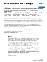
Determination of the optimal number of reference genes for normalizationFigure 1
Determination of the optimal number of reference genes for normalization. The GeNorm program calculates aver-
age expression stability (M) and the expression stability value by the calculation of the pair wise variation. For example V5/V6
indicates the variation in normalization factor with 5 versus 6 reference genes. A and C: Menghini NaCl. B and D: Menghini
water.
Determ ination of the optimal number of control genes for normalization
0.043
0.048 0.048
0.066
0.077
0.105
0.147
0.000
0.020
0.040
0.060
0.080
0.100
0.120
0.140
0.160
V2 / 3 V3/4 V4/ 5 V5/6 V6 / 7 V7/8 V8 /9
Pairwise Variations
Determination of the optimal number of control genes for norm alization
0.057
0.079
0.240
0.072
0.100
0.023
0.104
0.000
0.050
0.100
0.150
0.200
0.250
0.300
V2 / 3 V3/4 V4/ 5 V5/6 V6 / 7 V7/8 V8 /9
Pairwise Variations
Average expres s ion stability values of rem aining control genes
0.1
0.2
0.3
0.4
0.5
0.6
0.7
0.8
ACTB HNRPH GUSB B2M RPL8 RPS19 G3P HPRT
RPS5
<::::: Lea s t s t ab le g e ne s M o s t s t able g e ne s ::::>
Average expre ssion s tability value s of re m aining control genes
0.2
0.25
0.3
0.35
0.4
0.45
0.5
0.55
0.6
0.65
HNRPH HPRT GUSB RPL8 ACTB B2M RPS19 G3P
RPS5
<::::: Least st able g e nes Most stable g enes ::::>
A
B
C
D
Comparative Hepatology 2009, 8:3 />Page 5 of 8
(page number not for citation purposes)
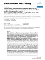
Liver histologyFigure 2
Liver histology. A) Normal liver, dog #1, portal area and periportal parenchyma. The tissue architecture is well preserved,
with good contrast and sufficient cellular morphology reflected in distinct cellular and nuclear membranes, and sufficient cyto-
plasmic details. Needle biopsy, 1 h formalin fixation, HE staining, bar 50 μm. B) Normal liver, dog #5, portal area with bile duct
(arrow) and periportal parenchyma. The tissue is well conserved, and there is mild cellular pronounciation and slightly
enhanced eosinophilic appearance of all cells save erythrocytes. Needle biopsy, 8 hrs Boonfix fixation at room temperature, HE
staining, bar 50 μm. C) Normal liver, dog #5, portal area and periportal parenchyma. Insufficient conservation of tissue archi-
tecture in the central part of the biopsy, to the right hand side of the arrow, with ill defined cellular borders, strong eosi-
nophilia and shrinkage of hepatocytes, pycnotic nuclei and artificially widened sinusoids. Needle biopsy, 8 hrs RNAlater fixation
at room temperature, HE staining, bar 50 μm. D) Copper related chronic active hepatitis, dog #9, parenchyma, control tissue.
Many, black staining copper granules appear in the cytoplasm of hepatocytes and Kupffer cells. Wedge biopsy, 24 hrs formalin
fixation, rhodanine acid stain, bar 50 μm. E) Liver with copper storage, dog #6, parenchyma. Intracytoplasmic copper granules
stain yellow-brown, therefore no reliable differentiation between copper and lipofuscin granules can be made. Needle biopsy, 8
hrs Boonfix fixation, rubeanic acid stain, bar 50 μm. F) Normal liver, dog #2, portal area and periportal parenchyma. Cholangi-
ocytes in the portal tract (asterisk) display a strong signal (brown) in the cytoplasm with negligable aspecific background stain-
ing. Also, the parenchyma contains one small, isolated positive periportal cell (arrow), interpreted as a progenitor cell. Needle
biopsy, 1 h formalin fixation, K-7 immunohistochemistry, bar 20 μm. G) Normal liver, dog #5, portal area and periportal paren-
chyma. All hepatocytes feature strong cytoplasmic reactivity, all other cells are negative. Needle biopsy, 1 h formalin fixation,
Hepar1 immunostaining, bar 50 μm. H) Normal liver, dog #8, parenchyma, control tissue. Strong signal (brown) is elicited
along the canalicular membranes of all hepatocytes, insignificant background staining. Wedge biopsy, 24 hrs formalin fixation,
MRP-2 immunostaining, bar 20 μm.
&
'
,
Comparative Hepatology 2009, 8:3 />Page 6 of 8
(page number not for citation purposes)
on blood plasma (Figure 2G). However, 8 hrs formalin
fixed biopsies displayed an irregularly dispersed signal
throughout the slide, while the biopsy fixed over 5 days
reacted as the biopsies fixed up to 4 hrs. The control tissue
revealed strongly increased reactivity in individual peri-
portal hepatocytes, which was less obvious in the Meng-
hini biopsies. Both Boonfix and RNAlater fixed
specimens, also after minus 20°C storage, showed a
strong signal in the periphery of the biopsy, but reacted
very poorly in the centre.
MRP-2
In 24 hrs formalin fixation, the positive control wedge
biopsy exhibited a strong brown signal along the canalic-
ular membranes of all hepatocytes for MRP-2, with negli-
gible background staining (Figure 2H). Increase in
fixation time up to 5 days significantly decreased reactivity
in a wedge biopsy. Menghini biopsies fixed from 1 h up to
5 days generally proved negative, with some faint signal at
4 hrs. All Boonfix treated specimens were negative.
RNAlater preserved specimens had a moderate to strong
signal at the periphery of the biopsy, unless stored at
minus 20°C after which no signal was present.
Discussion
In search for an easy-to-use method to acquire, fix and
store canine liver biopsies, we used the stability of 18S
and 28S rRNA as markers for totalRNA and mRNA stabil-
ity. Histological evaluation was based on HE, reticulin,
rhodanine and rubeanic acid stains and three different
immunohistochemical stains.
RNA quality was best guaranteed by the combination of a
Menghini biopsy with NaCl, followed by RNAlater preser-
vation and RNAeasy mini kit extraction. Under optimal
biopsy conditions (as was the case for the surplus dog
used to compare Menghini NaCl and Menghini water in
one single dog), no differences in RIN-values between the
two techniques were observed. Whether this reflects the
fact that exactly the same liver was used, or whether time
delay between the biopsy and the actual RNAlater storage,
as usually occurs under clinical situations, causes this dif-
ference remains unknown. In favor for the first explana-
tion accounts that in the clinical setting the difference was
consistent over a large number of biopsies. The evaluation
of the optimal number of reference genes needed to
obtain reliable data strengthened the observation that the
combination of a Menghini NaCl biopsy followed by
RNAlater preservation and an RNAeasy mini kit extraction
yields optimal RNA quality from canine liver biopsies.
The size of the biopsy needle used in this study was based
on a previous study on rat liver biopsy techniques, and
turned out to be an optimal balance between quantity and
quality of the biopsy and the health risks for the animal
[12]. This approach of RNA retrieval proved to be a rapid
and feasible method for storage for further molecular
analysis, and is in agreement with the findings of others
for yeast, human renal and uterine myometrial tissues
[15-17]. The quality of the obtained RNA in our approach
was feasible for micro-array analysis, which requires the
highest possible RNA quality, preferential a RIN value
above 8.0. Unfortunately our results show that optimal
RNA stabilization was only achieved with media that were
unsuitable for histology or immunohistochemistry. His-
tology of RNA later treated biopsies, evaluated in HE and
reticulin staining turned out to be of insufficient quality;
furthermore, for the antibodies tested either the back-
ground staining was too high or central staining appeared
very poor.
The best fixative for (immuno)histochemistry proved to
be 10% neutral buffered formalin. Boonfix fixation gave
good morphology and results in routine HE and reticulin
staining, but was suboptimal for the tested immunohisto-
chemical staining methods. RNAlater fixation yielded
poor morphology in routine histology and in immuno-
histochemistry. Most likely, these shortages in morpho-
logical evaluation of RNAlater treated specimens were
related to insufficient tissue fixation. Boonfix treated spec-
imens generally evoked less intense reactivity immunohis-
tochemically, but as all tested methods were optimized
for use in formalin fixed (24 hrs) wedge biopsy speci-
mens, they might perform better in a study where the pro-
tocols are tailor-made to the fixative. Storage in minus
20°C for Boonfix and RNAlater, as required for molecular
purposes, significantly worsened tissue morphology.
In our experience staining artefacts more frequently occur
in small formalin fixed paraffin embedded biopsies. We
hypothesized that in the relatively small biopsies overfix-
ation could easily occur. Therefore an effect of the dura-
tion of formalin fixation was assessed with subsequent
immunohistochemical evaluation of antibodies to pro-
teins at three different (sub)-cellular locations in addition
to routine histological staining methods. Differences of
the immunohistochemical reactivity for all three antibod-
ies were found between wedge biopsies and the smaller
Menghini tissue samples in this study. The observation
was most pronounced in MRP-2 stained slides where only
a very weak signal was evoked in the smaller biopsies. In
addition prolonged fixation in formalin caused a signal
reduction for K-7, but did not affect routine HE and reti-
culin staining. The difference is most likely due to changes
in epitopes required for immunohistochemistry, but less
for routine HE and reticulin staining. Indications for pos-
sible overfixation by formalin were present in K-7 and
possibly in MRP2 staining. Signal reduction in K-7 stained
biopsies was associated with increased fixation time and
was also present in the periphery of wedge biopsies (24
hrs and 5 days fixation). In both situations, prolonged
Comparative Hepatology 2009, 8:3 />Page 7 of 8
(page number not for citation purposes)
exposure to formalin could explain epitope masking due
to protein cross linking of the tissues antigens. Conse-
quently, this antigen masking could result in decreased
antigen-antibody reactivity. Occurrence and intensity of
this effect will vary per antibody as not all epitopes will be
affected similarly [18]. Immunohistochemical reactivity
was optimal after formalin fixation and replacement of
the formalin by ethanol 70% within 1 – 4 hrs.
Formalin fixation proved necessary for assessment of cop-
per accumulation in liver tissue. Routine rubeanic acid
staining was sufficient in a wedge biopsy (24 hrs) as well
as in a Menghini biopsy (8 hrs). Reliable rhodanine stain-
ing was limited to a wedge biopsy only. RNAlater or Boon-
fix treated slides did not produce a sufficient signal in any
of the investigated copper stains. Interestingly, previous
exposure to HCl damp in rubeanic acid staining, as was
suggested to enhance copper staining [18], completely
inhibited the signal in all slides and therefore proved to be
ineffective.
Conclusion
Summarized, in the search to decrease the number of
biopsies needed for molecular and (immuno)histochem-
ical analysis, it turned out that at least two biopsies (10%
neutral buffered formalin and RNAlater) are needed.
Since both biopsies can be dispersed in relatively non-
toxic liquid preservatives, this combination can easily pro-
vide researchers with material for high throughput expres-
sion analysis. Moreover it nicely resembles the sample
preparation protocols that are commonly used in clinics
today. Since biopsies fixed in either RNAlater or formalin
remain stable at room temperature, transport is easy from
the clinical situation to the research facility for further
processing as well as prolonged storage. Results of our
study showed that a reduction of the formalin fixation
time to 1 to 4 hrs will generally reduce formalin induced
reduced staining and staining artifacts. Therefore, any
extension of the formalin fixation period should be dis-
couraged when immunohistochemistry is considered.
In view of the large similarities between human and
canine liver diseases [19], it is conceivable that the proto-
cols described here can be easily translated into the
human biomedical field. Consequently, unique and rare
human liver biopsies can be obtained, stored and subse-
quently handled without loss of information.
Methods
Animals
All procedures were approved by the responsible ethical
committees according to Dutch legislation.
For this study, liver tissue was obtained from seven dogs.
In addition two archival specimens were used as positive
controls for staining during histologic examinations. Sur-
plus animals from orthopedic research revealed, histolog-
ically confirmed, healthy livers. These dogs were
euthanized immediately prior to extirpation of the liver,
using an overdose of pentobarbital via the cephalic vein.
Liver biopsies
Liver biopsies were taken according to the Menghini tech-
nique described by Rothuizen [20] and by use of a 16-
gauge biopsy needle using an automatic biopsy device
(Pro-Mag Ultra Automatic Biopsy Instrument, PBN Medi-
cals, Stenløse, Denmark). Liver biopsies retrieved by use
of the Menghini technique were kept in physiologic saline
solution (0.9% NaCl in sterile water, group Menghini
NaCl) or sterile water (group Menghini water) until trans-
fer into according preservatives. Liver biopsies retrieved
with the True-cut gun were kept at room air until transfer
into the different storage media.
After fixed time periods the material was further processed
with either one of the following four methods: snap freez-
ing and subsequent storage at minus 70°C, transfer into a
sterile 1.5 ml vial containing 1 ml of RNAlater (Applied
Biosystems, Nieuwerkerk a/d lJssel, the Netherlands),
Boonfix (Finetec, Tokyo, Japan) or B-RLT (QIAGEN,
Venlo, the Netherlands). Biopsies in these vials were kept
at 4°C for 2 hrs, and later transferred to minus 20°C and
minus 70°C freezing for long-term storage (2 weeks to 18
months). Additional biopsies retrieved exclusively for his-
tologic examinations were retrieved by the Menghini-
NaCl method, and immediately deposited at room tem-
perature (RT) per three in 6 ml containers filled with 10%
neutral buffered formalin. Wedge biopsies (1 × 1 × 1 cm)
were put in a larger container, containing at least 10 cm
3
of formalin.
Isolation of RNA, reversed transcriptase and quantitative
RT-PCR
RNA isolations with the RNAeasy kit (QIAGEN) or Trizol
reagent (Invitrogen, Leek, the Netherlands) were per-
formed according to the manufactures instructions. RNA
yields were quantified spectrophotometrically using the
Nanodrop ND-1000 (Isogen Life Science, IJsselstein, the
Netherlands) device and set to a 0.1 μg/μl concentration.
One microgram of each total RNA sample was used to
synthesize cDNA with an MMLV-derived reverse tran-
scriptase according to manufacturer's protocol (iScript
cDNA synthesis kit, Bio-rad, Veenendaal, the Nether-
lands). Details were described previously [19].
RNA quality was measured in two independent ways: By
means of the A260/A280 ratio, which estimates the
amount of protein contamination, and by means of the
Agilent 2100 Bioanalyzer (Agilent Technologies,
Amstelveen, the Netherlands), which displays RNA Integ-
rity Number (RIN-values) indicating the percentage of
intact 18S and 28S rRNA.
Comparative Hepatology 2009, 8:3 />Page 8 of 8
(page number not for citation purposes)
A SYBR Green based quantitative PCR was performed on
a Bio-Rad My-IQ detection system as described previously
[8].
Histology
After the specified fixation times (range 1 hr to 5 days),
formalin was replaced by 70% ethanol until further
processing. Other tissues were immersed in RNAlater (8
hrs) and Boonfix (2, 4, 8 hrs). In addition, also a biopsy
fixed in RNAlater or Boonfix was kept in a minus 20°C
freezer prior to further processing. After the different fixa-
tion procedures and replacement of preservatives by etha-
nol all tissue samples of one individual animal were
simultaneously dehydrated and paraffin embedded. Par-
affin blocks were stored at 4°C until use.
Routine histology performed on 3 μm sections included
HE (all animals save two controls), and the reticulin stain-
ing according to Gordon and Sweet (5 dogs). Primary his-
tological evaluation was based on the 24 hrs formalin
fixed wedge biopsies. Two cases with known hepatic cop-
per storage were also subjected to routine rhodanine and
rubeanic acid stains for copper accumulation. Moreover,
two enhancement methods of rubeanic acid staining [18]
were performed by 1): washing the slides 5 min. in 10%
neutral buffered formalin previous to rubeanic acid stain-
ing, or 2): after de-waxing, slides were placed face down-
wards over a beaker of HCl 37% for 15 min., followed by
15 min. wash in ethanol 90% and routine rubeanic acid
staining. The copper scoring system was described previ-
ously [21]. Single immunohistochemical staining for K-7,
Hepar1, and MRP2 was performed as previously described
[13,14].
Abbreviations
B2M: beta-2 microglobulin; GAPDH: glyceraldehyde-3-
phosphate dehydrogenase; GUSB: β-Glucuronidase; HE:
hematoxilin-Eosin; hnRNPH: Heterogeneous nuclear
ribonucleoprotein H; HPRT: Hypoxanthine phosphoribo-
syltransferase; K-7: cytokeratin-7; MRP-2: multi drug
resistance protein-2; Q-PCR: quantitative real-time PCR;
RPL8: ribosomal protein L8; RPS19: ribosomal protein
S19; RPS5: ribosomal protein S5; SNP: single nucleotide
polymorphism.
Competing interests
The authors declare that they have no competing interests.
Authors' contributions
GH performed the biopsies and wrote the first draft of this
manuscript. JIJ performed the IHC and co-wrote the first
draft of this manuscript. BB and BAS did the molecular
analysis. TSGAMvdI evaluated the histology. LCP and JR
designed the experimental set-up and co-wrote the final
version. All authors have read and approved this manu-
script.
References
1. Neff MW, Rine J: A fetching model organism. Cell 2006,
124(2):229-231.
2. Lindblad-Toh K, Wade CM, Mikkelsen TS, Karlsson EK, Jaffe DB,
Kamal M, Clamp M, Chang JL, Kulbokas EJ 3rd, Zody MC, et al.:
Genome sequence, comparative analysis and haplotype
structure of the domestic dog. Nature 2005,
438(7069):803-819.
3. Parker HG, Kim LV, Sutter NB, Carlson S, Lorentzen TD, Malek TB,
Johnson GS, DeFrance HB, Ostrander EA, Kruglyak L: Genetic
structure of the purebred domestic dog. Science 2004,
304(5674):1160-1164.
4. Sargan DR, Aguirre-Hernandez J, Galibert F, Ostrander EA: An
extended microsatellite set for linkage mapping in the
domestic dog. J Hered 2007, 98(3):221-231.
5. Wayne RK, Ostrander EA: Lessons from the dog genome. Trends
Genet 2007, 23(11):557-567.
6. Parker HG, Ostrander EA: Canine genomics and genetics: run-
ning with the pack. PLoS Genet 2005, 1:e58.
7. Sutter NB, Ostrander EA: Dog star rising: the canine genetic
system. Nat Rev Genet 2004, 5(12):900-910.
8. Brinkhof B, Spee B, Rothuizen J, Penning LC: Development and
evaluation of canine reference genes for accurate quantifica-
tion of gene expression. Anal Biochem 2006, 356(1):36-43.
9. Etschmann B, Wilcken B, Stoevesand K, Schulenburg A von der,
Sterner-Kock A: Selection of reference genes for quantitative
real-time PCR analysis in canine mammary tumors using the
GeNorm algorithm. Vet Pathol 2006, 43(6):934-942.
10. Peters IR, Peeters D, Helps CR, Day MJ: Development and appli-
cation of multiple internal reference (housekeeper) gene
assays for accurate normalisation of canine gene expression
studies. Vet Immunol Immunopathol 2007, 117(1–2):55-66.
11. Fleige S, Pfaffl MW: RNA integrity and the effect on the real-
time qRT-PCR performance. Mol Aspects Med. 2006, 27(2-
3):126-139.
12. Takemura F, Inaba N, Miyoshi E, Furuya T, Terasaki H, Ando S, Kon-
oshita N, Ogawa Y, Toniguchi N, Ito S: Optimization of liver
biopsy RNA sampling and use of reference RNA for cDNA
microarray analysis. Anal Biochem 2005, 337(2):224-234.
13. Ijzer J, Kisjes J, Penning LC, Rothuizen J, van den Ingh TS: The pro-
genitor cell compartment in the feline liver: An
(immuno)histochemical investigation. Vet Path 2009,
46(4):614-21.
14. Mekkonnen GA, Ijzer J, Nederbragt H: Tenascin-C in chronic
canine hepatitis: Immunohistochemical localization and cor-
relation with necro-inflammatory activity, fibrotic stage,
alpha-SMA, K-7 and CD3+ cells. Vet Path 2007, 44(6):803-813.
15. Dekairelle AF, Vorst S Van der, Tombal B, Gala JL: Preservation of
RNA for functional analysis of separated alleles in yeast:
comparison of snap-frozen and RNALater((R)) solid tissue
storage methods. Clin Chem Lab Med 2007, 45(10):1283-1287.
16. Roos-van Groningen MC, Eikmand M, Baelde HJ, de Heer E, Bruijn JA:
Improvement of extraction and processing of RNA from
renal biopsies. Kidney Int 2004, 65(1):97-105.
17. Mutter Gl, Zahrieh D, Liu C, Neuberg D, Finkelstein D, Baker HE,
Warrington JA: Comparison of frozen and RNAlater solid tis-
sue storage methods for use in RNA expression microarrays.
BMC Genomics 2004, 5(1):88.
18. Werner M, Chott A, Fabiano A, Battifora H: Effect of formalin tis-
sue fixation and processing on immunohistochemistry. Am J
Surg Pathol 2000, 24(7):1016-1019.
19. Spee B, Arends B, van den Ingh TS, Brinkhof B, Nederbragt H, Ijzer J,
Roskams T, Penning LC, Rothuizen J: Transforming growth factor
β-1 signalling in canine hepatic diseases: new models for
human fibrotic liver pathologies. Liver Int 2006, 26(6):716-725.
20. Stockhaus C, Ingh TSGAM van den, Rothuizen J, Teske E: A Multi-
step Approach in the Cytologic Evaluation of Liver Biopsy
Samples of Dogs with Hepatic Diseases. Vet Pathol 2004,
41(5):461-470.
21. van den Ingh TS, Rothuizen J, Cupery R: Chronic active hepatitis
with cirrhosis in the Doberman Pinscher. Vet Q 1988,
10(2):84-89.