Báo cáo y học: "An insertion mutation in ABCB4 is associated with gallbladder mucocele formation in dogs" docx
Bạn đang xem bản rút gọn của tài liệu. Xem và tải ngay bản đầy đủ của tài liệu tại đây (840.22 KB, 7 trang )
Mealey et al. Comparative Hepatology 2010, 9:6
/>Open Access
RESEARCH
© 2010 Mealey et al; licensee BioMed Central Ltd. This is an Open Access article distributed under the terms of the Creative Commons
Attribution License ( which permits unrestricted use, distribution, and reproduction in
any medium, provided the original work is properly cited.
Research
An insertion mutation in ABCB4 is associated with
gallbladder mucocele formation in dogs
Katrina L Mealey*
1,4
, Jonathan D Minch
1
, Stephen N White
2,3,4
, Kevin R Snekvik
3
and John S Mattoon
1
Abstract
Background: ABCB4 functions as a phosphatidylcholine translocater, flipping phosphatidylcholine across hepatocyte
canalicular membranes into biliary canaliculi. In people, ABCB4 gene mutations are associated with several disease
syndromes including intrahepatic cholestasis of pregnancy, progressive familial intrahepatic cholestasis (type 3),
primary biliary cirrhosis, and cholelithiasis. Hepatobiliary disease, specifically gallbladder mucocele formation, has been
recognized with increased frequency in dogs during the past decade. Because Shetland Sheepdogs are considered to
be predisposed to gallbladder mucoceles, we initially investigated ABCB4 as a candidate gene for gallbladder mucocele
formation in that breed, but included affected dogs of other breeds as well.
Results: An insertion (G) mutation in exon 12 of canine ABCB4 (ABCB4 1583_1584G) was found to be significantly
associated with hepatobiliary disease in Shetland Sheepdogs specifically (P < 0.0001) as well as other breeds (P <
0.0006). ABCB4 1583_1584G results in a frame shift generating four stop codons that prematurely terminate ABCB4
protein synthesis within exon 12, abolishing over half of the protein including critical ATP and a putative substrate
binding site.
Conclusions: The finding of a significant association of ABCB4 1583_1584G with gallbladder mucoceles in dogs
suggests that this phospholipid flippase may play a role in the pathophysiology of this disorder. Affected dogs may
provide a useful model for identifying novel treatment strategies for ABCB4-associated hepatobiliary disease in people.
Background
Bile is produced by the collective actions of a number of
transporters located on the canalicular membrane of
hepatocytes [1]. Active transport of biliary solutes creates
an osmotic force that attracts water through tight junc-
tions and aquaporins in the hepatocyte membrane [2,3].
Bile salts are the most important biliary solute. Other
important solutes of bile include cholesterol and phos-
pholipids. The presence of phospholipids, phosphatidyl-
choline (PC) in particular, in the biliary lumen is crucial
for protecting the epithelial cell membranes lining the bil-
iary system from the cytotoxic detergent actions of bile
salts [3-5]. Bile salt cytotoxicity is substantially reduced in
the presence of PC owing to the formation of mixed
micelles (PC + bile salts) rather than simple micelles (bile
salts only). Thus, a decrease in the amount of biliary PC
leads to injury of epithelial cells lining the biliary system
[6].
ABCB4 functions exclusively as a phospholipid translo-
cator [6]. ABCB4 is expressed on cannalicular mem-
branes of hepatocytes where it translocates PC from the
hepatocyte to the biliary canalicular lumen [7]. Proper
function of ABCB4 is critical for maintaining hepatobil-
iary homeostasis as evidenced by the myriad of diseases
that occur when polymorphisms of ABCB4 cause com-
plete or partial protein dysfunction. ABCB4 deficiency is
associated with a variety of hepatobiliary disorders in
people including progressive familial intrahepatic
cholestasis (PFIC type 3), cholelithiasis, and cholestasis of
pregnancy [4,8-10]. Abcb4-/- mice, in which Abcb4 func-
tion is lacking entirely, also develop severe hepatobiliary
disease that starts at a few weeks of age and progresses
throughout life [11,12].
Hepatobiliary disease in dogs has been recognized with
increased frequency during the past several years. In par-
ticular, gallbladder mucoceles (mucinous hyperplasia or
mucinous cholecystitis) have been documented to be an
* Correspondence:
1
Department of Veterinary Clinical Sciences, College of Veterinary Medicine,
Washington State University, Pullman, WA 99164-6610, USA
Full list of author information is available at the end of the article
Mealey et al. Comparative Hepatology 2010, 9:6
/>Page 2 of 7
increasingly important cause of hepatobiliary disease in
dogs [13-15]. Histopathologic findings associated with
ABCB4 associated diseases in people, including intrahe-
patic cholestasis, cholecystitis, and periportal inflamma-
tion [13,16,17], are not commonly reported in dogs with
gall bladder mucoceles. Additionally, gallbladder muco-
celes are not a component of ABCB4 linked syndromes in
people or mice. Gallbladder mucoceles, which occur
rarely in people, are often associated with extrahepatic
bile duct obstruction. The etiology of gallbladder muco-
celes in dogs has not yet been identified, but extrehepatic
bile duct obstruction is not commonly associated with
this disorder [14,15]. Gallbladder mucoceles may result
from chronic injury to the epithelial lining of the biliary
system since hypersecretion of mucin is the typical physi-
ologic response of any epithelial lining to injury.
Recently Shetland Sheepdogs were identified as a breed
that is predisposed to gallbladder mucocele formation,
suggesting a genetic predisposition [13]. Because ABCB4
dysfunction is associated with hepatobiliary disease in
people and mice, we postulated that a defect in canine
ABCB4 might be responsible for gallbladder mucocele
disease in dogs, and Shetland Sheepdogs in particular.
Therefore, we sequenced canine ABCB4 in affected and
unaffected Shetland Sheepdogs as well as affected and
unaffected dogs of other breeds.
Methods
Collection of DNA from affected and unaffected individuals
All work was approved by the institutional Animal Care
and Use Committee. Collection of DNA from affected
Shetland Sheepdogs was accomplished by soliciting own-
ers' cooperation. In order to cast a wide net, owners of
dogs with confirmed (ultrasound, surgery, or histopathol-
ogy) or suspected (elevated liver enzymes - alkaline phos-
phatase, alanine aminotransferase and/or gamma
glutamyl transferase -, total bilirubin, cholesterol and/or
triglycerides) gallbladder disease were asked to submit a
cheek swab, copy of the dog's pedigree, and copy of the
dog's medical record. Contact of Shetland Sheepdog
owners was made through the American Shetland Sheep-
dog Association. For collection of unaffected Shetland
Sheepdogs, an additional request for DNA from healthy
Shetland Sheepdogs (with confirmatory medical records)
was made. For collection of DNA from affected dogs of
any breed, records from the Washington Animal Disease
Diagnostic Laboratory were searched for canine patients
with histopathologic confirmation of gallbladder muco-
cele. For collection of DNA from unaffected dogs of any
breed, a specific solicitation through the Washington
State University College of Veterinary Medicine was
made for healthy dogs (no history of gallbladder disease)
over 9 years of age. In order to increase our confidence in
designating a dog as "unaffected", we recruited dogs
(Shetland Sheepdogs and other breeds) greater than 9
years of age. While this may have limited the number of
dogs included in the study, it more accurately reflected a
dog's true phenotype (affected vs. unaffected). A dog was
considered 'affected' if a gallbladder mucocele was diag-
nosed using previously established criteria[13], which
included at least one of the following (in order of increas-
ing stringency); ultrasound report by a boarded veteri-
nary radiologist (n = 3), surgical report (n = 5), or
histopathologic report (n = 7). Dogs with no evidence of
gallbladder disease as determined by a normal serum
chemistry panel and no apparent physical examination
abnormalities were considered 'unaffected'.
Sequencing of canine ABCB4
Exons 1 through 26 of canine ABCB4 were sequenced
after PCR amplification of genomic DNA from affected
and unaffected Shetland Sheepdogs. Table 1 contains the
sequences of the oligonucleotide primers. Purified PCR
amplicons were sequenced with an Applied Biosystems
ABI 3730 sequencer (Foster City, CA). Affected and unaf-
fected dogs of other breeds (non-Shetland Sheepdogs)
were sequenced only at exon 12. DNA from all dogs
except the 3 affected non-Shetland Sheepdogs was
extracted from cheek swab samples. Formalin-fixed, par-
affin embedded liver tissue was used for extraction of
DNA from these 3 dogs. Samples were processed first
using the RiboPure RNA extraction kit (Ambion, Foster
City, CA) until step C3. The interphase from this step
(containing DNA and protein) was then subjected to
DNA extraction using the DNeasy Blood and Tissue Kit
(Qiagen, Alameda, CA).
Allele specific PCR
In order to confirm the insertion mutation in exon 12
(ABCB4 1583_1584G), allele specific primers were
designed (mutant: forward 5'- CCTGGTTCGCAACCC
TAAGATCCG, reverse 5'- GCAATGTGGCCTGACAG
AAAGGGGAAATC; wildtype: forward 5'- CCTGGTTC
GCAACCCTAAGATCC, reverse 5'- GCAATGTGGCCT
GACAGAAAGGGGAAATC) to amplify a 202 bp ampli-
con. This also allowed confirmation of individual geno-
type.
Statistics
Association of genotype and gallbladder mucocele status
was analyzed using the frequency procedure of SAS 9.2
(SAS Institute, Cary, NC), specifying Fisher's exact test
and exact confidence intervals for the odds ratio.
Results
Collection of affected and unaffected individuals
Samples from 15 affected and 21 unaffected Shetland
Sheepdogs were sequenced. Diagnosis of gallbladder
mucocele was confirmed by ultrasound in 3 dogs, by sur-
Mealey et al. Comparative Hepatology 2010, 9:6
/>Page 3 of 7
gery in 5 dogs, and by histopathology in 7 dogs (Figure 1).
Median age of Shetland Sheepdogs with a diagnosis of
gallbladder mucocele was 9 years (range 5-12), which is
similar to previous reports [13,15]. Ages for all the 21
unaffected Shetland Sheepdogs were not available, but
the median age for those dogs whose ages were known (n
= 12) was 9.5 years of age (range 5-14). Ages and breeds
of the 3 affected non-Shetland Sheedogs are as follows:
Cairn Terrier (11 years), Cocker Spaniel (13 years) and
Pomeranian (11 years). Ages and breeds of the 20 unaf-
fected non-Shetland Sheepdogs are indicated in Table 2.
Sequencing of Canine ABCB4
Sequencing of all exons (1 to 26) of canine ABCB4 was
performed on genomic DNA from cheek swab samples
(Shetland Sheepdogs) or from archived liver tissue
(affected dogs that were not Shetland Sheepdogs). A sin-
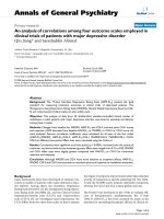
gle base pair insertion (G) was identified in exon 12 (Fig-
ure 2) in 14 of 15 affected Shetland Sheepdogs, 1 of 21
unaffected Shetland Sheepdogs, and 3 affected dogs of
other breeds (Cairn Terrier, Cocker Spaniel, and Pomera-
nian). The insertion mutation (ABCB4 1583_1584G) is
significantly associated (P < 0.0001) with the diagnosis of
gallbladder mucocele in Shetland Sheepdogs, with an
odds ratio of 280 (95% CI 12.7-12,350). In other dog
breeds, ABCB4 1583_1584G is also significantly associ-
ated with the diagnosis of gallbladder mucocele (P <
0.0006). The frame shift generated by the insertion results
in 4 premature stop codons within exon 12. The full
canine ABCB4 gene contains 26 exons which encode
essential structural elements that characterize ABC
transporters: two ATP binding domains and two sub-
strate binding sites. Essential structural elements of
ABCB4 normally contained within exon 12 and subse-
quent exons include both ATP binding sites and a sub-
strate binding site.
Table 1: Primers used for amplifying canine ABCB4.
Exon Forward Primer Reverse Primer Product Size
1 TTC AGT TGG CTA TGA AAC ATT TGG AGA CTA TCT TAA AGC ACT GAC TCC 165
2 CCA AAA AAC ATA TAG TTT TGG GGA GTC ATC TAG AAG TGC AAA CCA TTA AAC 302
3 CCT AGT AAC ACC TAT TAA TAG TTC AGC C CTC TGT AAG TTT GCA ATT ATT CTC 202
4 CTT CCT GAA AGA GAT GAA TAA AGA AC CAA AAG TAT GAC ATA AAT GAT ACA CTT AC 225
5 GAA GAC CTC CTG CCT GTA ACC ACT CAC ATG TGA AAA TGT TCC CGT TTC 201
6 CAT GAA TGT TTC TTC TCT GTC CAG GGT TCT TTG AAC CAG TGG AC 143
7 GGC TAT GAT TAT GGA CTG TTT TCT TG GGT TTC TTC ACG AAT ATT AGA AAG AC 208
8 GCT TAT AAC TTC TTC TTG TGT TCT TTT G GTG CAA GCC TCA AGG AAT TTT TTT TG 143
9 CCT TAA AAG TGC AGT TGG TTG GAA ATA AAA CCT GCC ACA GG 249
10 CGT GAA GAG TGT TCT CTT TCT CTC GCA GGG CTA ATT GGT AGC 177
11 CTT GAT GCT TTA GAT GTC AGA TGG CTC ACT TGC CTG AAG TCA AAG 278
12 GAG ATA CAT CAG GAG CTC CTC C CAG GTG TTT CGG GTT GAC TG 189
13 GTA ACC CTG TTG CAT CAC AC CTC AGC ATG GCA TTA GCT GC 239
14 CAA CTT AAC ATT TTC TCT TCT TTC AG GGA ATC ACT TGT GCC TGC 256
15 CCA CTT TCT CCT GAT TCT CCT G GGT GAA GCT GGC ATG AGA AC 219
16 CTC TCT CTG GCT CTC ATG CTC TAA TAG AAT GTG GAC TCG AG 188
17 CTG ATG ATC AAA AGG GAC AAT C GGA CTT CTC AAG TGC ACA C 118
18 GAA GGT GTG TTT TGT GCC ACA G CCC TTT CTG TCT CTC AAA TGG G 141
19 CAT GGC TCC CTC TTT GCT TTT GC CTC ACT GAA GCC TTC TTT GAC CCA C 212
20 CGT TAT CCA GAA GTA AAA GCC C CCT CAG GAA AGT ACT AGG GTC 159
21 CCA GTC AAC TAC ACT AGA AGC TG GAA CAA GTG AGT TTT TTC CAC CC 260
22 GGT AAG CAC TAT GTC TTT GGA C CAT TCA CCA GAC AGC AGA GAA C 222
23 CAG ACC AAT TAT AAT AGC AAC ATT AAC GCC TTA AAT AAG GTA CTA ACT TAA GC 227
24 GAT ACC CAC ATG TCA CAA TGT TCC TCC TGG TGC CAC TAC ATA GAC 402
25 GTC CTA TAC CAA GTC ATG AGG AC GGA AAC AGA GTG GAA AGA CC 179
26 GGA ACT AAC TGT AGA CTA TAA TGC GCT ATC TTA TCA ACA CCA AAT GG 393
Mealey et al. Comparative Hepatology 2010, 9:6
/>Page 4 of 7
A missense mutation in exon 15 of canine ABCB4 was
identified in the one affected Shetland Sheepdog that did
not harbor ABCB4 1583_1584G. This SNP results in a
nonhomologous amino acid substitution (alanine to ser-
ine) in exon 15 which may affect tertiary protein struc-
ture. However, this mutation was also present in 9 of the
21 unaffected Shetland Sheepdogs and 10 of the 15
affected Shetland Sheepdogs, so its significance is
unclear. No obvious differences were apparent in disease
severity or biochemical parameters in the affected dogs
with the mutation in exon 15.
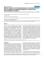
Confirmation of Insertion by Allele Specific PCR
To confirm the presence of ABCB4 1583_1584G as well as
determine the genotype of each dog, allele specific prim-
ers were designed and used to amplify the region of inter-
est in exon 12 (Figure 3). All dogs harboring the insertion
were heterozygous at the mutant allele suggesting a dom-
inant mode of inheritance with incomplete penetrance.
None of the dogs in the study were homozygous for the
mutant allele. Genotype frequencies are shown in Table 3.
Discussion
Over three dozen disease-causing mutations in human
ABCB4 have been described [5,7,9,10]. The disease spec-
trum ranges from severe (debilitating diseases of young
children that require liver transplantation) to mild. Dis-
ease severity often depends on the nature of the muta-
tion. Milder disease occurs when the ABCB4 gene
mutation reduces but does not eliminate transport activ-
ity of the protein. Similarly, milder forms of disease exist
in patients that are heterozygous for mutations that elim-
inate transporter activity (i.e., truncations).
The canine ABCB4 insertion mutation reported here
results in a truncation that eliminates more than 50% of
the protein. This mutation was significantly associated
with the diagnosis of gallbladder mucocele in Shetland
Sheepdogs as well as other dog breeds. The etiology of
gallbladder mucoceles in dogs is currently unknown, but
extrahepatic bile duct obstruction is not a common com-
ponent of the disease (as has been reported in people
with gallbladder mucoceles) [18]. The results reported
here provide evidence that dysfunction of ABCB4 is likely
involved. Hepatocyte PC transport, and therefore bile PC
content, in dogs that harbor ABCB4 1583_1584G would
be decreased compared to wildtype dogs. Biliary epithe-
lial lining cells would be subjected to bile salt-induced
injury because of diminished ability to form mixed
Table 2: Breed and age of unaffected dogs (non Shetland
Sheepdogs).
Breed Number of Dogs Age(years)
Afghan Hound 3 9.5; 10; 10
Asluki 1 12
Australian Shepherd 1 10
Brittany Spaniel 1 11
Corgi 1 9
English Cocker Spaniel 1 12
Golden Retriever 1 9.5
Jack Russell Terrier 1 9
Kelpie 1 13
Labrador Retriever 3 9; 9.5; 9.5
Miniature Pinscher 2 10; 13.5
Mixed Breed 1 10
Pitt Bull 1 15
Shih Tzu 1 14
Standard Poodle 1 10
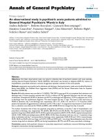
Figure 1 Gall Bladder. There is distention of the gall bladder with
abundant luminal accumulations of mucus interspersed with scant
amounts of bile. The mucosa of the gall bladder is lined by moderately
hyperplastic columnar epithelial cells with accentuation of the normal
folds by accumulations of mucus. Within the lamina propria and the
tunica muscularis there are occasional multifocal to perivascular accu-
mulations of lymphocytes and rare plasma cells. Hematoxylin and eo-
sin staining. Bar = 250 μm.
Figure 2 Electropherograms for wildtype and mutant canine
ABCB4. The insertion is indicated by an arrow.
Mealey et al. Comparative Hepatology 2010, 9:6
/>Page 5 of 7
micelles [19]. A universal physiologic response of epithe-
lial linings to injury is mucinous hyperplasia, a histo-
pathologic finding frequently described in dogs
diagnosed with gallbladder mucocele. Furthermore,
exposure to bile salts has been shown to stimulate mucin
secretion in cultured canine gallbladder epithelial cells
[20]. Thus, gallbladder epithelium in dogs that harbor
ABCB4 1583_1584G undergoes greater exposure to
unneutralized bile salts than that of wildtype dogs, result-
ing in greater mucin secretion, mucinous hyperplasia,
and eventually mucocele formation.
Because gallbladder mucoceles are a relatively new dis-
ease condition in dogs, a "gold standard" diagnosis has
not yet been defined. Inclusion criteria used in previous
publications consist of surgical or necropsy diagnosis
(macroscopic appearance), ultrasonographic diagnosis,
and/or histopathological diagnosis (microscopic appear-
ance) [14,15,21]. Each of these criteria has limitations for
diagnosing gallbladder mucoceles. A number of ultra-
sonographic findings have been associated with gallblad-
der mucocele, and there is sometimes disagreement
among ultrasonographers as to what constitutes a gall-
bladder mucocele. Additional confusion is created by ter-
minology such as "early" or "developing" gallbladder
mucocole. Because of the gallbladder's universal physio-
logical response to irritation (e.g., mucus secretion), some
might argue that even a histopathological diagnosis of
gallbladder mucocele may generate some speculation. It
seems reasonable, therefore, to entertain the possibility
that our study population ("affecteds") might contain false
positives and that our control population ("unaffecteds")
might contain false negatives despite the fact that cur-
rently acceptable criteria were used to identify these pop-
ulations. However, the statistical difference between
groups was so dramatic (based on current criteria) that
statistical relevance would still hold even if some errors
exist in the study or control population based on diagnos-
tic criteria that may be defined in the future. The associa-
tion of ABCB4 1583_1584G with gallbladder mucoceles
in dogs represents an important advancement in our
understanding of the disease.
A number of other potential etiologies have been sug-
gested for gallbladder mucoceles in dogs. These include
primary or secondary motility disorders of gallbladder
motility, a secondary complication of dyslipidemias
(Shetland Sheepdogs and Miniature Schnauzers) in par-
ticular, and primary disorders of mucus-secreting cells
[13]. Recently, hyperadrenocorticism was reported to be
significantly associated with the diagnosis of gallbladder
mucocele in dogs [21]. Our findings do not rule out other
Table 3: ABCB4 genotype frequencies in gallbladder mucocele affected and unaffected animals.
Shetland Sheepdog (affected) Shetland Sheepdog (unaffected)
ABCB4 1583_1584G (wildtype) 1 20
ABCB4 1583_1584G (heterozygous) 14 1
ABCB4 1583_1584G (homozygous) 0 0
Other breeds (affected) Other breeds (unaffected)
ABCB4 1583_1584G (wildtype) 0 20
ABCB4 1583_1584G (heterozygous) 3 0
ABCB4 1583_1584G (homozygous) 0 0
Figure 3 Representative gels containing amplified DNA of canine
ABCB4 from 3 affected (diagnosed with gallbladder mucocele)
and 3 unaffected Shetland Sheepdogs. Allele specific primers am-
plified both wildtype (A) and mutant (B) alleles in affected Shetland
Sheepdogs, but only wildtype sequence was amplified in unaffected
Shetland Sheepdogs.
Mealey et al. Comparative Hepatology 2010, 9:6
/>Page 6 of 7
potential etiologies, and it is certainly possible that
ABCB4 1583_1584G could be one of many contributing
factors to gallbladder mucoceles in dogs.
Many of the dogs from our study and other studies
were severely affected at the time of diagnosis with some
dogs dying of their disease despite surgical intervention
[13,15]. Our discovery of the insertion mutation in canine
ABCB4 allows early identification of dogs predisposed to
gallbladder mucocele formation. This creates a number of
beneficial applications for dogs. Genotyping of young
dogs for ABCB4 1583_1584G would allow veterinarians
to closely monitor for development of a gallbladder
mucocele in affected dogs. Surgical intervention could be
performed earlier in the disease process before disease-
induced morbidity places the patient at higher risk for
intra- and post-operative complications.
Another benefit of genotyping dogs for the ABCB4
1583_1584G is the possibility of medical or dietary man-
agement to prevent or at least delay the onset of mucocele
formation. Currently, no medical treatment options have
been systematically evaluated for managing dogs with
gallbladder mucoceles primarily because information
regarding the etiology of the disease has been lacking.
However, ursodeoxycholic acid has been suggested [22].
Some human patients with ABCB4-associated biliary dis-
ease benefit from treatment with ursodeoxycholic acid, a
relatively hydrophilic and much less cytotoxic bile acid
than most endogenous bile salts [4]. Studies to determine
bile composition in wildtype dogs and dogs with the
ABCB4 1583_1584G mutation should be performed in
order to further characterize the disease. One would
expect affected dogs to have bile with lower phospholipid
concentrations than wildtype dogs, and thus a greater
proportion of simple micelles rather than mixed micelles.
These studies would also be important to determine how
useful affected dogs would be as a model for the various
biliary diseases in people that result from similar ABCB4
mutations.
The authors speculate that occurrence of gallbladder
mucoceles in dogs is inherited in a dominant fashion with
incomplete penetrance, however further research is
required to confirm the mode of inheritance. While it is
possible that the one unaffected carrier of the ABCB4
1583_1584G insertion may develop biliary disease in the
future, there was no evidence of disease at 9 years of age.
No dogs in this study population were homozygous for
the mutation. Because a more severe phenotype is
observed in people homozygous for mutations resulting
in elimination of ABCB4 protein function, one would
speculate that the same would be true for dogs. In people
with PFIC (type 3), the disease manifests during early
childhood and is fatal without a liver transplant [4]. It is
possible that homozygosity for the mutation results in
death of affected dogs either during embryonic develop-
ment or in early puppyhood.
In conclusion, the ABCB4 1583_1584G is strongly asso-
ciated with the diagnosis of gallbladder mucocele in dogs.
Results of this study provide the first spontaneous animal
model for studying a number of potentially lethal or
severely debilitating hepatobiliary diseases in people that
are also associated with ABCB4 dysfunction. This canine
model may be useful for studying potential medical and/
or dietary treatments for ABCB4-associated hepatobil-
iary diseases in people.
List of abbreviations
ABC: adenosine triphosphate-binding cassette; ABCB4:
adenosine triphosphate-binding cassette, subfamily B,
member 4; PC: Phosphatidylcholine, G: guanine.
Competing interests
The authors declare that a patent application has been filed by Washington
State University listing two of the authors as inventors (KLM, JDM).
Authors' contributions
JDM performed experiments; JSM and KRS assisted in acquiring and interpret-
ing data; SNW performed statistical analysis; KLM conceived and designed the
research project. All authors made critical revision of the manuscript for impor-
tant intellectual content. All authors read and approved the final manuscript.
Acknowledgements
The authors would like to thank Mary B. Mahaffey, DVM for promoting sample
submission within the American Shetland Sheepdog Association. The authors
would also like to thank all dog owners for donating samples and sharing data
from their dogs' medical records. This work was supported by a Washington
State University College of Veterinary Medicine Intramural Grant and Proceeds
from the Veterinary Clinical Pharmacology Laboratory at Washington State
University.
Author Details
1
Department of Veterinary Clinical Sciences, College of Veterinary Medicine,
Washington State University, Pullman, WA 99164-6610, USA,
2
USDA-ARS
Animal Disease Research Unit, Pullman, WA 99164-6630, USA,
3
Department of
Veterinary Microbiology & Pathology, College of Veterinary Medicine,
Washington State University, Pullman, WA 99164, USA and
4
Center for
Integrated Biotechnology, Washington State University, Pullman, WA, 99164,
USA
References
1. Pellicoro A, Faber KN: Review article: The function and regulation of
proteins involved in bile salt biosynthesis and transport. Aliment
Pharmacol Ther 2007, 26:149-160.
2. Elferink RO, Groen AK: Genetic defects in hepatobiliary transport.
Biochim Biophys Acta 2002, 1586:129-145.
3. Coleman R, Iqbal S, Godfrey PP, Billington D: Membranes and bile
formation. Composition of several mammalian biles and their
membrane-damaging properties. Biochem J 1979, 178:201-208.
4. Oude Elferink RP, Paulusma CC: Function and pathophysiological
importance of ABCB4 (MDR3 P-glycoprotein). Pflugers Arch 2007,
453:601-610.
5. Davit-Spraul A, Gonzales E, Baussan C, Jacquemin E: Progressive familial
intrahepatic cholestasis. Orphanet J Rare Dis 2009, 4:1.
6. Trauner M, Fickert P, Wagner M: MDR3 (ABCB4) defects: a paradigm for
the genetics of adult cholestatic syndromes. Semin Liver Dis 2007,
27:77-98.
Received: 20 November 2009 Accepted: 3 July 2010
Published: 3 July 2010
This article is available from: 2010 Mealey et al; licensee BioMed Central Ltd. This is an Open Access article distributed under the terms of the Creative Commons Attribution License ( which permits unrestricted use, distribution, and reproduction in any medium, provided the original work is properly cited.Comparative Hepatology 2010, 9:6
Mealey et al. Comparative Hepatology 2010, 9:6
/>Page 7 of 7
7. Dean M, Annilo T: Evolution of the ATP-binding cassette (ABC)
transporter superfamily in vertebrates. Annu Rev Genomics Hum Genet
2005, 6:123-142.
8. Delaunay JL, Durand-Schneider AM, Delautier D, Rada A, Gautherot J,
Jacquemin E, Ait-Slimane T, Maurice M: A missense mutation in ABCB4
gene involved in progressive familial intrahepatic cholestasis type 3
leads to a folding defect that can be rescued by low temperature.
Hepatology 2009, 49:1218-1227.
9. Gonzales E, Davit-Spraul A, Baussan C, Buffet C, Maurice M, Jacquemin E:
Liver diseases related to MDR3 (ABCB4) gene deficiency. Front Biosci
2009, 14:4242-4256.
10. Nakken KE, Labori KJ, Rodningen OK, Nakken S, Berge KE, Eiklid K, Raeder
MG: ABCB4 sequence variations in young adults with cholesterol
gallstone disease. Liver Int 2009, 29:743-747.
11. Smit JJ, Schinkel AH, Oude Elferink RP, Groen AK, Wagenaar E, van
Deemter L, Mol CA, Ottenhoff R, van der Lugt NM, van Roon MA, van der
Valkc MA, Offerhausd GJA, Bernsc AJM, Borst P: Homozygous disruption
of the murine mdr2 P-glycoprotein gene leads to a complete absence
of phospholipid from bile and to liver disease. Cell 1993, 75:451-462.
12. Baghdasaryan A, Fickert P, Fuchsbichler A, Silbert D, Gumhold J, Horl G,
Langner C, Moustafa T, Halilbasic E, Claudel T, Trauner M: Role of hepatic
phospholipids in development of liver injury in Mdr2 (Abcb4)
knockout mice. Liver Int 2008:948-958.
13. Aguirre AL, Center SA, Randolph JF, Yeager AE, Keegan AM, Harvey HJ, Erb
HN: Gallbladder disease in Shetland Sheepdogs: 38 cases (1995-2005).
J Am Vet Med Assoc 2007, 231:79-88.
14. Besso JG, Wrigley RH, Gliatto JM, Webster CR: Ultrasonographic
appearance and clinical findings in 14 dogs with gallbladder
mucocele. Vet Radiol Ultrasound 2000, 41:261-271.
15. Pike FS, Berg J, King NW, Penninck DG, Webster CR: Gallbladder mucocele
in dogs: 30 cases (2000-2002). J Am Vet Med Assoc 2004, 224:1615-1622.
16. Worley DR, Hottinger HA, Lawrence HJ: Surgical management of
gallbladder mucoceles in dogs: 22 cases (1999-2003). J Am Vet Med
Assoc 2004, 225:1418-1422.
17. Newell SM, Selcer BA, Mahaffey MB, Gray ML, Jameson PH, Cornelius LM,
Downs MO: Gallbladder mucocele causing biliary obstruction in two
dogs: ultrasonographic, scintigraphic, and pathological findings. J Am
Anim Hosp Assoc 1995, 31:467-472.
18. Nahrwold D: Textbook of Surgery: The Biological Basis of Modern Surgical
Practice Philadelphia: W. B. Saunders; 1991.
19. Anwer MS, Meyer DJ: Bile acids in the diagnosis, pathology, and therapy
of hepatobiliary diseases. Vet Clin North Am Small Anim Pract 1995,
25:503-517.
20. Klinkspoor JH, Yoshida T, Lee SP: Bile salts stimulate mucin secretion by
cultured dog gallbladder epithelial cells independent of their
detergent effect. Biochem J 1998, 332:257-262.
21. Mesich ML, Mayhew PD, Paek M, Holt DE, Brown DC: Gall bladder
mucoceles and their association with endocrinopathies in dogs: a
retrospective case-control study. J Small Anim Pract 2009, 50:630-635.
22. Walter R, Dunn ME, d'Anjou MA, Lecuyer M: Nonsurgical resolution of
gallbladder mucocele in two dogs. J Am Vet Med Assoc 2008,
232:1688-1693.
doi: 10.1186/1476-5926-9-6
Cite this article as: Mealey et al., An insertion mutation in ABCB4 is associ-
ated with gallbladder mucocele formation in dogs Comparative Hepatology
2010, 9:6