Báo cáo y học: "The human allergens of mesquite (Prosopis juliflora)" pptx
Bạn đang xem bản rút gọn của tài liệu. Xem và tải ngay bản đầy đủ của tài liệu tại đây (241.76 KB, 5 trang )
BioMed Central
Page 1 of 5
(page number not for citation purposes)
Clinical and Molecular Allergy
Open Access
Research
The human allergens of mesquite (Prosopis juliflora)
Sue Killian*
1,2
and John McMichael
1
Address:
1
Dept. Research, The Institute for Therapeutic Discovery, P.O. Box 127, Delanson, New York, 12053, USA and
2
The Institute for
Therapeutic Discovery, 628 Kerns Mt. Ln., New Market, VA, 22844, USA
Email: Sue Killian* - ; John McMichael -
* Corresponding author
Abstract
Background: A computerized statistical analysis of allergy skin test results correlating patient
reactivities initiated our interest in the cross-reactive allergens of mesquite tree pollen. In-vitro
testing with mesquite-sensitized rabbits and a variety of deciduous tree pollens revealed so many
cross-reactivities that it became apparent there could be more allergens in mesquite than
previously described in the world literature. Our purpose was to examine the allergens of mesquite
tree pollen (Prosopis juliflora) which elicit an IgE response in allergic humans so that future research
could determine if these human allergens cross-react with various tree pollens in the same manner
as did the mesquite antiserum from sensitized rabbits.
Methods: Proteins from commercial mesquite tree pollen were separated by polyacrylamide gel
electrophoresis in the presence of sodium-dodecyl-sulphate. These mesquite proteins were
subjected to Western blotting using pooled sera from ten mesquite-sensitive patients and goat anti-
human IgE. The allergens were detected using an Amplified Opti-4-CN kit, scanned, and then
interpreted by Gel-Pro software.
Results: Thirteen human allergens of mesquite pollen were detected in this study.
Conclusion: The number of allergens in this study of mesquite exceeded the number identified
previously in the literature. With the increased exposure to mesquite through its use in "greening
the desert", increased travel to desert areas and exposure to mesquite in cooking smoke, the
possible clinical significance of these allergens and their suggested cross-reactivity with other tree
pollens merit further study.
Background
Mesquite (Prosopis juliflora) is a major cause of allergic dis-
ease in the southwestern United States [1,2], Mexico [3],
Saudi Arabia, South Africa [4,5], Kuwait [6], United Arab
Emirates (UAE) [7], and India [8]. Prosopis juliflora is a leg-
ume with several variations [2] that has been used for the
reclamation of desert lands and as a wood resource [4,8],
with the end result that its easily dispersed and its far-
traveling pollen [1,4,8] is an abundant and significant
source of allergens [5]. Novey [1] reported that mesquite
was the most prevalent pollen sensitizing 100 of his
patients in a California study, while Bener et al. [7], report
that 45% of the patients they tested in the UAE were sen-
sitive to Prosopis. In addition to pollen exposure, the burn-
ing of mesquite wood and its resulting smoke may be
another source of exposure to some of these same aller-
gens [9,10].
Our interest in mesquite was initiated by a clinician who
observed that many of his allergy patients (1598 out of
Published: 05 July 2004
Clinical and Molecular Allergy 2004, 2:8 doi:10.1186/1476-7961-2-8
Received: 23 April 2004
Accepted: 05 July 2004
This article is available from: />© 2004 Killian and McMichael; licensee BioMed Central Ltd. This is an Open Access article: verbatim copying and redistribution of this article are permit-
ted in all media for any purpose, provided this notice is preserved along with the article's original URL.
Clinical and Molecular Allergy 2004, 2:8 />Page 2 of 5
(page number not for citation purposes)
4361 patient cases) were sensitized to mesquite pollen
even though most of them had no known direct exposure
to mesquite (Personal communication with Allan D. Lie-
berman, M.D., The Center for Occupational and Environ-
mental Medicine, 7510 Northforest Dr., N. Charleston,
SC, USA). A computerized-analysis of skin test data from
this medical practice showed mesquite correlating with a
high number of other seemingly unrelated commercial
antigens, leading us to believe it might have a proclivity
for cross-reactivity [11]. Tree pollen cross-reactivity with
mesquite was confirmed using rabbit antiserum for both
Ouchterlony (unpublished study) testing and Western
blotting [12], but the relevance to human allergy needed
to be established. The aim of the current study was to
amplify the present knowledge of mesquite allergens
which affect humans preliminary to investigating the
impact of mesquite pollen cross-reactivity.
Methods
Proteins from a 1:2 dilution of mesquite tree pollen
extract (Prosopis juliflora var. glandulosa)(Greer Laborato-
ries, Inc., Lenoir, North Carolina) in Laemmli Sample
buffer (Bio-Rad) were separated by polyacrylamide gel
electrophoresis in the presence of sodium-dodecyl-sul-
phate (SDS-PAGE) according to the method of Laemmli
[13]. The protein concentration of the mesquite extract
was as determined by Greer Laboratories using the Kjeld-
hal Method [14]. Six lanes on each of 4 replicate gels were
loaded with 8 µg of mesquite extract protein. A Mini-Pro-
tein 3 Electrophoresis Apparatus (Bio-Rad, Hercules, Cal-
ifornia) and a pH 8.3 Tris-glycine buffer were used.
Polyacrylamide concentration of the precast 10 lane gels
was 4% for the stacking gel and 12% for the resolving gel.
Electrophoretic migration was performed at 21°C and
150 v constant voltage for 35 minutes until the bands
migrated to the lower edge of the gel. Protein bands were
visualized by Brilliant Blue R-250 (Fisher Biotechnology,
Inc., Fair Lawn, NY) staining. Dual Color Precision Plus
Protein Standards expressing calibration points of 10, 15,
20, 25, 37, 50, 75, 100, 150 and 250 kd (Bio-Rad) were
included in the electrophoretic separation. Protein bands
were destained and scanned at 150 pixels per inch (ppi)
setting by a Hewlett Packard ScanJet 4100C and inter-
preted using Gel-Pro 3.1 computer software (Media
Cybernetics, Silver Spring, Maryland) to determine their
molecular weights, relative optical densities and approxi-
mate protein distribution.
Immunoblotting was performed using a Mini-Trans-Blot
Electrophoretic Transfer Cell (Bio-Rad) according to the
method of Towbin et al. [15]. Unstained mesquite pollen
extract (Greer Laboratories) electrophoresis gels were elec-
troblotted onto 0.2 µm Trans-Blot Transfer Media (nitro-
cellulose membranes) (Bio-Rad). Membranes were
blocked with 5% blotting grade nonfat milk (Bio-Rad) in
wash buffer of PBST (phosphate buffered saline: 10 nM
sodium phosphate, 150 nM sodium chloride and 1%
Tween-20 (Bio-Rad).
Sera from fifteen mesquite-positive patient donors were
tested for their IgE responses using this electrophoresis
and Western blotting protocol and all produced positive
IgE responses to mesquite pollen allergens. Ten of these
patients were then chosen to participate in the study
because they not only tested mesquite-positive but also
provided at least 250 ml of their sera, the minimum need
for sequential experiments. The five patients who were not
included in the study provided insufficient quantities of
sera for the present experimental replicates and future
studies. These sera were obtained from both commercial
sources and private physicians (Table 1). Each donor was
given a research number to protect his/her confidentiality
and each donor from a private physician's practice signed
a consent form.
The pooled sera from these 10 patients were placed in a
55°C water bath for 30 minutes to destroy complement.
These pooled sera were diluted 1:2 in the wash buffer with
0.05% blotting grade nonfat milk and incubated with the
nitrocellulose blot overnight. Bound IgE was detected by
incubating the blot for 5 hours, using a 1:1000 dilution of
goat anti-human IgE conjugated to peroxidase (Sigma,
Saint Louis, Missouri). An Amplified Opti-4-CN Kit (Bio-
Rad) was used for visualization of the IgE responses.
Between each step the nitrocellulose blots were washed
three times for five minute periods with PBST. When dry,
the blots were scanned into the computer at both 150 and
1200 ppi and interpreted by Gel-Pro 3.1 software, as were
the electrophoresis gels
Human serum, determined by the supplier to be non-
allergenic by the Pharmacia Method, was purchased for
use as a negative control (PlasmaLab International, Ever-
ett, Washington). Six mesquite lanes on three replicate
blots were incubated with the non-allergic serum and goat
anti-human IgE as previously described, providing nega-
tive controls.
As a separate control for non-specific background, mes-
quite antigen was subjected to electrophoresis and blotted
and detected as previously described except for the omis-
sion of the human sera.
Results
Fourteen mesquite protein bands, visualized by SDS-
PAGE using Brilliant Blue-250 dye, were detected by Gel
Pro 3.1 software (Figure 1). The molecular weights of
these gel bands were 11, 14, 16, 17, 18, 19, 20, 27, 30, 36,
44, 56, 71 and 99 kd. The 19 kd band, detected by Gel-Pro
3.1 was not visible to the naked eye. Twelve of these gel
Clinical and Molecular Allergy 2004, 2:8 />Page 3 of 5
(page number not for citation purposes)
bands produced an IgE response when subjected to West-
ern blotting (Figure 1). Gel-Pro 3.1 software interpreted
these blot bands as 11, 16, 17, 18, 20, 27, 30, 36, 44, 56,
71, and 99 kd. An additional IgE response by a 64 kd band
not present on the gel was detected on the blot resulting
in thirteen total blot bands (Figure 1). The remaining 14
kd gel band produced only a slight image when blotted.
The total protein loaded into each lane of the gel was 8.0
µg but only 6.3 µg total protein was detected by Gel-Pro
software in each lane. The protein in each band varied
from 0.22 µg to 0.70 µg with the average being 0.44 µg.
The control blot using the same mesquite antigen and
purchased non-allergic human serum (PlasmaLab Inter-
national, Everett, WA) showed no binding at all when
blotted. The control for non-specific background pro-
duced a faint set of bands in the 18 to 20 kd area. The opti-
cal densities of the non-specific background in the control
blots were 30 times less intense than the test band of 18
kd and 89 times less intense than the test band of 20 kd.
Discussion
Literature describing the allergens of Prosopis spp. is
sparse, with the majority of published research coming
from Thakur and fellow scientists in India. Using gel filtra-
tion chromatography and polyacrylamide gel electro-
phoresis with 7.5% gels, Thakur and Sharma separated
Prosopis juliflora into six fractions of 13, 20, 27.5, 41, 55.5
and 81 kd [16]. In a guinea pig skin prick test using each
of these fractions, they found the 20 kd fraction had major
allergenic activity [16]. Using sensitized rabbits, Thakur
[8] found that the 10 and 20 kd fractions were both
glycoproteins. Thakur reported a 45% success rate in using
both the mesquite crude allergen extract and the 20 kd
glycoprotein fraction in human desensitization to Pro-
sopis.
More et al. [9] used 10–20% gels to investigate the aller-
gens of mesquite in Arizona, USA, and reported IgE
responses to 59 and 66 kd proteins in the pollen, wood
and wood smoke of mesquite. They commented that
more allergens of mesquite pollen were present but did
not give further descriptions [9].
Table 1: Sources of allergic sera and testing methods determining mesquite sensitivity.
Sources of
Mesquite-Allergic Sera
Diagnostic Testing of Donors Donor Sensitivities to Other Allergens
1. PlasmaLab International, Everett, WA Pharmacia CAP Method : IgE Class 4
response* (very high) of 20.4 kU/L specific to
mesquite pollen
pollens (trees, grass and weeds), molds,
epidermals, and foods
2. PlasmaLab International, Everett, WA Pharmacia CAP Method : IgE Class 3
response (high) of 17.0 kU/L (kilo units per
liter) specific to mesquite pollen
pollens (trees and weeds), molds, epidermals
and foods
3. PlasmaLab International, Everett, WA Pharmacia CAP Method : IgE Class 3
response (high) of 12.4 kU/L specific to
mesquite pollen
pollens (trees, grass and weeds), molds,
epidermals and foods
4. Cliniqa Corporation, Fallbrook, CA RAST Testing: Class 4 (501–1500 SIE units
using the THABEST IgE Scoring System)**
pollens (trees, grass and weeds), molds,
epidermals and foods
5. Cliniqa Corportion, Fallbrook, CA RAST Testing: Class 3 (151–500 SIE units
using the THABEST IgE Scoring System)
pollens (trees, grass and weeds), molds,
epidermals and foods
6. Private Physician, Scottsdale, AZ Positive scratch test using mesquite antigen
from Greer Laboratories, Inc.
pollens and molds
7. Private Physician, Winchester, VA Positive (severely allergic) intradermal skin
test using mesquite antigen from Greer
Laboratories, Inc.
no other known allergies
8. Private Physician, Winchester, VA Positive (moderately allergic) intradermal skin
test using mesquite antigen from Greer
Laboratories, Inc.
milk allergy; no other known allergies
9. Private Physician, Winchester, VA Positive (moderately allergic) intradermal skin
test using mesquite antigen from Greer
Laboratories, Inc.
pollens (trees, grass and weeds), molds,
epidermals and foods
10
.
Seraplex, Inc., Duarte, CA Pharmacia CAP Method Class 3 response
(high) of 12.5 kU/L specific to mesquite
pollen.
pollens (types unavailable) and foods
*Obtained from the class description in the package insert of Pharmacia CAP Method. **Obtained from the class description on patient records,
Cliniqa, Inc.
Clinical and Molecular Allergy 2004, 2:8 />Page 4 of 5
(page number not for citation purposes)
In comparing our results (using 12% gels and pooled
human sera for Western blotting) to Thakur's research, we
concur with the 20 kd band. The non-specific background
in the 20 kd area detected in our negative controls
appeared negligible in this study since the optical inten-
sity of the 20 kd band was 89 times stronger than the
control.
It is possible that these bands of the present research and
Thakur's [17] (in parentheses) are the same bands: 11 kd
(13 kd), 27 kd (27.5 kd), 44 kd (41 kd), 56 kD (55 kd),
71 and 99 kd (81 kd). Discrepancies could be due to the
differences in pollen extracts, the concentrations of the
gels, the sera used [18], the accuracy of molecular weight
standards or the sensitivity of the detection systems.
In addition to the bands described by Thakur, we found
bands at 17, 18, 36 and 64 kd.
The strong 64 kd band present on the blot but not on the
gel could be explained by the sensitivities of the detection
systems of the gel (500 ng) and the blot (5 pg), differing
by a magnitude of 1000. A faint 19 kd band was visible on
the blot and on the gel but we did not list it as an separate
allergen because it was not distinct from the 20 kd band.
This could have been the non-specific background
detected in our negative controls. Gel band 14 blotted
only faintly and was not included in the total numbers of
mesquite allergens in this study.
Before pooling the sera we noticed variation in the reactiv-
ity patterns of several of the 10 serum donors at the 56 –
65 levels. When pooled this seemed to result in a blurred
area between 56 and 65 kd whereas in individual donors
there were clear bands present.
Some donors' sera were missing a band present in other
donors. It is possible patients simply have different reac-
tivity patterns to the same pollen or this could be caused
by their exposures to variations in Prosopis pollen species.
Varieties of Prosopis juliflora in the southwest United States
include – glandulosa, Torreyana, and velutina [2]. Prosopis
species are morphologically variable, are considered a
synagameon (habitual hybridization), and these hybrids
are fertile [3]. Patient exposure to mesquite varieties or
hybrids could result in different IgE banding patterns,
especially when comparing worldwide distributions of
Prosopis.
Mesquite (Prosopis juliflora) is considered a serious aller-
gen. Exposure to this pollen in arid areas (both naturally
occurring and by intentional plantings), through interna-
tional travel and military deployment is significant. In
addition to pollen exposure, mesquite smoked foods are
popular and exposure to mesquite antigens may occur
both in food preparation and consumption.
The relevence of cross-reactive mesquite allergens to
humans needs serious consideration. The human
allergens of mesquite visualized in this study compare
favorably to the cross-reactive mesquite allergens we pre-
viously described [12] using mesquite-sensitized rabbits.
While this does not necessarily mean that the human
allergens to mesquite are cross-reactive, it does suggest the
possibility.
Conclusion
This research suggests that there are at least thirteen
human allergens in mesquite tree pollen. The significance
of the human allergens of mesquite and their possible
cross-reactivities with other tree pollens, as suggested by a
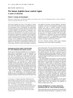
Electrophoresis gel lane of mesquite antigen (left) and mes-qite allergens demonstrated by Western blotting (right)Figure 1
Electrophoresis gel lane of mesquite antigen (left) and mes-
qite allergens demonstrated by Western blotting (right).
Pooled allergic human serum produced thirteen IgE
responses to mesquite tree pollen (Prosopis juliflora var. glan-
dulosa) when tested by Western blotting. The molecular
weights of these allergens ranged from 11 to 99 kd when
they were blotted from a 12% electrophoresis gel onto
nitrocellulose as detected by the Amplified Opti-4-CN sys-
tem (Bio-Rad).
NG
NG
NG
NG
NG
NGNG
NG
NGNG
NGNG
NG
NG
NGNG
NGNG
NG
NG
NG
NG
NG
NG
NG NG
Publish with BioMed Central and every
scientist can read your work free of charge
"BioMed Central will be the most significant development for
disseminating the results of biomedical research in our lifetime."
Sir Paul Nurse, Cancer Research UK
Your research papers will be:
available free of charge to the entire biomedical community
peer reviewed and published immediately upon acceptance
cited in PubMed and archived on PubMed Central
yours — you keep the copyright
Submit your manuscript here:
/>BioMedcentral
Clinical and Molecular Allergy 2004, 2:8 />Page 5 of 5
(page number not for citation purposes)
previous mesquite-sensitized rabbit cross-reactivity study,
merit further research.
References
1. Novey HS, Roth M, Wells ID: Mesquite pollen – an aeroallergen
in asthma and allergic rhinitis. J Allergy Clin Immunol 1977,
59:359-363.
2. Bieberdorf FW, Swinny B: Mesquite and related plants in
allergy. Ann Allergy 1952, 10:720-724.
3. Bessega C, Ferreyra JC, Vilardi JC, Saidman BO: Unexpected low
genetic differentiation among allopatric species of section
Algarobia of Prosopis (Leguminosae). Genetica 2000,
109:255-266.
4. Al-Frayh A, Hasnain SM, Gad-elRab MO, Al-Turk T, Al-Mobeireek K,
Al-Sedairy ST: Human sensitization to Prosopis juliflora antigen
in Saudi Arabia. Ann Saudi Med 1999, 19:331-336.
5. Ezeamuzie DI, Thomson MS, Al-Ali S, Dowaisan A, Khan M, Hijazi Z:
Asthma in the desert: spectrum of the sensitizing
aeroallergens. Allergy 2000, 55:157-162.
6. Davis RR: Spore concentrations in the atmosphere at
Ahmadi, a new town in Kuwait. J Gen Microbiol 1969, 25:643-648.
7. Bener A, Safa W, Abdulhalik S, Lestringant GG: An analysis of skin
prick test reactions in asthmatics in a hot climate and desert
environment. Allerg Immunol (Paris) 2002, 34:281-286.
8. Thakur IS: Purification and characterization of the glycopro-
tein allergen from Prosopis juliflora pollen. Biochem Int 1991,
23:449-459.
9. More D, Whisman L, Whisman B, Jordan-Wagner D: Identification
of specific IgE to mesquite wood smoke in individuals with
mesquite pollen allergy. J Allergy Clin Immunol 2002, 110:814-816.
10. Johns RE, Lee JS, Agahian B, Gibbons HL, Reading JC: Respiratory
effects of mesquite broiling. J Occup Med 1986, 28:1181-1184.
11. Killian S, Fretwell SD, McMichael J: Antigenic cross-reactivity sug-
gested by intradermal skin test correlations. J Nutr Environ Med
1997, 7:237-251.
12. Killian S, McMichael J: Cross-reactivity of mesquite tree pollen
with deciduous tree pollens [abstract]. Allergy Clin Immunol
2002, 109:s137.
13. Laemmli UK: Cleavage of structural proteins during the
assembly of the head of the bacteriophage T4. Nature 1970,
227:680-685.
14. The Association of Official Analytical Chemists: Official Methods of
Analysis of the AOAC 14th edition. Washington DC; 1984.
15. Towbin H, Staehelin T, Gordon J: Proc Nat Acad Sci 1970,
76:4350-4354.
16. Thakur IS, Sharma JD: Isolation and characterization of aller-
gens of Prosopis juliflora pollen grains. Biochem Int 1985,
11:903-912.
17. Thakur IS: Fractionation and analysis of allegens from Prosopis
juliflora pollen. Int Arch Allergy Appl Immunol 1989, 90:124-129.
18. Caballero T, Pascual C, Garcia-Ara MC, Ojeda JA: IgE crossreactiv-
ity between mugwort pollen (Artemisia vulgaris) and hazlenut
(Abellana nux) in sera from patients with sensitivity to both
extracts. Clin Exp Allergy 1997, 27:1203-1211.
19. Thakur IS: Fractionation and immunochemical characteriza-
tion of Prosopis juliflora pollen allergen. Biochem Int 1986,
13:951-960.