Báo cáo Y học: The human b-globin locus control region A center of attraction potx
Bạn đang xem bản rút gọn của tài liệu. Xem và tải ngay bản đầy đủ của tài liệu tại đây (223.65 KB, 11 trang )
REVIEW ARTICLE
The human b-globin locus control region
A center of attraction
Padraic P. Levings and Jo¨ rg Bungert
Department of Biochemistry and Molecular Biology, Gene Therapy Center, Center for Mammalian Genetics, College of Medicine,
University of Florida, Gainesville, FL, USA
The human b-globin gene locus is the subject of intense
study, and over the p ast two decades a wealth of infor mation
has accumulated on how tissue-specific and stage-specific
expression of its genes is ach ieved. T he data are extensive and
it would be d ifficult, if not imposs ible, to formulate a com-
prehensive model integrating every aspect of what is cur-
rently known. In this review, w e introduce the fundamental
characteristics of globin locus regulation as well as questions
on which much of the current research is predicated. We then
outline a hypothesis that encompasses m ore recent results,
focusing on the m odification of h igher-order chromatin
structure a nd recruitment of transcription complexes t o the
globin l ocus. The essence of this hypothesis i s that t he locus
control region (LCR) is a genetic entity highly accessible t o
and capable of recruiting, with great efficiency, chromatin-
modifying, coactivator, and transcription complexes. These
complexes are used to establish accessible chromatin
domains, allowing basal factors to be loaded on to specific
globin gene promoters in a d evelopmental stage-specific
manner. We conceptually divide this process into four steps:
(a) generation of a highly accessible LCR holocomplex;
(b) recruitment of transcription and chromatin-modifying
complexes to the LCR; (c) e stablishment o f chromatin
domains permissive fo r transcription; (d) transfer of tran-
scription c omplexes to globin g ene p romoters.
Keywords: chromatin domains; globin g enes; intergenic
transcription; locus control region; tran scription.
ORGANIZATION AND STRUCTURE
OF THE HUMAN b-G L O B I N L O C U S
The five genes of the human b-globin locus are arranged in a
linear array on chromosome 1 1 and are e xpressed in a
developmental stage-specific manner i n e rythroid cells
(Fig. 1) [1]. The e-globin gene is transcribed in the embry-
onic yolk sac and located at the 5 ¢ end. After the switch in
the s ite of h ematopoiesis from the yolk s ac to the fetal liver,
the e-gene is repressed and the two c-globin genes, l ocated
downstream o f e, are ac tivated. In a second switch,
completed shortly after birth, the bone marrow becomes
the major site of hematopoiesis, coincident with activation
of the adult b-globin g e ne, while the c-globin genes become
silenced. The d-globin gene is also activated in erythroid cells
derived from bone marrow hematopoiesis but is only
expressed at levels less t han 5% of t hat of the b-globin gene.
The complex program of transcriptional regulation
leading to the differentiation and developmental stage-
specific expression in th e globin l ocus is mediated by DNA-
regulatory sequences located both proximal and distal to the
gene-coding regions. The most prominent distal r egulatory
element in the human b-globin locus is the locus control
region (LCR), located f rom a bout 6 to 22 kb upstream of
the e-globin gene [2–4]. The LCR is composed of several
domains that exhibit extremely high sensitivity to DNase I
in erythroid cells (called h ypersensitive, or HS, s ites), and is
required for high-level globin gene expression at all develop-
mental stages [5].
The entire b-globin locus remains i n an i nactive DNase
I-resistant chromatin conformation in cells in which the
globin g enes are not expressed. In erythroid cells, t he entire
locus shows a higher degr ee of sensitivity t o DNase I,
indicating that it is in a more open and accessible chromatin
configuration [6]. Studies analyzing t he human b-globin
locus i n t ransgenic mice have shown that sensitivity to
DNase I in specific regions of the globin locus varies and
depends on the developmental s tage of erythropoiesis (yolk
sac, fetal liver, adult spleen ) [ 7]. T he LCR remains sensitive
to DNase I at all developmental stages, whereas sensitivity
to DN ase I in the region containing the e-globin and
c-globin genes is higher in embryonic c ells, and DNase I
sensitivity in the region containing the d-globin and b-globin
gene is higher in adult erythroid cells [7].
This review focuses on the regulation of the human
b-globin gene l ocus, and we would like to refer the reader t o
another r ecent review that compares the regulation of
different complex gene loci [8].
DEVELOPMENTAL STAGE-SPECIFIC
EXPRESSION OF THE GLOBIN GENES
The stage-specific activation and repression of the individual
globin genes during development is regulated by various
Correspondence to J. Bungert, Department of Biochemistry and
Molecular Biology, Gene Therapy Center, Cent e r for Mammalian
Genetics, College of Medicine, University of Florida, 1600 SW Archer
Road, Gainesville, FL 32610, USA. Fax: +352 392 2953,
Tel.: + 352 392 0121, E-mail: fl.edu
Abbreviations: LCR, locus control region; HS, hypersensitive; EKLF,
erythroid kru
¨
ppel-like factor; MEL cells, murine erythroleukemia
cells; ICD, interchromosomal domain; HLH, helix–loop–helix.
(Received 1 5 November 2001, revised 16 January 200 2, accepted
21 January 2002)
Eur. J. Biochem. 269, 1589–1599 (2002) Ó FEBS 2002
mechanisms. First, genetic information g overning the s tage -
specificity for all b-like globin g enes is located in g ene
proximal regions. These elements represent transcription
factor-binding sites that recruit proteins or protein com-
plexes in a stage-specific manner. Examples exist for the
presence of both positive and negative acting factors that
turn genes on or off at a specific developmental stage [1].
The most extensively studied stage-specific activator is
EKLF (erythroid kru
¨
ppel like factor), which is crucial for
human b-globin gene expression [9]. Gene-ablation studies
in mice have shown that EKLF deficiency leads to a specific
reduction in adult b-globin gene expression, with a
concomitant increase in expression of the fetal genes [10–
12]. Associated with the d ramatic decrease in adult b-globin
gene expression is a reduction in DNase I HS site formation
in the b-globin gene promoter as well as in LCR element
HS3 [13]. These results demonstrate t hat E KLF is critically
required for the expression of the adult b-globin gene and
suggest that EKLF may exert part of its function by
changing chromatin structure. Indeed, A rmstrong et al.[14]
showed that EKLF recruits chromatin-remodeling factors
to the adult b-globin promoter and that this remodeling
activity was sufficient to activate b-globin gene expression in
an erythroid-specific manner in vitro. EKLF acts in a
sequence-specific context to activate transcription of the
b-globin gene [ 15]. Although both t he e-globin a nd b-globin
gene promoters h arbor binding sites for EKLF, only the
b-globin gene is expressed at definitive stages of erythro-
poiesis. Disruption of direct repeat elements flanking the
e-promoter EKLF binding site leads to expression of the
e-globin gene at the adult stage [15]. This observation
indicates that repression of the e-globin gene at the definitive
stage is in part due to proteins that interfere with the
interaction of the transcriptional activator EKLF.
There is also increasing evidence for the presence of stage-
specific factors regulating t he expression of the two c-globin
genes. In particular, it has been shown that C ACCC and
CCAAT motifs are required for activation of the c-globin
genes. The C ACCC element is bound by members of the
family of kru
¨
ppel-like zinc finger (KLF) proteins [16].
Potential candidates for proteins acting through this
element are EKLF, FKLF, FKLF-2, and BKLF [17]. The
CCAAT box interacts with the heterotrimeric protein NF-Y
[18], which appears to play a r ole similar to EKLF a nd may
recruit c hromatin-remodeling activities to t he c-globin g ene
promoters at the fetal stage.
The combined data demonstrate that stage-specific
factors interacting with individual globin gene promoters
play important roles in the regulation of local chromatin
structure and stage-specific gene expression.
Another important parameter regulating t he stage-speci-
fic activity of the globin g enes is the relative position of t he
genes with respect to the LCR [19,20]. Inverting t he genes
relative to the L CR leads to an i nappropriate expression of
the adult b-globin gene at the embryonic stage and the
absence of e-globin gene expression at all stages [21].
Although the mechanistic basis for the importance of gene
order in the globin locus is not entirely clear, it is in
agreement with the hypothesis that t he genes in the globin
locus are competitively regulated by the LCR [22,23] and
suggests that repressors restrict the a bility of the LCR to
activate transcription of only one or two genes at specific
developmental stages. These factors could either modulate
the chromatin structure around the inactive genes [7] or
interact with globin gene promoters to prevent t he interac-
tion of a gene with the LCR in a developmental stage-
specific manner [15].
STRUCTURE AND FUNCTION
OF THE LCR
The overall organization of the LCR is c onserved among
several vertebrate species. The conservation of individual
factor-binding sites within the HS core elements implies that
these sites are important for LCR function [24]. However,
this by no means leads to the conclusion that transcription
factor-binding sites t hat are not conserved are functionally
irrelevant. Some o f these nonconserved s ites may mediate
novel functions acquired during evolution. For example, the
developmental p attern of globin gene e xpression in humans
is quite different from that in mice (Fig. 1) [25].
Whereas almost all studies agree that the human b-globin
LCR is required for high-level transcription of all b-like
globin genes, the question of whether the LCR also
regulates the chromatin structure over the whole l ocus is a
matter of debate. Deletion of the complete LCR from either
the murine or human locus does not appear to change the
overall general sensitivity to DNase I of the locus, indicating
that the LCR is not required for unfolding of higher-order
chromatin structure [26–28]. Our understanding of the
structural basis for general DNase I sensitivity of chromatin
is limited. Loci permissive for transcription are within
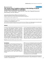
Fig. 1. Diagrammatic representation of t he h uman b-globin gene locus
(not d rawn to s cale). The five genes of the human b-globin gene l ocus
are arranged in linear order reflecting their expression during develop-
ment. The LCR is represented as the sum of the five HS sites. It should
be no ted that a dditional HS sites w ere mapped 5 ¢ to HS5 [ 95], but i t
is currently not known whether these sites participate in globin gene
regulation or whether they are associated with the regulation of
genes l ocated upstream o f the globin locus. The HS co re elements a re
200–400 bp in size and separated from each other by more than 2 kb.
During no rmal human development, the e-globin g ene is expressed in
the first trim ester in e rythroid cells derived from yolk sac hematopoi-
esis. The c-globin g enes are expressed in erythroid cells generated in the
fetal liver un til around birth. The adu lt b-globin gene i s expressed
around birth predominantly in cells derived from bone marrow
hematopoiesis. The expression pattern of the human globin genes is
somewhat different when analyzed in the context of transgenic mice
[96], where the e-globin and c-globin genes are coexpressed in the
embryonic yolk sac and the b-globin gene is expressed at high levels in
fetal liver and circulating erythroid cells from bone marrow.
1590 P. P. Levings and J. Bungert (Eur. J. Biochem. 269) Ó FEBS 2002
domains of general DNase I sensitivity. However, the
presence of a DNase I-sensitive domain does not indicate
that all of the genes residing within the domain are
transcribed or even that they are permissive for transcription
[28]. In t his respect the L CR could be i nvolved in regulating
chromatin structure beyond the formation of a g eneral
DNase I-sen sitive domain, for example by regulating the
modification o f histone tails (methylation, acetylation,
phosphorylation) [29].
It is unquestionable that the LCR p rovides a n open and
accessible chromatin structure at ectopic sites in transgenic
assays [5]. Whether this is t rue for all c hromosomal
positions is not known, because there are no data availab le
that demonstrate LCRs function from within a defined
heterochromatic environment. However, globin gene
expression constructs reveal strong position-of-integration
effects in transgenic assays in the absence of the LCR,
suggesting that at most sites the LCR is able to confer an
accessible chromatin structure. It is important to understand
that any model describing globin gene regulation must
address the LCR’s ability to open chromatin and en hance
globin gene expression at ectopic sites.
Current models propose that the individual HS c ore
elements interact to form a higher-order structure, com-
monly referred to as the LCR holocomplex [30,31].
Evidence supporting the holocomplex model came from
the genetic analys is of mutant LCRs in transgenic assays
[31–34]. Deletion o f individual L CR HS elements in single-
copy YAC transgenes led to strong reductions in globin
gene expression and also i mpaired the formation of
DNase I HS sites associated with the LCR and the globin
gene promoters. These data suggest that LCR HS site
deletions render the LCR unable to protect from position-
of-integration effects in transgenic studies [32]. In contrast
with these findings, the consequence of deleting HS sites
from the endogenous mouse locus on globin gene expression
is much milder and does not appear to affect the formation
of remaining HS sites [35–37]. The different results from
studies of globin l ocus transgenes vs. endogenous loci could
be explained in several ways [38]. F irst, the differences could
solely be based o n t he observation that an incomplete LCR
is not able to confer pos ition-independent chromatin
opening and gene expression in the globin locus at ectopic
sites. Secondly, differences in the size of the deleted
fragments could result in different phenotypes. The most
severe effects on globin gene expression were observed in
those transgenes in which only t he 200–400-bp ÔcoreÕ
enhancer elements were deleted. All the experiments in the
endogenous murine globin locus removed the cores together
with the flanking sequences. Finally, it i s possible t hat the
endogenous murine globin l ocus contain s sequences in
addition to the LCR that are able to provide an open
chromatin configuration.
Recently, Hardison and c olleagues analyzed the function
of LCR HS s ites in the p resence o r absence of the HS core
flanking sequences in murine erythroleukemia (MEL) cells
using recombination mediated cassette exchange [39]. At
several fixed positions, the inclusion of the flanking
sequences leads to a synergistic enhancement of expression
by the combination of HS units, whereas combining the
core HS elements only additively enhanced reporter gene
expression. Similarly, May et al. [40] showed that the
combination of H S2, 3, and 4 led to therapeutic leve ls of
b-globin gene expression in b-thalassemic mice only in the
presence of sequences flanking the L CR HS cores. Taken
together, t he data suggest t hat t he HS units interact with
each other to g enerate an L CR holocomplex, f ormation of
which is required for high-level b-globin g ene expression.
The flanking sequences could be important in positioning
the HS core elements in ways that facilitate their interactions
[39].
LCR INTERACTING PROTEINS
Knowledge about the proteins th at interact with the LCR
in vivo is very limited. Here we will focus on more recent
results describing the activities of specific proteins or protein
complexes implicated in LCR function. For a more
comprehensive summary of proteins interacting with regu-
latory sequences throughout the globin locus, w e would like
to refer the reader to previous reviews [1,24].
The DNA sequence motifs that a re most conserved
among different species are M ARE ( maf r ecognition
element) and GATA sequences in HS2, 3 and 4, KLF-
binding sites in HS2 and HS3, and an E-box motif in HS2
[24]. MARE sequences are bound in vitro by a large number
of different proteins that all heterodimerize with small maf
proteins [41]. Individual members of this family are
characterized by the presence of leucine zipper motifs, the
founding member being NF-E2 (p45) [42]. Other members
of this family also expressed i n erythroid cells are Bach1,
NRF1 and NRF2 ( NF-E2 related factor 1 and 2) [43–45].
A variety of data suggest a pivotal role for p45 in LCR
function [42,46]. However, g ene ablation studies have
shown that erythropoiesis is not affected in mice lacking
NF-E2 (p45), N RF1 or N RF2, suggesting functional
redundancy among the NF-E2 family members in erythroid
cells [47–49].
It should be noted that, although the NF-E2-like proteins
are a ll thought to interact with the same DNA-binding site,
they are structurally different. Bach1 for example contains a
BTB/Poz domain and forms oligomers while bound to
DNA in vitro [50]. This observation prompted investigators
to analyze whether Bach1/small maf heterodimers could
simultaneously bind to HS2, 3, and 4 and mediate the
interaction between the core elements [51]. Using atomic
force microscopy, i t was shown that Bach1-containing
heterodimers could indeed cross-link H S s ites in vitro,
indicating that proteins exist t hat bind to t he LCR a nd are
able to mediate the interaction of HS sites. Importantly, this
activity of Bach1 depends on the presence o f the BTB/Poz
domain.
The CACCC sites in HS2 and H S3 are probably bound
in vivo by EKLF. First, transgenic mice containing the
human b-globin locus and lacking EKLF exhibit a reduc-
tion in the formation of HS3 [13]. In addition, using the
Pin-Point assay, Lee et al. [52] d emonstrated that EKLF
binds to both HS2 and HS3 in vivo. Interestingly, the
binding of EKLF to HS3 i s r educed in the absence of HS2,
suggesting some f orm of c ommunicatio n between these two
elements [52].
The GATA sites are bound by either GATA-1 or
GATA-2, the only two members o f the GATA family of
transcription factors known to be expressed in erythroid
cells [53]. GATA-1 is one of the e arliest markers i n r ed cell
differentiation and is detectable in progenitor cells that do
Ó FEBS 2002 Multistep model for locus control region function (Eur. J. Biochem. 269) 1591
not yet e xpress the globin g enes [54]. Interestingly, L CR HS
sites are already detectable in these undifferentiated precur-
sor c ells [55]. These results suggest that GATA-1 may b e
involved in the regulation o f chromatin structure at an early
stage of erythroid differentiation.
The E-box in HS2 i nteracts with helix–loop–h elix (HLH)
proteins in vitro, and both USF and Tal1 were shown to
interact with this element [56,57]. USF is a ubiquitously
expressed member o f the HLH family of proteins and binds
to DNA as a heterodimer usually composed of USF1 and
USF2. USF has been implicated in the r egulation of m any
genes and normally acts as a transcriptional activator.
However, it has also been reported to function through
initiator e lements, in which c ase i t m ediate s t he recruitment
of Pol II transcription complexes [58,59]. Tal1 is hemato-
poietic specific and appears to function at an early step
during the specification of hematopoietic progenitor cells
[60].
Protein–protein interactions probably play important
roles in LCR function. We have already discussed the
multimerization of Bach/maf heterodimers. Other protein–
protein interactions known t o occur among LCR-binding
proteins involve those between the GATA factors and
between GATA factors and EKLF, LMO2/Tal1, and Sp1
[61–63]. In addition, GATA-1, EKLF and NF-E2 (p45)
were shown to interact with coactivators and acetyltrans-
ferase activities [64,65]. EKLF has also been d emonstrated
to interact with members of t he Swi/SNF f amily of
chromatin-remodeling complexes [14]. These results show
that most proteins binding to one LCR core element have
the potential to interact with proteins binding to another
LCR core HS site, which could initiate and stabilize an LCR
holocomplex. In a ddition, the results also demon strate that
LCR-interacting p roteins recruit macromo lecular c om-
plexes involved in chromatin r emodeling and histone
acetylation.
REPLICATION AND CHROMATIN
STRUCTURE
The human b-globin l ocus replicates early in erythroid cells
and l ate in nonerythroid cells. E arlier s tudies suggested that
the LCR regulates the timing and usage of an origin of
replication located between the d-globin and b-globin gene
[66]. This interpretation was based on the observation that a
large deletion in the human b-globin locus, starting
immediately upstream of HS1 and spanning about 30 kb,
inactivates the entire globin locus [66]. The globin genes
linked to this deletion are not transcribed, the locus becomes
late replicating, and remains in a DNase I-resistant and
inaccessible configuration. However, recent analysis of the
consequence of a targeted deletion of the LCR demonstrates
that the LCR regulates neither the timing of replication i n
the g lobin l ocus nor the usage of the replication o rigin [ 67].
Thus, a putative element regulating replication timing in the
human b-globin locus must be located 5¢ to the L CR.
An important question that has to be addressed is
whether activation of the globin locus and LCR function
requires replication. During differentiation of erythroid
cells, the locus undergoes various transitions, the first of
which is the formation of DNase I HS sites in the LCR [55].
Does the formation of HS sites a t this early stage in
differentiation r equire replication? In other words, do the
proteins responsible for HS site formation require a window
of opportunity after replication to bind and then prevent the
generation of repressive chromatin structure or do these
proteins recruit chromatin-remodeling activities that change
the chromatin structure in a replication-independent man-
ner? Experiments that i ndirectly addressed this issue were
those in which investigators generated heterokaryons with
MEL cells, which represent definitive erythroid cells that
express the adult b-globin gene, and human K562 cells,
which represent primitive erythroid cells that express the
e-globin gene [68]. These studies showed that trans-acting
factors in t he MEL cells are able to activate t ranscription of
the human b-globin g ene. Interestingly, the onset of
b-globin gene expression in these experiments occurred
about 12 h after fusion. Because the globin l ocus replicates
early in e rythroid cells, t hese results c ould be interpreted to
mean t hat rep lication i s required f or t rans-activation of the
human b-globin genes in the heterokaryons. On the other
hand, this experiment could also lead to the interpretation
that the human locus can be activated by transcription
factors and accessory proteins already present i n t he adult
(MEL) erythroid cells. This mode of regulation would b e
similar to the induction of genes by hormone receptors [69].
However, differences in the two systems may exist, as the
globin locus is a developmentally regulated locus, the
expression of which c hanges as the cell d ifferentiates. Genes
regulated by hormone and orphan receptors are transcribed
in mature cells and t heir expression is regulated b y e xternal
stimuli, i.e. hormones. Obviously more studies are needed
that examine the relationship between replication and
chromatin structure in the globin locus. For example, it
would be interesting to examine t he binding of chromatin
components and transcription factors during the cell cycle in
erythroid cells.
INTERGENIC TRANSCRIPTS
IN THE GLOBIN LOCUS
In 1992, Tuan et al. [70] reported that long transcripts
initiate within LCR HS2 and proceed in a unidirectional
manner toward the globin genes. Further studies by the
same group led to the startling observation that transcrip-
tion always proceeds in the direction o f a linked gene,
independent from the orientation of HS2 [71]. This r esult
suggests some form of communication between the promo-
ter a nd LCR H S2 in these experiments. Subsequent studies
in the laboratories of Proudfoot [72] and Fraser [7] identified
noncoding transcripts over the entire LCR and in between
the g lobin gene c oding regions. Interestingly, the pattern of
intergenic transcription during development a ppears to
correlate with the pattern of general DNase I sensitivity [7].
Mutations that delete the start site of the adult-specific
intergenic transcripts l ead t o a decrease in ge neral DNase I
sensitivity and b-globin g ene transcription, suggesting that
intergenic transcription modulates the chromatin structure
of globin locus subdomains. Intergenic transcripts appear to
be generated in a cell-cycle-dependent manner, detectable
during early S-phase but predominantly present in G1 [7].
These results provide evidence for the hypothesis that
intergenic transcription is transient. Recently Plant et al.
[73] analyzed intergenic transcripts across the globin locus
by nuclear run-on analysis and did not find any evidence for
the stage-specific generation of these transcripts. The
1592 P. P. Levings and J. Bungert (Eur. J. Biochem. 269) Ó FEBS 2002
discrepancy between this study and that of Gribnau et al.[7]
is not understood at the moment, but it is possible that at
certain stages of t he cell c yc le, the entire locus i s transcribed
for a short period of time. A subsequent step could then shut
off transcription in silenced domains, but reduced tran-
scription could still be detectable by the more sensitive assay
employed by Plant et al. [73].
INSULATORS
The chicken b-globin locus is flanked by i nsulator elements
which mark clear boundaries between active and inactive
chromatin [74,75]. No such sequences have been conclu-
sively identified i n the human or murine globin locus, a nd it
appears that the DNase I-sensitive domain in these loci
extend far 5¢ of the L CR and far 3¢ of the b-globin gene.
Recent experiments distinguish between insulator sequences
that block the action of an enhancer or silencer an d t hat o f
boundary elements that separate open and closed chromatin
domains [74]. The 5¢ most HS site of the chicken LCR, HS4,
appears to harbor both activities [ 75]. In this sense it is quite
possible that the human b-globin locus contains insulator
elements that restrict the action of the LCR to within
specific domains. Some evidence suggests that HS5 may
harbor insulator activity. First, HS5 harbors a binding site
for the protein CTCF, which is largely responsible for
insulator function of chicken H S4 [76]. Secondly, inversion
of the entire LCR with respect to the g enes reduces globin
gene expression to less than 30% of wild-type levels [21].
Thirdly, an e-globin gene placed upstream of the LCR is not
transcribed [21]. Finally, HS5 was shown to exhibit
insulator activity in cell culture expe riments [77].
NUCLEAR LOCALIZATION
Recent data suggest that enhancer and other regulatory
elements affect the position of genes within the nucleus
[78,79]. For example, it was shown that in t he absence of
an enhancer, the b-globin gene is located close to
centromeric heterochromatin, an environment within the
nucleus th at is incompatible with transcription [80]. In the
presence of LCR element HS2, the b-globin gene localizes
away from centromeric heterochromatin, suggesting that
activities associated with HS2 are able to relocate the
transgene to a transcriptionally permissive nuclear region
[80]. This phenomenon has been most intensively a nalyzed
in yeast, in which specific protein complexes appear to
direct the location of genes into active or inactive regions
of the nucleus [81]. However, Milot et al.[32]showedthat
a wild-type globin locus that integrated close to centro-
meric h eterochromatin was still active, suggesting that, in
the presence of the LCR, the globin locus is active even
when situated close to a defined heterochromatic envi-
ronment.
Recent a dvances in fluorescent labeling of chromatin as
well as three-dimensional fluorescent microscopy indicate
that chromosomes occupy distinct regions, or domains,
within the cell nucleus [82]. These chromosome domains
may be composed of up to 1 Mb of chromatin supported by
the nuclear architecture and appear to contain loops of
about 50–200 kb of DNA possessing one or several gene
loci that may or may not be co-regulated. The spaces
between these territories are believed to b e occupied by a
Ômatrix Õ-like structure, consisting of filamentous proteins,
which is defined as the interchromosomal domain (ICD).
Active gene loci are located at the surface of chromosomal
domains in direct contact w ith the IC D, whereas inactive
loci are located away from the ICD within chromosomal
domains. I t is proposed that macromolecular protein
complexes involved i n chromatin remodeling, tr anscription,
and splicing are enriched in the ICD, whereas single proteins
or smaller protein complexes can diffuse into regions of the
chromatin domains that are not in contact w ith the ICD.
The former ideas are based on indirect observations using
microscopy and fluorescent l abeling. We can therefore only
describe the existence of chromosome territories and the
ICD as speculative at best. However, it is safe to say that
gene loci are located in specific regions of the nucleus and
that the r elative position of these loci changes on activation.
If applied to the regulation of the globin genes, the ICD
model could explain why deletion of the LC R in the
endogenous human or murine globin loci silences globin
gene expression without altering the e stablishment of
DNase I a nd hyperacetylated c hromatin. It is p ossible that
transcription factors could gain access to t he globin locus
and change higher-order chromatin structure, but that the
LCR is required to organize the globin locus in a way that it
is located in c lose proximity to the ICD. The situation is
similar in concept to mechanisms described for the regula-
tion of gene loci d uring differentiation of B-lymphocytes.
Fisher and colleagues [79] have shown that specific gene loci
relocate to inactive regions in the nucleus of cycling B -cells.
The relocation and inactivation is r egulated by the DNA-
binding protein Ikaros, which m ediates the association of
gene loci with centromeric heterochromatin.
A MULTISTEP MODEL FOR HUMAN
b-GLOBIN GENE REGULATION
Step 1: generation of a highly accessible
LCR holocomplex
We propose that the first step towards activation of the
globin genes during differentiation is the partial unfolding of
the chromatin structure containing the globin locus into a
DNase I-se nsitive domain (Fig. 2A). This step may or may
not require replication. The initial unfolding of the
chromatin structure is mediated by the diffusion of eryth-
roid-specific proteins into chromosomal domains that are
not permissive for transcription. These proteins bind to
sequences throughout the globin locus leading to the par tial
unfolding and perhaps hyper-acetylation of the chromatin.
If replication is required for globin locus activation, we
propose that erythroid-specific proteins bind to the globin
locus after DNA synthesis , prevent the formation o f
repressive chromatin, and mark the locus by modification
of histone tails.
GATA factors may be involved in the initial step of
globin l ocus activation, as their binding sequences are
located throughout the globin locus. In addition, GATA-1
is one of the earliest marke rs of red cell differentiation [54]
and is known to associate with proteins containing histone
acetyltransferase activities. The partial unfolding into a
DNase I-sensitive structure does not require activities
associated with the LCR. This is shown by the fact that
even in the absence of an intact LCR, the r est o f t he globin
Ó FEBS 2002 Multistep model for locus control region function (Eur. J. Biochem. 269) 1593
locus is r endered nuc lease s ensitive [28] and exhibits
increased histone H4 acetylation [83]. We propose that all
subsequent steps require activities recruited to the LCR. It is
possible that the initial invasion of the globin locus by
erythroid factors could mark the locus for relocation to an
area that is close to the ICD. Proteins normally associated
with het erochromatin, such as SUV39H1, M3 3, and B M-1,
could be involved in regulating the accessibility and location
of the globin locus [84].
The reorganization of the chromosomal domain, which
renders the globin locus accessible to chromatin-remodeling,
coactivator and transcription complexes present in the ICD,
Fig. 2. Multistep model for human b-globin gene r egulation. The m ode l depicts four steps p ro posed to be involved in th e regulation of c hrom atin
structure and gene expression in the human b-globin locus. The model focuses on the regulation of the human globin locus in the context of
transgenic mice, but it is assumed that the same principal mechanisms govern the correct expression of the b-globin genes during human
development, except that the timing of e xpression of the genes is so mewhat different (see Fig. 1). (A) Generation of a highly accessible LCR
holocomplex. We propose that the initial events in activating the h uman globin gene locus during differentiation involves the partial unfolding of the
chromatin structure into a DNase I-sensitive domain and the binding of protein complexes to the LCR HS sites. This will then generate the LCR
holocomplex, t he protein-mediated interaction of HS s ites. ( B ) Re cruitm ent of chromatin- remodeling, coactivator an d t ranscriptio n complexes.
Once the LCR holocom plex is ge nerate d, the glob in locus i s relocated to an area of the nucleus enriche d for macro molecu lar comp lexes in volved in
coactivation, c hromatin remodeling (or modification of histone tails) and transcription. These complexes are recruited to the LCR, which provides a
highly accessible platform for recruiting these activities. (C) Establishment of chromatin domains permissive for transcription. The macromolecular
protein complexes recruited to the LCR will initially be used to establish chromatin domains that allow transcription of the genes. Specifically, we
propose that the LCR recruits elongation-competent transcription complexes (or complexes that are rendered elongation competent at the LCR)
that track along the DNA and modify the chromatin structure. This reorganization of the chromatin structure will render the promoters accessible
for activating proteins and components of the preinitiation complex. Data published by Gribnau et al . [7] suggest that intergenic transcription and
chromatin reorgan ization i s s tage-spe cific and restricted to t he genes that a re e xpressed e ith er a t t he embryonic or ad ult stage. (D) Transfer of
macromolecular protein complexes to individual globin gene p romoters. Once active chromatin d omains are established, the LC R recruits
elongation-incompeten t transcription complexes, which are transferred to the individual globin gene promoters present in the accessible chromatin
domains. The p olymerases are then rendered elongation-competent, possibly through phosphorylation of t he C-terminal domain [88].
1594 P. P. Levings and J. Bungert (Eur. J. Biochem. 269) Ó FEBS 2002
is regulated by elements within the LCR. Once the l ocus
becomes accessible to macromolecular complexes in the
ICD, protein complexes aggregate at the LCR HS core
elements. In vitro experiments suggest that HS site forma-
tion occurs even in the ab sence of regular chromatin
structure and may involve the generation of S1-sensitive
segments within the core HS sites [85]. Therefore, it is
proposed that protein complexes bind to the HS core sites
and bend or disturb the structure of the DNA. The
formation of pro tein aggregates and t he subsequent distur-
bance of DNA structure at the LCR HS core elements could
lead to a highly accessible region in the b-globin locus.
The generation of a n LCR holocomplex probably
involves interactions between protein complexes at the
different HS units including the cores and the flanking
sequences. In early differentiation stages, NF-E2 sites may
be occupied by Bach1/maf heterodimers, which may
facilitate interactions between HS sites, b ut may also hold
the LCR in an inactive configuration. Heme-mediated
inhibition of Bach1/maf binding at later stages of differen-
tiation would allow the binding of other members of the
NF-E2 family of proteins [86].
Step 2: recruitment of chromatin-remodeling
and transcription complexes to the LCR
Formation of the LCR holocomplex results in the massive
disruption of chromatin structure and a high density of
DNA-bound proteins (Fig. 2 B). The consequence of this
shift i n s tructure is that ac tivi ties tha t are normally
associated with tr anscriptionally active chr omatin w ill
gravitate to the LCR. We propose that proteins bound to
the c ore HS s ites, namely members of the NF-E2 family,
GATA factors and EKLF, recruit chromatin-remodeling
complexes and coactivators. The recruitment of RNA
polymerase II may involv e HLH proteins as they have been
shown to mediate transcription complex formation on
TATA-less genes [58,59].
Initially the LCR c ould recruit elongation-competent
transcription complexes associated with chromatin-remode-
ling activities that would initiate the establishment of
transcriptionally permissive chromatin domains within the
locus. Orphanides & Reinberg [87] have proposed the
presence of ÔpioneerÕ polymerases which are involved in
the modulation of chromatin structure. Such a ÔpioneerÕ
polymerase may be recruited to the LCR, associate with
chromatin-modifying activities, and track along the DNA
to modify the nucleosome structure of chromatin domains
in the globin locus. Once active chromatin domains are
established, the LCR could re cruit elongation-incompetent
transcription complexes. These complexes could then be
delivered to individual globin g ene promoters and w ould
then be rendered elongation-competent, possibly through
phosphorylation of the C-terminal domain of RNA
polymerase II [88].
Step 3: establishment of chromatin domains
permissive for transcription
Recent studies have shown that the b-globin locus under-
goes dynamic changes in both DNase I sensitivity and
histone acetylatio n patterns during development [83,89].
The changes in chromatin structure as well as the presence
of intergenic transcripts have been used to separate the
globin locus into developmental stage-specific chromatin
domains [7]. Although the exact mechanism by which the
developmental patterns of chromatin structure and inter-
genic transcription are established i s unknown, it is likely
that the recruitment o f c hromatin-modifying and transcrip-
tion complexes to the LCR would initiate the processes
involved (Fig. 2C).
There are three lines of evidence suggesting that
intergenic transcription modifies the chromatin structure
within the g lobin l ocus subdomains . F irst, L CR transcripts
initiate both upstream or within the LCR a nd proceed in a
unidirectional manner toward the genes [7,71,72]. Sec-
ondly, deletion of a region containing the adult-specific
transcription initiation site leads to a decrease in general
DNase I sensitivity within the s ubdomain and a decrease in
expression of the adult b-globin gene [7]. Finally, it is
feasible that chromatin-modifying activities associate with
Ôpioneer Õ polymerase complexes at the LCR, which would
initiate transcription and modify the c hromatin structure o f
globin locus subdomains [87]. In vivo, nucleosomes in
transcribed r egions of chromatin are unfolded exposing the
cysteinyl-thiol groups of histone H3, and this unfolding
was observed only i n the presence of active transcription
[90,91]. Furthermore, these unfolded nucleosomes were
associated with highly acetylated histones. The fact that
reconstitution of nucleosomes with hyperacetylated
histones could not recapitulate the unfolded structure led
the authors to conclude that acetylation was not a
requirement of nucleosome unfolding. More recently it
was found that histone acetylation was required to
maintain the unfolded nucleosome structure that resulted
from transcriptional elongation [92]. This result suggests
that transcription can modify the chromatin of an active
gene domain so as t o distinguish it from that of an
accessible but otherwise inactive one [92].
Two models have been proposed to explain how the
LCR enhances globin gene transcription, the looping or
linking model [6,22]. According to the linking model,
activities recruited by the LCR would be transmitted to
the globin genes through an array of proteins binding
along the DNA. T he looping model proposes direct
interactions between the LCR and individual genes with
the intervening DNA looping out. The establishment of
transcriptionally permissive chromatin domains in the
globin locus can be explained according to both models.
It is possible that the LCR and segments of the adult-
specific chromatin domain are in direct contact and that
transcription complexes and chromatin-modifying activit-
ies are transferred b y a looping mechanism. On the other
hand, the observation that a certain fraction of adult cells
coexpress the c-globin and b-globin genes, which are
located in different chromatin domains [7], could suggest
that at a certain stage, the whole locus is ÔopenÕ and that
the repression of the e/c-chromatin domain is a secondary
process in volving the deacetylation a nd inactivation of the
embryonic domain. This idea is supported by the data of
Forsberg et al. [89] showing that the pattern of histone
acetylation across the globin locus varies during develop-
ment. These authors suggest th at dynamic changes in the
acetylation patterns, initiated by the recruitment of h istone
acetyltransferase a nd deacetyltransferase to the LCR, may
affect globin gene expression by regulating th e chromatin
Ó FEBS 2002 Multistep model for locus control region function (Eur. J. Biochem. 269) 1595
structure of stage-specific chromatin domains. However,
the authors point out that histone acetylation alone is not
likely to regulate transcription because inhibition of
histone deacetylase activity did not reactivate a develop-
mentally silenced globin gene.
Step 4: transfer of transcription complexes
to individual globin genes
The establishment of stage-specific domains within the
globin locus would restrict the action of the LCR to either
the embryonic/fetal genes or the adult genes. Several lines of
evidence suggest that the LCR directly communicates with
the genes to transfer transcription and/or chromatin
remodeling complexes to the promoters (Fig. 2D) [85,93].
First, studies have shown that LCR-dependent promoter
activation is associated with hyperacetylation o f histone H3
in both the LCR and the active gene [83]. Given that H3 and
H4 histone acetylation at a level above that of a n i nactive
locus is observed even in the absence of the LCR in these
studies, one could conclude that LCR-dependent hyper-
acetylation o f active g enes is the result o f d irect interactions
between the LCR and the genes. This interaction could
result in the transfer of chromatin remodeling and tran-
scription c omplexes from the LCR to the p romoter.
Secondly, RNA PolII is recruited to LCR elements HS2
and HS3 in vitro [85] and in vivo [93]. Johnson et al.[93]
recently reported that RNA PolII is located at both LCR
element HS2 and the b-globingeneinMELcells.InMEL
cells lacking N F-E2 (p45), PolII is still recruited to the LCR
but is no longer detectable at the b-globin gene. This resu lt
suggests that p45 is involved in the transfer of PolII
transcription complexes from the LCR to the adult b-globin
gene promoter. Indeed, S awado et al. [94] recently showed
that p45 could be cross-linked in vivo to the b-globin gene
promoter.
It is likely that transcription factors interacting with
individual globin promoters direct the LCR to specific genes
within transcriptionally permissive domains. But why would
this transfer be required, why w ould the transcription
complexes not be loaded directly to the p romoter regions?
We believe that the answer to these questions lies in the
assumption that in vivo theglobingenepromotersarenotas
accessible as the LCR. It is possible that transcription o f the
globin genes requires local remodeling of the nucleosome
structure and that the activities required f or chromatin
remodeling a re first recruited to t he LCR and then targeted
to individual globin genes.
Our model describing gene regulation of the human
b-globin locus focuses on the ability of the LCR to act as
a c enter of a ttraction for various regulatory activities found
in the cellular milieu. The LCR nucleates and perpetuates
dynamic changes in chromatin structure and transcriptional
activity throughout the locus to produce the elegant pattern
of developmental stage-specificity characteristic of globin
gene expression.
ACKNOWLEDGEMENTS
We thank our colleagues in the laboratory, Sung-Hae Lee Kang, Kelly
Leach, Karen Vieira, and Christof Dame for stimulating discussions
and Mike Kilberg (UF) and Doug Engel (Northwestern U niversity)
for critically r eading the manuscript. We also t h ank t he reviewers f or
helpful suggestions. The projects in the authors’ laboratory are
supported by grants f rom the A merican Heart Assoc iation and from
the NIH (DK 58209 and DK 52356).
REFERENCES
1. Stamatoyannopoulos, G. & Nienhuis, A. (1994) Hemoglobin
switching. In The M olecular Basis o f B lood Diseases (Stama-
toyannopoulos, G., Nienhuis, A., Majerus, P. & Varmus, H., e ds),
pp. 107–155. W.B. Saunders, Philadelphia, PA.
2. Tuan, D., Solomon, W., Li, Q. & London, I.M. (1985) The Ôb-like
globin domainÕ in human erythroid cells. Proc. Natl Acad. Sci.
USA 82, 6384–6388.
3. Forrester, W.C., Takegawa, S., Papayannopoulos, T., Stama-
toyannopoulos, G. & Gro udin e, M. ( 1987) Evidence for a locus
activation region: the formation of developmentally stable
hypersensitive sites in globin-expressing h ybrids. Nucleic Acids
Res. 15, 1 0159–10177.
4. Grosveld, F., van Assendelft, G.B., Greaves, D .R. & Kollias, G .
(1987) Position-independent, high-level expression of the human
b-globingeneintransgenicmice.Cell 51, 975 –985.
5. Higgs, D. (1998) Do LCRs open chromatin domains? Cell 95,
299–302.
6. Bulger, M. & Groudine, M. (1999) Looping versus linking: toward
a model for long-d istance gene a ctivation. Genes D ev. 13 , 2465–
2477.
7. Gribnau, J., Diderich, K., Pruzina, S ., Calzolari, R. & Fraser, P.
(2000) Intergenic tra nscription a nd deve lopmental r emodeling of
chromatin structure in the human b-globin locus. Mol. Cell. 5,
377–386.
8. Bonifer, C. (2000) Developmental r egulation of eukaryotic gene
loci: whi ch cis -regulatory information is re quired? Tre nds Genet.
16, 310–315.
9. Miller, I.J. & Bieker, J .J. (1993) A novel, erythroid cell-specific
transcription factor that binds to the C ACC element related to
the kru
¨
ppel family of nuclear proteins. Mol. Cell. Biol. 13,
2776–2786.
10.Nuez,B.,Michalovich,D.,Bygrave,A.,Ploemacher,R.&
Grosveld, F. ( 1995) Defective h aematopoiesis in fetal liver r esult-
ing from inactivation of the EKLF gene. Nature (London) 375,
316–318.
11. Perkins, A.C., S harpe, A.H . & Orkin, S.H. ( 1995) Lethal
b-thallassemia in mice lacking the erythroid CACCC-transcription
factor EKLF. Natu re (London) 37 5 , 318–322.
12. Perkins, A.C., Gaensler, K.M. & Orkin, S.H. (1996) Silencing of
human fetal globin expression is i mpaired in the absence of the
adult b-globin gene activator protein EKLF. Proc.NatlAcad.Sci.
USA 93, 12267–12271.
13. Wijgerde, M ., Gribnau, J., Trimborn, T., Nuez, B. , Philipsen, S.,
Grosveld, F. & Fraser, P. (1996) The role of EKLF in b-globin
gene competition. Genes Dev. 10 , 2894–2902.
14. Armstrong, J.A., Bieker, J .J. & Emerson, B.M. (1998) A Swi/
SNF-related chromatin-remo deling comp lex, E-RC1, is req uired
for tissue-specific transcriptional regulation by EKLF in vitro. Cell
95, 93–104.
15. Tanimoto,K.,Liu,Q.,Grosveld,F.,Bungert,J.&Engel,J.D.
(2000) Context-dependent EKLF responsiveness defines the
developmental specificity of the human e-globingeneinerythroid
cells o f YAC transgenic mice. Genes Dev. 14, 2778–2794.
16. Bieker, J.J. (2001) Kru
¨
ppel-like factors: three fingers in many pies.
J. Biol. Chem. 276 , 34355–34358.
17. Asano, H., Li, X.S. & Stamatoyannopoulos, G . (1999) FKLF, a
novel Kru
¨
ppel-like factor that activates hu man embryonic and
fetal b-like globin g enes. Mol. Cell. Biol. 19, 3571–3579.
18. Duan, Z., Stamatoyannopoulos, G. & Li, Q. (2001) Role of
NF-Y in in vivo regulation of the c-globin gen e. M o l. Cell. Bi ol . 21,
3083–3095.
1596 P. P. Levings and J. Bungert (Eur. J. Biochem. 269) Ó FEBS 2002
19. Dillon, N., Trimborn, T., Strouboulis, J ., Fraser, P. & Grosveld ,
F. (1997) The effect of distance on long–range chromatin inter-
actions. Mol. Cell 1, 131– 139.
20. Peterson, K.R. & S tamatoyannopoulos, G. (1993) Role of gene
order in developmental control of human gamma- and beta-globin
gene ex pression. Mol. Cell. Biol. 13, 4 836–4843.
21. Tanimoto, K., Liu, Q., Bungert, J. & Engel, J.D. (1999) Effects of
altered gene order or orien tatio n of the locus c ontro l region on
human b-globin gene expression in mice. Nature (London) 398,
344–348.
22. Engel, J.D. & Tanimoto, K. (2000) Looping, link ing and c hro-
matin activity: new insights into b-globin locus regulation. Cell
100, 4 99–502.
23. Choi, O. & Engel, J.D. (1988) Developmental regulation of
b-glo bin gene s witch ing. Cell 55 , 17–26.
24. Hardison, R., Slightom, J.L., Gumucio, D.L., Goodman, M.,
Stojanovich, N. & Miller, W. (1997) Locus control re gions of
mammalian beta-globin gene clusters: combining phylogenetic
analysis and experimental results to gain functional insights. Gene
51, 73– 94.
25. Trimborn, T., Gribnau, J., Grosveld, F. & Fraser, P. (1999)
Mechanisms of developmental control of transcription in the
murine a-andb-globin loci. Genes Dev. 13 , 112–124.
26. Epner, E., Reik, A., Cimbora, D., Telling, A., Bender, M.A.,
Fiering, S., Enver, T., Martin, D.I.K., Kennedy, M., Keller, G.
& Groudine, M. (1998) The b-globin L CR is not ne cessary for
an o pen chromatin structure or developmentally regulated t rans-
cription of the native mouse b-globin locus. Mol. Cell 2, 447–455.
27. Reik, A., Telling, A., Zitnik, G., Cimbora, D., Epner, E. &
Groudine, M. (1998) The locus control r egion is necessary for gene
expression in t he h uman b-globin locus but not f or the mainten-
ance of an open chromatin structure in erythroid c ells. Mol. Cell .
Biol. 18, 5 992–6000.
28. Bender, M.A., Bulger, M., Close, J. & Groudine, M. (2000)
Globin g ene swit ching and DNaseI se nsitivity of t he endogenous
b-glo bin l ocus in mice does not require the locu s c ontrol region.
Mol. Cell 5, 387–393.
29. Rice, J.C. & Allis, C.D. (2001) Histone methylation versus histone
acetylation: new i nsights into epigenetic regulation. Curr. Opin.
Cell Bi ol. 13, 2 63–273.
30. Wijgerde, M., Grosveld, F. & Fraser, P. (1995) T ranscription
complex s tabilityandchromatindynamics in vivo.Nature (London)
377, 2 09–213.
31. Bungert, J., Dave, U., Lim, K C., Lieuw, K.H., Shavit, J.A., Liu,
Q. & Engel, J.D. (1995) Synergistic regulation of human b-globin
gene switching by locus control region elements HS3 and HS4.
Genes Dev. 9, 3083–3096.
32. Milot, E., Strouboulis, J., Trimborn, T., Wijgerde, M., de Boer, E .,
Langeveld, A., T an-un, K., Vergeer, W., Yannoutsos, N.,
Grosveld, F. & F raser, P . ( 1996) Heterochromatin effects on t he
frequency and duration of LCR-mediated gene transcription. Cell
87, 105 –114.
33. Navas, P.A., Peterson, K.R., Li, Q., Skarpidi, E., Rohde, A.,
Shaw, S.E., Clegg, C.H ., A sano, H. & Stamatoyannopoulos, G .
(1998) Developmental specificity of the interaction between the
locus control region and embryonic or fetal globin genes in
transgenic mice with an HS3 c ore deletion. Mol. Cell. Biol. 17,
4188–4196.
34. Bungert,J.,Tanimoto,K.,Patel,S.,Liu,Q.,Fear,M.&Engel,
J.D. (1999) Hypersensitive site 2 specifies a unique function within
the human b-globin l ocus control r egion to stimulate globin ge ne
transcription. Mol. Cell. B iol. 19, 3 062–3072.
35. Fiering, S., Epner, E., Robinson, K., Zhuang, Y., Telling, A., Hu,
M., Martin, D.I.K., Enver, T., Ley, T.J . & Groudine, M. (1995 )
Targeted deletion o f 5¢HS2 of the murine b-globin LCR reveals
that it is not essential for proper regulation of the b-like globin
locus. Genes Dev. 9, 2 203–2213.
36. Hug, B., Wesselschmidt, R.L., Fiering, S., Bender, M.A., Grou-
dine, M. & Ley, T.J. (1996) Analysis of mice containing a targeted
deletion of the b-globin locus control region 5 ¢HS3. Mol. Cell.
Biol. 16, 2 906–2912.
37. Bender, M.A., Mehaffey, M.G., Telling, A., Hug, B., Ley, T.J.,
Groudine, M. & Fierin g, S. (2000) Inde pendent fo rmat ion of
DNaseI hypersensitive s ites in the murine b-globin locus co ntrol
region. Blood 95, 3 600–3604.
38. Engel, J.D. (2001) Simp le math for t he beta-globin l ocus control
region. Blood 98, 2 000.
39. Molete, J.M., Petrykowska, H., Bouhassira, E.E., Feng, Y Q.,
Miller, W. & Hardison, R. (2001) Sequences flanking hyper-
sensitive sites of the b-globin locus con trol region are required
for s ynergistic enhancement. Mol. Cell. Biol. 21, 2969 –2980.
40. May, C., Rivella, S., Callegari, J., Keller, G., Gaensler, K.M.,
Luzzatto,L.&Sadelain,M.(2000)Therapeutichemoglobin
synthesis in beta-thallassaemic mice e xpressing lentivirus-encoded
human beta-globin. N ature (London) 406 , 82–86.
41. Motohashi, H., Shavit, J.A., Igarashi, K., Yamamoto, M. &
Engel, J.D. (1997) The world according to maf. Nucleic Acids Res.
25, 295 3–2959.
42. Andrews, N .C., Erdjument-Bromage, H., Davidson , M.B.,
Tempst, P. & Orkin, S.H. (1993) E rythroid transcription factor
NF-E2 is a hematopoietic-specific basic-leucine zipper protein.
Nature ( London) 362, 722–728.
43. Oyake, T., Itoh, K., Motohashi, H., Hayashi, N., Hoshino, H.,
Nishizawa, M., Y amamoto, M. & Igarashi, K. (1996) Bach p ro-
teins belong to a novel family of BTB-basic leucine zipper tran-
scription factors that interact with mafK and regulate
transcription through the NF-E2 site. Mol. Cell. Biol. 16, 6083–
6095.
44. Chan, J.Y., Han, X L. & Kan, Y W. (1993) Cloning of Nrf1, an
NF-E2 related tran scription facto r, by genet ic s election in yeast .
Proc. N atl Acad. Sci. USA 90 , 11371–11375.
45. Moi, P., Chan, K., Asunis, I., Cao, A. & Kan, Y W. (1994)
Isolation of N F-E2-related f actor 2 (NRF2), a NF-E 2 like basic
leucine zipper transcriptional ac tivator that b inds to t he tandem
NF-E2/Ap1 repeat of beta-globin locus control region. Proc. Natl
Acad.Sci.USA91, 9 926–9930.
46. Forsberg, E.C., Downs, K.M. & Bresnick, E .H. (2000) D irect
interaction of NF-E2 with hypersensitive site 2 of the b-globin
locus control region in living cells. Blood 96, 334–339.
47. Shivdasani, R.A., Rosenblatt, M.F., Zucker-Franklin, D.C.,
Jackson, W., Hunt, P., Saris, C.J. & Orkin, S.H. (1995) Tran-
scription factor N F-E2 is required for p latelet formation
independent of the actions of thrombopoietin/MGDF in mega-
karyocyte development. Cell 81 , 695–701.
48. Farmer, S.C., Sun, C.W., Winnier, G.E., Hogan, B.L. & Townes,
T.M. (1997) The bZIP transcription factor LCR-F1 is esse ntial
for mesoderm formation in mouse development. Genes Dev. 11,
786–798.
49. Chan, K., Lu, R., Chan, J.C. & Kan, Y W. (1996) NRF2, a
member of the NF-E2 family of transcription factors, is not
essential for murine erythropoiesis, growth, and development.
Proc. N atl Acad. Sci. USA 93 , 13943–13948.
50. Igarashi, K., Hoshino, H., Muto, A., Suwabe, N., Nishikawa, S.,
Nakauchi,H.&Yamamoto,M.(1998)MultivalentDNAbinding
complex generated by small Maf and B ach1 as a possible bio-
chemical b asis for b-globin l o cus control region complex. J. Biol.
Chem. 27 3, 11783–11790.
51. Yoshida, C., Tokumasu, F., Hohmura, K.I., Bungert, J., Hayashi,
N., N agasawa, T., Engel, J.D., Yamamoto, M., Takeyasu, K . &
Igarashi, K. ( 1999) Lo ng range interaction of cis-D NA e lem ents
mediated by architectural transcription factor Bach1. Genes C ells
4, 643–655.
52. Lee, J S., Ngo, H., Kim, D. & Chung, J.H. (2000) Erythroid
Kru
¨
ppel-like factor is recruited to the CACCC box in the b-globin
Ó FEBS 2002 Multistep model for locus control region function (Eur. J. Biochem. 269) 1597
promoter but n ot to the CACCC box in the c-globin promoter:
The role of neighboring promoter elements. Proc.NatlAcad.Sci.
USA 97, 2468–2473.
53. Leonhard, M .W., Lim, K C. & Enge l, J.D. (1993 ) Expression of
the GATA transcription factor family during early erythroid
development and differentiation. Development 119, 5 19–531.
54. Hu, M., Krause, D., Greaves, M., Sharkis, S., Dexter, M.,
Heyworth, C. & Enver, T. (1997) Multilineage gene expression
precedes com mitm ent in the hemopoietic system. Genes D ev. 11 ,
774–785.
55. Jiminez,G.,Griffiths,S.D.,Ford,A.M.,Greaves,A.M.&Enver,
T. (1992) Activation of the b-globin locus control region precedes
commitment to the erythroid l ineage. Proc.NatlAcad.Sci.USA
89, 10618–10622.
56. Elnitski, L., Miller, W. & Hardison, R. (1997) Conserved E-boxes
function as p art of the enhancer in hypersensitive site 2 of the beta-
globin locus control region: role of basic-helix-loop-helix proteins.
J. Biol. Chem. 27 2, 369–378.
57. Bresnick, E.H. & Felsenfeld, G. (1993) Evidence that the tran-
scription factor USF is a component of the human beta-globin
locus control region heteromeric protein c omp lex. J. Biol. Chem.
268, 18824–18834.
58. Du, H., Roy, A.L. & Roeder, R.G. (1993 ) H uman tra nscription
factor USF stimulates transcription through the initiator elements
of t he HIV-1 and the Ad ML promoters. EMBO J. 12, 501– 511.
59. Bungert, J., Kober, I., During, F. & Seifart, K.H. (1992) Tran-
scription factor e USF is a n essential c omponent of isolated t ran-
scription complexes on the duck histone H5 gene and it mediates
the interac tion o f T FIID with a TATA-defi cient promoter. J. Mol.
Biol. 223, 8 85–898.
60. Shivdasani, R.A., Mayer, E.L. & Orkin, S.H. (1995) Absence o f
blood formation in mice lacking the T-cell leukaemia oncoprotein
tal-1/SCL. Nature (London) 373, 432–434.
61. Wadman, I.A., Osada, H., Grutz, G.G., Agulnick, A.D.,
Westphal,H.,Foster,A.&Rabbitts,T.H.(1997)TheLIM-only
protein Lmo2 is a bridging molecule assembling an erythroid,
DNA-binding complex which includes TAL1, E47, GATA-1 and
Ldb1/NL1 proteins. EMBO J. 16, 3145–3157.
62. Crossley, M., Merika, M. & Orkin, S.H. (1995) Self-association of
the erythroid transcription factor GATA-1 mediated by its zinc
finger domains. Mol. Cell. Biol. 15, 2 448–2456.
63. Merika, M. & Orkin, S.H. (1995) Functional synergy and physical
interactions of the erythroid transcription factor GATA-1 with the
Kru
¨
ppel proteins Sp 1 and EKLF. Mol. Cell. Biol. 15, 2437 –2447.
64. Blobel, G. (2000) CREB-binding protein and p300: molecular
integrators of h ematopoie tic transcription. Blood 95, 7 45–755.
65. Zhang, W. & Bieker, J.J. (1998) Acetylation and modulation of
erythroid Kru
¨
ppel-like factor (EKLF) activity by interaction with
histone acetyl transferases. Proc.NatlAcad.Sci.USA95, 9855–
9860.
66. Forrester, W .C., Epner , E., Driscoll, M.C., Enver, T ., Br ice, M.,
Papayannopoulos , T. & Groudine, M. (1990) A deletion of the
human b-globin locus activation region causes a major alteration
in chromatin s tructu re a nd r eplication a cro ss the e ntire b-globin
locus. Genes D ev. 4, 1637–1649.
67. Cimbora, D.M., Schu
¨
beler, D., Reik, A., Hamilt on, J., Francastel,
C.,Epner,E.M.&Groudine,M.(2000)Long-distancecontrolof
origin choice a nd replication t iming in the human b-globin l ocus
are independent of the locus control region. Mol. Cell. Biol. 18,
5581–5591.
68. Baron, M. & Maniatis, T. (1986) Rapid reprogramming of globin
gene expression in transient h eterokaryo ns. Cell 46, 5 91–602.
69. Beato, M., Herrlich, P. & Schu
¨
tz, G . (1995) Steroid h ormone
receptors: many actors in search of a p lot. Cell 83, 851–857.
70. Tuan, D., Kong, S. & Hu, K. (1992) Transcription of the HS2
enhancer in erythroid cells. P roc. Natl Acad. Sci. U SA 89,
11219–11223.
71. Kong, S., Bohl, D., Li, C. & Tuan, D. (1997) Transcription of the
HS2 enhancer toward a cis-linked gene i s i ndependent of t he o r-
ientation, po sition, and distance of the enh ancer relative to t he
gene. Mol. Cell. Biol. 17 , 3955–3965.
72. Ashe,H.L.,Monks,J.,Wijgerde,M.,Fraser,P.&Proudfoot,N.J.
(1997) Intergenic transcription and transinduction of the h uman
b-globin l ocus. Genes D ev. 11, 2494–2509.
73. Plant,K.E.,Routledge,S.J.&Proudfoot,N.J.(2001)Intergenic
transcription in the human beta-globin gene cluster. Mol. Cell
Biol. 21, 650 7–6514.
74. Bell, A.C., West, A.G. & Felsenfeld, G. (2001) Insulators and
boundaries: versatile regulatory elements in the eukaryotic
genome. Science 291, 4 47–450.
75. Chung, J.H., Whiteley, M. & Felsenfeld, G. (1993) A 5¢element of
the chick en b-globin domain serves as an insulator in human
erythroid cells and protects against position effects i n Drosophila.
Cell 74, 5 05–514.
76. Bell, A.C., West, A.G. & Felsenfeld, G. (1999) The protein CTCF
is req uired for the enhancer bloc king activity of vertebrate
insulators. Cell 98, 387–396.
77. Li, Q. & Stamatoyannopoulos, G. ( 1994) Hypersensitive site 5 of
the human beta locus con trol region functions as a chrom atin
insulator. Blood 84, 1 399–1401.
78. Gasser, S.M. (2000) Positions of potential: nuclear organization
and gene expression. Cell 104, 6 39–642.
79. Brown, K .E., Baxter, J., Graf, D., M erkenschlager, M. &
Fisher, A.G. (1999) Dynamic r epositioning of genes in the
nucleus of lymphocytes preparing for cell division. Mol. Cell 3,
207–217.
80. Francastel, C., Walters, M.C., Groudine, M. & Martin, D.I.K.
(1999) Stable transgene expression requires positioning away from
the heterochromatin compartmen t and the binding of transcrip-
tion factors to the enh ance r. Cell 99 , 259–269.
81. Dubrana, K., Perrod, S. & Gasser, S.M. (2001) Turning telomeres
off and o n. Curr. O pin. Cell Biol . 13, 281–289.
82. Cremer, T., Kreth, G., K oester, H ., Fink, R .H.A., Heintzmann,
R., C remer, M., S olovei, I., Zink, D. & Cremer, C . (2000) Chro-
mosome territories, interchromatin domain compartment, and
nuclear matrix: an integrated view of the functional nuclear
matrix. Crit. R ev. Eukaryot. Gene Expr. 12, 1 79–212.
83. Schu
¨
beler, D., Francastel, C., Cimora, D.M., Reik, A., Martin,
D.I.K. & Groudine, M. (2000) Nuclear localization and
histone acetylation: a pathway for chromatin opening and
transcriptional activation of the human b-globin locus. Genes Dev.
14, 940–950.
84. McMorrow, T., van den Wijngaard, A., Wollenschlaeger, A., van
de Corput, M., Monkhorst, K., Trimborn, T., Fraser, P ., van
Lohuizen, M., Jenuwein, T. , Djabali, M., P hilipsen, S., Grosveld,
F. & Milot, E. (2000) Activation of the b-globin locus by tran-
scription f actors and chromatin modifiers. EMBO J. 19,
4986–4996.
85. Leach, K.M., N ightingale, K., Igarashi, K., Levings, P.P.,
Engel, J.D., Becker, P.B. & Bungert, J. (2001) Reconstitution
of human b-globin locus control region hypersensitive sites
in the absence of chromatin assembly. Mol. Cell. Biol. 21,
2629–2640.
86. Ogawa, K., Sun, J., Taketani, S., Nakajima, O., Nishitani, C.,
Sassa, S., Hayashi, N., Yam amoto, M., Shibahara, S., Fujita, H.
& Igarashi, K. (2001) Heme mediates derepression of a Maf
recognition e lement through d irect binding to trans cription
repressor Bach1. EMBO J. 20, 2835 –2843.
87. Orphanides, G. & Reinberg, D. (2000) RNA polymerase II
elongation through c hromatin . Nature (London) 407, 4 71–476.
88. Riedl, T. & Egly, J.M. (2000) Phosphorylation in transcription: the
CTD and more. Ge ne Expr. 9, 3–13 .
89. Forsberg, E.C., Downs, K.M., Christensen, H.M., Im, H., Nuzzi,
P.A. & Bresnick, E.H. (2000) Developmentally dynamic histone
1598 P. P. Levings and J. Bungert (Eur. J. Biochem. 269) Ó FEBS 2002
acetylation pattern of a tissue-specific chromatin domain. Proc.
Natl Acad . Sci. U SA 97, 1 4494–14499.
90.Chen,T.A.,Smith,M.M.,Le,S.Y.,Sternglanz,R.&
Allfrey, V.G. (1991) Nucleosome fractionation by mercury
affinity chromatography: contrasting distribution of transcrip-
tionally active and acetylated histones in nucleosome fractions
of wild-type yeast cells and cells expressing a histone H3 ge ne
altered to encode a cysteine 110 residue. J. Biol. Chem. 266,
6489–6498.
91. Bazett-Jones, D.P., Mendez, E., Czarnota, G.J., Ottensmeyer,
F.P. & Allfrey, V.G. (1996) Visualization and analysis of unfolded
nucleosomes associated with transcribing chromatin. Nucleic
Acids Res. 24, 321–329.
92. Walia, H., Chen, H .Y., Sun, J.M., Holth, L.T. & Davie, J.R.
(1998) Histon e acetylation is required to maintain the unfolded
nucleosome structure a ssociated w ith t ranscribing DNA. J. Biol.
Chem. 27 3, 14516–14522.
93. Johnson, K., Christensen, H., Z hao, B. & B resnick, E.H. (2001)
Distinct mechanisms control RNA polymerase II recruitment to a
tissue-specific locus cont rol region and a do wnstream promoter.
Mol. Cell 8, 465–471.
94. Sawado, T., Igarashi, K. & Groudine, M. (2001) Activation of
b-major globin gene transcription is associated with recruitment of
NF-E2 to the b-globinLCRandgenepromoter.Proc. Natl Acad.
Sci. USA 98, 10226–10231.
95. Bulger, M ., von Doorninck, J.H., Saitoh, N., Telling, A., Farrell,
C., Bender, M.A., Felsenfeld, G., Axel, R. & Groudine, M. (1999)
Conservation of sequence and structure flanking the mouse and
human b-globin loci: the b-globingenesareembeddedwithinan
array of odorant receptor genes. Proc. Natl A cad. Sci. USA 96,
5129–5134.
96. Strouboulis, J ., Dillon, N . & Grosveld, F. (1992) Developmental
regulation of a complete 7 0 kb b-globin locus in transgenic mice.
Genes Dev. 6, 1857–1864.
Ó FEBS 2002 Multistep model for locus control region function (Eur. J. Biochem. 269) 1599