Báo cáo y học: "Lysis with Saponin improves detection of the response through CD203c and CD63 in the basophil activation test after crosslinking of the high affinity IgE receptor FcεRI" ppsx
Bạn đang xem bản rút gọn của tài liệu. Xem và tải ngay bản đầy đủ của tài liệu tại đây (2.39 MB, 9 trang )
BioMed Central
Page 1 of 9
(page number not for citation purposes)
Clinical and Molecular Allergy
Open Access
Research
Lysis with Saponin improves detection of the response through
CD203c and CD63 in the basophil activation test after crosslinking
of the high affinity IgE receptor FcεRI
Hans Jürgen Hoffmann*
1
, Mette Bøgebjerg
1,2
, Lars Peter Nielsen
2
and
Ronald Dahl
1
Address:
1
Department of Pulmonary Medicine, Aarhus University Hospital, Aarhus University, DK 8000 Aarhus C, Denmark and
2
Institute of
Pharmacology, Aarhus University, DK 8000 Aarhus C, Denmark
Email: Hans Jürgen Hoffmann* - ; Mette Bøgebjerg - ;
Lars Peter Nielsen - ; Ronald Dahl -
* Corresponding author
Abstract
Background: The basophil activation test (BAT), in which translocation of markers to the surface
of blood basophils is measured in response to allergen by flow cytometry, is a rapid assay that is
gaining popularity. Two markers are currently being evaluated for the BAT; CD63 and the lineage-
specific CD203c. In a recent report, detection of CD203c after lysis with Saponin was shown to be
superior to detection of CD63 after lysis with formic acid. We wanted to compare a) lysis with
formic acid and lysis with Saponin, b) the response through CD203c and CD63, and c) the
definition 10% activated cells above background with the probability binning metric T(χ) > 4, on
sets of data generated with blood basophils stimulated with varying concentrations of anti-FcεRI
antibody.
Methods: Blood from volunteers was incubated with serial logarithmic dilutions of anti-FcεRI and
subsequently with antibodies to CD203c PE and CD63 FITC. Sets of samples set up in parallel were
lysed with either Saponin based Whole Blood Lysing reagent or with formic acid based
Immunoprep/Q-prep. Samples were acquired on a FACS Calibur, but were compensated and
analysed offline. Responders were defined as persons who had 10% or more activated basophils
above background, or a T(χ) > 4, for two consecutive dilutions of anti-FcεRI antibody.
Results: More basophils (median 1164 vs. median 397) and better discrimination of upregulated
CD203c and CD63 amongst responders were obtained after lysis with Saponin than after lysis with
formic acid. We suggest that CD203c may be a more sensitive marker for the BAT than CD63, as
6/11 responders were found with CD203c, compared with 3/11 with CD63. Most responders (7/
11) were identified with probability binning.
Conclusion: A combination of lysis with Saponin and the markers CD203c and CD63 computed
by probability binning may be the most sensitive method of detecting activation of basophils after
stimulation through FcεRI.
Published: 04 July 2005
Clinical and Molecular Allergy 2005, 3:10 doi:10.1186/1476-7961-3-10
Received: 11 April 2005
Accepted: 04 July 2005
This article is available from: />© 2005 Hoffmann et al; licensee BioMed Central Ltd.
This is an Open Access article distributed under the terms of the Creative Commons Attribution License ( />),
which permits unrestricted use, distribution, and reproduction in any medium, provided the original work is properly cited.
Clinical and Molecular Allergy 2005, 3:10 />Page 2 of 9
(page number not for citation purposes)
Background
The basophil activation test (BAT), in which an allergen-
specific response is measured by flow cytometry (reviewed
in Ebo et al [1]), is gaining popularity as an ex vivo diag-
nostic tool. It is a rapid test with relatively high sensitivity
and specificity that relies on surface translocation of trans-
membrane markers by regulated exocytosis in response to
a stimulus through the high affinity IgE receptor (FcεRI).
Crosslinking by anti-IgE of IgE bound to FcεRI [2,3], or
stimulation with fMLP [4] serve as positive control. A
third option is to crosslink FcεRI with a monoclonal anti-
body [5]. Concentrations of allergens selected to elicit a
graded response are used to test for response to allergen.
We regard the BAT as an attractive tool in the arsenal of
the allergologist to identify culprit allergens.
Two markers are currently evaluated for the BAT – CD63
with a broad expression profile [6] and more recently
CD203c, a lineage marker for CD34+ progenitor cells,
mast cells and basophil granulocytes [7]. As CD203c is a
unique marker for basophils and mast cell precursors, it
may be sufficient for identification and detection of acti-
vation of basophils. When using CD63 as a metric, it is
common to use antibodies to IgE [2,8-10], sometimes
with CD45 [11,12] to identify basophils. An alternative
method uses CD123 and HLA DR [13].
Most reports on the test have used either one of the mark-
ers, but in a recent publication [14] these markers were
directly compared – with the caveat that response through
CD63 was evaluated after lysis with Q-prep (based on for-
mic acid), and the response through CD203c was evalu-
ated after lysis with Whole Blood Lysing reagent (WBL,
based on Saponin), both from Coulter. Although
Hauswirth et al [7] have shown that there is good con-
cordance between CD63 and CD203c, authors that estab-
lished their experience base with CD63 contested the
publication of data suggesting that CD203c is superior to
CD63 [5,15]. We have compared the two markers CD63
and CD203c after lysis with WBL or Immunoprep/Q-
prep, the manual kit from Coulter using the same chemis-
try as Q-prep, and find that lysis with the Saponin-based
WBL is superior to lysis with Immunoprep/Q-prep, and
that the response through CD203c after lysis with
Saponin is stronger and more distinct than that through
CD63. We have also tested probability binning condition
T(χ) > 4 as an algorithm to identify a response, and found
it comparable to "baseline + 10% activated cells", the
method we used to define positive events [14].
Methods
Stimulation and flow cytometry
The method used was designed to be rapid for use in rou-
tine diagnosis. Heparinised blood (4 ml) was obtained
from 11 informed volunteers, of which 2 had allergic air-
way disease. The procedure had been approved by the Eth-
ics Committee of Aarhus County. Aliquots (100 µl) were
incubated at 37°C for 5 minutes with increasing amounts
of antibody to FcεRI CRA1 (Kyoto Pharmaceutical Indus-
try Co., Japan) [16](spanning 7 orders of magnitude from
0,01 pg/µl to 10 ng/µh). CD203c PE (Immunotech,
France) and CD63 FITC (Caltag, USA) were added to the
same tube (titrated to 5 µl for each antibody) and incuba-
tion at 37°C continued for 10 minutes. The time point at
15 minutes was selected on the basis of published optimal
times of response for CD203c [7,17] and CD63 [17]. The
reaction was stopped by addition of lysing reagent, and
after lysis, fixation and a wash, the samples were analysed
on a FACS Calibur (Becton-Dickinson, Irvine, CA, USA)
without hardware compensation. Samples were lysed
with either WBL or Immunoprep/Q-prep, (both from
Coulter Corporation, Hialeah, FL, USA) according to the
manufacturer's instructions. Standards for software com-
pensation were acquired by labelling one drop of Comp
beads (Becton-Dickinson, Irvine, CA, USA) with 5 µl of
antibody.
Data analysis and statistics
Data files were compensated and analysed with FlowJo
version 6.1 (Treestar Corp, USA, Figure 1). The lym-
phocyte region containing CD203c
+
basophils was con-
firmed by the dynamic backgating function of FlowJo
(Figure 2), and basophils were identified as CD203c
+
cells. In a separate dot plot, basophil expression of CD63
and CD203c were plotted. Thresholds were set at 2% on
histograms of CD203c and CD63 (Figure 3). Figures 1, 2,
3 were generated on the same representative dataset. For
probability binning analysis [18] of cells in the basophil
gate, unstimulated samples were set as reference, and all
samples stimulated with CRA1 were compared to that
sample. Normal distribution of the data sets (% positive
cells) was confirmed (SPSS v 10), and data was analysed
with the Students t test. P < 0,05 was assumed to be
significant.
Results
More basophils are detected with WBL than with
Immunoprep/Q-prep
The yield of basophils from the WBL lysis (median 1164
cells for 250 000 events acquired) was significantly better
than the yield with Immunoprep/Q-prep (median 397
cells for 250 000 events acquired) for 7/11 data sets (Table
1). In the four sets where the difference was not signifi-
cant, the yield of basophils in the WBL assay was lower
than the median, but still better than with Immunoprep/
Q-prep. When plotting the cell number against the
amount of CRA1 added, there was a trend toward an
increased yield at high concentrations of CRA1 after lysis
with Immunoprep/Q-prep, suggesting that basophils
Clinical and Molecular Allergy 2005, 3:10 />Page 3 of 9
(page number not for citation purposes)
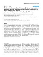
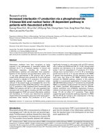
Analysis of Basophil activationFigure 1
Analysis of Basophil activation. Analysis of basophils after lysis by Immunoprep/Q-prep (a–f) or WBL (g–l) of a represent-
ative donor. By dynamic backgating (illustrated in figure 2), the region in a forward scatter vs side scatter plot in which
basophils are located was optimised (a & g). CD203c+ cells were identified in this gate (b & h), and the expression of CD203c
and CD63 was evaluated (c & i). CD203c vs CD63 expression at differend concentrations of CRA1 is shown after both lysis
conditions (1 ng/ul panels c, g, 0,001 ng/ul in panels f & l, 0, 0,0001 in panels e & k; pbs in panels d & j).
Clinical and Molecular Allergy 2005, 3:10 />Page 4 of 9
(page number not for citation purposes)
were detected more easily when they express high levels of
CD203c. This trend was much less pronounced after lysis
with WBL.
CD203c is more sensitive than CD63 at detecting signalling
through Fc
ε
RI
When defining a positive response as two consecutive
responses of more than 10% above baseline [14], fewer
responders were recorded with CD63 (3/11 data sets)
than with CD203c (6/11 data sets). All responders
through CD63 respondent also through CD203c. Lysis
procedure had no effect on CD63, but there was one more
response detected with CD203c after lysis with WBL than
after lysis with Immunoprep/Q-prep (Table 2). The partic-
ipants were split into three groups on the basis of >10%
positive cells at two consecutive dilution (Table 2):
responders with both CD63 and CD203c (n = 3),
responder with CD203c only (4 for WBL, n = 3 for Immu-
noprep/Q-prep) and non responders (n = 5).
Probability binning offers an integrative alternative to
using either only CD203c or only CD63
When analyzing the same dataset by defining that the
probability binning metric T(χ)
CD203c, CD63
> 4 for two
consecutive dilutions as a response, a similar classification
emerged for the data set obtained after lysis with WBL (7/
11 data sets), and to some part with Immunoprep/Q-prep
(4/11 data sets, Table 2). Discrimination of T(χ)
CD203c,
CD63
was significantly better after lysis with WBL than after
lysis with Immunoprep/Q-prep (4/11 data sets).
The ratio of activation was higher for CD203c than for
CD63
The degree of activation detected through CD203c and
CD63 after lysis with either Immunoprep/Q-prep or WBL
was compared by dividing the fraction of positive cells in
CRA1-activated samples of responders by the fraction of
positive cell at baseline (Figure 4). The signal was better
with WBL (Figure 4b & 4d) than with Immunoprep/Q-
prep (Figure 4a & 4c) and was slightly better with CD203c
(Figure 4c & 4d) than with CD63 (Figure 4a & 4b). Lysis
with WBL was significantly better for both CD203c (3/6
data sets) and for CD63 (1/6 data sets) (Table 3). Detec-
tion with CD203c was significantly better after lysis with
WBL (1/6 data sets) and Immunoprep/Q-prep (2/6 data
sets). Detection of CD203c was significantly better in 4/6
experiments when comparing the lysis condition used by
Boumiza et al (Immunoprep/Q-prep for detecting CD63
and WBL for detection of CD203c) [14].
Discussion
The BAT is an exiting development in applied functional
flow cytometry, and a number of laboratories have devel-
oped independent procedures to use it The first common
approach to standardization is a EAACI working paper
. We chose to use stimulation of
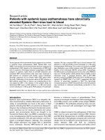
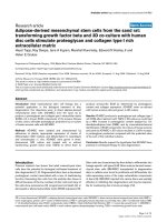
Backgated basophil populationsFigure 2
Backgated basophil populations. Backgated basophils identified after lysis with Immunoprep/Q-prep (a) and WBL (b).
Clinical and Molecular Allergy 2005, 3:10 />Page 5 of 9
(page number not for citation purposes)
blood basophils through FcεRI with the antibody CRA1
[16] to compare different lysis methods. Recently,
Boumiza published a controversial comparison of
responses through CD63 after lysis with Immunoprep/Q-
prep and CD203c after lysis with WBL [14] that was
contested by groups with experience in detecting CD63.
Other reports that so far have compared CD63 and
CD203c [7,17] give anecdotal evidence of a similar
response through the markers, but have not compared
them stringently. We had noticed that lysis with Saponin
(on which WBL is based) gives appreciably better results
than lysis with ammonium chloride (unpublished), and
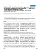
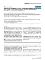
Histograms of CD63 and CD203c expression on basophils and control cellsFigure 3
Histograms of CD63 and CD203c expression on basophils and control cells. Histograms of expression of CD203c (a
& c) and CD63 (b & d) on basophils at baseline (red line) and after maximal stimulation (green line), and on lymphocytes (blue
line) after lysis with Immunoprep/Q-prep (a & b) and WBL (c & d). Markers were set to include ~2% of unstimulated basophils.
Clinical and Molecular Allergy 2005, 3:10 />Page 6 of 9
(page number not for citation purposes)
chose to compare the lysis methods (WBL, with Saponin,
and Immunoprep/Q-prep lysing reagent, with formic
acid) and markers (CD63 FITC and CD203c PE) used for
the report)[14].
The yield was remarkably better for WBL (involving
Saponin) than for Immunoprep/Q-prep (involving for-
mic acid). Although the minimum number of basophils
for a useful test has been reported to be 100 [19], we pre-
fer to have more than 500, which is within reason as we
could obtain approximately 500 basophils from 100 µl
blood from most donors using lysis with WBL (Table 1).
The threshold for a positive response has in the past been
set by an empirically determined fraction of basophils
detected above background. The threshold for detection
of allergens by a BAT should be set using Receiver
Operating Curves [20]. For the present study it was
deemed to be stringent to set it to be 10% above the
unstimulated control experiment in two consecutive
dilutions of allergen or, in this case, antibody to FcεRI to
be comparable to the previous study comparing CD63
and CD203c [14]. Using this threshold, 6 of 11 persons
mounted a positive response to cross linking of FcεRI. In
other studies, the threshold is set empirically between 6%
and 17% (EAACI Position paper at
).
As there is evidence that CD203c and CD63 are translo-
cated to the basophil cell surface by different mechanisms
and with different kinetics [17], it may be of interest to
monitor them simultaneously. Probability binning [21] is
an algorithm by which variance in the control distribution
is minimised by varying bin size before a normalised chi-
square value is computed for each sample distribution
Table 1: Basophil cell yields. Average of numbers detected after lysis at 7 different concentrations of CRA1 from 250000 (normalized)
events after lysis with either WBL or Immunoprep/Q-prep (average ± SD, tested with a paired t test). + = atopics, - = non atopics
Donor Allergy WBL ± SD Immunoprep ± SD p-value
1 - 1714 ± 314 1108 ± 449 <0,015
2 - 1979 ± 132 369 ± 274 <0,001
3 + 1638 ± 66 406 ± 262 <0,001
4 - 568 ± 84 182 ± 107 <0.001
5 - 1646 ± 282 371 ± 248 <0,001
6 + 483 ± 113 436 ± 138 0,494
7 - 1164 ± 70 213 ± 160 <0.001
8 - 1132 ± 372 582 ± 150 0,003
9 - 1397 ± 207 306 ± 133 <0,001
10 - 995 ± 481 577 ± 415 0,107
11 - 407 ± 128 397 ± 183 0,904
Median 1164 397
Table 2: Responders as defined by Boumiza et al [14] or by T(χ) > 4 for two consecutive dilutions of CRA1. Y = responder
CD63 CD203c T(χ)
63,203c
Donor WBL Immunoprep WBL Immunoprep WBL Immunoprep
1
2
3
4 Y
5
6YYY
7 YYYY
8 YYY
9YYYYY
10YYYYYY
11YYYYYY
Clinical and Molecular Allergy 2005, 3:10 />Page 7 of 9
(page number not for citation purposes)
using the same bins. This has two advantages: 1. it com-
bines the information residing in cell number in a given
bin with median fluorescence intensity and 2. the bins
constructed during the analysis can be extended from a
univariate analysis to be rectangles on a dot plot of
CD203c vs CD63 containing an even number of events of
the unstimulated control, and a single chi-square value
can then be computed that incorporates change in both
markers [18]. We show that the result of probability bin-
ning with CD63 and CD203c as dimensions is similar to
the result of the method of assigning a threshold at base-
line + 10%. This suggests that T(χ)
CD203c CD63
is as sensitive
as the conditions defined by Boumiza et al [14] (back-
ground + 10% positive).
The relative sensitivity of CD203c and CD63 was com-
pared by calculating the relative increase in ratio of posi-
tive cells over baseline conditions at each concentration of
FcεRI antibody. As lysis with Immunoprep/Q-prep results
in a lower basophil yield and immunofluorescence than
WBL, and CD63 appears to be upregulated to a lesser
extent than CD203c, the combination used in [14],
Comparison of response of CD63 and CD203c with different lysis methodsFigure 4
Comparison of response of CD63 and CD203c with different lysis methods. Degree of activation at varying concen-
trations of CRA1. The % positive cells at a given concentration are plotted against the amount of anti-FceRI antibody. The low-
est data point is labelled 1e-8 for the purpose of representation on a log scale. (a) Immunoprep/Q-prep lysis, detection with
CD63, (b) Immunoprep/Q-prep lysis, detection with CD203c, (c) WBL lysis, detection with CD63, (d) WBL lysis, detection
with CD203c. In panel d, one responder achieved significantly higher activation ratios than 35.
Clinical and Molecular Allergy 2005, 3:10 />Page 8 of 9
(page number not for citation purposes)
detecting CD63 after lysis with Immunoprep/Q-prep (Fig-
ure 4a) and detection of CD203c (Figure 4d) after lysis
with WBL, accentuated differences between the markers.
Conclusion
We present data that supports the claim that WBL is a bet-
ter lysis method than the automated Immunoprep/Q-
prep (shown here), and that CD203c is more sensitive
than CD63 at detecting FcεRI-mediated activation and
uniquely identifies basophils in human blood. As the pre-
sented data were obtained with an antibody to FcεRI, the
results need to be confirmed after stimulation with aller-
gen. Probability binning offers an approach that com-
bines CD63 and CD203c into one metric that has a high
response. A combination of lysis with Saponin and the
markers CD203c and CD63 [17] computed by probability
binning may be the most sensitive method of detecting
activation of basophils after stimulation through FcεRI.
Competing interests
The author(s) declare that they have no competing
interests.
Authors' contributions
HJH conceived the project, analysed the data and wrote
the manuscript. BMH recruited patients and did the exper-
iments, LPN contributed to project design and writing of
the manuscript. RD contributed to the design of the study
and writing of the manuscript. All authors read and
approved the final manuscript.
Acknowledgements
This study was financed by The Danish Velux Foundation, Hørslev-Fonden,
Augustinus-fonden, C. C. Klestrup og Hustru Henriette Klestrups Mindel-
egat and Danish Medical Research Council.
References
1. Ebo DG, Hagendorens MM, Bridts CH, Schuerwegh AJ, De Clerck LS,
Stevens WJ: In vitro allergy diagnosis: should we follow the
flow? Clin Exp Allergy 2004, 34:332-339.
2. Sanz ML, Sanchez G, Gamboa PM, Vila L, Uasuf C, Chazot M, et al.:
Allergen-induced basophil activation: CD63 cell expression
detected by flow cytometry in patients allergic to Dermat-
ophagoides pteronyssinus and Lolium perenne. Clin Exp Allergy
2001, 31:1007-1013.
3. Sainte-Laudy J, Sabbah A, Drouet M, Lauret MG, Loiry M: Diagnosis
of venom allergy by flow cytometry. Correlation with clinical
history, skin tests, specific IgE, histamine and leukotriene C4
release. Clin Exp Allergy 2000, 30:1166-1171.
4. Ebo DG, Lechkar B, Schuerwegh AJ, Bridts CH, De Clerck LS, Stevens
WJ: Validation of a two-color flow cytometric assay detecting
in vitro basophil activation for the diagnosis of IgE-mediated
natural rubber latex allergy. Allergy 2002, 57:706-712.
5. De Weck AL, Sanz ML: For allergy diagnostic flow cytometry,
detection of CD203c instead of CD63 is not at all an
improvement in other hands. Clin Exp Allergy 2003, 33:849-852.
6. Nieuwenhuis HK, van Oosterhout JJ, Rozemuller E, van Iwaarden F,
Sixma JJ: Studies with a monoclonal antibody against activated
platelets: evidence that a secreted 53,000-molecular weight
lysosome-like granule protein is exposed on the surface of
activated platelets in the circulation. Blood 1987, 70:838-845.
7. Hauswirth AW, Natter S, Ghannadan M, Majlesi Y, Schernthaner GH,
Sperr WR, et al.: Recombinant allergens promote expression
of CD203c on basophils in sensitized individuals. J Allergy Clin
Immunol 2002, 110:102-109.
8. Moneret-Vautrin DA, Sainte-Laudy J, Kanny G, Fremont S: Human
basophil activation measured by CD63 expression and LTC4
release in IgE-mediated food allergy. Ann Allergy Asthma Immunol
1999, 82:33-40.
9. Schuerwegh AJ, Ebo DG, Bridts CH, De Clerck LS, Stevens WJ:
CD63 expression on basophils of nonallergic controls and
patients allergic to wasp. J Allergy Clin Immunol 2001,
108:150-152.
10. Erdmann SM, Heussen N, Moll-Slodowy S, Merk HF, Sachs B: CD63
expression on basophils as a tool for the diagnosis of pollen-
associated food allergy: sensitivity and specificity. Clin Exp
Allergy 2003, 33:607-614.
11. Abuaf N, Rajoely B, Ghazouani E, Levy DA, Pecquet C, Chabane H, et
al.: Validation of a flow cytometric assay detecting in vitro
basophil activation for the diagnosis of muscle relaxant
allergy. J Allergy Clin Immunol 1999, 104:411-418.
12. Monneret G, Benoit Y, Debard AL, Gutowski MC, Topenot I, Bien-
venu J: Monitoring of basophil activation using CD63 and
CCR3 in allergy to muscle relaxant drugs. Clin Immunol 2002,
102:192-199.
13. Sturm GJ, Bohm E, Trummer M, Weiglhofer I, Heinemann A, Aberer
W: The CD63 basophil activation test in Hymenoptera
venom allergy: a prospective study. Allergy 2004, 59:1110-1117.
14. Boumiza R, Monneret G, Forissier MF, Savoye J, Gutowski MC, Pow-
ell WS, et al.: Marked improvement of the basophil activation
test by detecting CD203c instead of CD63. Clin Exp Allergy
2003, 33:259-265.
15. Ebo DG, Lechkar B, Schuerwegh AJ, Bridts CH, De Clerck LS, Stevens
WJ: Comments regarding 'Marked improvement of the
Table 3: Difference in activation for combinations of WBL, Immunoprep/Q-prep and CD63 and CD203c amongst responders The p-
value was calculated with the Students t-test. ns = not significant
WBL vs Immunoprep CD203c vs CD63
CD63 CD203c Immunoprep WBL, CD203c WBL vs
CD63 Immunoprep
6 ns 0,025 ns ns ns
7 ns 0,016 0,027 0,016 0,012
8 0,016 ns 0,004 ns 0,019
9 nsnsnsnsns
10 ns 0,018 ns ns 0,019
11 ns ns ns ns 0,043
Publish with BioMed Central and every
scientist can read your work free of charge
"BioMed Central will be the most significant development for
disseminating the results of biomedical research in our lifetime."
Sir Paul Nurse, Cancer Research UK
Your research papers will be:
available free of charge to the entire biomedical community
peer reviewed and published immediately upon acceptance
cited in PubMed and archived on PubMed Central
yours — you keep the copyright
Submit your manuscript here:
/>BioMedcentral
Clinical and Molecular Allergy 2005, 3:10 />Page 9 of 9
(page number not for citation purposes)
basophil activation test by detecting CD203c instead of
CD63' by Boumiza et al. Clin Exp Allergy 2003, 33:849-3.
16. Jensen BM, Hansen JB, Dissing S, Gerwien J, Skov PS, Poulsen LK:
Monomeric immunoglobulin E stabilizes FcepsilonRIalpha
from the human basophil cell line KU812 by protecting it
from natural turnover. Clin Exp Allergy 2003, 33:655-662.
17. Buhring HJ, Streble A, Valent P: The basophil-specific ectoen-
zyme E-NPP3 (CD203c) as a marker for cell activation and
allergy diagnosis. Int Arch Allergy Immunol 2004, 133:317-329.
18. Roederer M, Moore W, Treister A, Hardy RR, Herzenberg LA:
Probability binning comparison: a metric for quantitating
multivariate distribution differences. Cytometry 2001, 45:47-55.
19. Erdmann SM, Sachs B, Hoffmann-Sommergruber K, Scheiner O, Merk
H: Regarding Ebo DG, Hagendorens MM, Bridts CH, Schuer-
wegh AJ, De Clerck LS & Stevens WJ. In vitro allergy diagno-
sis: should we follow the flow? Clin Exp Allergy 2004; 34:332–
9. Clin Exp Allergy 2004, 34:1498-1499.
20. Hemery ML, Arnoux B, Dhivert-Donnadieu H, Rongier M, Barbotte
E, Verdier R, et al.: Confirmation of the diagnosis of natural
rubber latex allergy by the Basotest method. Int Arch Allergy
Immunol 2005, 136:53-57.
21. Roederer M, Treister A, Moore W, Herzenberg LA: Probability
binning comparison: a metric for quantitating univariate dis-
tribution differences. Cytometry 2001, 45:37-46.