Báo cáo y học: "CCR3, CCR5, CCR8 and CXCR3 expression in memory T helper cells from allergic rhinitis patients, asymptomatically sensitized and healthy individual" docx
Bạn đang xem bản rút gọn của tài liệu. Xem và tải ngay bản đầy đủ của tài liệu tại đây (386.95 KB, 6 trang )
BioMed Central
Page 1 of 6
(page number not for citation purposes)
Clinical and Molecular Allergy
Open Access
Research
CCR3, CCR5, CCR8 and CXCR3 expression in memory T helper
cells from allergic rhinitis patients, asymptomatically sensitized and
healthy individuals
Mille Holse, Kristian Assing and Lars K Poulsen*
Address: Laboratory for Medical Allergology 7542, National University Hospital, Blegdamsvej 9, DK-2100 Copenhagen, Denmark
Email: Mille Holse - ; Kristian Assing - ; Lars K Poulsen* -
* Corresponding author
Abstract
Background: Chemokine receptors have been suggested to be preferentially expressed on CD4+
T cells with CCR3 and CCR8 linked to the T helper (Th) 2 subset and CCR5 and CXCR3 to the
Th1 subset, however this remains controversial.
Objective: Our aim was to compare the CCR3, CCR5, CCR8 and CXCR3 expression in memory
Th cells from allergic, asymptomatically sensitized and healthy individuals.
Methods: Peripheral blood mononuclear cells from 8 pollen allergic rhinitis patients, 10
asymptomatically sensitized and 10 healthy individuals were stimulated for 7 days with allergen or
tetanus toxoid. CCR3, CCR5, CCR8, CXCR3, CD4 and CD45RO were detected by flow
cytometry.
Results: No differences in chemokine receptor expression were observed between the three
groups on day 0, and seven days of unstimulated culture did not change the expression. Both
antigenic stimuli increased the chemokine receptor expression, tetanus toxoid being the most
potent. No differences in percentage chemokine receptor positive memory Th cells were observed
between the three groups on day 7. Only a change in MFI for CCR5 was significantly different
between the three groups after allergen stimulation of the Th cells.
Conclusion: We conclude that even though allergen and antigen induced increased chemokine
receptor expression, no differences in profiles were identified in memory Th cells from patient
groups with different atopic status.
Introduction
The prevalence of allergy is increasing in the westernized
part of the world with estimates suggesting that 20–30%
of the population is affected [1]. However, unlike the reac-
tion of most IgE-sensitized individuals who upon re-expo-
sure to the allergen develop symptoms due to activation
and release of mediators from various immune cells,
some individuals seem to exhibit an IgE positive pheno-
type without having any allergic symptoms. These indi-
viduals have been described in the literature as
asymptomatically sensitized and are phenotypically consid-
ered to be a group between the allergic and the healthy
individuals with an increased risk of developing allergy
[2,3].
Published: 19 April 2006
Clinical and Molecular Allergy 2006, 4:6 doi:10.1186/1476-7961-4-6
Received: 29 November 2005
Accepted: 19 April 2006
This article is available from: />© 2006 Holse et al; licensee BioMed Central Ltd.
This is an Open Access article distributed under the terms of the Creative Commons Attribution License ( />),
which permits unrestricted use, distribution, and reproduction in any medium, provided the original work is properly cited.
Clinical and Molecular Allergy 2006, 4:6 />Page 2 of 6
(page number not for citation purposes)
The chemokines and their receptors play a pivotal role in
leukocyte migration and chemotaxis. It is still controver-
sial whether these receptors can function as phenotypic
markers on certain cell subsets. CCR3 and CCR8 have
been suggested as Th2 markers whereas CXCR3 is men-
tioned in the literature as a Th1 marker [4-6]. The CCR3
ligand CCL11/eotaxin is upregulated in nasal mucosa of
allergic rhinitis patients during the pollen season [7].
CCL1/I-309, which is the only CCR8 ligand, is upregu-
lated in patients with atopic dermatitis [8] and IL-12
inhibit its production [9]. Also, CCL1/I-309 is released by
mast cells in response to IgE cross-linking [10] indicating
a role in allergic inflammation. On the contrary, the IFN-
γ-inducible CXCR3 ligands and some CCR5 ligands are
increased in autoimmune diseases [11-14]. However,
other findings show that no correlation exists between
Th1/Th2 cytokine profile and chemokine receptor expres-
sion on a single cell level [15] and also suggest that the
chemokine receptor profile can be changed without a con-
comitant change in cytokine profile [16] questioning the
use of chemokine receptors as markers for T cell subsets.
As the chemokine receptor profile determines the migra-
tory patterns of leukocytes, we wanted to compare this
profile with respect to CCR3, CCR5, CCR8 and CXCR3 in
memory Th cells from allergic, asymptomatically sensi-
tized and healthy individuals to obtain knowledge about
their migratory potential and any differences in expres-
sion patterns that might exist between these three groups.
Methods
Patients
10 healthy, 5 asymptomatically birch pollen sensitized, 5
asymptomatically grass pollen sensitized, 5 birch pollen
allergic and 3 grass pollen allergic volunteers with sea-
sonal hay fever symptoms were examined during the birch
or grass pollen season respectively. Skin prick test (Solu-
prick, ALK-Abello, Hørsholm, Copenhagen), histamine
release [17] and specific IgE against birch and grass pollen
using the CAP-system (Pharmacia, Uppsala, Sweden)
were determined for all volunteers (Table 1). The skin
prick test was performed according to the guidelines of
European Academy of Allergy and Clinical Immunology
[18]. Three of the allergic patients had allergic asthma. The
allergic subjects received no corticosteroid treatment for
three months prior to the study. The asymptomatically
sensitized and healthy control subjects took no hay fever
medicine. All subjects came from the area of greater
Copenhagen (Storkøbenhavn). The study was approved
by the local Ethical Committee and the clinical features of
the patients are described in detail elsewhere [19].
Cell stimulation
Peripheral blood mononuclear cells (PBMCs) were iso-
lated from whole blood by gradient centrifugation on
Lymphoprep (Nycomed, Roskilde, Denmark). The
PBMCs were cultured (3 × 10
6
) in 6 well plates with anti-
gen in 6 ml of low endotoxin RPMI1640 medium con-
taining 10% heat-inactivated autologous serum, 25 mM
HEPES, 2 mM L-glutamine, 50 µM β-mercaptoethanol,
100 U/ml streptomycin/penicillin for 7 days in the pres-
ence of either 15 µg/ml birch or grass allergen (ALK-
Abello, Hørsholm, Copenhagen), 10 µg/ml Tetanus tox-
oid (TTx) (Statens Serum Institut, Copenhagen, Den-
mark) or no antigen as a control. On day 7, the cells were
harvested and used for flow cytometric analysis. The
lipopolysaccharide level in both allergen extracts was < 7
EU/mg and after 24 hours of stimulation of PBMCs from
Table 1: Patient characterization.
Birch Grass
Patients Age
(range)
Sex
Male/Female
Symptoms Spt IgE class
Median(range)
HR class
Median(range)
Symptoms Spt IgE class
Median(range)
HR class
Median(range)
Healthy 25
(22–43)
3/7 0/10 0/10 0
(0)
0
(0)
0/10 0/10 0
(0)
0
(0)
AS Birch 25
(24–27)
1/4 0/5 5/5 0
(0–2)
2
(0–3)
0/5 0/5 0
(0)
0
(0)
AS Grass 25
(22–31)
1/4 0/5 0/5 0
(0)
0
(0)
0/5 5/5 0
(0–2)
0
(0–3)
Allergic
Birch
27
(25–43)
4/1 5/5 5/5 3
(2–4)
3
(2–3)
2/5 2/5 0
(0–4)
2
(0–3)
Allergic
Grass
26
(24–41)
3/0 1/3 1/3 0
(0–3)
0
(0–3)
3/3 3/3 4
(2–4)
3
(0–3)
AS: asymptomatically sensitized. Spt: skin prick test. HR: histamine release
Skin prick tests (performed in duplicate) were considered positive when mean wheal diameter >3 mm. Allergic symptoms were reported in diary
cards during the relevant pollen season. Symptoms were considered as pollen allergy when lasting > 7 days or when symptoms were repeatedly
elicited when pollen counts exceeded a certain (individual) level [2]. The skin prick test was performed according to the guidelines of European
Academy of Allergy and Clinical Immunology [18]. n = 10 for the healthy controls, n = 10 for the asymptomatically sensitized individuals and n = 8
for the allergic individuals.
Clinical and Molecular Allergy 2006, 4:6 />Page 3 of 6
(page number not for citation purposes)
healthy individuals no detectable amounts of TNF-α were
observed.
Flow cytometry
Surface markers were detected using primary labeled anti-
bodies: CCR3-FITC, CCR5-FITC, CCR8-FITC, CXCR3-
FITC (R&Dsystems, Abingdon, UK), CD4-PE-Cy5 and
CD45RO-PE (Dakocytomation, Glostrup, Denmark).
Three-color flow cytometry was performed on day 0 and
day 7 on a FACScan (Becton Dickinson, Heidelberg, Ger-
many) using WinList (Verity Software House, Topsham,
USA) software for analysis. Isotype control cut-off values
were set to > 98% and 10.000 PBMC were acquired. Gat-
ing was done by firstly applying a CD4+ gate followed by
determination of the percentage and mean fluorescence
intensity (MFI) of the CD45RO and chemokine receptor
double positive population. CD45RO is a marker of effec-
tor and memory T cells, however throughout this article
these CD4+ CD45RO+ T cells will be mentioned as mem-
ory T helper cells for convenience.
Statistical analysis
Samples were compared using non-parametric statistics
(Kruskall-Wallis test or Wilcoxon's test for matched pairs).
Values of P < 0.05 were considered significant.
Results
Day 0
Immediate after isolation of the PBMCs, the cells were
subjected to flow cytometric analysis. No significant dif-
ferences in the percentage of CCR3+, CCR5+, CCR8+ and
CXCR3+ memory Th cells from allergic, asymptomatically
sensitized and healthy individuals were observed (Figure
1). Likewise, no differences in MFI were observed between
the three donor groups (results not shown).
Day 7
Effect of stimulation
No significant differences in chemokine receptors (neither
expressed as the percentage of positive cells nor as MFI)
were observed between day 0 and the cells having been
kept in antigen-free medium for 7 days as controls. Thus
the medium alone and the experimental set-up did not
influence the chemokine receptor expression. After TTx
stimulation a significant increase in MFI was observed for
CCR3 in the allergic and asymptomatically sensitized
individuals, but not in the healthy control group (Table
2). However, no increases in the percentage of CCR3+
memory Th cells were observed in any of the groups.
CCR5 increased both as MFI and the percentage of CCR5+
memory Th cells in the asymptomatically sensitized and
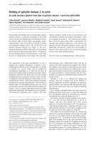
Percentage chemokine receptor positive memory T helper cells day 0Figure 1
Percentage chemokine receptor positive memory T helper cells day 0. Percentage CCR3+, CCR5+, CCR8+ and
CXCR3+ memory Th cells from allergic (dots), asymptomatically sensitized (triangles) and healthy control (crosses) individuals
on day 0 immediate ex vivo. - = median value. n = 10 for the healthy controls, n = 10 for the asymptomatically sensitized and n
= 8 for the allergic individuals except for CCR8 where n = 6 for the asymptomatically sensitized and allergic individuals. 10.000
PBMCs were acquired for the analysis. Isotype control cut-off values were set to >98%. Samples were run in monocates. For
experimental design and analysis see Methods.
Clinical and Molecular Allergy 2006, 4:6 />Page 4 of 6
(page number not for citation purposes)
healthy control group whereas no changes in CCR5 was
observed in the allergic individuals. Also, an increase in
the percentage of CCR8+ memory Th cells was observed in
the healthy control group, but no significant changes in
MFI were observed for this receptor. An increase in the
percentage of CXCR3+ memory Th cells was observed in
all three groups after TTx stimulation, however increases
in MFI were only observed in the healthy control group.
After stimulation with allergen, increases in the percent-
age of CCR5+ memory Th cells were observed in healthy
controls and in MFI in allergic individuals. Allergen stim-
ulation did not induce any changes in CCR3, CCR8 and
CXCR3 expression. When pooling all 28 patients in the
statistical analysis, TTx was able to induce expression of all
receptors both seen as a significant increase in the percent-
age of chemokine receptor positive cells and as MFI. Aller-
gen stimulation only induced a significant increase in the
percentage of CCR5+ memory Th cells.
Group differences
To compare the chemokine receptor expression between
the three groups, the Day 7
no antigen
receptor level was sub-
tracted from either the Day 7
allergen
or the Day 7
TTx
sample
to obtain the change in receptor expression (∆Chemokine
receptor).
No differences in ∆Chemokine receptor for the percentage
of chemokine receptor positive cells were observed
between the three groups after stimulation with TTx or
allergen. When comparing the ∆Chemokine receptor for
the MFI, the change in the CCR5 after allergen stimulation
was significantly different between the three groups (P =
0.02).
Discussion
Other studies have linked certain diseases with aberrant
expression of one or more chemokine receptors
[12,20,21]. However, very few studies have been con-
ducted with regard to the phenotype of asymptomatically
sensitized individuals and, to our knowledge none on
chemokine receptor profiles.
In this study, no differences were found in receptor
expression patterns immediate ex vivo for CCR3, CCR5,
CCR8 and CXCR3 in memory Th cells from allergic,
asymptomatically sensitized and healthy individuals
despite the fact that the study was carried out in the pollen
season.
Our findings are in agreement with other studies report-
ing equal mRNA levels of CCR3 and CCR5 in PBMCs [22]
and same levels of CXCR3+ peripheral blood Th cells [21]
in patients with atopic dermatitis and healthy controls,
but in disagreement with other findings showing
decreased percentage of CCR5+ and CXCR3+ memory Th
cells in the blood from patients with atopic dermatitis
compared to healthy controls [23].
Changes in chemokine receptor expression were observed
after stimulation with both antigens (Table 2). CCR5
expression was induced after TTx stimulation, but only in
Table 2: Chemokine receptor expression in memory T helper cells induced by 7 days of antigenic stimuli. Median chemokine receptor
expression in memory Th cells in percentage and MFI after 7 days of stimulation with antigen (allergen (15 µg/ml) or TTx (10 µg/ml))
or no antigen as a control. n = 10 for the healthy controls, n = 10 for the asymptomatically sensitized individuals and n = 8 for the
allergic individuals except for CCR8 where n = 6 for the asymptomatically sensitized and allergic individuals. 10.000 PBMCs were
acquired for the analysis. Isotype control cut-off values were set to >98%. Samples were run in monocates. For experimental design
and analysis see Methods.
% median (range) MFI median (range)
No Ag Allergen TTx No Ag Allergen TTx
CCR3 Allergic 10.6 (4.7–32) 17.3 (9.8–29.5) 15.6 (4.4–52.2) 21 (16.2–32.3) 19.4 (16.9–37.2) 26.1* (16.6–40.5)
AS 10.3 (3.3–19.7) 8.7 (2.8–23.2) 11.9 (5–28.8) 20.5 (14.7–27.2) 18.6 (16.4–32.3) 23.4 * (18.4–37.4)
Healthy 8.7 (1.5–45) 11.2 (3.2–36.8) 23.4 (4.5–44.3) 19.7 (12.6–45.4) 17.8 (12.5–32.2) 23.7 (12.5–38.4)
CCR5 Allergic 21.3 (7.8–31.5) 25 (4.1–53.9) 28.3 (12.3–61.1) 21.4 (16.8–27.9) 25.9 * (22.4–48.5) 26.1 (16.1–42.5)
AS 15.2 (2.6–36.6) 17 (3–33.5) 16.9 * (5.2–65.9) 18.3 (15.2–24.5) 17.5 (15.4–20.5) 22.5 * (18.2–58.2)
Healthy 11.7 (5.6–34.5) 13.5 * (6.7–29.7) 20.2 * (10.1–74.1) 16.7 (12.7–22) 17.3 (13.5–25.6) 25.1 * (13.4–72)
CCR8 Allergic 10.4 (1.6–20.4) 12.8 (3.3–20) 12.7 (2.2–27.4) 24.3 (16.8–37.6) 27.9 (18.8–40.3) 28.2 (17.8–82.6)
AS 3.7 (1.8–16.6) 3.2 (1.7–14.7) 6.3 (1.4–34.9) 21.9 (17–26.1) 22.8 (17.2–44.6) 24.9 (16.2–36.6)
Healthy 5.7 (0.7–16.4) 3.9 (1.5–14) 9.3 * (3.8–35.7) 19 (12.6–49.6) 19.2 (11.7–46.6) 23.9 (12.4–48.2)
CXCR3 Allergic 42.9 (22–47.5) 42.1 (19.2–67.4) 49.2 * (27–74.2) 57.4 (33.1–65.3) 54.4 (48.4–72.4) 60.6 (36.9–81.8)
AS 28.1 (18–56.6) 27.8 (21–56.8) 29 * (20.8–77.4) 39.7 (29.3–59.7) 42.3 (30.6–54.5) 46.5 (33.5–115)
Healthy 28.8 (19.8–46.5) 31.4 (17.3–46.9) 35.9 * (25.8–82.9) 37.8 (32–65.2) 39.3 (30.1–63.9) 57.6 * (32–162.4)
Bold text and * indicates significant differences between the antigen (allergen or TTx) stimulated samples and control samples where no antigen was
added.
Ag: antigen AS: asymptomatically sensitized individuals.
Clinical and Molecular Allergy 2006, 4:6 />Page 5 of 6
(page number not for citation purposes)
asymptomatically sensitized and healthy individuals. The
reason why allergic individuals do not upregulate this
receptor even when stimulated with a type 1 antigen is
speculative, but one reason might be due to their Th2
biased reaction pathway. However, they do show signifi-
cant increases in percentage CXCR3+ memory Th cells
after TTx stimulation, in accordance with this receptor's
much stronger link to the Th1 phenotype [24].
Only when grouping all individuals, did the recall antigen
TTx induce significant increases in expression of all recep-
tors. The reason for the less clear effect as observed in the
individual groups might be due to the great inter-individ-
ual variation in chemokine receptor expression level, an
observation also described by others [25]. Nevertheless,
TTx induced more changes than the allergenic stimuli, an
effect that is likely due to the higher frequency of TTx spe-
cific T cells compared to allergen specific T cells in periph-
eral blood (Glue, unpublished results).
CCR5 appears to be the most allergen susceptible recep-
tor. However, the apparent overlap in expression levels
between the groups would exclude the use of this receptor
as a diagnostic tool and thus is of no major clinical inter-
est.
When comparing the three groups after antigen stimula-
tion, we found no differences in expression patterns
between the three groups except for the change in CCR5
MFI which was significantly different between the three
groups. This is the only observed difference between the
three groups but as discussed above the great overlap in
receptor levels would not make this finding of any clinical
relevance.
In spite of the apparent lack of differences between the
three groups with respect to chemokine receptor profile, a
parallel study using a somewhat larger sample size
showed that allergen stimulation induced significantly
more proliferation of memory Th cells in the allergic indi-
viduals compared to the asymptomatically sensitized and
healthy individuals as well as a different cytokine profile
[19].
Conclusion
In conclusion, both antigenic stimuli were able to induce
changes in chemokine receptor expression. TTx seemed to
be a more potent stimulus with regard to changes in
chemokine receptor expression in all three groups com-
pared to the pollen allergens. No major differences in
CCR3, CCR5, CCR8 and CXCR3 were found between
allergic, asymptomatically sensitized and healthy individ-
uals and thus chemokine receptor expression in periph-
eral blood memory Th cells does not seem to be linked to
patient status. No major differences were seen between
the three groups after antigenic stimulation and thus we
conclude that pollen allergic, asymptomatically pollen
sensitized and healthy individuals cannot be distin-
guished by means of chemokine receptors expression in
memory Th cells and thus the migratory potentials of the
memory Th cells seem to be the same between the three
groups.
Abbreviations
Ag: antigen AS: asymptomatically sensitized MFI: mean
fluorescence intensity PBMC: peripheral blood mononu-
clear cell Th: T helper TTx: Tetanus toxoid
Competing interests
The author(s) declare that they have no competing inter-
ests.
Authors' contributions
All authors participated in the design of the study. KA con-
ducted the patient contact and characterization, cell isola-
tion and stimulation assays. MH conducted the flow
cytometry and analyzed the data. All authors contributed
towards the manuscript preparation with MH as the main
author of the article.
References
1. Parronchi P, Brugnolo F, Sampognaro S, Maggi E: Genetic and envi-
ronmental factors contributing to the onset of allergic disor-
ders. Int Arch Allergy Immunol 2000, 121:2-9.
2. Bodtger U, Poulsen LK, Malling HJ: Asymptomatic skin sensitiza-
tion to birch predicts later development of birch pollen
allergy in adults: a 3-year follow-up study. J Allergy Clin Immunol
2003, 111:149-154.
3. Bodtger U: Prognostic value of asymptomatic skin sensitiza-
tion to aeroallergens. Curr Opin Allergy Clin Immunol 2004, 4:5-10.
4. Sallusto F, Lenig D, Mackay CR, Lanzavecchia A: Flexible programs
of chemokine receptor expression on human polarized T
helper 1 and 2 lymphocytes. J Exp Med 1998, 187:875-883.
5. Zingoni A, Soto H, Hedrick JA, Stoppacciaro A, Storlazzi CT, Sini-
gaglia F, D'Ambrosio D, O'Garra A, Robinson D, Rocchi M, et al.: The
chemokine receptor CCR8 is preferentially expressed in Th2
but not Th1 cells. J Immunol 1998, 161:547-551.
6. Sallusto F, Mackay CR, Lanzavecchia A: Selective expression of
the eotaxin receptor CCR3 by human T helper 2 cells. Science
1997, 277:2005-2007.
7. Pullerits T, Linden A, Praks L, Cardell LO, Lotvall J: Upregulation of
nasal mucosal eotaxin in patients with allergic rhinitis during
grass pollen season: effect of a local glucocorticoid. Clin Exp
Allergy 2000, 30:1469-1475.
8. Gombert M, Dieu-Nosjean MC, Winterberg F, Bunemann E, Kubitza
RC, Da Cunha L, Haahtela A, Lehtimaki S, Muller A, Rieker J, et al.:
CCL1-CCR8 interactions: an axis mediating the recruitment
of T cells and Langerhans-type dendritic cells to sites of
atopic skin inflammation. J Immunol 2005, 174:5082-5091.
9. Iellem A, Colantonio L, Bhakta S, Sozzani S, Mantovani A, Sinigaglia F,
D'Ambrosio D: Inhibition by IL-12 and IFN-alpha of I-309 and
macrophage-derived chemokine production upon TCR trig-
gering of human Th1 cells. Eur J Immunol 2000, 30:1030-1039.
10. Gilchrest H, Cheewatrakoolpong B, Billah M, Egan RW, Anthes JC,
Greenfeder S: Human cord blood-derived mast cells synthe-
size and release I-309 in response to IgE. Life Sci 2003,
73:2571-2581.
11. Ueno A, Yamamura M, Iwahashi M, Okamoto A, Aita T, Ogawa N,
Makino H: The production of CXCR3-agonistic chemokines
by synovial fibroblasts from patients with rheumatoid arthri-
tis. Rheumatol Int 2005, 25:361-367.
Publish with BioMed Central and every
scientist can read your work free of charge
"BioMed Central will be the most significant development for
disseminating the results of biomedical research in our lifetime."
Sir Paul Nurse, Cancer Research UK
Your research papers will be:
available free of charge to the entire biomedical community
peer reviewed and published immediately upon acceptance
cited in PubMed and archived on PubMed Central
yours — you keep the copyright
Submit your manuscript here:
/>BioMedcentral
Clinical and Molecular Allergy 2006, 4:6 />Page 6 of 6
(page number not for citation purposes)
12. Balashov KE, Rottman JB, Weiner HL, Hancock WW: CCR5(+) and
CXCR3(+) T cells are increased in multiple sclerosis and
their ligands MIP-1alpha and IP-10 are expressed in demyeli-
nating brain lesions. Proc Natl Acad Sci U S A 1999, 96:6873-6878.
13. Sorensen TL, Tani M, Jensen J, Pierce V, Lucchinetti C, Folcik VA, Qin
S, Rottman J, Sellebjerg F, Strieter RM, et al.: Expression of specific
chemokines and chemokine receptors in the central nervous
system of multiple sclerosis patients. J Clin Invest 1999,
103:807-815.
14. Patel DD, Zachariah JP, Whichard LP: CXCR3 and CCR5 ligands
in rheumatoid arthritis synovium. Clin Immunol 2001, 98:39-45.
15. Nanki T, Lipsky PE: Lack of correlation between chemokine
receptor and T(h)1/T(h)2 cytokine expression by individual
memory T cells. Int Immunol 2000, 12:1659-1667.
16. Aarvak T, Strand E, Teigland J, Miossec P, Natvig JB: Switch in
chemokine receptor phenotype on memory T cells without
a change in the cytokine phenotype. Scand J Immunol 2001,
54:100-108.
17. Hansen KS, Khinchi MS, Skov PS, Bindslev-Jensen C, Poulsen LK,
Malling HJ: Food allergy to apple and specific immunotherapy
with birch pollen. Mol Nutr Food Res 2004, 48:441-448.
18. Dreborg S, Frew A: Position Papers. Allergen standardization
and skin tests. Allergy 1993, 48:9-82.
19. Assing K, Nielsen CH, Poulsen LK: Immunological characteris-
tics of subjects with asymptomatic skin sensitization to birch
and grass pollen. Clin Exp Allergy 2006, 36:283-292.
20. Teleshova N, Pashenkov M, Huang YM, Soderstrom M, Kivisakk P,
Kostulas V, Haglund M, Link H: Multiple sclerosis and optic neu-
ritis: CCR5 and CXCR3 expressing T cells are augmented in
blood and cerebrospinal fluid. J Neurol 2002, 249:723-729.
21. Wakugawa M, Nakamura K, Kakinuma T, Onai N, Matsushima K,
Tamaki K: CC chemokine receptor 4 expression on peripheral
blood CD4+ T cells reflects disease activity of atopic derma-
titis. J Invest Dermatol 2001, 117:188-196.
22. Hatano Y, Katagiri K, Takayasu S: Decreased levels of CXCR3
transcripts in peripheral blood mononuclear cells from
patients with atopic dermatitis and with cutaneous diseases
associated with eosinophilia. Arch Dermatol Res 2001,
293:319-322.
23. Okazaki H, Kakurai M, Hirata D, Sato H, Kamimura T, Onai N, Mat-
sushima K, Nakagawa H, Kano S, Minota S: Characterization of
chemokine receptor expression and cytokine production in
circulating CD4+ T cells from patients with atopic dermati-
tis: up-regulation of C-C chemokine receptor 4 in atopic der-
matitis. Clin Exp Allergy 2002, 32:1236-1242.
24. Kim CH, Rott L, Kunkel EJ, Genovese MC, Andrew DP, Wu L,
Butcher EC: Rules of chemokine receptor association with T
cell polarization in vivo. J Clin Invest 2001, 108:1331-1339.
25. Campbell JD, Stinson MJ, Simons FE, Rector ES, HayGlass KT: In vivo
stability of human chemokine and chemokine receptor
expression. Hum Immunol 2001, 62:668-678.