Báo cáo y học: "Arctigenin from Arctium lappa inhibits interleukin-2 and interferon gene expression in primary human T lymphocytes" docx
Bạn đang xem bản rút gọn của tài liệu. Xem và tải ngay bản đầy đủ của tài liệu tại đây (473.78 KB, 8 trang )
RESEARCH Open Access
Arctigenin from Arctium lappa inhibits interleukin-2
and interferon gene expression in primary human
T lymphocytes
Wei-Jern Tsai
1
, Chu-Ting Chang
2
, Guei-Jane Wang
1
, Tzong-Huei Lee
3
, Shwu-Fen Chang
4
, Shao-Chun Lu
5
and
Yuh-Chi Kuo
2*
Abstract
Background: Arctium lappa (Niubang), a Chinese herbal medicine, is used to treat tissue inflammation. This study
investigates the effects of arctigenin (AC), isolated from A. lappa, on anti-CD3/CD2 8 Ab-stimulated cell proliferation
and cytokine gene expression in primary human T lymphocytes.
Methods: Cell proliferation was determined with enzyme immunoassays and the tritiated thymidine uptake
method. Cytokine production and gene expression were ana lyzed with reverse transcription-polymerase chain
reaction.
Results: AC inhibited primary human T lymphocytes proliferation activated by anti-CD3/CD28 Ab. Cell viability test
indicated that the inhibitory effects of AC on primary human T lymphocyte proliferation were not due to di rect
cytotoxicity. AC suppressed interleukin-2 (IL-2) and interferon-g (IFN-g) production in a concentration-dependent
manner. Furthermore, AC decreased the IL-2 and IFN-g gene expression in primary human T lymphocytes induced
by anti-C D3/CD28 Ab. Reporter gene analyses revealed that AC decreased NF-AT-mediated reporter gene
expression.
Conclusion: AC inhibited T lymphocyte proliferation and decreased the gene expression of IL-2, IFN-g and NF-AT.
Background
The central event in the generation of immune responses
is the activation and clonal expansion of T cells. Interac-
tion of T cells with antigens initiates a cascade of bio-
chemical events and gene expression that induces the
resting T cells to activate and proliferate [1]. Activation
of nuclear factor of activated T cells (NF-AT) and a series
of genes such as interleukin-2 (IL-2) and interferon-g
(IFN-g) are pivotal in the growth of T lymphocytes
induced by antigen s [2,3]. Thus, growth modulators or
other external events affecting T cell proliferation are
likely to act by controlling the expression or functi on of
the products of these genes [4]. The immune responses
to invasive organisms, if inappropriat ely intense or pro-
longed, may paradoxically aggravate the injury or even
cause death. The use of immunomodulatory medications
must therefore be discreet. Re gulation of T lymphocyte
activation and proliferation and cytokine production is
one of the action mechanisms [5,6].
Chinese medicinal herbs are now widely acknowledged
for their immunomo dulatory activities [1]. A me mber of
the Compositae family, Arctium la ppa (Niuba ng)is
regarded as an effective Chinese medicine for alleviation
of rheumatic pain and fever [7]. Arctigenin (AC), a
bioactive component of A. lappa, has various biological
activities including: (1) inhibition of nitric oxide, interlu-
kin-6 and tumor nec rosis factor-a production in macro-
phages [8,9]; (2) anti-proliferative activity against
leukemia cells [10]; and (3) protective effects on hepato-
cytes from CCl
4
injury [11]. Definitive evidence for its
effects on T cell-mediated immune responses has been
scarce.
The present study aims to elucidate the effects of
AC on T lymphocytes proliferation, production and
gene expression of IL-2 and IFN-g in T lymphocytes
* Correspondence:
2
Institute of Life Science, Fu-Jen University, Taipei, 24205, Taiwan
Full list of author information is available at the end of the article
Tsai et al. Chinese Medicine 2011, 6:12
/>© 2011 Tsai et a l; licensee BioMed Central Ltd. This is an Open Access article distributed under the terms of the Creative Commons
Attribution License ( which permits unrestricted use, distribution, and reproduction in
any medium, provided the original work is properly cited.
induced by anti-CD3/CD28 antibodies (Ab) and NF-AT
activation.
Methods
Preparation of arctigenin (AC)
ACwasisolatedfromdriedgroundofA. lappa L. by
using reported methods [12]. Briefly, ground A. lappa
(1 kg) was extracted with ethanol (2L × 3) at room tem-
perature. The solvent was removed under reduced pres-
sure and the residue was partitioned between H
2
Oand
ethyl acetate (EtOAc). The concentrated EtOAc extracts
(60 mg) were subjected to chromatography over silica gel
andelutedwithn-hexane/EtOAc (4:1), n-hexane/EtOAc
(1:1) and EtOAc successively. AC (4.5 mg; C
21
H
24
O
6
;
MW 372; Figure 1) was purified from EtOAC fraction
with bioassay-guided separation. Mass and NMR spectral
data for this compound were identical with those pre-
viously reported [12]. AC, with the purity above 98%, was
dissolved in dimethylsulfoxide (DMSO) to a concentra-
tion of 100 mM and then stored at 4°C until use.
Participants
Ten healthy male participants aged between 20 and 32
years (mean 26) were selected for this study. The experi-
mental protocol were reviewed and approved by the
institutional human experimentation committee of Fu-
Jen University. Written informed consent w as obtained
from all participants.
Preparation of primary human T lymphocytes
Heparinized human peripheral bloods (80 ml) were
obtained from healthy donors. The peripheral blood was
centrifuged at 850 × g (Sorvall Legend RT, Kendro,
Germany) at 4°C for ten minutes to remove the plasma.
The blood cells were diluted with phosphate buffered
saline (PBS) and then centrifuged in a Ficoll-Hypaque
discontinuous gradient (specific gravity 1.077) at 420 × g
(Sorvall Legend RT, Kendro, Germany) for 30 minutes.
The peripheral blood mononuclear cell (PBMC) layers
were collected and washed with cold distilled water and
10× Hanks’ buffer saline solution (HBSS) to remove red
blood cells. T lymphocytes were separated from PBMC
by nylon wool columns (Wako Chemicals, USA). Puri-
fied T lymphocytes had >87% CD3
+
cells and <0.5%
CD14
+
or CD19
+
cells. The cells were re-suspended to a
concentration of 2 × 10
6
cells/ml in RPMI-1640 medium
supplemented with 2% fetal calf serum (FCS), 100 U/ml
penicillin and 100 μg/ml streptomycin [4].
Lymphoproliferation test
The l ymphoproliferation test was modified from a pre-
viously described method [13]. Briefly, the density of T
lymphocytes was adjusted to 2 × 10
6
cells/ml before use.
Cell suspension (100 μl) was applied into each well of a
96-well flat-bottomed plate (Nunc 167008, Nunclon,
Denmark) with or without anti-CD3 (1 μg /ml)/CD28
(3 μg/ml) antibody (eBioscience, USA). Cyclosporin
A (CsA , 2.5 μM), an immuno-s uppressor, was used as a
reference drug [14]. AC was added to the cells at var-
ious concentrations (6.25, 12.5 and 25 μM). The plates
were incubated in 5% CO
2
-air humidified atmosphere at
37°C for three days. Subsequently, tritiated thymidine
(1 μCi/well, New England Nuclear, USA) was added
into each well. After incubated for 16 hours, the cells
were harvested on glass fiber filters by an automatic har-
vester (Dynatech, Multimash 2000, UK). Radioactivity
(counting per minute, CPM) in the filters was measured
by a scintillation counter (LS 6000IC, Beckman Instru-
ments Inc., USA). The inhibitory activity of AC on T
lymphocytes proliferation was calculated according to
the following formula:
Inhibitory activity (%) = [Control group (CPM) -
Experiment group (CPM)]/Control group (CPM) × 100%
Determination of IL-2 and IFN-g production
Prim ary human T lym phocytes (2 × 10
5
cells/well) were
cultured with anti-CD3/CD28 Ab alone or in combina-
tion with cyclosporin A (CsA) or various concentrations
of AC for three days. The cell supernatants were then
collected and assayed for IL-2 and IFN-g concentrations
by enzyme immunoassays (EIAs; R&D systems, USA).
Determination of cell viability
Resting or anti-CD3/CD28 Ab-activated T lymphocytes
were cultured in a medium, namely DMSO (0.1%), or
various concentrations of AC (6.25, 12. 5 and 25 μM) for
2
&+
+2
2
+
&2
2&+
2
+
Figure 1 Chemical structure of AC.
Tsai et al. Chinese Medicine 2011, 6:12
/>Page 2 of 8
four days. After stained by trypan blue, total, viable and
non-viable cell numbers were counted with a hemocyt-
ometer under microscope. The percentage of viable cells
was calculated according to the following formula:
Viability (%) = (Viable Cell Number/Total Cell Num-
ber) × 100%
Extraction of total cellular RNA
T lymphocytes (5 × 10
6
) were activated with or without
anti-CD3/CD28 Ab and co-cultured with 6.25, 12.5 or
25 μM of AC for 18 hours. T lymphocy tes were col-
lected and lysed by RNA-Bee™ (Tel-Test, USA). After
centrifugation with 12000 × g (Sigma 2K15, B Braun,
Germany) at 4°C for 15 min, the supernatants were
extracted with a phenol-chloroform mixture. The
extracted RNA was precipitated with 100% cold ethanol.
The total cellular RNA was pelleted by centrifugation
and re-dissolved in diethyl pyrocarbonate (DEPC)-trea-
ted water. The concentration of RNA was calculated
according to its optical density at 260 nm.
Reverse transcription-polymerase chain reaction (RT-PCR)
RT-PCR was carried out according to a previously
described method [15]. Briefly, RNA (1 μg) was reverse-
transcribed to cDNA by the Advantage™ RT-for-PCR
kit(Clontech,USA)according to the manufacturer’s
instructions. Briefly, 10 μlofcDNAwasmixedwith
0.75 μM primers, four units of Taq polymerase, 10 μlof
reaction buffer consisting of 2 mM Tris-HCl (pH8.0),
0.01 mM ethylenediaminetetraacetate (EDTA), 0.1 mM
dithiothreitol (DTT), 0.1% Triton X-100, 5% glycerol
and 1.5 mM MgCl
2
,and25μlofwatermakingupa
total v olume of 50 μl. All primer pairs for t he glyceral-
dehyde-3- phosphate dehydrogenase (GAPDH), IL-2, and
IFN-g were designed according to the published human
cDNA sequence data (T able 1). Settings of the PCR
thermocycler were as follows: denaturing at 94°C for
1 minute, annealing at 60°C for 1 minute and elongation
at 72°C for 80 seconds for the first 35 cycles and finally
elongation at 72°C fo r 10 mi nutes. After the reacti on,
the amplified products were run on 1.8% agarose gel for
electrophoresis.
Luciferase assay
Jurkat cells (5 × 10
4
) were transfected by pGL4.30
(luc2P/NFAT-RE/Hygro) with Lipofectamin™ 2000
(Invitrogen, USA) for 24 h ours according to the manu-
facturer’s instructions. Then, the cells were cultured
with anti-CD3 (1 μg/ml)/CD28 (3 μg/ml) Ab in the pre-
sence or absence of AC (6.25 , 12.5 and 25 μM) or CsA
(2.5 μM) for four hours. Total cell lysates were extracted
with 1× reporter lysis buffer (Promega, USA). Total cell
lysates (10 μg) were used to determine luciferase activity
by the Luciferase Assay System (Promega, USA).
Statistical analysis
Data were presented as mean ± standard deviation (SD).
The differences between groups were assessed with stu-
dent’s t test and corrected with the Bonferroni test. Cor-
relations between AC concentration and activity
parameters were calculatedwithPearsonproduct-
moment correlation test. P < 0.05 was considered statis-
tically significant.
Results
Effects of AC on primary human T lymphocytes
proliferation
Using indicated concentrations of AC isolated from
A. lappa, we treated resting cells or cells activated with
anti-CD3/CD28 Ab. Cell proliferation was determined by
tritiated thymidine uptake. As shown in Figure 2A, treat-
ment with anti-CD3/CD28 A b for three days increased
cell proliferation by about 11-fold (P = 0.002). Neither the
resting or the activated cells was affected by DMSO treat-
ment in terms of the tritiated thymidine uptake. While AC
had little effect on tritiated thymidine uptake in resting
T lymphocytes, the enhanced uptake observed in the acti-
vated cells was significantly suppresse d by 6.25, 12.5 and
25 μMAC(P = 0.006, P =0.007andP = 0.002 respec-
tively). The inhibition of AC on the activated cells were in
a dose-dependent manner (r = -0.963, P = 0.0374). At
6.25 μM, the inhibitory percentage of AC was 37.0 ± 5.0%
on T lymphocytes proliferation activated by anti-CD3/
CD28 Ab. The corresponding degree of inhibition for
12.5 μM was 52.1 ± 2.9% whereas that for 100 μMwas
78.0 ± 4.0%. The IC
50
of AC on activated primary human
T lymphocytes proliferation was 15.7 ± 3.2 μM.
Viability of primary human T lymphocytes treated with
various concentrations of AC
We examined the viabilities of resting or anti-CD3/
CD28 activated T lymphocytes treated with 6.25, 12.5
Table 1 Oligonucleotide sequences of the primers used for amplification of IL-2, IFN-g and GAPDH mRNA in primary
human T lymphocytes
Cytokine Sequence Predicted size (bp)
IL-2 5’-GTC ACA AAC AGT GCA CCT AC-3’ 5’-GAA AGT GAA TTC TGG GTC CC-3’ 262
IFN-g 5’-GCA GAG CCA AAT TGT CTC CT-3’ 5’-ATG CTC TTC GAC CTC GAA AC-3’ 320
GAPDH 5’: TGA AGG TCG GAG TCA ACG GAT TTG GT 3’: CAT GTG GGC CAT GAG GTC CAC CAC 983
Tsai et al. Chinese Medicine 2011, 6:12
/>Page 3 of 8
and 25 μM respectively for four days. AC had no cyto-
toxicity, ie the viabilities of resting or activated cells
were not significantly decreased after treatment with
various concentrations of AC for four days (Figure 2B).
In comparison with the medium-treated group, neither
the viability of the resting T lymphocytes nor that of the
anti-CD3/CD28-activated T lymphocy tes was reduced
by DMSO, indicating that decreased T lymphocytes pro-
liferation by AC was not related to direct cytotoxicity.
Effects of AC on IL-2 and IFN-g production in primary
human T lymphocytes
Production of IL-2 and IFN-g is a hallmark of activated
T lymphocytes [16]. To investigate whether AC affected
IL-2 and IFN-g productions in T lymphocytes, we stimu-
lated the cells with anti-CD3/CD28 Ab in the presence
or absence of various concentratio ns of AC (6.25 , 12.5
and 25 μM) for three days. Supernatants were then col-
lected and the productions of IL-2 and IFN-g were deter-
mined with EIA. Treatment with anti-CD3/CD28 Ab for
three days stimulated IL-2 and IFN-g productions in pri-
mary human T lymphocytes by abou t 29-fold (P = 0.004)
and 23-fold (P = 0.006) respectively (Figure 3). By con-
trast, the stimulated production of IL-2 and IFN-g in acti-
vated primary human T lymphocytes was significantly
suppressed by 6.25, 12.5 and 25 μM AC (IL-2: P = 0.001,
P = 0.001 and P = 0.001 respec tively; IFN -g: P =0.001,P
= 0.001 and P = 0.005 respectively) . The inhibitory activ-
ity of AC was in a dose-dependent manner (IL-2: r=-
0.972, P = 0.0278; IFN-g: r = -0.936, P = 0.0642). AC
impaired IL-2 and IFN-g productions in primary human
T lymphoctyes induced by anti-CD3/CD28 Ab.
Inhibitory effects of AC on IL-2 and IFN-g gene expression
in primary human T lymphocytes
To determine whether AC reduced IL-2 and IFN-g pro-
duction was related to gene expression, we extracted
total cellular RNA from activated primary human T
lymphocytes in the presence or absence of AC for 18
hours, ready for RT-PCR. The results of RT-PCR are in
Figure 4. The mRNA for GAPDH was detectable in the
samples treated with medium (Lane 1), DMSO (0.1%;
Lane 2), AC (6.25, 12.5 and 25 μM; Lanes 3 to 5), anti-
CD3/CD28 Ab (Lane 6), DMSO and anti-CD3/CD28 Ab
(Lane 7), and AC and anti-CD3/CD28 Ab ( Lanes 8 to
10) respectively (Figure 4A and 4B). The results indi-
cated that the levels of IL-2 (P = 0.003) and IFN-g (P =
0.001) mRNA in T lymphocytes were significantly
induced by anti-CD3/CD28 Ab. By contrast, PCR pro-
ducts for both cytokines amplified from activated T lym-
phocytes RNA pre parations were reduced by AC. The
laser densitometry analysis demonstratedthattheratio
of IL-2 to GAPDH mRNAs in anti-CD3/CD28 Ab-acti-
vated T lymphocytes were significantly decreased by
6.25, 12.5 and 25 μMAC(P =0.001,P = 0.001 and P =
0.001 respectively). AC (25 μM) also significantly ame-
liorated the ratio of IFN- g to GAPDH mRNAs in acti-
vated T lymphocytes (P = 0.001). Thus, AC inhibited
IL-2 and IFN-g production.
Inhibitory effects of AC on NF-AT activation
We used the luciferase assay to determine effects of AC
on one major transcription factor, NF-AT, induced by
CD3/CD28 signaling and involved in IL-2 and IFN-g
gene regulation [17]. The reporter cells, Jurkat cells
transfected with pGL4.30 (luc2P/NFAT-RE/Hygro), were
cultured in the presence of AC (6.25, 12.5 and 25 μM)
for four hours. The cellular proteins were then extracted
from the cells and subjected to the luciferase activity
assay. As shown in Figure 5, anti-CD3/CD28 Ab
induced a 4.6-fold increase in luciferase activity (P =
0.001) whereas t he vehicle (0.1% DMSO) did not affect
this induction. CsA significantly interrupted the lucifer-
ase activity in activated T cells (P = 0.001). However,
treatment with 6.25, 12.5 and 25 μM of AC significantly
decreased the luciferase activity of anti-CD3/CD28 Ab-
activated cells in a dose-dependent manner (r = -0.958,
P = 0.0418). Thus, AC modulated NF-AT activation.
Effects of CsA on IL-2, IFN-g and cell proliferation in T
lymphocytes activated with anti-CD3/CD28 Ab
To determine whether AC decreased NF-AT activation,
gene expression of IL -2 and IFN-g and cell proliferation
in T lymphocytes, we added CsA (2.5 μM), an NF-AT
inhibitor, into T lymphocytes and ana lyzed gene expres-
sion of IL-2 and I FN-g as well as cell proli feration.
While IL-2 (P = 0.001) and IFN-g mRNA (P = 0.002)
were significantly induced in anti-CD3/CD28 Ab-acti-
vated T lymphocytes, CsA signigicantly blocked IL-2 (P
= 0.001) and IFN-g (P = 0.008) expression in the cells
(Figures 4C and 4D). CsA also significantly reduced IL-2
(P =0.001)andIFN-g (P = 0.003) production in the
activated cells (Figures 3A and 3B). Furthermore, the T
lymphocyte proliferation stimulated by anti-CD 3/CD28
Ab was significantly suppressed by CsA (Figure 2A; P =
0.003).
Discussion
Several pharmacological effects were identified in
A. lappa su ch as anti-bacterial infection, scavenging free
radicals [18], binding platelet-activ ating factors [19] and
inhibiting acute ear swelling [20]. This study showed
that AC from A. lappa had a profound inhibitory effect
on the proliferation of primary human T lymphocytes
stimulated by anti-CD3/CD28 Ab. The proliferation-
suppressive actions of AC were not explained by a drug-
induced reduction in cell viability. We observed that AC
decreased production and mRNA expression of IL-2 and
Tsai et al. Chinese Medicine 2011, 6:12
/>Page 4 of 8
IFN-g and activation of NF-AT in human T lympho-
cytes induced by anti-CD3/CD28 Ab.
Apart from A. lappa, AC is found in various plants
such as Bardanae fructus, Saussurea medusa, Torreya
nucifera and lepomea cairica. AC prevents leukocytes
from recruitment into the inflamed tissue [21]. AC
blocks TNF-a production by impairments of AP-1 acti-
vation [9]. The present study demonstrated that AC
suppres sed proliferation and IL-2 and IFN-g production
in primary human T lymphocytes activated by anti-
CD3/CD28 Ab. AC is a potent inducer of apoptosis for
HL-60 T le ukemia cells, MH60 B lymphoma cells and
SW480 colon cancer cells [22]. Thus, we coul d not rule
out the possibility that AC inhibited the p roliferation of
primary human T lymphocytes via the apoptosis path-
way. The possible inhibitory effect of DMSO on primary
human T lymphocytes was also studied in these experi-
ments. The cell proliferation and v iability were not
changed by DMSO. Therefore, the inhibitory function of
AC was unlikely related to DMSO.
Interaction of T lymphocytes with antigens init iates a
cascade of genes expression such as IL-2 and IFN-g
mRNA inducing the resting T cells to proliferate [23].
0HGLXP '062 &V$
+7K\PLGLQHuSWDNH&30
5HVWLQJFHOOV
$QWL&'&'$EDFWLYDWHGFHOOV
P
PP
P0
0HGLXP '062
9LDELOLW\
5HVWLQJFHOOV
$QWL&'&'$EDFWLYDWHGFHOOV
P
PP
P0
AC concentration
AC concentration
A
B
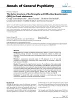
Figure 2 Effects of AC on cell proliferation and cell viability in
primary human T lymphocytes. Primary human T lymphocytes (2
×10
5
/well) were stimulated with or without anti-CD3 (1 μg/ml)/
CD28 (3 μg/ml) Ab and treated with medium, 0.1%DMSO, or the
indicated concentration of AC, or CsA (2.5 μM). (A) After incubated
for 72 hours, the proliferation of T lymphocytes was detected by
tritiated thymidine uptake (1 μCi/well). After incubated for 16 hours,
the cells were harvested by an automatic harvester, then
radioactivity was measured by liquid scintillation counting. (B) After
96 hr incubation, T cells were harvested and numbers of total,
viable, and nonviable cells were counted after trypan blue staining.
Each bar represents the mean ± SD of three independent
experiments.
##
P < 0.01: vs. the cells treated with DMSO.
**P < 0.01: vs. the cells treated with DMSO and anti-CD3/CD28 Ab.
0HGLXP '062 &V$
5HVWLQJFHOOV
$QWL&'&'$EDFWLYDWHGFHOOV
P
PP
P0
0HGLXP '062 &V$
,)1
J
J
J
J
5HVWLQJFHOOV
$QWL&'&'$EDFWLYDWHGFHOOV
P
PP
P0
AC concentration
AC concentration
A
B
SURGXFWLRQSJPO
,/SURGXFWLRQSJPO
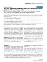
Figure 3 IL -2 and IFN- g production in primary human T
lymphocytes treated with AC. Primary human T lymphocytes C (2
×10
5
/well) were treated by 0, 6.25, 12.5 and 25 μM of AC or CsA
(2.5 μM) with or without anti-CD3 (1 μg/ml)/CD28 (3 μg/ml) Ab for
three days. Then the cell supernatants were collected and IL-2 and
IFN-g concentrations were determined by EIA, respectively. Each bar
is the mean ± SD of three independent experiments.
##
P < 0.01: vs.
the cells treated with DMSO. **P < 0.01: vs. the cells treated with
DMSO and anti-CD3/CD28 Ab.
Tsai et al. Chinese Medicine 2011, 6:12
/>Page 5 of 8
$&&RQFHQWUDWLRQP
PP
P0
0HGLXP '062
,/P51$*$3'+P51$
5HVWLQJFHOOV
$QWL&'&'$EDFWLYDWHGFHOOV
$&&RQFHQWUDWLRQP
PP
P0
0HGLXP '062
,)1
J
J
J
J
P51$*$3'+P51$
5HVWLQJFHOOV
$QWL&'&'$EDFWLYDWHGFHOOV
'062 &V$ '062 &V$
,/P51$*$3'+P51$
$QWL&'&'$E
'062 &V$ '062 &V$
,)1
J
J
J
J
P51$*$3'+P51$
$QWL&'&'$E
,/
*$3'+
ES
ES
,/
*$3'+
ES
ES
,)1J
JJ
J
*$3'+
ES
ES
ES
ES
,)1J
JJ
J
*$3'+
$&FRQFHQWUDWLRQP
PP
P0
0HGLXP '062
,/P51$*$3'+P51$
5HVWLQJFHOOV
$QWL&'&'$EDFWLYDWHGFHOOV
$&FRQFHQWUDWLRQP
PP
P0
0HGLXP '062
,)1
J
J
J
J
P51$*$3'+P51$
5HVWLQJFHOOV
$QWL&'&'$EDFWLYDWHGFHOOV
'062 &V$ '062 &V$
,/P51$*$3'+P51$
$QWL&'&'$E
'062 &V$ '062 &V$
,)1
J
J
J
J
P51$*$3'+P51$
$QWL&'&'$E
,/
*$3'+
ES
ES
,/
*$3'+
ES
ES
,)1J
JJ
J
*$3'+
ES
ES
ES
ES
,)1J
JJ
J
*$3'+
AC
BD
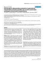
Figure 4 Effects of AC on IL-2 and IFN-g transcripts in primary human T lymphocytes induced by anti-CD3/CD28 Ab. Pri mary human T
lymphocytes (5 × 10
6
) activated with or without anti-CD3 (1 μg/ml)/CD28 (3 μg/ml) Ab in the presence or absence of 6.25, 12.5 or 25 μMACor
CsA (2.5 μM) for 18 hr. The total cellular RNA was isolated from T lymphocytes and aliquots of 1 μg of RNA were reverse-transcribed for
synthesis of cDNA. Briefly, 10 μl of cDNA was applied for the PCR test. The PCR was done as described in Materials and Methods. After the
reaction, the amplified product was taken out of the tubes and run on 2% agarose gel. (A) and (B): Lane 1 - medium, Lane 2 - 0.1% DMSO,
Lanes 3 to 5 - 6.25, 12.5 and 25 μM AC, Lane 6 - anti-CD3/CD28 Ab, Lane 7 - DMSO and anti-CD3/CD28 Ab, Lanes 8 to 10 - 6.25, 12.5 or 25 μM
AC and anti-CD3/CD28 Ab. (C) and (D): Lane 1 - 0.1% DMSO, Lane 2 - CsA, Lane 3 - DMSO and anti-CD3/CD28 Ab, Lane 4 - CsA and anti-CD3/
CD28 Ab. Graphical representation of laser densitometry of IL-2 and IFN-g mRNA expression in resting or anti-CD3/CD28 Ab-stimulated PBMC in
the presence or absence of AC or CsA. Each band was quantitated using laser-scanning densitometer SLR-2D/1D (Biomed Instruments, USA). The
ratio of each cytokine mRNA to GAPDH mRNA was calculated. Each bar is the mean ± SD of three independent experiments.
##
P < 0.01: vs. the
cells treated with DMSO. **P < 0.01: vs. the cells treated with DMSO and anti-CD3/CD28 Ab.
Tsai et al. Chinese Medicine 2011, 6:12
/>Page 6 of 8
This study showed that AC inhibited IL-2 and IFN-g pro-
ductions in primary human T lymphocytes stimulated by
ant i-CD3/CD28 Ab. The impairments of IL-2 and IFN-g
production were related to the suppression of their mRNA
transcriptions by AC. Since T lymphocyte proliferation is
primarily mediated by IL-2, inhibition of IL-2 production is
a central mechanism of action of several immunosuppres-
sants such as CsA. This study also demonstrated that CsA
inhibited IL-2 a nd IFN-g gene expression and cell prolifera-
tion in primary human T lymphocytes induced by anti-
CD3/CD28 Ab, suggesting that AC actions are similar to
those of CsA which induces arr est activation and prolifera-
tion of T cell s by inhibiting IL-2 transcription [14]. Fur ther-
more, the preliminary data from immunofluorescence
staining indicated that AC had no effect on IL-2 receptor
expression in primary human T lymphocytes activated by
anti-CD3/CD28 Ab (data not shown), suggesting that the
reduction of proliferation in AC-treated T lymphocytes was
not caused by down-regulation of IL-2 receptor expression.
Failure to produce IL-2 and IFN-g may be the reason why
primary human T lymphocytes do not proliferate.
NF-AT is a major player in the control of T lympho-
cytes activation a nd proliferation [2]. After anti-CD3/
CD28 Ab stimulation, calcium-dependent phosphatase
calcineurin binds to NF-AT, dephosphorylates NF-AT
and causes nuclear import of NF-AT. The binding
domain of NF-AT is Rel similarity domain located in
numerous cytokine promoters. IL-2 and IFN-g gene
expressions in T lymphocytes are controlled by NF-AT-
dependent promoters/enhancers [24]. This study found
that AC decreased NF-AT activation. NF-AT is a target
for the immunosuppressants CsA and FK506 which are
efficient inhibitors of T cell activation [14]. This study
also demonstrated that CsA blocked NF-AT activation,
suggesting that AC inhibited I L-2 and IFN-g production
and cell proliferati on in prima ry human T lymphocytes
by modulation of NF-AT activation. Interleukin-10 is
mainly produced by regulatory T lymphocytes and regu-
lates other immune cells [24]. We also showed that AC
(25 μM) did not affect IL-10 production in primary
human T lymphocytes induced by anti-CD3/CD28 Ab
(453 ± 88 vs. 412 ± 75pg/ml).
Conclusion
AC inhibited T lymphocyte proliferation and decreased
the gene expression of IL-2, IFN-g and NF-AT.
Abbreviations
Ab: antibody; AC: arctigenin; RT-PCR: reverse transcription-polymerase chain
reaction; IL-2: interleukin-2; IFN-γ: interferon-γ; PBMC: peripheral blood
0HGLXP '062 &V$
/XPLQHVFHQFH
UHODWLYHOLJKWXQLWV
5HVWLQJFHOOV
$QWL&'&'$EDFWLYDWHGFHOO
V
$&FRQFHQWUDWLRQP
PP
P0
Figure 5 Effects of AC on NF-AT activation. Jurkat cells (5 × 10
4
) were transfected with pGL4.30 (luc2P/NFAT-RE/Hygro) by Lipofectamin™
2000 (Invitrogen, USA) for 24 hours according to the manufacturer’s instructions. Then, the cells were cultured with anti-CD3 (1 μg/ml)/CD28
(3 μg/ml) Ab in the presence or absence of AC (6.25, 12.5 and 25 μM) or CsA (2.5 μM) for four hours. Total cell lysates were extracted with
1× reporter lysis buffer (Promega, USA), then 10 μg of total cell lysates were used to determine luciferase activity by the Luciferase Assay System
(Promega, USA). Each bar is the mean ± SD of three independent experiments.
##
P < 0.01: vs. the cells treated with DMSO. **P < 0.01: vs. the
cells treated with DMSO and anti-CD3/CD28 Ab.
Tsai et al. Chinese Medicine 2011, 6:12
/>Page 7 of 8
mononuclear cells; EtOAc: ethyl acetate; DMSO: dimethylsulfoxide; PBS:
phosphate buffered saline; HBSS: Hanks’ buffer saline solution; FCS: fetal calf
serum; CsA: cyclosporin A; NF-AT: nuclear factor of activated T cells; EIA:
enzyme immunoassays; DEPC: diethyl pyrocarbonate; EDTA:
ethylenediaminetetraacetate; DTT: dithiothreitol; GAPDH: glyceraldehyde-3-
phosphate dehydrogenase.
Acknowledgements
This study was partially supported by grants from Council of Agriculture (97-
1.2.1-al-22), National Science Council (NSC96-2320-B-030-006-MY3; NSC 99-
2320-B-030-004-MY3), Committee on Chinese Medicine and Pharmacy
(CCMP96-RD-207) and Fu-Jen University (9991A15/10993104995-4), Taiwan.
Author details
1
National Research Institute of Chinese Medicine, Taipei, 11221, Taiwan.
2
Institute of Life Science, Fu-Jen University, Taipei, 24205, Taiwan.
3
Graduate
Institute of Pharmacology Science, Taipei Medical University, Taipei, 11031,
Taiwan.
4
Graduate Institute of Medical Sciences, Taipei Medical University,
Taipei, 11031, Taiwan.
5
Department of Biochemistry and Molecular Biology,
College of Medicine, National Taiwan University, Taipei, 10051, Taiwan.
Authors’ contributions
WJT and CTC designed and conducted the experiments. GJW and THL
isolated and purified AC from A. lappa. SFC and SCL constructed pGL4.30
(luc2P/NFAT-RE/Hygro) plasmids. YCK supervised the study and revised the
manuscript. All authors read and approved the final version of the
manuscript.
Competing interests
The authors declare that they have no competing interests.
Received: 14 October 2010 Accepted: 25 March 2011
Published: 25 March 2011
References
1. Kuo YC, Yang NS, Chou CJ, Lin LC, Tsai WJ: Regulation of cell proliferation,
gene expression, production of cytokines and cell cycle progression in
primary human T lymphocytes by piperlactam S isolated from Piper
kadsura. Mol Pharmacol 2000, 58:1057-1066.
2. Rao A, Luo C, Hogan PG: Transcription factors of the NFAT family:
regulation and function. Annu Rev Immunol 1997, 15:707-747.
3. Rochman Y, Spolski R, Leonard WJ: New insights int o the regulation of
T cells by gamma (c) family cytokines. Nat R ev Immunol 2009,
9:480-49 0.
4. Kuo YC, Weng SC, Chou CJ, Chang TT, Tsai WJ: Activation and proliferation
signals in primary human T lymphocytes inhibited by ergosterol
peroxide isolated from Cordyceps cicadae. Br J Pharmacol 2003,
140:895-906.
5. Hoyer KK, Dooms H, Barron L, Abbas AK: Interleukin- in the development
and control of inflammatory disease. Immunol Rev 2008, 226:19-28.
6. Liu CP, Kuo YC, Lin YL, Liao JF, Shen CC, Chen CF, Tsai WJ: (S)-Armepavine
inhibits human peripheral blood mononuclear cells activation by
regulating Itk and PLC γ activation in a PI3K-dependent manner. J Leukoc
Biol 2007, 81:1276-1286.
7. Holetz FB, Pessini GL, Sanches NR, Cortez DA, Nakamura CV, Filho BP:
Screening of some plants used in the Brazilian folk medicine for the
treatment of infectious diseases. Mem Inst Oswaldo Cruz 2002,
97:1027-1031.
8. Zhao F, Wang L, Liu K: In vitro anti-inflammatory effects of arctigenin, a
lignan from Arctium lappa L., through inhibition on iNOS pathway.
J Ethnopharmacol 2009, 122:457-462.
9. Cho MK, Jang YP, Kim YC, Kim SG: Arctigenin, a phenylpropanoid
dibenzylbutyrolactone lignan, inhibits MAP kinases and AP-1 activation
via potent KK inhibition: the role in TNF-alpha inhibition. Int
Immunopharmacol 2004, 4:1419-1429.
10. Matsumoto T, Hosono-Nishiyama K, Yamada H: Antiproliferative and
apoptotic effects of butyrolactone lignans from Arctium lappa on
leukemic cells. Planta Med 2006, 72:276-278.
11. Kim SH, Jang YP, Sung SH, Kim CJ, Kim JW, Kim YC: Hepatoprotective
dibenzylbutyrolactone lignans of Torreya nucifera against CCl
4
-induced
toxicity in primary cultured rat hepatocytes. Biol Pharm Bull 2003,
26:1202-1205.
12. Liu S, Chen K, Schliemann W, Strack D: Isolation and identification of
arctiin and arctigenin in leaves of Burdock (Arcticum lappa L.) by
polyamide column chromatography in combination with HPLC-ESI/MS.
Phytochem Anal 2005, 16:86-89.
13. Wu MH, Tsai WJ, Don MJ, Chen YC, Kuo YC: Tanshinlactone A from Salvia
miltiorrhiza modulates interleukin-2 and interferon-γ gene expression. J
Ethnopharmacol 2007, 113:210-217.
14. Schreiber SL, Crabtree GR: The mechanism of action of cyclosporin A and
FK 506. Immunol Today 1992, 13:136-142.
15. Chen YC, Tsai WJ, Wu MH, Lin LC, Kuo YC: Suberosin inhibits human
peripheral blood mononuclear cells proliferation through the
modulation of NF-AT and NF-κB transcription factors. Br J Pharmacol
2007, 150:298-312.
16. Seko Y, Cole S, Kasprzak W, Shapiro BA, Ragheb JA: The role of cytokine
mRNA stability in the pathogenesis of autoimmune disease. Autoimmun
Rev 2006, 5:299-305.
17. Crabtree GR: Contingent genetic regulatory events in T lymphocyte
activation. Science 1989, 243:355-361.
18. Lin CC, Lu JM, Yang JJ, Chuang SC, Ujiie T: Anti-inflammatory and radical
scavenge effects of Articum lappa. Am J Chin Med 1996, 24:127-137.
19. Iwakami S, Wu JB, Ebizuka Y, Sankawa U: Platelet activating factor (PAF)
antagonists contained in medicinal plants: lignans and sesquiterpenes.
Chem Pharm Bull (Tokyo) 1992, 40:1196-1198.
20. Knipping K, van Esch ECAM, Wijering SC, van der Heide S, Dubois AE,
Garsen J: In vitro and in vivo anti-allergic effects of Arctium lappa. Exp Biol
Med 2008, 233:1469-1477.
21. Kang HS, Lee JY, Kim CJ: Anti-inflammatory activity of arctigenin from
Forsythiae fructus. J Ethnopharmacol 2008, 116:305-312.
22. Yoo JH, Lee HJ, Kang K, Jho EH, Kim CY, Baturen D, Tunsag J, Nho CW:
Lignans inhibit cell growth via regulation of Wnt/beta-catenin signaling.
Food Chem Toxicol 2010, 48:2247-2252.
23. Arai K, Lee F, Miyajima A: Cytokines: Coordinators of immune and
inflammatory response. Annu Rev Biochem 1990, 59:783-836.
24. Serfling E, Berberich-Siebelt F, Chuvpilo S, Jankevics E, Klein-Hessling S,
Twardzik T, Avots A: The role of NF-AT transcription factors in T cell
activation and differentiation. Biochim Biophys Acta 2000, 1498:1-18.
doi:10.1186/1749-8546-6-12
Cite this article as: Tsai et al.: Arctigenin from Arctium lappa inhibits
interleukin-2 and interferon gene expression in primary human T
lymphocytes. Chinese Medicine 2011 6:12.
Submit your next manuscript to BioMed Central
and take full advantage of:
• Convenient online submission
• Thorough peer review
• No space constraints or color figure charges
• Immediate publication on acceptance
• Inclusion in PubMed, CAS, Scopus and Google Scholar
• Research which is freely available for redistribution
Submit your manuscript at
www.biomedcentral.com/submit
Tsai et al. Chinese Medicine 2011, 6:12
/>Page 8 of 8