ECG Learning Center - part 4 pps
Bạn đang xem bản rút gọn của tài liệu. Xem và tải ngay bản đầy đủ của tài liệu tại đây (1.7 MB, 38 trang )
ecg_vent_pace.html
Ventricular Pacing in Atrial Fibrillation - Marquette-
KH
Marquette Electronics Copyright 1996
[5/11/2006 9:39:59 AM]
ecg_12lead010.html
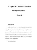
Atrial Flutter With 2:1 AV Conduction-KH
Frank G.Yanowitz, M.D.
In this example of atrial flutter with 2:1 AV conduction the flutter waves are very hard to see. Atrial
flutter with 2:1 block must be considered, however, because the heart rate is about 150 bpm. A careful
look at V1 shows the two flutter waves for each QRS complex complex. One flutter wave immediately
follows the QRS and the other is just before the QRS.
[5/11/2006 9:40:00 AM]
ecg_12lead008.html
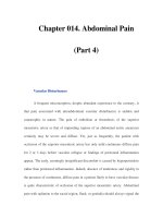
Atrial Flutter With 2:1 AV Conduction-KH
Frank G.Yanowitz, M.D.
Atrial flutter with 2:1 AV block is one of the most frequently missed ECG rhythm diagnoses because the
flutter waves are often hard to find. In this example two flutter waves for each QRS are best seen in lead
III and V1. The ventricular rate at 150 bpm should always prompt us to consider atrial flutter with 2:1
conduction as a diagnostic consideration.
[5/11/2006 9:40:01 AM]
ecg_12lead009.html
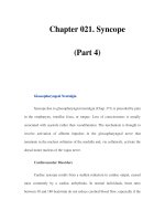
Atrial Flutter With 3:2 AV Conduction-KH
Frank G.Yanowitz, M.D.
This 12-lead ECG shows a subtle bigeminal rhythm resulting from atrial flutter with a 3:2 AV
conduction ratio; RR intervals alternate by a small duration. This is uncommon! The impulses from the
atrial flutter conduct through the AV junction in a Wenckebach sequence; for every 3 flutter waves the
second conducts more slowly than the first, and the third flutter wave is blocked.
[5/11/2006 9:40:02 AM]
ecg_12lead009z.html
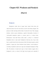
Atrial Flutter with 3:2 Conduction Ratio: Frontal
Plane Leads-KH
Frank G.Yanowitz, M.D.
Note the subtle bigeminy in the RR intervals. The best way to identify the flutter waves in this example is
to imagine what lead III would look like if the QRS complexs disappeared; what remains is a reasonable
"saw-tooth" pattern characteristic of atrial flutter with a flutter rate of about 300 bpm.
[5/11/2006 9:40:03 AM]
ecg_477.html
Atrial Flutter With Variable AV Block And Rate-
Dependent LBBB-KH
Frank Yanowitz Copyright 1996
The basic rhythm is atrial flutter with variable AV block. When 2:1 conduction ratios occur there is a rate-
dependent LBBB. Don't be fooled by the wide QRS tachycardia on the bottom strip. It's not ventricular
tachycardia, but atrial flutter with 2:1 conduction and LBBB. Lidocaine is not needed because there is no
ventricular ectopy.
[5/11/2006 9:40:03 AM]
ecg_12lead011z.html
Atrial Flutter With 2:1 AV Conduction: Leads II, III,
V1-KH
Frank G.Yanowitz, M.D.
In leads II and III, the one of the flutter waves occurs at the end of the QRS complex and might be
mistaken for part of the QRS itself; i.e., the S wave. In lead V1, the two flutter waves for every QRS are
more easily identified.
[5/11/2006 9:40:04 AM]
ecg_12lead051.html
LBBB and Atrial Flutter with 2:1 AV Block
Frank G. Yanowitz, M.D. copyright 1997
The LBBB is obvious by the monophasic R wave in leads I and aVL; the atrial flutter is less obvious, but
in lead V1 atrial activity at 280/min can be seen in a 2:1 conduction pattern.
[5/11/2006 9:40:05 AM]
ecg_478.html
Atrial Flutter With 2:1 and 4:1 Conduction and Rate
Dependent LBBB-KH
Frank Yanowitz Copyright 1996
In this example of atrial flutter with variable AV conduction, the faster rates are associated with rate-
related LBBB. Don't confuse this for ventricular tachycardia.
[5/11/2006 9:40:06 AM]
ecg_12lead010z.html
Atrial Flutter With 2:1 AV Conduction: Lead V1-KH
Frank G.Yanowitz, M.D.
The arrows point to two flutter waves for each QRS complex. Atrial rate = 280; ventricular rate = 140.
[5/11/2006 9:40:07 AM]
ecg_12lead011.html
Atrial Flutter With 2:1 AV Conduction-KH
Frank G.Yanowitz, M.D.
Flutter waves are best seen in lead V1; one immediately follows the QRS and the other precedes the next
QRS. The regular ventricular rate of 150 bpm should always prompt us to condider this diagnosis.
[5/11/2006 9:40:07 AM]
ecg_atrial_flutter.html
Atrial Flutter With Variable AV Block - Marquette-
KH
Marquette Electronics Copyright 1996
[5/11/2006 9:40:08 AM]
ecg_12lead008z.html
Atrial Flutter With 2:1 Conduction: Leads II, III, V1-
KH
Frank G.Yanowitz, M.D.
[5/11/2006 9:40:09 AM]
ecg_428.html
Massage Parlor Games-KH
Frank Yanowitz Copyright 1996
When unsure of the mechanism of a supraventricular tachycardia, carotid sinus massage may help make
the diagnosis. In this example, carotid sinus massage causes marked AV block which permits easy
recognition of the rapid, regular atrial flutter waves.
[5/11/2006 9:40:09 AM]
ecg_junctional.html
Junctional Escape Rhythm-KH
Marquette Electronics Copyright 1996
[5/11/2006 9:40:10 AM]
ecg_accelerate.html
Accelerated Junctional Rhythm-KH
Frank G. Yanowitz, M.D., copyright 1997
[5/11/2006 9:40:11 AM]
ecg_500.html
Junctional Tachycardia With Exit Block: A
Manifestation of Digitalis Intoxication-KH
Frank Yanowitz Copyright 1996
The "ladder diagram" says it all: the atria are fibrillating; there is complete heart block in the AV junction;
a junctional tachycardia focus is firing at about 130 bpm, but not all junctional impulses reach the
ventricles due to 2nd degree exit block.
[5/11/2006 9:40:12 AM]
ecg_493.html
Digitalis Intoxication: Junctional Tachycardia With
and Without AV Block-KH
Frank Yanowitz Copyright 1996
In a patient with longstanding atrial fibrillation being treated with digoxin, a regular tachycardia, as seen in
'A', with a RBBB suggests a junctional or supraventricular tachycardia. Group beating, in 'B', is likely due
to a 2nd degree, Type 1, exit block below the ectopic junctional focus. This is highly suggestive of
digitalis intoxication.
[5/11/2006 9:40:12 AM]
ecg_494.html
Digitalis Intoxication: Junctional Tachycardia With
and Without Exit Block-KH
Frank Yanowitz Copyright 1996
In 'A' the rhythm is junctional tachycardia with RBBB. In 'B' there is 2nd degree exit block with a 3:2
conduction ratio; i.e., every 3rd junctional impulse fails to reach the ventricles at least for the first two
groupings on 1.4sec.
[5/11/2006 9:40:13 AM]
ecg_v_fib.html
Ventricular Fibrillation - Marquette-KH
Marquette Electronics Copyright 1996
[5/11/2006 9:40:14 AM]
ecg_0280_mod.html
1st Degree AV Block
Frank Yanowitz Copyright 1996
The normal PR interval is 0.12 - 0.20 sec, or 120 -to- 200 ms. 1st degree AV block is defined by PR
intervals greater than 200 ms. This may be caused by drugs, such as digoxin; excessive vagal tone;
ischemia; or intrinsic disease in the AV junction or bundle branch system.
[5/11/2006 9:40:15 AM]
ecg_0311_mod.html
ECG Of The Century: A Most Unusual 1st Degree
AV Block
Frank Yanowitz Copyright 1996
On Day 1, at a heart rate of 103 bpm the P waves are not clearly defined suggesting an accelerated
junctional rhythm. However, on Day 2, at a slightly slower heart rate the sinus P wave suddenly appears
immediately after the QRS complex. In retrospect, the sinus P wave in Day 1 was found burried in the
preceding QRS; note the notch on the downstroke of the QRS. On Day 3 a normal PR interval was seen.
(1 of 2) [5/11/2006 9:40:15 AM]
ecg_0311_mod.html
How long can the PR interval get in 1st degree AV block??? No one knows.
(2 of 2) [5/11/2006 9:40:15 AM]
ecg_12lead019.html
Left Atrial Abnormality & 1st degree AV Block-KH
Frank G.Yanowitz, M.D.
The P-wave is notched, wider than 0.12s, and has a prominent negative (posterior) component in V1 - all
criter for left atrial abnormality or enlargement (LAE). The PR interval >0.20s. Minor ST-T wave
abnormalities are also present.
[5/11/2006 9:40:17 AM]
ecg_0283_mod.html
A Very Subtle 1st Degree AV Block
Frank Yanowitz Copyright 1996
Where are the P waves??? They are hiding in the T waves as indicated by the arrows. How do we know?
The PVC unmasked the sinus P wave, and now it is seen in the pause following the PVC. The PR
interval is, therefore, about 500 ms.
[5/11/2006 9:40:17 AM]