Essential Cardiac Electrophysiology Self Assessment - Part 2 pptx
Bạn đang xem bản rút gọn của tài liệu. Xem và tải ngay bản đầy đủ của tài liệu tại đây (218.34 KB, 31 trang )
16 Essential Cardiac Electrophysiology
• Lidocaine and other class 1B agents block the slow component of sodium current
and decrease QT in patients with LQT3
5
.
• Negative inotropy by sodium channel blockers may be due to blockage of the
slow sodium channel current.
• Slowing of heart rate produced by class 1B agents is due to blocking of
background sodium current that contributes to the phase 4 of pacemaker AP.
• β-Adrenergic stimulation reverses the effects of class I drugs.
• Proarrhythmia from class IC drugs develops during increased heart rate when
sympathetic activity is enhanced. Beta blockers may reverse this phenomenon
5
.
• Angiotensin II increases the frequency of reopening of the sodium channel and
increases the Na current.
1.3 CALCIUM CHANNELS AND CURRENTS
14–17
• The process of channel opening and closing is called gating.
• Open channels are active. Closed channels are inactive. Calcium and sodium
channels open in response to depolarization and enter the nonconducting state
during repolarization, a gating process known as inactivation.
• Alpha 1 subunit of the Ca channel contains the binding site for calcium channel
blocking drugs.
• Calcium channels are very selective and allow Ca permeability 1000-fold
faster.
• There are four types of calcium channels:
i L-type expressed on surface membrane.
ii T-type expressed on surface membrane.
iii Sarcoplasmic reticulum (SR) Ca release channel.
iv Inositol triphosphate (IP3) receptor channels are present on internal
membrane.
L-type calcium channel (L = Large and lasting)
• It is a major source of Ca entry into the cell. It opens when depolarization reaches
positive to −40 mV.
• It is responsible for excitation in sino atrial node (SAN) and atrio-ventricular
node (AVN). It produces inward current that contributes to depolarization in
SAN and AVN.
• It produces inward current responsible for plateau of AP.
• Increased calcium current prolongs depolarization and increases the height of
the AP plateau.
• Calcium channel dependent inward current is responsible for EAD.
• I
CaL
is responsible for excitation, contraction, and coupling. Blockade of these
channels results in negative inotropic effects.
• In AF decrease activity of the I
CaL
channel shortens APD and perpetuates
arrhythmia (electrical remodeling).
Ions, Channels, and Currents 17
Regulation of pacemaker and Ca currents
β-Adrenergic receptor stimulation
• It increases L-type calcium channel activity.
• This results in increased contractility, heart rate, and conduction velocity.
• Stimulation of receptors activates guanosine triphosphate binding protein Gs,
which in turn stimulates adenylyl cyclase activity, thus increasing the cAMP
level.
• β-Blockers have no direct effect on calcium channel.
• Sympathetic stimulation may also activate alpha1 receptors.
Parasympathetic stimulation
• It decreases L-type calcium activity through muscarinic and cholinergic
receptors.
• Acetylcholine, through G protein, activates inwardly rectifying I
Kach
, which
makes MDP more negative and decreases the slope of diastolic depolarization.
This results in slowing of the heart rate.
• Magnesium acts as an L-type calcium channel blocker.
T-type calcium channel
• These are found in cardiac and vascular smooth muscles, including coronary
arteries.
i It opens at more negative potential.
ii It rapidly inactivates (Transient T).
iii It demonstrates slow deactivation.
iv Has low conductance (tiny T).
• It is found in high density in SAN and AVN.
• It does not contribute to AP upstroke which is dominated by sodium channel.
• It is implicated in cell growth.
• T-type Ca channel density is increased in the presence of the growth hormone,
endothelin-1, and pressure overload.
• Failing myocytes also demonstrate increase density of T-type Ca channels.
• Drugs and compounds that block T-type Ca channels include the following:
i Amiloride
ii 3,4-Dichrobenzamil
iii Verapamil
iv Diltiazem
v Flunarizine
vi Tetradrine
vii Nickel
viii Cadmium
ix Mibefradil
• T-type Ca channel is up regulated by norepinephrine, alpha agonist (phenyleph-
rine), extracellular ATP, and LVH.
18 Essential Cardiac Electrophysiology
Sarcoplasmic calcium release channels (also called
Ryanodine receptors)
• These are intracellular channels that are regulated by calcium.
• These channels mediate the influx of calcium from SR into cytosol.
• It provides calcium for cardiac contraction. SR controls the cytoplasmic Ca level
by release or uptake during systole and diastole, respectively.
• Calcium release from SR is triggered by increase in intracellular calcium,
produced by L-type Ca channel. It is called calcium-induced calcium release
(CICR).
• When a cell is calcium overloaded SR releases calcium spontaneously and
asynchronously causing DAD (delayed after depolarization) seen in digitalis
toxicity.
• Caffeine releases calcium from SR.
• Doxorubicin decreases cardiac contractility by depleting SR calcium.
• Magnesium and ATP potentiates channel flux.
• In ischemia decreased intracellular ATP decreases calcium release and causes
ischemic contractile failure.
• Verapamil has no effect on sarcoplasmic Ca release channel (SCRC).
• SR also has potassium, sodium, and hydrogen channels.
Inositol triphosphate receptors (IP3)
• These receptors are found in smooth muscles and in specialized conduction
tissue.
• These are up regulated by angiotensin II and α-adrenergic stimulation.
• Stimulation of myocytes angiotensin II receptor by angiotensin increases intra-
cellular IP3.
• The arrhythmogenic effect of angiotensin II in CHF may be due to elevated IP3.
• These receptors have been implicated in apoptosis.
Tetrodotoxin (TTX) sensitive calcium channel
• It produces inward current. It is blocked by TTX.
• The channel that carries this current is permeable to both sodium and calcium.
• Elevated intracellular Na may activate reverse Na/Ca exchange, thus increasing
the levels of intracellular Ca which may trigger SR calcium release.
• It may contribute to cardiac arrhythmias.
Sodium and calcium exchange
• Opening of voltage operated calcium channel, during the plateau phase of APD,
increases the flux of calcium into cytoplasm. This causes CICR from SR.
• During diastole calcium is removed from the cell by sodium/calcium exchange
located in the cell membrane.
• Lowering of pH blocks sodium/calcium exchange.
Ions, Channels, and Currents 19
• SR calcium ATPases, Sarcolemmal calcium ATPases and sodium/calcium
exchange decrease cytoplasmic calcium from elevated systolic level to baseline
diastolic level by pumping Ca back into SR or by extruding Ca out of the cell.
• During calcium removal inwardly directed current is observed, which may cause
DAD.
• DAD occurs when there is pathologically high calcium load either due to digitalis
toxicity or following reperfusion.
• Na/Ca exchange is able to transport calcium bi-directionally. Reverse mode will
increase intracellular calcium, which may trigger SR calcium release.
Effect of antiarrhythmic drugs on calcium channel
• Most Na and K channel blocking drugs also affect Ca channels.
• Quinidine, Disopyramide, Lidocaine, Mexiletine, Diphenylhydantoin, Flecain-
ide, Propafenone, Moricizine, and Azimilide suppress L-type calcium current.
• Amiodarone blocks both L and T-type Ca currents.
• Sotalol has no effect on Ca channel.
• Digoxin inhibits sodium/potassium ATPases. This inhibition results in an
increase in intracellular Na, which in turn leads to an increase in intracellular
Ca through Na/Ca exchange.
• Verapamil blocks Ca current and decreases calcium activated chloride current.
References
1 Delisle BP. Anson BD. Rajamani S. January CT. Biology of cardiac arrhythmias: ion
channel protein trafficking. Circ Res. 94:1418–28, 2004.
2 Rosati B. McKinnon D. Regulation of ion channel expression. Circ Res. 94:874–83, 2004.
3 Priori SG. Inherited arrhythmogenic diseases: The complexity beyond monogenic
disorders.Circ Res. 94:140–5, 2004.
4 Enkvetchakul D. Nichols CG. Gating mechanism of KATP channels: Function fits form.
[Review] [100 refs] J Gen Physiol. 122:471–80, 2003.
5 Sah R. Ramirez RJ. Oudit GY. Gidrewicz D. Trivieri MG. Zobel C. Backx PH. Regulation
of cardiac excitation–contraction coupling by action potential repolarization: Role of the
transient outward potassium current (Ito). J Physiol. 546(Pt1):5–18, 2003.
6 Kass RS. Moss AJ. Long QT syndrome: Novel insights into the mechanisms of cardiac
arrhythmias. J Clin Invest. 112:810–5, 2003.
7 Gross GJ. Peart JN. KATP channels and myocardial preconditioning: An update. Am J
Physiol Heart Circ Physiol. 285:H921–30, 2003.
8 Clancy CE. Kass RS. Defective cardiac ion channels: From mutations to clinical
syndromes. J Clin Invest. 110:1075–7, 2002.
9 Hubner CA. Jentsch TJ. Ion channel diseases. Hum Mol Genet. 11: 2435–45, 2002.
10 Schram G. Pourrier M. Melnyk P. Nattel S. Differential distribution of cardiac ion channel
expression as a basis for regional specialization in electrical function. Circ Res. 90:939–50,
2002.
11 Clancy CE. Kass RS. Defective cardiac ion channels: From mutations to clinical
syndromes. J Clin Invest. 110:1075–7, 2002.
20 Essential Cardiac Electrophysiology
12 Towbin JA. Friedman RA. Provocation testing in inherited arrhythmia disorders: Can we
be more specific? Heart Rhythm. 2:147–8, 2005.
13 Fish JM. Antzelevitch C. Role of sodium and calcium channel block in unmasking the
Brugada syndrome. Heart Rhythm. 1:210–17, 2004.
14 Dolphin AC. G protein modulation of voltage-gated calcium channels. Pharmacol Rev.
55:607–27, 2003.
15 Yamakage M. Namiki A. Calcium channels – basic aspects of their structure, function and
gene encoding; anesthetic action on the channels – a review. Can J Anaesth. 49:151–64,
2002.
16 Marks, AR. Cardiac intracellular calcium release channels: Role in heart failure. Circ Res.
87:8–11, 2000.
17 Grossman E. Calcium antagonists. Prog Cardiovasc Dis. 47:34–57, 2004.
2 Electrophysiologic Effects of
Cardiac Autonomic Activity
Self-Assessment Questions
1 Which one of the following is the likely cardiac manifestation of β
3
receptor
stimulation?
A Increase in contractility
B Decrease in contractility
C Decrease in heart rate
D Increase in heart rate
2 Which one of the following muscarinic receptors is predominantly found in the
heart?
A M
1
B M
2
C M
3
D M
4
3 What is the likely cardiac effect of muscarinic receptor stimulation by
acetylcholine?
A Coronary vasoconstriction
B Positive chronotropic response
C Enhanced inotropic response
D Negative dromotropic effect
4 Which one of the following is likely to occur with cardiac adenosine receptors
stimulation?
A Negative chronotropic effect
B Positive dromotropic effect
C Enhanced contractility
D Coronary vasoconstriction
21
22 Essential Cardiac Electrophysiology
5 Which one of the following currents is activated by purinergic agonists?
A K
Ach,Ado
B I
Na
C I
CL
D I
Ca(T)
6 Which of the following effects is due to adenosine?
A Increase in Ca current
B Decrease in atrial APD and refractory period
C Stimulation of P
2
receptors
D Decrease in I
Katp
current
7 Which of the following electrophysiologic effects is least likely to occur with
vagal denervation of the atrium?
A Increases atrial APD and ERP
B Abolishes sinus arrhythmia
C Decreases heart rate variability and baroreflex sensitivity
D Decreases the ventricular effective refractory period
8 Which one of the following observations may suggest that in the treat-
ment of CHF nonselective β blockers are likely to be superior to selective
β blockers?
A β1 receptors are up regulated
B β2 and β3 receptors are up regulated
C Peripheral vascular resistance is increased
D Glomerular filtration rate is decreased
Electrophysiologic Effects of Cardiac Autonomic Activity 23
2.1 ADRENERGIC RECEPTORS
1–4
The human adrenergic receptor family consists of nine subtypes originating from
different genes: α1A, α1B, α1D, α2A, α2B, α2C, β1, β2, and β3.
β-adrenergic receptors
• β
1
is a predominant adrenergic receptor in the myocardium. 75% of the total β
receptor population is β
1
.
• β
1
stimulation causes positive inotropic, chronotropic, and lusitropic
(relaxation) response. cAMP-dependent activation of protein kinase A (PKA)
phosphorylates and activates β-adrenergic receptor.
• Even in the presence of continuing β stimulation cAMP response wanes. This
phenomenon is called receptor desensitization. Persistent agonist stimulation
decreases the total number of receptors (receptor down regulation).
• In ageing heart, β
1
receptor is down regulated and β
2
becomes dominant.
• In congestive heart failure (CHF) sustained adrenergic stimulation leads to
desensitization and down regulation of β
1
receptors. β
2
receptor expression is
preserved. α1 receptor subtypes remain constant or may even be up-regulated.
Under these conditions β
2
and α1 stimulation results in atrial and ventricular
arrhythmias.
• This supports the notion that nonselective β blockers reduce cardiac mortality
in post-myocardial infarction (MI) and CHF patients.
• In general, the type-2 adrenergic receptors (α2 and β2) are found at the pre-
junctional site in the central and peripheral sympathetic nervous system, where
activation of α2 receptors inhibits and activation of β2 receptors enhances
norepinephrine release (Table 2.1).
• Presynaptically localized α2A-receptor and α2C-receptor subtypes are import-
ant in decreasing sympathetic activity in the central nervous system as well
as in decreasing the norepinephrine release in cardiac sympathetic nerve
terminals.
• β
2
receptor is up regulated in denervated, transplanted heart.
• Stimulation of the β
2
receptors of sino atrial node (SAN) results in sinus
tachycardia.
• β
2
receptor stimulation elevates intracellular pH, which increases responsiveness
to calcium.
• β-Adrenergic stimulation increases I
K
.
• In cardiomyocytes, endothelial or smooth muscle cells, the type-2 adrener-
gic receptors are also present postsynaptically together with α1, β1, and β3
receptors.
• Acute changes in myocardial function are exclusively governed by the β
receptors.
• α1-Receptor contribution is negligible in humans under normal conditions.
24 Essential Cardiac Electrophysiology
Table 2.1 Characteristics of the subtypes of adrenergic receptors
Receptor Agonist Antagonist Tissue Responses
α
1
Epi > NE Iso Prazocin Heart ↑Contractility,
phenylephrine arrhythmias
Intestinal SM Relaxation
Urinary and vascular SM Contraction
Liver Glycogenolysis
Gluconeogenesis
α
2
Epi > NE Iso, Yohimbine Pancreatic β cells ↓Insulin
clonidine Aggregation
Platelets ↓NE
Nerve terminal Contraction
Vascular SM
β
1
Iso > Epi = NE, Metoprolol Heart ↑Inotropy, chronotropy
dobutamine and AV conduction
Juxtaglomerular ↑Renin
β
2
Iso > Epi NE Propranolol Heart ↑Inotropy
terbutaline Automaticity
Arrhythmias
Vascular GI GU Relaxation
bronchial SM
Skeletal muscle Glycogenolysis
K uptake
Liver Glycogenolysis
β
3
Iso = NE > Epi Adipose tissue Lipolysis
Heart ↓Contractility
Epi, epinephrine; NE, norepinephrine; Iso, isoproterenol; SM, smooth muscle; GI, gastrointestinal;
GU, genitourinary; ↑, increased release; ↓, decreased release.
• Although all three types of α1 receptors are expressed in the heart, the α1A is
the dominant subtype.
• No direct α2-receptor mediated effects are discernible on the myocardium.
• α1-Adrenergic receptor stimulation induces growth.
β3 receptors
• β3 receptor is an important regulator of adipose tissue and gastrointestinal
tract. It is also present in human heart and is implicated as an inhibitor of
cardiac contractile function. In normal heart β3-adrenoceptors protects myocar-
dium from the deleterious effects of excess catecholamines that may occur in
hyperadrenergic states including heart failure.
Electrophysiologic Effects of Cardiac Autonomic Activity 25
• The negative inotropic effects of β3-adrenoceptors are mediated through
activation of constitutively expressed endothelial nitric oxide synthase. This
action opposes the positive inotropic effects of catecholamines on β1- and
β2-receptors, which are mediated via cyclic adenosine monophosphate
(cAMP).
• Whereas β3 receptor activation may protect against cardiac myocyte damage due
to catecholamine excess during the early stage of heart failure, β3-adrenoceptor
up-regulation may contribute to decrease contractility in the later phases of
disease.
• β3-adrenoceptors are desensitization-resistant and their action may exceed
that of impaired, down-regulated or desensitized β1- and β2-adrenoceptors.
This may result in depression of contractility and exacerbation of heart
failure.
• This supports the observation that non selective beta blockers reduce cardiac
mortality in post MI and CHF patients.
2.2 CHOLINERGIC RECEPTORS
5,6
• Cholinergic receptors are nicotinic or muscarinic depending on their ability to
interact with nicotine or muscarine.
• Cholinergic receptors are activated by acetylcholine (Ach) from parasympathetic
nerve terminals.
• The effects of Ach that are mimicked by muscarine and blocked by atropine are
called muscarinic effects. Other effects of Ach that are mimicked by nicotine and
are not antagonized by atropine but are blocked by tubocurarine are labeled as
nicotinic effects.
• Cardiac action of Ach is mediated by muscarinic cholinergic receptors.
• Five types of (M1–M5) muscarinic receptors have been identified.
• M1 and M3 receptors cause mobilization of intracellular Ca by activating
phospholipase C. M2 and M4 receptors inhibit adenylyl cyclase and enhance
K conductance through K channels.
• M1 receptor is found in autonomic ganglion and central nervous system.
• M2 is a dominant muscarinic receptor of cardiac myocytes.
• M3 is a predominant receptor in smooth muscle cells, where its stimulation
causes contraction, and in secretory glands.
• Inhibitory effects of Ach on calcium current and contractility are due to M2
receptors and can be blocked by M2 antagonist.
• ACh is hydrolyzed by acetylcholinestrase. Cardiac effects of ACh are character-
ized by vasodilatation, negative chronotropic effect, negative dromotropic effect
[decrease in conduction in SAN and atrio-ventricular node (AVN)], and negative
inotropic effect.
• Commonly used synthetic choline derivatives are Methacholine, Carbachol, and
Bethanechol.
26 Essential Cardiac Electrophysiology
• Muscarine, pilocarpine, and arecholine are naturally occurring alkaloids with
pharmacological properties similar to Ach.
• Atropine and scopolamine are naturally occurring alkaloids that act as
muscarinic receptor antagonist.
2.3 PURINERGIC RECEPTORS
1,7
• Autonomic nonadrenergic and noncholinergic nerves were suggested to
contain ATP.
• Nerves utilizing ATP as their principal transmitter were named “purinergic”
based on the actions of purine nucleotides and nucleosides in a wide variety
of tissues; P1- and P2-receptor classification was proposed.
Purinergic receptors
• There are two types of purinergic receptors, P1–P2.
• P1 is activated by adenosine.
• P2 is activated by extracellular ATP.
• Two types of P2 receptors have been identified. P2X are ion channels, while P2Y
are G protein coupled receptor.
• P1 purinoceptors are much more sensitive to adenosine and AMP than to ADP
and ATP. The reverse is true for P2 purinoceptors.
• P2 receptors are unaffected by Methylxanthines such as caffeine and theophyl-
line that selectively and completely inhibits the P1 purinoceptors.
• Subclasses of P1 adenosine receptors were introduced when their stimulation
was shown to inhibit (A1 subtype) or activate (A2 subtype) adenylyl cyclase
activity.
Adenosine
• There are four subtypes of adenosine (P1 purinergic) receptors: A
1
,A
2
A, A
2
B,
and A
3
. All four are expressed in the heart.
• A
1
and A
2
receptors are antagonized by xanthines.
• Electrophysiologic actions of adenosine on the heart are mediated by A
1
recept-
ors. Blockade of this receptor abolishes negative chronotropic, dromotropic,
inotropic and anti-β-adrenergic effects of adenosine.
• A
1
receptors inhibit adenylyl cyclase and activate K current and phospholipase C.
• A
2
receptors activate adenylyl cyclase.
• A
2
A receptors are present in vascular endothelium and smooth muscles of
coronary arteries and cause adenosine induced coronary vasodilatation.
• Effects of extracellular ATP, before its degradation to adenosine, are mediated
by P
2
purinergic receptors.
Electrophysiologic Effects of Cardiac Autonomic Activity 27
• Stimulation of muscarinic and adenosine receptors causes activation of inhibit-
ory guanine (G) proteins. This leads to production of cAMP mediated activation
of I
CaL
, I
K
, and I
CL
.
• Density of A
1
adenosine and M
2
muscarinic cholinergic receptor is greater in
the atrium.
Acetylcholine and adenosine sensitive potassium currents
• Cholinergic and purinergic agonists activate inwardly rectifying potassium
currents I
KAch,Ado
.
• Inwardly rectifying means it is easier for the current to flow inwards than out-
wards through I
KAch,Ado
channels. Inward rectification is attributed to voltage
dependent block of potassium channels by intracellular Mg and polyamines
(Spermine and Spermidine).
• These currents are also activated by extracellular ATP, arachidonic acid
(Prostacycline), somatostatin, and sphingosine 1 phosphate.
• Lipooxygenase potentiates and cyclooxygenase inhibits I
KAch,Ado
.
• Hyperpolarizing current (I
f
) is an inward current and is responsible for diastolic
depolarization of SAN. This is a time-dependent, nonspecific cation current.
• Ach and adenosine inhibit I
f
. This effect is due to inhibition of adenylate cyclase.
• Adenosine has no effect on ventricular AP of myocytes.
• In the SAN and AVN adenosine and Ach cause hyperpolarization and reduce the
rate of diastolic depolarization. These actions result in slowing of the heart rate
(negative chronotropy) and delay in AVN conduction (negative dromotropy).
• Adenosine’s effects occur at the atrial-His and nodal region, it has no effect on
the nodal-His bundle region (NH) of the AVN.
• Ach and adenosine decrease atrial action potential duration (APD) and con-
tractile force (in the absence of β-adrenergic stimulation) due to activation of
I
KAch,Ado
and inhibition of I
CaL
.
• Adenosine and Ach attenuate β-adrenergic stimulated I
CaL
in ventricular and
atrial myocytes and inhibit transient inward current and delayed after depolariz-
ation (DAD). Adenosine may be effective in catecholamine sensitive ventricular
tachycardia by abolishing cAMP-dependent triggered activity.
• Ach and adenosine decrease atrial action potential duration (APD) and effective
refractory period (ERP) and facilitate induction of atrial fibrillation (AF).
• During AV nodal ischemia local production of adenosine may be responsible for
bradycardia seen in patients with inferior wall MI.
• Extracellular ATP causes DAD, and early after depolarization by stimulating P
2
receptors and inducing I
CaL
(Table 2.2).
Cardiac autonomic innervations
1
• Vagal postganglionic neurons to sinus node are located in the left pulmonary
vein left atrial junction, and neurons to AVN are found in the IVC-LA fat pad.
28 Essential Cardiac Electrophysiology
Table 2.2 Effect of Ach, adenosine, and extracellular
ATP on cardiac currents
Ach Adenosine ATP
Receptor M
2
A
1
P
2
I
KAch,Ado
↑↑ ↑
I
K
No effect No effect ↑
I
KATP
↑↑ ↑
I
CaL
↓↓ ↑In atria ↓ in V
I
Na
No effect No effect Variable
I
f
↓↓ No effect
I
CL
No effect ↓ I
f
stimulated ↑
↑,increase; ↓, decrease; V, ventricle.
• SVC/aorta fat pad innervates both atria.
• Vagal denervation of atria prevents induction of acute AF, abolishes sinus
arrhythmias, decreases heart rate variability, and eliminates baroreflex
sensitivity without affecting vagal innervation to the ventricle.
• Efferent vagal fibers to ventricle do not travel through three epicardial fat pads.
• Changes in sinus node function may not reflect changes at the ventricular
level.
• Sympathetic efferent fibers are epicardial along coronary arteries, and parasym-
pathetic efferent fibers are subendocardial in the ventricle.
• Block of nitric oxide synthase attenuates cholinergic action and increases calcium
current.
L-Arginine reverses these effects.
•
L-Arginine also reduces the effects of sympathetic stimulation such as shortening
of ERP and induction of ventricular arrhythmias.
• Vagal stimulation facilitates AF.
• Decrease in refractory period may be due to activation of sodium/hydrogen
exchanger induced by atrial ischemia during AF.
• After MI there is regional sympathetic denervation which results in post-
denervation supersensitivity to circulating catecholamines and predisposes to
ventricular arrhythmias.
• Beneficial effects of β blockers in reducing the incidence of sudden cardiac
death in post-MI patients are due to reduction in the heart rate and other
anti-sympathetic activities.
• Initial sympathetic stimulation and subsequent withdrawal resulting in hypo-
tension and bradycardia may be responsible for neurocardiogenic syncope.
• Pure I
Kr
blockers may not reduce sudden death (SWORD and DIAMOND)
because sympathetic stimulation may activate I
Ks
and I
K
.
• I
Ks
blockers prolong APD and refractoriness; however, this effect is lost with
sympathetic stimulation.
• Alpha blocking agents have no antifibrillatory effects.
Electrophysiologic Effects of Cardiac Autonomic Activity 29
• In post-MI patients LCSD confers the same survival benefit as β blockers (3%
mortality). This provides an alternative to patients who cannot take β blockers.
• Vagal stimulation decreases the heart rate and lethal arrhythmia during
ischemia. These effects are reversed by atropine.
• Muscarinic agonists such as Morphine, Methacholine, and Oxotremorine reduce
ischemic ventricular arrhythmias.
• Exercise training increases parasympathetic tone, increases heart rate variability
(HRV), baroreflex sensitivity and decreases the density of β-adrenergic receptor.
In HRV low frequency component reflects sympathetic and high frequency
component reflects vagal activity.
References
1 Kirstein SL. Insel PA. Autonomic nervous system pharmacogenomics: A progress report.
Pharmacol Rev. 56 (1): 31–52, 2004.
2 Taylor MR. Bristow MR. The emerging pharmacogenomics of the beta-adrenergic
receptors. Congest Heart Fail. 10 (6): 281–8, 2004.
3 Reiter MJ. Cardiovascular drug class specificity: Beta-blockers. Prog Cardiovasc Dis. 47 (1):
11–33, 2004.
4 Metra M. Nodari S. Dei Cas L. Beta-blockade in heart failure: Selective versus nonselective
agents. Am J Cardiovasc Drugs. 1: 3–14, 2001.
5 Harvey RD. Belevych AE. <:2524/entrez/query.fcgi?cmd=
Retrieve&db=pubmed&dopt=Abstract&list_uids=12871825> Muscarinic regulation of
cardiac ion channels. Br J Pharmacol. 139: 1074–84, 2003.
6 Brodde OE. Bruck H. Leineweber K. Seyfarth T. <:2524/entrez/
query.fcgi?cmd=Retrieve&db=pubmed&dopt=Abstract&list_uids=11770070> Presence,
distribution and physiological function of adrenergic and muscarinic receptor subtypes
in the human heart. Basic Res Cardiol. 96: 528–38, 2001.
7 Vassort G. Adenosine 5-Triphosphate: A P2-purinergic agonist in the myocardium.
Physiological reviews. 2: 81, 2001.
3 Mechanisms of Arrhythmias
Self-Assessment Questions
1 Which one of the following statements about the Osborn wave is incorrect?
A It is due to transmural voltage gradient
B It may occur in hypothermia
C It may be seen in hypercalcemia
D It is a manifestation of intracranial bleed
2 Which of the following statements about DAD is incorrect?
A It is caused by inward currents produced by Ca overload
B Most common cause of DAD is Digoxin
C Occurs during phase 2 of APD
D Lidocaine may decrease the tendency for DAD
3 Which of the following statements about EAD is correct?
A Occurs after the completion of the AP
B Occurs during tachycardia
C It is caused by reactivation of the I
CaL
during the plateau phase
D It is caused by Na channel blockers
4 Which of the following statements about reentry is correct?
A It occurs when there is prolongation of phase 2 of AP
B It is caused by spontaneous depolarization during phase 4
C It is a result of sarcoplasmic reticulum Ca overload
D It requires an area of slow conduction and unidirectional block
30
Mechanisms of Arrhythmias 31
Conduction and block
Electrophysiology of action potential (AP)
• Normal APD is 180 milliseconds.
• Maximum negative membrane potential is defined as a resting potential.
• I
k
repolarizes the membrane to the resting potential.
• When the membrane potential reaches the threshold level it results in the
onset of AP. When the threshold stimulus excites the cell, I
Na
depolarizes the
membrane at 382 V/s. This produces phase 0 of AP. Phase 1 of AP is a result of
early repolarization produced by I
to
. Plateau phase is phase 2 of AP.
• I
CaL
(inward current) supports AP plateau against repolarizing (outward
current) I
k
. I
CaL
triggers calcium release from sarcoplasmic reticulum (SR)
through calcium induced calcium release (CICR).
• I
Na/Ca
pump, during repolarization, extrudes calcium. It takes in three sodium
ions for each calcium ion that is removed. This causes significant inward current,
which slows repolarization and prolongs APD.
• I
Kr
increases during the early phase of AP. Its level of activity is low due to
instantaneous inward rectification. I
Kr
is potassium selective.
• I
Ks
attains a large magnitude during a plateau. It is a major repolarizing current.
• The end of the plateau phase heralds the beginning of phase 3 of AP. During
this phase depolarizing Ca and Na currents decline and K currents enhance
repolarization. The end of phase 3 occurs when the resting potential is reached.
• Cells capable of producing spontaneous depolarization initiate phase 4 of AP.
Depolarizing and repolarizing currents
• Inward movement of positive ions depolarizes the cell by increasing the positive
charge on the inner surface of the cell membrane as compared with the outer
surface of the membrane. Outward movement of positive ions repolarizes the cell
membrane by making the inner surface more negative than the outer surface.
• Potassium currents are outward currents.
• During the plateau phase of AP current flow activity is reduced. It separates
phase 2 from phase 3 of AP.
• The length of the plateau phase of the AP determines the strength and duration
of cardiac contraction and produces a cardioprotective window during which
reexcitation by sodium and calcium channel cannot occur.
• A small net current is needed to maintain the plateau and a small change in the
current markedly influences the time course of the plateau.
• Repolarization begins when the net current flow becomes outward either by an
increase in outward or by a decrease in inward currents.
Supernormal conduction
• During a recovery phase of APD supernormal conduction may exist when a
subthreshold stimulus may evoke a response. The same stimulus may fail to
produce a response before or after the supernormal conduction period.
32 Essential Cardiac Electrophysiology
• An Impulse arriving during supernormal excitability conducts better than
expected or conducts when the block was expected. It is not faster than normal.
• Supernormal conduction depends on supernormal excitability and exists only
in Purkinje fibers, not in His bundle or myocardium. It may be noted in the
presence of a complete AV block.
• Supernormal conduction is recorded in diseased cardiac tissue.
• Improved AV conduction (not supernormal conduction) may be due to gap
phenomenon, peeling of refractory period and dual AV nodal physiology.
• The period of supernormal excitability correlates with the end of the T wave and
the beginning of diastole.
• Supernormal conduction may manifest as unexpected normalization of bundle
branch block at a shorter RR interval.
• The PR interval remains unchanged or is shorter, thereby excluding equal delay
in both bundles as a cause of normalization of the QRS.
• In the presence of acceleration dependent aberration during atrial fibrillation
(AF) or sinus rhythm with premature atrial contractions (PACs), supernormal
conduction may manifest as normalization of QRS.
• Atrial impulse may propagate during a supernormal period and result in
displacement of the subsidiary pacemaker due to concealed conduction.
• The duration of the preceding cycle length (CL) determines the location of
supernormal conduction. Longer CL shifts the supernormal period to the right.
• During AV block a sinus impulse may conduct or an electronic pacemaker may
capture during the period of supernormal excitability.
Concealed conduction
• It is characterized by unexpected behavior of a subsequent impulse in response
to incomplete conduction of a preceding electrical impulse. Its diagnosis is made
by deductive analysis and exclusion of the other conduction abnormalities such
as block of conduction.
• A concealed impulse may become intermittently manifest in other parts of the
same tracing.
• ECG reflects conduction and electrophysiologic properties of the myocardium.
• Propagation of an impulse through specialized conduction tissue is not recorded
on a surface electrocardiogram but can be inferred.
• Concealment is commonly encountered at the level of AVN or bundle branches.
Exit block
• It is commonly seen in sino atrial, junctional, and ventricular pacemaker.
• A repetitive pattern or group beating of type I or II periodicity is suggestive of
exit block.
• Type I exit block presents with gradual shortening of PP or RR interval and
failure to record P or R resulting in a pause that is less than the sum of two basic
CLs. This is typical of Wenckebach periodicity.
• Atypical Wenckebach delay may resemble sinus arrhythmia.
Mechanisms of Arrhythmias 33
• Type II exit block is characterized by a pause that is a multiple of basic CL.
• In a parasystole, failure of the impulse to manifest may be due to an exit block
or physiologic refractoriness.
• If a tachycardia presents with two different CLs in which short CL is longer than
basic CL and long CL is less than the sum of two basic CL and the sum of short
and long CL equals three basic CL then3:2type I exit block is present.
Gap junction
• Gap junctions are for intercellular transfer of current. Connexion 43 (CX43) is
a major gap junction protein. It is found in human atrium and ventricles.
• A 50% reduction in CX43 produces a marked slowing of conduction velocity.
• These are located at or near the ends of working atrial and ventricular myocytes
where intercalated discs connect adjacent cells.
• Longitudinal conduction velocity in myocardium is 0.7 m/s and transverse
velocity of 0.2 m/s. This produces the substrate for anisotropic conduction.
• In crista terminalis the ratio of longitudinal to transverse conduction is 10 : 1.
• Slow conduction in the SA and AV nodes is due to small and sparsely distributed
gap junctions.
• Gap junctions in Purkinje tissue facilitate rapid conduction.
• CX40 is a major conductor of intercellular currents in atrium. CX43 is a major
conductor of intercellular currents in ventricle.
Continuous and discontinuous conduction
• The difference between the electrical potential of excited and nonexcited tissue
produces a flow of current.
• As the current flows through the cell membrane it shifts membrane potential to
threshold potential by activating sodium and calcium currents.
• During normal propagation, sodium (inward) current provides the charge for
membrane depolarization except in atrio-ventricular node (AVN) and sino atrial
node (SAN).
• Delay in propagation may occur in the gap junction. Calcium inward current is
essential for allowing the current to pass through the cell junction.
• Atrial trabeculation may contribute to conduction discontinuities.
• In ventricle connective tissue, hypertrophy, a scar from myocardial infarction
(MI) may add to discontinuities.
• Impedance of a scar is lower than that of normal myocardium.
• Capillaries may contribute to anisotropic conduction.
Electrical heterogeneity
• AP of ventricular epicardium and M cells shows a prominent notch due to I
to
mediated phase 1.
• Absence of a notch in endocardium correlates with weaker I
to
.
• Spike and dome morphology of AP is absent in the neonate. Its gradual
appearance correlates with the appearance of I
to
.
• I
to2
is a calcium activated chloride current.
34 Essential Cardiac Electrophysiology
• The magnitude of I
to
and spike and dome in right ventricular epicardium is more
prominent than left ventricular epicardium.
I
to
and J wave
• Transmural voltage gradient between epicardium and endocardium due to I
to
results in J wave (Osborn wave).
• Prominent J waves may occur in the presence of hypothermia and
hypercalcemia.
• Occurrence of elevated J point may be due to transmural gradient and dispersion
of early repolarization.
Automaticity
• Automaticity occurs due to spontaneous diastolic depolarization as a result of
inward Na currents or due to decay of outward K currents during phase 4 of AP.
• Normal automaticity occurs during physiologic conditions utilizing the currents
that are normally involved in impulse generation. It is present in SA node, some
atrial tissue such as crista terminalis and Bachmann’s bundle, AV node and His
Purkinje fibers.
• Ionic currents which are normally not involved in pacemaker current may
produce these currents in the presence of disease state and cause abnormal
automaticity and induce arrhythmias.
• The rate of impulse formation in the pacemaker cell depends on resting potential,
slope of diastolic depolarization and threshold (take-off) potential.
• Norepinephrine accelerates spontaneous depolarization of SAN by stimulat-
ing β
1
receptors. Acetylcholine (Ach) slows spontaneous depolarization via
muscarinic receptors.
• Increase in adenylyl cyclase and cyclic adenosine monophosphate (cAMP) and
I
f
current activity accelerates and decrease in the activity slows the sinus rate.
• Ach slows the sinus rate by opening I
KAch
. This results in more negative
maximum diastolic potential (MDP).
• Ventricular myocytes also have pacemaker current (I
f
). β Stimulation increases
pacemaker current in Purkinje fiber. Ach reverses the effects of β stimulation by
reducing cAMP generated by β stimulation; it has no direct effect on pacemaker
current.
• Pacemaker cell depolarization is inhibited when it is driven faster than its
intrinsic rate (overdrive suppression). This is mediated by sodium/potassium
exchange pump and possible inactivation of I
CaL
current.
• Atrial and ventricular myocytes do not have spontaneous diastolic depolarization.
• When membrane potential is depolarized to less than −60 mV spontaneous
depolarization may occur (abnormal automaticity). This is mediated by slow
inward calcium current.
• Decrease in membrane potential may be due to ischemia.
• Extracellular Na and Ca may affect abnormal automaticity.
• Overdrive suppression is less likely to affect abnormal automaticity.
Mechanisms of Arrhythmias 35
Pacemaker channels
• Pacemaker channels are dominant in SAN and subsidiary pacemakers.
• Subsidiary pacemakers are located in AVN and His purkinje system
(HPS).
• Cells from SAN demonstrate steeper diastolic depolarization and therefore reach
the threshold earlier than subsidiary pacemakers. This may cause overdrive
suppression of subsidiary cells.
• Diastolic depolarization is caused by activating inward currents or by deactivating
outward currents. At least one of these currents is time dependent.
• Delayed rectifier (I
K
I
Kr
I
Ks
) and I
f
are present in SAN.
• I
f
is a hyperpolarizing current. Its conductance is increased by extracellular
hyperkalemia.
• L- and T-type calcium currents are also present in pacemaker cells.
• Inward rectifying I
K1
is present in purkinje cells but not in SAN. This
current increases with elevated extracellular potassium. This may contribute
to suppression of pacemaker activity in HPS during hyperkalemia.
• Decay of I
K
appears to facilitate pacemaker current.
Autonomic regulation of pacemaker currents
• β Agonist stimulates G protein through the β receptors located on the cell mem-
brane. This increases the level of cAMP and promotes phosphorylation of I
CaL
,
I
f
, I
K
, and I
Na/K
pump through protein kinase A. These actions promote increase
in heart rate as a result of increase in MDP and increase in the slope of diastolic
depolarization.
• Parasympathetic agonist Ach activates muscarinic receptors.
• Ach, through G protein, activates inwardly rectifying I
KAch
, which makes MDP
more negative and decreases the slope of diastolic depolarization. This results in
slowing of the heart rate.
Triggered activity
1,2
• Triggered activity is initiated by after-depolarization. There are two types of
after-depolarizations, early and delayed.
• Early after-depolarization (EAD) occurs before and delayed after depolarization
(DAD) occurs after the completion of AP repolarization.
Delayed after-depolarization
• DAD occurs after repolarization of AP. It is caused by inward currents produced
by an increase in the intracellular Ca load.
• DADs are associated with fast heart rate and Ca overload.
• Increase in the level of catecholamines and cAMP enhances Ca uptake and causes
DAD in atrial and ventricular myocytes.
• Catecholamines increase sarcolemmal calcium by stimulating sodium/calcium
exchange.
36 Essential Cardiac Electrophysiology
• The most common cause of DAD is Digoxin. It inhibits the sodium/potassium
pump and leads to an increase in intracellular calcium.
• Increase in the extracellular ATP levels potentiates the DAD effect of
catecholamines.
• Withdrawal of cholinergic stimulation increases calcium in atrial myocytes and
may cause DAD.
• Longer APD favors DAD. Longer CL allows for more calcium entry into cells.
The amplitude of the DAD depends on CL.
• Quinidine may increase DAD by prolonging APD. Lidocaine shortens APD and
thus decreases DAD.
Sodium and calcium exchange and DAD
• Opening of voltage-operated calcium channels, during the plateau phase of APD,
increases the flux of calcium into cytoplasm. This causes CICR from SR.
• During diastole calcium is removed from the cell by the sodium/calcium
exchange pump, located in the cell membrane.
• Lowering of pH blocks sodium/calcium exchange.
• SR calcium ATPase, Sarcolemmal calcium ATPase, and sodium/calcium
exchange decrease cytoplasmic calcium from the elevated systolic level to the
baseline diastolic level by pumping Ca back into SR or by extruding Ca out of
the cell.
• During calcium removal, inwardly directed current I
Na/Ca
is observed, which
may cause DAD.
• DAD occurs when there is a pathologically high calcium load either due to
digitalis toxicity or fallowing reperfusion.
• Na/Ca exchange is able to transport calcium bi-directionally. Reverse mode will
increase intracellular calcium, which may trigger SR calcium release.
• DAD is induced by a spontaneous release of calcium from the overloaded SR,
which in turn activates the Na/Ca exchanger to extrude Ca from the cell. This
generates, because of the 3 : 1 ratio of Na/Ca exchange, a large inward current
that causes depolarization and DAD. I
CaL
does not participate in DAD.
Early after-depolarizations
Phase 2 EAD
• EAD occurs when a large inward current during the plateau phase occurs,
resulting in prolongation of plateau. This provides time for reactivation of I
CaL
.
It is this second phase of reactivation of inward I
CaL
that produces EAD by
depolarizing the membrane.
• EAD does not require a spontaneous release of calcium from SR and does not
require inward activation of I
Na/Ca
.
• I
CaL
is a primary depolarizing factor responsible for EAD.
• The delicate balance between depolarizing and repolarizing currents controls the
plateau phase of the AP. An increase in the inward current and/or a decrease in
Mechanisms of Arrhythmias 37
the outward current may induce EADs. Examples include persistent inward I
Na
in LQT3 and reduced I
Kr
and I
Ks
in LQT2 and LQT1, respectively.
• Once the plateau is prolonged, reactivation of I
CaL
induces EAD. This mechanism
applies to phase 2 (plateau) EAD.
• EAD occurs in Purkinje fibers and M cells of the myocardium.
• Other conditions that can cause EAD are bradycardia, which reduces the out-
ward current caused by delayed rectifier I
K
, hypokalemia, and increase in
calcium current induced by sympathetic stimulation in the presence of ischemia
or injury.
• Pharmacological agents such as potassium channel blockers (Quinidine, Sotalol),
in the presence of hypokalemia and bradycardia prolong repolarization and
induce EAD.
• I
Kr
blockers such as Erythromycin, Piperidine derivatives that block histamine
H
1
receptors such as Astemizole and Terfenadine, and Cisapride increase APD
and cause EAD.
• Magnesium, Flunarizine, and Ryanodine can abolish EAD by decreasing the
intracellular calcium load.
• EAD caused by inward sodium current are abolished by sodium channel blocker
Mexiletine.
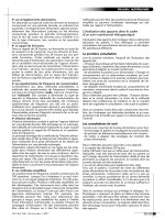
Phase 3 EAD
• These occur during fast repolarization and share the mechanism of DAD (spon-
taneous Ca release from SR and activation of the I
Na/Ca
). They are called EAD
because they occur before the completion of AP repolarization (Fig. 3.1).
• EAD may occur during the plateau phase and are caused by L-type Ca current.
EAD that occur during phase 3 of APD are due to the Na/Ca exchange current.
Torsades De Pointes (TDP)
• TDP is a polymorphic ventricular tachycardia (VT) associated with long QT
syndrome (LQTS).
• Quinidine and hypokalemia produce EAD and triggered activity resulting in TDP.
Initial event in TDP is EAD-induced triggered activity.
• TDP often follows short long short CL.
Excitability and conduction
• Excitability is dependent on the availability of sodium channels; thus reduced
Na channel activity will reduce excitability and conduction velocity.
• Reduced membrane excitability and reduced gap junction coupling slows
conduction, which may predispose to reentrant arrhythmias.
• Reduced gap junction coupling also slows conduction velocity, which may allow
I
CaL
to induce inward depolarizing currents.
• I
CaL
plays a dominant role in maintaining conduction in the setting of reduced
coupling. While I
Na
controls excitability, I
CaL
influences conduction during
reduced coupling.
38 Essential Cardiac Electrophysiology
Phase 2 EAD
Phase 3 EAD
DAD
C
I
R
C
A
i
n
S
R
R
e
a
c
t
i
v
a
t
i
o
n
o
f
I
C
a
L
Fig 3.1 Mechanisms of EAD and DAD.
Summary
• DADs occur during calcium overload due to Ca release from SR. This activates
I
Na/Ca
.
• EAD is generated by recovery and reactivation of I
CaL
.
• Phase 3 EAD shares the mechanism of DAD.
• Slow conduction could be due to decreased membrane excitability or reduced
gap junction coupling.
• I
Na
causes slow conduction, due to decreased excitability.
• Slow conduction due to decreased gap junction coupling requires contribution
of I
CaL
.
Reentry
• The size of reentry circuit depends on tissue excitability.
• It overdrive suppresses normal pacemaker cells.
• Decrease in conduction velocity, increase in the refractory period and/or
decrease in the circuit length will make reentry less likely.
• Decreasing wavelength will increase the tendency to cause reentry arrhythmias.
• Restitution is described as the recovery of excitability after the refractory period.
• Multiple reentries may result in fibrillation.
• The reentry wave dies when it reaches the border of the tissue.
• The spiral of reentry wave may drift or it could be fixed (pin) around the obstacle.
Mechanisms of Arrhythmias 39
• Reentry can be classic (anatomical) or functional.
• If a stimulus enters a vulnerable window of an anatomical reentry, it
terminates it.
• Functional reentry is not terminated by entry of the stimulus inside the
vulnerable window.
• Pacing induces a drift in the reentry circuit.
• Decrease in tissue excitability, by slowing conduction, eliminates reentry.
• When an electrical shock is applied to the heart, the tissue near the cathode
(negative electrode) is depolarized (positive charge on the membrane) and the
tissue near the anode (positive electrode) is hyperpolarized (negative charge on
the membrane). This terminates the arrhythmia.
• For initiation and maintenance of reentry, whether anatomic or functional,
unidirectional conduction block and the presence of excitable tissue ahead of
propagating wave front (excitable gap) are essential. Slowing of conduction or
shortening of the refractory period or both facilitate the excitable gap.
• Half of all cell-to-cell connections are side to side and the other half are end to
end. The gap junction membrane provides resistance to current flow that results
in slower conduction transversely than longitudinally. This results in anisotropic
conduction through the myocardium. Reduction in myocardial CX43 results in
slowing of conduction velocity.
• During myocardial ischemia slowing of the conduction occurs due to changes in
ion channel function and increased resistance at gap junctions. After 60 minutes
of ischemia irreversible damage occurs to the gap junction membrane and CX43.
This results in slowing and non-uniform conduction.
• Crista terminalis and pectinate muscle produce anisotropic conduction and act
as facilitators of reentry. Conduction along the longitudinal axis of the crista and
pectinate muscle is faster than along the horizontal axis.
• Crista and Eustachian ridge act as anatomic barrier (isthmus) during reentrant
activation.
• Discordant activation of atrial epicardium and endocardium at a faster rate
promotes reentry.
• Crista, pectinate muscles, and Backman bundle propagate sinus impulses rapidly.
• Ectopic beat may alter normal conduction and produce changes in refractoriness
which promotes reentry.
Phase 2 reentries
2
• Prominent outward current due to I
to
results in shortening of the APD.
• This may occur during ischemia and may result in a decrease of the plateau phase
of AP. These changes may occur nonuniformly throughout the myocardium and
cause dispersion of repolarization and phase 2 reentries.
• When I
to
, an outward current, is dominant it results in APD shortening and loss
of plateau of AP in some epicardial sites producing dispersion of repolarization.
This results in local reexcitation and premature beats. This mechanism is termed
as phase 2 reentry.
40 Essential Cardiac Electrophysiology
• Phase 2 reentry may occur in the presence of potassium channel opener
Pinacidil, sodium channel blockers Flecainide, increase in extracellular calcium,
and ischemia.
• I
to
blockers restore homogeneity and abolish reentrant activity.
• I
to
is present in ventricular epicardium but not in endocardium. It is responsible
for spike and dome morphology of AP in epicardium.
• Reduced activity of I
Ks
in M cells prolongs APD. Bradycardia and class III drugs
prolong APD in M cells and predispose to arrhythmias.
• Repolarization is sensitive to changes in the heart rate.
Pharmacological differences in epicardium and endocardium
• Ach may alter the epicardial repolarization pattern by blocking I
Ca
or activating
I
KAch
. It has no effect on I
to
.
• Isoproterenol causes epicardial AP abbreviation more than it does in the
endocardium.
• Organic calcium channel blockers (Verapamil) and inorganic calcium channel
blocker MnCl
2
can cause loss of AP plateau phase in epicardium but only a slight
abbreviation of AP in endocardium.
• Sodium current block decreases APD in epicardium.
• Block or decrease in calcium current leaves outward currents unopposed, which
may result in shortening of APD.
• I
to
block may establish electrical homogeneity and abolish arrhythmias due
to dispersion of repolarization caused by drugs and ischemia. Quinidine
inhibits I
to
.
• Amiloride, a potassium sparing diuretic, prolongs APD and refractoriness.
• M cells, found in mid-myocardium of anterior, lateral wall, and outflow tract
exhibit marked AP prolongation in response to bradycardia and on exposure to
class III agents.
• This may be due to weaker I
Ks
activity and stronger late I
Na
activity.
• At slower rates M cells may contribute to pump efficiency because prolonged
depolarization permits longer and more efficient contraction.
• Epicardium and endocardium electrically stabilize and abbreviate APD of
M cells.
• Loss of either layer by infarction will lead to prolongation of APD. This may be
the mechanism by which QT prolongation and dispersion occurs in non-Q wave
MI. These differences could be aggravated by drugs that prolong the QT interval
or in patients with LQTS.
• M cells play an important role in the inscription of T waves by producing a
gradient between epicardium, endocardium, and M cells.
• U waves are due to repolarization of His Purkinje cells.
Post myocardial infarction arrhythmias
• Post MI hypertrophy of non-infracted myocytes occurs by three weeks due to
volume overload.