Essential Cardiac Electrophysiology Self Assessment - Part 3 doc
Bạn đang xem bản rút gọn của tài liệu. Xem và tải ngay bản đầy đủ của tài liệu tại đây (1.23 MB, 31 trang )
Sinus Node Dysfunction and AV Blocks 47
• Successful radiofrequency (RF) ablation of IST or sinus node modification
remains difficult. Although short-term success rates may be favorable (range
76–100%), long-term outcomes are disappointing.
• The endpoint of successful sinus node ablation remains unclear. Heart
rate below 80–90 bpm, with or without isoproterenol infusion (usually
1–2 μg/min), at the conclusion of the procedure is considered a reasonable end
point.
• Most of the cardiac and extra-cardiac symptoms persist despite documented
slower heart rates, suggesting that sinus tachycardia and symptoms of
palpitations are likely secondary manifestations of autonomic dysregulation.
• In the absence of atrial or other supraventricular tachycardia and autonomic and
other multisystem symptoms RF ablation can be considered.
• Three-dimensional mapping or intracardiac echocardiography in localizing the
crista may improve the outcome of the ablation.
• Surgical or RF ablation of the SAN and insertion of a permanent pacemaker may
be considered. Fatigue, awareness of the paced rhythm and other symptoms may
persist in spite of rate reduction.
• To avoid diaphragmatic paralysis high output pacing should be performed
with an ablation catheter along the crista terminalis before delivering RF
current.
• The clinical features of IST significantly overlap with postural orthostatic
tachycardia syndrome (POTS).
• A multidisciplinary approach involving neurologist, cardiovascular rehabilita-
tion, and psychiatrist may be necessary in managing patients with IST.
Sinus node dysfunction (SND)
Causes of sinus node dysfunction
Intrinsic (primary) SND may result from fibrosis and ageing-related loss of
pacemaker cells.
Extrinsic (secondary) causes of SND are listed in Box 4.1.
Pathophysiology
• The sinus node is located near the superior anterolateral portion of RA near the
SVC junction and the superior end of the crista terminalis.
• Cells in the SAN demonstrate diastolic depolarization and its AP is calcium
channel dependent. Phase 0 demonstrates slow upstroke velocity.
• SAN cells do not have connexion 43 (×43) gap junctions.
• Impulses may originate along the crista. With sympathetic stimulation the source
of impulse formation shifts more superiorly and with vagal stimulation it shifts
more inferiorly.
• The primary pacemaker area is located in the center of the node. Sympathetic
and parasympathetic nerves innervate it.
• The AP of the pacemaker cells is characterized by phase 4 depolarization,
relatively positive MDP and slow upstroke velocity.
48 Essential Cardiac Electrophysiology
Box 4.1 Extrinsic causes of SND
Hypothyroidism
SAN or atrial ischemia
Post-atrial surgery
Medications: Antiarrhythmics: Class I agents, Amiodarone
Beta blockers: Ca channel blockers
H2 receptor blockers: Ranitidine, Cimetidine
Psychotropic drugs: Lithium, Tricyclic antidepressants, Phenothiazines
Neurologic diseases: Myotonic dystrophy, Emery–Dreifuss syndrome, Tuberous sclerosis
Infiltrative disorders: Amyloidosis, Hemochromatosis, Systemic Lupus, Sarcoidosis,
Lymphoma
Myocarditis
Familial
Carotid sinus hypersensitivity
Increased vagal tone
Jaundice
Hypothermia
Elevated intracranial pressure
• Phase 4 depolarization is the result of four different ionic currents:
i I
K
delayed rectifier.
ii Increase I
f
inward current.
iii Inward calcium current.
iv Background current.
• Sympathetic stimulation enhances phase 4 depolarization by increasing I
f
and
I
Ca
. Parasympathetic stimulation decreases phase 4 depolarization.
• Two potassium currents I
to
and I
K
and Na/Ca exchanger are responsible for
sinus node repolarization.
• Drugs causing negative chronotropic response aggravate SND.
• Sensitivity to parasympathetic transmitters increases with age.
• Increase in APD of atrial myocardium may cause bradycardia. This may be the
mechanism of bradycardia in LQTS.
• Bradycardia-related dispersion of refractoriness might cause tachyarrhythmia.
Clinical manifestations
• The SAN intrinsic rate decreases as the number of cells declines with
increasing age.
• Long pauses may occur due to exit block from SAN after premature atrial con-
tractions (PAC) or termination of the tachycardia. This may result in syncope or
near syncope. Bradycardia may cause fatigue and/or dyspnea.
• Atrial asystole may predispose to thromboembolic complications.
• SND may present as abnormality of impulse formation such as bradycardia or
sinus arrest or as abnormality of impulse conduction such as exit block or loss
of physiologic responsiveness such as chronotropic incompetence.
Sinus Node Dysfunction and AV Blocks 49
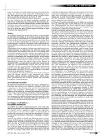
continuous tracing, lead I.
Fig 4.1 Sinus pause (sino atrial exit block) following rapid paroxysmal AF, followed by extreme
sinus bradycardia. Typical of Brady Tachy syndrome.
• SND could be intrinsic due to structural disease of the sinus node or it could be
due to extrinsic influences.
• Bradycardia, SA exit block, sinus arrest, chronotropic incompetence, atrial
fibrillation (AF), or arrhythmias are the common manifestations of SND.
• SND may appear intermittently.
• Extrinsic factors causing SND include carotid sinus syndrome, vasovagal
syncope, and increased vagal tone.
• Bradycardia may cause fatigue and dyspnea. Palpitation and embolism may
occur due to AF.
• Carotid sinus pressure or tilt test may uncover the abnormalities.
• Carotid sinus hypersensitivity, sternocleidomastoid denervation syndrome and
neurocardiogenic syncope may coexist with SND.
Electrocardiographic characteristics of SND
• Persistent sinus bradycardia, in the absence of drugs, is common.
• A sinus pause of 3 seconds or more may occur (Fig. 4.1). The duration of the
pause is not a multiple of the basic heart rate. Following the pause the heart rate
may accelerate (not seen in exit block). Non-conducted PAC may mimic sinus
pauses. (Fig. 4.1).
• SA exit block is a common manifestation of SND. In type 1 SA exit block there is
progressive shortening of the PP interval preceding the pause. The pause is less
than the sum of two preceding sinus cycle lengths (CLs).
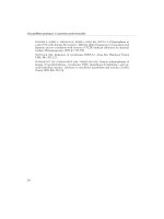
• In type 2 SA block the pause is equal to a multiple of sinus CL. (Fig. 4.2).
• In SND there may be concomitant suppression of subsidiary pacemaker.
Diagnosis
• Exercise test may uncover chronotropic incompetence.
• Administration of adenosine bolus, if results in slowing of sinus CL, by more
than 2 standard deviations, is indicative of SND.
50 Essential Cardiac Electrophysiology
N N M N N N
Fig 4.2 SA exit block.
• Sinus node recovery time (SNRT) is an interval from the last paced beat to the
first sinus impulse. It is considered abnormal if it exceeds 1400 milliseconds.
• Corrected SNRT is derived by subtracting base line sinus CL from SNRT. A CSNRT
of greater than 530 milliseconds is considered abnormal.
• CSNRT, if divided by 2, yields sino atrial conduction time (SACT). Normal SACT
ranges from 70 to 120 milliseconds.
• Atropine by decreasing the vagal tone and enhancing retrograde conduction into
SN may worsen SNRT.
• Low intrinsic heart rate after autonomic blockade is suggestive of SND.
• After complete autonomic blockade the intrinsic heart rate can be calculated by
118.1 −[0.57 × age].
• During electrophysiologic study a normal corrected SNRT does not exclude the
possibility of SND.
Prognosis and treatment
• Prognosis is good; however, occurrence of a stroke in the presence of AF,
congestive heart failure (CHF) and AV block may alter the outcome.
• Atrial based pacing for symptomatic bradycardia lowers the incidence of AF and
thromboembolic events and CHF.
• Atrial based pacing should be considered for symptomatic patients. The incidence
of AV block is 1% per year.
• Drugs responsible for bradycardia should be discontinued.
• DDD pacing should be considered for patients with His Purkinje disease and
neurocardiogenic syncope.
• Pacing may allow the use of antiarrhythmic drugs.
• Anticoagulation should be considered in the presence of atrial flutter/fibrillation.
• Anticholinergics, sympathomimetic, or methylated xanthines can be used for
patients with mildly symptomatic bradycardia.
• Discontinuation of the offending agents, treatment of hypothyroidism, use of
vagolytic agents or theophylline may be helpful in the short term.
• VVI pacemaker implant is the treatment of choice for patients who present with
a pause related syncope but otherwise have normal atrio-venticular node (AVN)
conduction.
AVN anatomy and electrophysiology
1
• The AVN is located in the triangle of Koch, bound by the tendon of Todaro,
orifice of the coronary sinus (CS) and the septal leaflet of the tricuspid valve.
Sinus Node Dysfunction and AV Blocks 51
• There are anterior and posterior inputs to the AVN from the atrium.
• There are two types of cells in the AVN: rod-and ovoid-shaped cells.
• Spontaneous activity correlates with I
f
current, which is far greater in the ovoid
cells.
• I
Na
and I
to
are present in the rod cells.
• AVN conduction delay is inversely related to the prematurity of the impulse that
it receives from the atrium.
• The occurrence of longer S2H2 interval with shorter S1S2 is due to the slow
recovery of excitability of N cells.
• Slow AVN pathway (posterior input) is located posteriorly and inferiorly
between the orifice of the CS and the septal leaflet of the tricuspid valve.
• Fast pathway is located anteriorly and superiorly in the interatrial septum. It
has a shorter distance to travel to the AVN but demonstrates longer effective
refractory period (ERP).
• A sudden change in the AH interval with a minimal change in the input inter-
val may be due to shift of conduction from anterior (fast) to posterior (slow)
pathway.
• After the successful elimination of the AVNRT, discontinuous conduction may
still be present.
• In AF slow pathway elimination may not alter the heart rate if impulses can
reach the AVN via another route.
• Subthreshold stimuli delivered in the triangle of Koch cause postganglionic
release of Ach, resulting in hyperpolarization of N cells and slowing AVN
conduction.
AV block AV dissociation
• AV dissociation can be due to AV block or due to physiologic refractoriness,
resulting in the failure of transmission of the atrial impulse to the ventricle.
• AV block is due to failure of conduction of the atrial impulse to ventricle in the
absence of physiologic refractoriness. It is generally due to interruption of the
normal conduction pathway or due to pathologic refractoriness.
• AV block can be proximal (above the His bundle) indicating block in the AVN
or it can be intra-Hisian or it can be distal to His bundle (infra-Hisian).
• The prognosis depends on the site of the AV block. Block distal to His bundle
implies poor prognosis.
Prolonged PR interval (first-degree AV block)
It is defined as PR interval of more than 200 milliseconds.
• This may represent conduction delay in the atrium, AVN or His Purkinje system
(HPS).
• All P waves are conducted to the ventricle with prolonged but constant interval.
The causes of variable PR interval are enumerated in Box 4.2.
• If the QRS duration is normal the delay is invariably in the AVN. Ninety percent
of these cases will demonstrate prolonged AH interval.
52 Essential Cardiac Electrophysiology
Box 4.2 Causes of variable PR interval
Intermittent conduction over slow pathway
Intermittent conduction over accessory pathway
Type I (Wenckebach) AV block
Concealed conduction from premature beats into AV junction
Intermittent junction rhythm and AV dissociation
High adrenergic tone
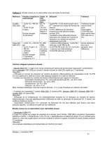
PR 170
RR 960 880
1740
860 1640
280 300 320
Fig 4.3 Type I AV block (intervals are in msecs).
• If the QRS duration is prolonged then the delay could be in the AVN (60%) or
in HPS.
• Very long PR intervals favor delay in the AVN.
• Prognosis of the patients with prolonged PR interval is good and no therapy is
indicated.
• In patients with prolonged PR interval and bifascicular block, the rate of progres-
sion to CHB is low and in asymptomatic patients pacing is not indicated even if
the patient requires general anesthesia.
• If the HV interval exceeds 100 milliseconds, prophylactic pacing is indicated.
Second-degree AV blocks
These are of two types.
• Type I AV block (Mobitz I or Wenckebach) is characterized by the following
(see Fig. 4.3):
i Progressive prolongation of the PR interval at decreasing increments.
ii Progressive shortening of the RR interval.
iii A pause encompassing the blocked P wave. The duration of the pause is less
than the sum of two PP intervals.
• Typical type I AV block is seen in 50% of cases, others are atypical and char-
acterized by a varying sequence of PR and RR intervals. For example, PR and
RR interval that terminates the cycle may be longest and PR interval may be
constant or decrease.
• Long Wenckebach cycles tend to be atypical.
• Concealed conduction may be the mechanism for prolongation of PR RR in
atypical sequence.
• Type I AV block, in asymptomatic subjects with normal heart, has excellent
prognosis and requires no treatment.
Sinus Node Dysfunction and AV Blocks 53
I
aVR
V1 V4
II
III
V1
II
V5
aVF
V2
V3
V5
V6
aVL
No Δ in PP Interval
RBBB
No Δ in PR
Fig 4.4 Type II AV block.
• Type I AV block with normal QRS complex is likely to be AV nodal; however,
if the QRS duration is prolonged the block may be in the AVN, His bundle or
infra-Hisian.
• Symptomatic patients with syncope, near syncope, worsening of CHF, or angina
due to bradycardia produced by type I AV block may require pacing.
• Type II AV block is characterized by the following:
i Constant PP and RR intervals.
ii Constant PR interval before the blocked P wave.
iii The pause encompassing the P wave is twice as long as the preceding PP
interval (Fig. 4.4).
• It often accompanies bundle branch block.
• Site of block is invariably in the His or infra-Hisian (Fig. 4.5).
• Second-degree AV block with narrow QRS complex is likely to be type I AV
block with minimal increments in the PR interval and may be mistaken for
type II block.
• 2 : 1 AV block with very long PR interval and narrow QRS suggests AV nodal
block.
• Constant PR interval of all captured complexes, in spite of varying RP interval,
suggests type II AV block. If the PR interval varies inversely with RP interval it
is likely to be due to type I AV block.
• Functional infra-Hisian block may occur in the presence of long short HH CLs
preceding the block. Rapid atrial pacing will not reproduce this type of block.
Pacing is not indicated for functional infra-Hisian AV block.
• Type II AV block often progresses to complete AV block and requires pacing even
in an asymptomatic patient.
• Type II AV block accompanied by alternating BBB would require a permanent
pacemaker.
54 Essential Cardiac Electrophysiology
I
II
VI
A
A
H
A
A
V
H
A
His
RV
H
IV
HRA
100
msec
A
Fig 4.5 Infra-Hisian type II AV block.
Fig 4.6 Complete AV block.
Third-degree AV block or complete AV block
• It could be congenital or acquired.
• The Site of the block could be AVN or below the His.
• It is characterized by the failure of all the P waves to conduct to the
ventricle.
• Escape rhythm can be junctional with a rate of 40–60 bpm and narrow
QRS complex or 20–40 bpm with wide QRS if it arises from the ventricle
(Fig. 4.6).
• Drug-induced AV block may persist after discontinuing the offending agent.
5
• Pacing is recommended for congenital AV block if
i The patient is symptomatic.
ii The QRS is wide.
iii The rate is less than 50 bpm.
iv The block is infra-Hisian.
Other causes of the AV block are listed in Box 4.3.
Paroxysmal AV block
8
• It presents as an abrupt and persistent AV block in the presence of normal AV
conduction.
• It is produced by blocked or conducted PAC or PVC.
• It occurs below the His.
Mechanisms include the following:
i Concealed conduction of non-conducted P waves into the AV junction.
ii Deceleration dependent depolarization of the lower AV junction.
Sinus Node Dysfunction and AV Blocks 55
Box 4.3 Causes of complete AV block
Drugs
5
Beta blockers, Ca Ch blockers, Quinidine, Procainamide Amiodarone
Degenerative diseases Lenegre disease, Lev disease, Sclerosis of conduction system
CAD MI, Ischemia
Infection Rheumatic fever, Myocarditis, Lyme disease, Chagas disease
Connective tissue Ankylosing spondylitis, Reiter disease, Polychondritis, Scleroderma,
diseases
6,7
Rheumatoid arthritis
Infiltrative disorders Amyloidosis, Sarcoidosis, Tumors, Hodgkin disease, Myeloma
Neurologic disorders Becker Muscular dystrophy, Myotonic dystrophy
Congenital Fibroelastosis, Transposition of vessels, Septal defects, Collagen
diseases in mother
Metabolic Hypoxia, Electrolyte disorders
Traumatic Surgical trauma, Cardiac contusion, Alcohol/surgical septal ablation
Resumption of normal AV conduction following paroxysmal block has been
attributed to the following:
i Wedensky facilitation, where properly timed retrograde impulse allows sub-
threshold antegrade impulse to conduct.
ii Peeling of refractory period (shortening of the refractory period with increasing
frequency of stimulation).
• These patients require permanent pacing.
AV block in patients with acute myocardial infarction (MI)
4,9
• It is common with inferior MI.
• It is probably due to increased vagal tone that accompanies early after an acute
inferior MI. It presents as prolonged PR or type I AV block or advanced AV block.
• It responds to atropine.
• AV block occurring late after an acute MI is secondary to ischemia of the AVN,
resulting in increased levels of adenosine in the AVN area. These effects can be
blocked by theophylline.
Characteristics of AV block in the setting of inferior and anterior MI differ and
are listed in Table 4.1.
AV dissociation
• During AV dissociation atrial and ventricular activity is independent.
• The mechanisms of AV dissociation could be physiologic or pathologic:
i Physiologic refractoriness with interference. (Impulse may conduct if occurs
during non-refractory window of CL).
• The Rate of the primary pacemaker (sinus) is slower than the subsidi-
ary (junctional) pacemaker, resulting in non-conduction of some of the
impulses due to physiologic refractoriness.
• Inappropriate acceleration of the subsidiary pacemaker. Accelerated
junctional rhythm or ventricular tachycardia.
56 Essential Cardiac Electrophysiology
Table 4.1 Characteristics of AV block in the setting of acute MI
Inferior MI Anterior MI
Characteristic Preceded by type I AV block Preceded by type II AV block
Onset Occurs in the first 2–3 days Occurs in the first week
Duration May last for 3–14 days Could be permanent
Site Nodal Infra nodal
Pathology AV nodal ischemia Necrosis of conduction tissue
QRS duration Narrow Wide. Bundle branch block
Treatment Temporary pacing Permanent pacing
Mortality (%) 10–20 60–80
• Due to physiologic refractoriness and physiologic AV conduction delay
when the primary pacemaker accelerates (Sinus or atrial tachycardia).
ii Pathologic failure of conduction of sinus impulses resulting in more P waves
than QRS complexes as in complete AV block. (Impulse does not conduct
even if it occurs during non-refractory period of the CL.)
References
1 James TN. Structure and function of the sinus node, AV node and His bundle of the
human heart: Part I-structure. Prog Cardiovasc Dis. 45:235–6, 2002.
2 Shen WK. Modification and ablation for inappropriate sinus tachycardia: Current status.
Card Electrophysiol Rev. 6:349–55, 2002.
3 Shen WK. How to manage patients with inappropriate sinus tachycardia. Heart Rhythm.
2:1015–19, 2005.
4 Brady WJ. Diagnosis and management of bradycardia and atrioventricular block associ-
ated with acute coronary ischemia. Emerg Med Clin North Am. 19:371–84, 2001.
5 Zeltser D. Justo D. Halkin A. Drug-induced atrioventricular block: prognosis after
discontinuation of the culprit drug. J Am Coll Cardiol. 44:105–8, 2004.
6 Clancy RM. Buyon JP. Autoimmune-associated congenital heart block: dissecting the
cascade from immunologic insult to relentless fibrosis. Anat Rec A Discov Mol Cell Evol Biol.
280:1027–35, 2004.
7 Qu Y. Xiao GQ. Chen L. Autoantibodies from mothers of children with congenital heart
block down regulate cardiac L-type Ca channels. J Mol Cell Cardiol. 33:1153–63, 2001.
8 Silvetti MS. Grutter G. Di Ciommo V. Paroxysmal atrioventricular block in young patients.
Pediatr Cardiol. 25:506–12, 2004.
9 Abidov A. Kaluski E. Hod H. Leor J. Vered Z. Gottlieb S. Behar S. Cotter G. Israel Working
Group on Intensive Cardiac Care: Influence of conduction disturbances on clinical out-
come in patients with acute myocardial infarction receiving thrombolysis (results from
the ARGAMI-2 study). Am J Cardiol. 93:76–80, 2004.
5 Supraventricular Tachycardia
Self-Assessment Questions
5.1 ATRIAL FLUTTER
1 A 54-year-old male presents with progressively increasing dyspnea. ECG is
shown below. Serum potassium was 3.2 mEq. Perfusion studies are normal.
Echocardiogram revealed biatrial enlargement, enlarged and diffusely hypokin-
etic left ventricle, and ejection fraction of 26%. Eight months ago on a routine
examination blood pressure of 120/70 and a heart rate of 70 b.p.m. were recor-
ded. Six months ago a spot check in a drug store revealed a blood pressure of
110/70 and a heart rate of 150 b.p.m. The patient was asymptomatic.
I aVR V1 V4
II
II
V5
aVL V2 V5
III
VI
aVF
V3 V6
What will you recommend?
A IV lidocaine
B Bi V defibrillator implant
C Radiofrequency ablation
D Ca channel blockers
57
58 Essential Cardiac Electrophysiology
2 A 36-year-old sales representative was found to have an atrial flutter. Physical
examination and echocardiogram were normal. Serum potassium was 3.9 mEq.
The next day he reported to hospital outpatient area for chemical cardioversion.
He received 1 mg of Ibutilide intravenously and converted to sinus rhythm
within 10 minutes. Two hours later he insisted on leaving the hospital. While
in a meeting he had an episode of syncope. He was in sinus rhythm and blood
pressure was normal.
What is the most likely cause of his syncope?
A Recurrence of atrial flutter with rapid ventricular response
B Neurocardiogenic syncope
C Polymorphic ventricular tachycardia
D Embolic stroke from left atrial clot
3 A 45-year-old male who has persistent atrial flutter, undergoes radiofrequency
ablation. During the second application of the energy flutter is terminated. What
will you consider as a satisfactory end point for this procedure?
A Termination of atrial flutter is a satisfactory end point
B Bidirectional block across isthmus should be demonstrated
C Rapid atrial pacing should be performed in an attempt to induce flutter
D Additional RF lesions should be delivered near CS ostium
4 A 77-year-old female had paroxysmal atrial fibrillation. Echocardiogram and
perfusion studies were normal. She was treated with propafenone 150 mg TID.
Five weeks later she came to the clinic complaining of palpitations. ECG revealed
persistent atrial flutter.
What will be your recommendation?
A Continue propafenone and consider ablation for atrial flutter
B Discontinue propafenone
C Consider ablation for atrial fibrillation
D Consider rate control using Ca channel blockers and β blockers
5.2 ATRIAL TACHYCARDIAS
1 A 56-year-old nurse presents with recurrent episodes of tachycardia. ECG is
shown.
V1
II
Supraventricular Tachycardia 59
What is the most likely diagnosis?
A Sinus tachycardia
B Atrial tachycardia
C AVRT
D Atypical AVNRT
2 Ablation at which of the following sites is likely to cure the tachycardia?
A Accessory pathway
B AV nodal slow pathway
C Atrial tachycardia focus
D AV junction
3 A 25-year-old man is referred to you because of sustained tachycardia. During
electrophysiologic study, following tracing was obtained.
Which of the following is the most likely diagnosis?
A Atrial tachycardia
B Atypical AV reentrant tachycardia using a slowly conducting pathway as the
retrograde limb
C Atypical (fast-slow) AV nodal reentrant tachycardia
D Sinus node reentrant tachycardia
5.3 ATRIAL FIBRILLATION
1 A 35-years-old female with mitral valve disease presents with history of sev-
eral episodes of atrial fibrillation that lasted for 2–4 hours and spontaneously
terminates. This could be classified as:
A Persistent AF
B Chronic AF
C Paroxysmal AF
B Lone AF
2 Most common symptom of the AF is:
A TIA/Stroke
B Fatigue and tiredness
C Syncope
D Chest pain
60 Essential Cardiac Electrophysiology
3 Which of the following is not a risk factor for thromboembolic complication
in a patient with AF and therefore not an indication for anticoagulation with
warfarin?
A The patient is 57 year old
B Hypertension
C LVF
D Diabetes
4 A 48-year-old patient presents with AF that has lasted for more than 48 hours.
TEE is negative for intracardiac clots. What will be your recommendation
following a successful cardioversion?
A Begin Warfarin and continue indefinitely
B Begin Aspirin
C Begin Warfarin for 3 weeks
D Patient does not need anticoagulation or antiplatelate therapy
5 Attempted cardioversion for AF is unsuccessful in a 60-year-old patient. His
EF is 25%. Should he undergo repeat cardioversion after administration of IV
ibutilide?
A True
B False
6 A 50-year-old female patient presents with paroxysmal AF. She was recently
diagnosed to have chronic active hepatitis and abnormal liver function test.
To maintain sinus rhythm which of the following drugs could be safely
prescribed?
A Sotalol
B Mexiletine
C Amiodarone
D Procainamide
7 A 76-year-old woman has had persistent atrial fibrillation for three years. She
also has hypertension and asthma. Current medications are warfarin; digoxin,
0.25 mg daily; atenolol, 25 mg twice daily; diltiazem, 30 mg every 8 hours;
and inhaled bronchodilators. During 24-hour ambulatory ECG monitoring, the
average ventricular response was 130 beats per minute (b.p.m.) with occasional
episodes of 150 b.p.m. Echocardiogram showsa6cmleft atrium and a dilated
left ventricle. Estimated left ventricular ejection fraction is 35%.
Which of the following is the best treatment plan?
A Increase the dose of atenolol
B Increase the dose of diltiazem
C Initiate amiodarone and consider elective cardioversion 4 weeks later
D Radiofrequency catheter ablation of the AV junction, and insertion of rate-
responsive ventricular pacemaker
Supraventricular Tachycardia 61
5.4 AUTOMATIC JUNCTIONAL TACHYCARDIA
1 To differentiate AJT from AVNRT which of the following maneuvers could be
helpful?
A PAC delivered near slow pathway when septal A has occurred
B Adenosine infusion
C Parahisian pacing
D Assessment of prematurity index
2 Which of the following therapeutic options is least likely to help in the treatment
of AJT?
A AV node ablation and insertion of permanent pacemaker
B Amiodarone
C Digoxin
D Propafenone
5.5 AV NODE REENTRY TACHYCARDIAS
1 Dual AV node physiology is defined as:
A 50 milliseconds increase in H
1
H
2
for 10 milliseconds decrease in A
1
A
2
interval
B 50 milliseconds increase in A
2
H
2
for 10 milliseconds decrease in A
1
A
2
interval
C 50 milliseconds increase in H
2
V
2
for 10 milliseconds decrease in A
1
A
2
interval
D Occurrence of atrial echo beats in response to any A
1
A
2
2 Which of the following properties is the result of distal insertion of fast pathway
into AV junction?
A Increased decremental conduction
B Increased response to AV node blocking agents
C Greater response to Na channel blocking agents
D Preexcitation on surface electrocardiogram
3 After slow pathway ablation, the presence of A
2
H
2
jump without induction of
tachycardia is indicative of unsuccessful ablation.
A True
B False
4 In which of the following conditions is the HA interval during tachycardia
likely to be shorter than the HA interval during RV pacing?
A AVNRT
B AVRT
C RVOT VT
D Atrial tachycardia
62 Essential Cardiac Electrophysiology
5 Which of the following conditions are likely to present with variable HA
interval? (Choose more than one).
A AVNRT
B AVRT with dual AV node physiology
C Atrial tachycardia
D Multiple concealed accessory pathways
6 A 25-year-old female had recurrent episodes of palpitation. During electro-
physiologic study wide complex tachycardia was induced. Tachycardia could
be entrained by right atrial pacing; however, on repeated occasions, on ter-
mination of atrial pacing tachycardia resumed as shown in the following
tracing.
What is the most likely mechanism of this tachycardia?
A AVNRT
B AVRT
C Atrial tachycardia
D LVVT (fascicular tachycardia)
7 A 50-year-old female had recurrent episodes of tachycardia. During electro-
physiologic study the following observations were made.
Supraventricular Tachycardia 63
Ablation at which of the following sites is mostly likely to cure the
tachycardia?
A Subeustachian isthmus
B Accessory pathway
C AV nodal slow pathway
D Atrial tachycardia focus
8 A 26-year-old woman, who is in the last trimester of pregnancy, complains of
palpitations. Episodes last several minutes and terminate spontaneously; they
are not associated with dizziness, chest pain, or syncope. One such episode
was recorded while in primary care physician’s office.
Tachycardia terminates spontaneously. Baseline ECG and echocardiogram are
normal.
Which of the following is most appropriate at this time?
A Deliver the fetus as soon as possible
B Begin flecainide
C Begin metoprolol
D Reassure the patient
9 A 67-year-old man has had palpitations for the last 10 years. Recently the
episodes have become more frequent and severe. Medical history includes
hypertension, diabetes, and hypercholesterolemia. ECG recorded in the office
was found to be normal.
64 Essential Cardiac Electrophysiology
Two days ago he came to hospital complaining of dyspnea, chest pressure,
and palpitations. He was diaphoretic; the heart rate was 140 per minute, the
blood pressure was 100/60 mm Hg. Electrocardiogram is shown.
I
aVR
aVL
aVF
V1
V2
V3
V4
V5
V6
II
III
Today during electrophysiologic study anterograde dual AV nodal pathway is
demonstrated; however, sustained supraventricular tachycardia (SVT) cannot
be induced.
Which one of the following should be done next?
A Repeat electrophysiologic study in 4 weeks
B Prescribe Metoprolol
C Ablate the AV nodal slow pathway
D Prescribe amiodarone
10 A 32-year-old woman undergoes electrophysiologic study for recurrent pal-
pitations. The following recording is obtained.
Supraventricular Tachycardia 65
Which of the following is the most likely diagnosis?
A AV reentrant tachycardia using an accessory pathway
B Atypical (fast-slow) AV nodal reentrant tachycardia
C Intraatrial reentrant tachycardia
D Typical (slow-fast) AV nodal reentrant tachycardia
5.6 AV REENTRANT TACHYCARDIAS
1 A 27-year-old female patient presents with AVRT utilizing left lateral AP. There
is a variation in the cycle length although the VA interval remains unchanged.
The most likely explanation of this observation is:
A Slowing of conduction in AP
B Change in conduction through the AV node due to dual AV node physiology
C Multiple accessory pathways
D Accelerated conduction through His Purkinje system
2 Which one of the following is least likely to present with short RP
tachycardia?
A AVNRT
B Ventricular tachycardia
C AVRT
D Sinoatrial reentrant tachycardia
3 On termination of the SVT the last complex recorded is a QRS. Which one of
the following is the most likely mechanism?
A AVNRT
B AVRT
C Atrial tachycardia
D None of the above
4 His synchronous PVC terminates the SVT without depolarizing the atrium. In
which of the following conditions is this phenomenon likely to be noticed?
A Atrial tachycardia
B AVNRT
C Automatic junctional tachycardia
D AVRT
5 In which of the following conditions is the VA interval likely to be shorter
during RV apex pacing than RV base pacing?
A AV node
B Septal AP
C Left posterior AP
D Atrioventricular connection (Mahaim fibers)
66 Essential Cardiac Electrophysiology
6 Which one of the following is likely to present with a constant VA interval
with or without His capture during parahisian pacing?
A Dual AV nodal physiology
B Septal AP
C Nodo-fascicular pathway
D Atriofascicular pathway
7 Which one of the following is unlikely to cause variable VA interval during
AVRT?
A Intermittent bundle branch block
B Multiple retrogradely conducting accessory pathways
C Decremental retrograde conduction through the AP
D Dual AVN physiology
8 SVT is entrained by RV pacing. On termination of V pacing the response is
AAV. This observation suggests the diagnosis of:
A Atrial tachycardia
B AVNRT
C AVRT
D Atrio fascicular tachycardia
9 SVT is entrained by RV pacing. On termination of V pacing the stimulus to A-VA
interval is >85 msec and PPI-TCL interval is >115 msec. These observations
suggests the diagnosis of:
A Atrial tachycardia
B Atypical AVNRT
C Atriofascicular pathway
D AVRT
10 Which of the following observations is least likely to suggest the presence of
antidromic tachycardia as opposed to antegrade bystander utilization of AP
during atrial tachycardia?
A Earliest retrograde atrial activation is through AV junction
B Late PAC advances the whole tachycardia circuit (V and following A)
C PVC delivered at the ventricular insertion site of the AP abolishes the
preexcitation but tachycardia continues
D Ventricles are the integral part of the tachycardia circuit
11 During a wide complex tachycardia it is noted that the atrium and ventricle
are an obligatory part of the circuit and retrograde atrial activation is eccentric.
These observations suggest presence of:
A Atrial tachycardia utilizing bystander AP
B VT utilizing bystander AP
C Antidromic tachycardia
D Bypass to bypass tachycardia
Supraventricular Tachycardia 67
12 PAC delivered during wide complex tachycardia advances the ventricular elec-
trogram and the next atrial electrogram without any change in the atrial
activation sequence. These findings are suggestive of:
A AVNRT utilizing bystander AP
B Atrial tachycardia utilizing bystander AP
C Antidromic tachycardia
D Ventricular tachycardia
13 During wide complex tachycardia, a short HV interval is recorded. This finding
suggests the presence of:
A Ventricular preexcitation due to bystander AP
B Antidromic tachycardia
C Atrio fascicular pathway mediated tachycardia
D BBRVT
14 A 30-year-old male, who is known to have preexcitation, presents to ER with
atrial fibrillation of 1 hour duration. The ventricular rate is 130–170 b.p.m.
The patient is hemodynamically stable. Which of the following can be safely
used to treat AF?
A IV Digoxin
B IV Verapamil
C IV procainamide
D IV metoprolol
15 A 19-year-old man who has paroxysmal supraventricular is referred to you for
evaluation and recommendation. The patient has had tachycardia since age 10.
Electrocardiogram obtained during sinus rhythm is shown. Echocardiogram is
normal.
At which site, will delivery of radiofrequency result in resolution of the
problem?
I
aVR
V1
V2
V3
V4
V5
V6
aVL
aVF
II
III
A Right anterior
B Posteroseptal
C Left lateral
D Anteroseptal
68 Essential Cardiac Electrophysiology
16 Surface and intracardiac electrograms recorded during tachycardia are shown
in the following tracing.
Which maneuver will help identify the correct mechanism of the
tachycardia?
A Atrial pacing
B Ventricular pacing
C Isoproterenol infusion
D Mapping in the left superior pulmonary vein
17 A 20-year-old male presented with recurrent episodes of palpitations. During
electrophysiologic study following arrhythmia was induced. Ablation at which
one of the following sites is likely to terminate the tachycardia?
FCS = closely spaced (2 mm) electrograms recorded from CS using femoral
approach. DCS, MCS and PCS are recorded from subclavian approach.
(electrode spacing 10-2 mm)
A Left lateral mitral annulus
B LVOT
C Right lateral tricuspid annulus
D Right bundle
Supraventricular Tachycardia 69
18 A 50-year-old woman has a long history of paroxysmal tachycardia despite
treatment with digoxin, atenolol, and flecainide. She underwent electro-
physiologic study and radiofrequency ablation (tracing A). Two months later
she had a recurrence of the tachycardia. Electrophysiologic study was repeated
(tracing B).
Tracing A
Tracing B
Which of the following best describes the mechanism(s) of the tachycardias?
A The patient had atypical AVNRT and now has typical AV nodal reentrant
tachycardia
B The patient had AVRT utilizing the anteroseptal accessory pathway and now
has atrial tachycardia
C The patient had AVRT utilizing the LLAP and now has typical AVNRT
D The patient had permanent junctional reciprocating tachycardia and now
has atypical AV nodal reentrant tachycardia
19 A 15-year-old female fainted in a physician’s office while the blood
was being drawn. There is no previous history of syncope, chest pain,
or palpitations. ECG rhythm strip recorded in the physician’s office is
70 Essential Cardiac Electrophysiology
shown below.
What will be your recommendation?
A Consider electrophysiologic study and RF ablation
B Begin procainamide
C Begin Metoprolol
D Reassure the patient
20 A 44-year-old man is referred to you because he has had three episodes
of palpitations in the past year. Each episode began suddenly, lasted up to
30 minutes, and was associated with severe lightheadedness. The patient has
not had syncope, dyspnea, or chest pain, and he has no family history of heart
disease.
The findings of the physical examination are normal. Electrocardiograms
recorded a few minutes apart are shown in tracings A and B.
I aVR V1 V4
II aVL V2 V5
III aVF V3 V6
Tracing A
Supraventricular Tachycardia 71
I aVR V1 V4
II aVL V2 V5
III aVF V3 V6
Tracing B
What is the most likely diagnosis?
A AVNRT
B Atrial tachycardia
C AVRT
D RVOT-VT and atrial tachycardia