Essential Cardiac Electrophysiology Self Assessment - Part 4 pps
Bạn đang xem bản rút gọn của tài liệu. Xem và tải ngay bản đầy đủ của tài liệu tại đây (631.62 KB, 31 trang )
78 Essential Cardiac Electrophysiology
-
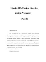
Atrial tachycardia (AT)
Focal AT
Automatic
Nonautomatic
Triggered activity
Mechanisms
Macroreentrant AT
Reentry
Isthmus dependent
Non-isthmus dependent
Microreentry
Fig 5.5 Classification of atrial tachycardia.
12
Table 5.1 Characteristics of the different mechanisms of focal AT
Automatic AT Triggered activity Microreentrant AT
PES No response CL dependent Reproducibly initiates
and terminates AT
Isoproterenol for induction Yes No No
Entrainment No No Yes
Warm up cool down Yes No No
Response to adenosine No
∗
Yes Yes
Response to propranolol,
verapamil
Propranolol Propranolol, verapamil Verapamil
Vagal maneuvers No response Terminates AT No response
PES, programmed electrical stimulation.
∗
Yes if AT is induced by Isoproterenol. Major drawback of
the above observations is the overlap of responses.
• Electrophysiologic mechanism of the atrial tachycardia can be reentry, abnormal
automaticity or triggered activity (Table 5.1).
• Focal tachycardias are characterized by centrifugal spread of electrical impulse
from a single focus.
• Activation covers less than 20% of the tachycardia cycle length (CL).
• Localization of the focal AT can be achieved by multielectrode catheters or point-
to-point exploration of the atrium after regionalizing the focus.
• Some focal AT origin sites may produce misleading activation results. These sites
include the following:
1 Right superior pulmonary vein (RSPV) focus could be mistaken for superior
right atrium (RA).
2 Superior vena cava (SVC) focus may be mistaken for right AT.
3 Focal AT arising from the left-hand side of the interatrial septum.
4 Epicardial focus and Marshall’s ligament. Tachycardia arising from these sites
may be difficult to ablate due to epicardial origin and can be mistaken for
tachycardia arising from the left pulmonary vein or atrial appendage.
Supraventricular Tachycardia 79
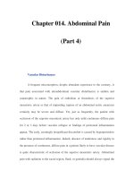
Pos P wave in LII, LIII, aVF
Neg P wave in LI aVL
Pos P . 80 msec in V1
Laterak LA, LSPV, LIPV
Neg P wave in LII, LIII & aVF
Neg P wave in V5 and V6
Inferomedial RA
Superolateral RA, RSPV
Neg P wave in LII, LIII & aVF
Inferolateral RA, Annulus,
CS RIPV
Fig 5.6 Site of origin of focal AT and respective P wave morphology.
Clinical presentation
14
• Patients present with palpitation, dyspnea, dizziness, or chest pain.
• Rapid firing of the focal AT may induce atrial fibrillation (AF), thus the patient
may present with AF.
Electrocardiographic characteristics of the focal atrial tachycardia
15,16
• In focal AT P waves are separated by an isoelectric line.
• P wave morphology may help identify the approximate origin of the AT
(Fig. 5.6).
• The commonest location of right AT in patients with structurally normal heart
is crista terminalis. P wave morphology is positive in LII, LIII, and aVF.
• The presence of anisotropy and automaticity in the cells of crista terminalis may
facilitate the occurrence of the tachycardia in this location.
• Tachycardias arising from the atrio-ventricular (AV) annulus or coronary sinus
(CS) os account for approximately 20% of all AT.
Ablation for atrial tachycardia
17–25
• AT arising from the atrial septum (right or left) or triangle of Koch may require
electrophysiologic study and multielectrode or three-dimensional (3D) mapping
for precise localization prior to ablation.
• Dense mapping in the area of interest, using a 3D sequential mapping system,
may help in precise localization of the focal AT. Incomplete mapping may result
in erroneous results and failed ablation.
• If earliest activation appears to be on the right side of the interatrial septum but
activation time from the onset of the P wave is less than 15 milliseconds, earliest
site appears to be near Bachmann bundle, or a narrow monophasic P wave
80 Essential Cardiac Electrophysiology
is present in V1, consider further mapping on the left side of the interatrial
septum.
• Tachycardia arising from the RSPV may be mistaken for right AT. When multiple
sites in posterior superior RA register the same activation time, it is likely that
the tachycardia is arising from RSPV.
• The presence of a diffuse area of activation near LSPV, LIPV, and the posterolat-
eral mitral annulus may suggest an epicardial focus.
• Focal ablation site can be further confirmed by the presence of fractionated
electrograms, negative unipolar atrial electrogram, or transient termination of
the tachycardia during mechanical pressure from the ablation catheter.
• AT should be differentiated from AVRT and atypical AVNRT (Table 5.1 in
Section 5.6).
• The target for ablation is the earliest activation preceding P wave by more than
30 milliseconds. Energy 30–50 W is delivered for 30–60 seconds.
• Acceleration of the tachycardia and termination within 10 seconds of radio-
frequency (RF) application is a sign of a successful outcome.
• Ablation in the lateral wall of the RA, crista terminalis, may result in phrenic
nerve damage.
• Ablation from the atrial septum, Koch’s triangle carries the risk of AV block.
Titrating energy delivery from 5 to 40 W and closely monitoring AV conduction
may avoid occurrence of AV block.
• Ablation of the AT arising from the annulus requires documentation of the small
A and larger V electrogram at the site of ablation.
• If ablating in venous structures, CS veins, SVC, lower power and temperature
not exceeding 50
◦
C may help avoid thrombus formation or stenosis.
• The success rate from RF ablation of the focal AT is 90% and the recurrence rate
is 10%.
• Predictors of lower success rate or recurrences include:
1 Left AT.
2 Multiple focal origin.
3 Older patients.
Macroreentrant atrial tachycardia
16–25
• Macroreentrant tachycardia can be classified into isthmus or non-isthmus
dependent type of AT (Fig. 5.7).
Isthmus dependent
Non-isthmus dependent
Atrial flutter
Scar Patch or other
anatomic barrier
Fig 5.7 Classification macroreentrant AT
Supraventricular Tachycardia 81
• Activation and recording of the fractionated electrograms identifies the area of
slow conduction. (Please refer to Section 5.1.)
• Pacing for concealed entrainment from the isthmus at CL 30 milliseconds shorter
than flutter CL results in acceleration of the tachycardia to pacing CL without
any change in the morphology of the P waves, recorded on surface ECG. On
termination of pacing, the post-pacing interval is the same as the tachycardia
CL. The sensitivity and specificity of this maneuver for the diagnosis of reentrant
isthmus dependent tachycardia is approximately 90%.
Entrainment mapping
18,23,25
• Entrainment mapping can be used to determine if the tachycardia is originating
from the RA or the left atrium (LA).
• If the PPI-TCL = <50 milliseconds in HRA and PCS then it is suggestive of right
atrial flutter.
• If the PPI-TCL = <50 milliseconds in HRA but >50 milliseconds in PCS then it
is suggestive of lateral RA tachycardia.
• If the PPI-TCL = >50 milliseconds in HRA and PCS then it is suggestive of left
PV tachycardia.
• If the PPI-TCL = >50 milliseconds in HRA and <50 milliseconds in PCS and
DCS consider left atrial flutter utilizing mitral annular isthmus.
• If the PPI-TCL = >50 milliseconds in HRA and <50 milliseconds in PCS and
>50 milliseconds in DCS consider right PV or septal tachycardia.
• Computerized 3D mapping allows recording of the isochronal maps of the
tachycardia circuit.
• Scar-related macroreentrant right ATs have been characterized as atypical atrial
flutter.
• Scar-related AT may require higher energy and temperatures for successful
ablation. This could be accomplished by a large/irrigated tip catheter.
• Combination of the electroanatomical and electrophysiologic mapping improves
ablation outcome.
• As opposed to focal AT, atrial activation during macroreentrant tachycardia
occupies 90% or more of the tachycardia CL. Earliest and latest activation tend
to be adjacent.
• Left atrial macroreentrant tachycardia is characterized by the following:
16
1 Negative P waves in LI and aVL.
2 Area of slow conduction between mitral annulus and anatomic barrier which
could be pulmonary vein, scar, or atrial appendage.
3 Post-pacing interval in the RA is >40 milliseconds longer than the tachycardia
CL at three or more sites including cavotricuspid isthmus, thus excluding right
atrial flutter or macroreentrant tachycardia.
4 CL variation in the LA precedes the RA.
5 Right atrial activation accounts for less than 50% of the tachycardia CL during
sequential catheter mapping.
82 Essential Cardiac Electrophysiology
• Macroreentrant tachycardias are common after surgical repair procedures such
as Mustard and Senning, Fontan or repair of tetralogy of Fallot.
• Identification and elimination of areas of slow conduction between the scars or
scars and anatomical barriers is the preferred approach during RF ablation.
• Incisional (scar-related) reentry may occur following:
1 Surgery for congenital heart disease.
2 Partially successful Maze procedure.
3 Catheter based ablation for AF.
• Following a patch repair of the ASD the isthmus for reentrant tachycardia may
be between the patch and the CS.
• Atriotomy scar-related macroreentrant tachycardia may occur from the scar that
extends from the atrial appendage to the inferoposterior right atrial free wall.
Incision typically does not extend to IVC or TA producing a narrow isthmus.
• Entrainment with concealed fusion can be demonstrated from the entry, mid
portion, or the exit site of this isthmus.
• Following observations are likely to identify the optimum site for successful
ablation:
1 PPI is the same as TCL.
2 Earliest electrogram precedes the onset of surface P wave by more than
50 msec.
3 On pacing from the site of the earliest electrogram, at cycle length 20 to
30 msec shorter, results in concealed entrainment.
4 Interval from the electrogram to the onset of P wave is same as the interval
from the stimulus to the onset of P wave. (Figs. 5.8 and 5.9.)
• Electroanatomical mapping can identify activation pattern, scar by using voltage
map, and sites for ablation.
I
II
aVF
v1
HRA
HIS–P
HIS–M
HIS–D
RA
200 msecs
25
26
Fig 5.8 Electrogram to onset of P wave 200 milliseconds.
I
38 39
II
aVF
V1
HRA
HIS-P
HIS-M
HIS-D
S
1
S
1
200 msec
S
1
Fig 5.9 Stimulus to onset of P wave 200 milliseconds. Tachycardia is entrained without a change
in activation or P wave morphology.
Supraventricular Tachycardia 83
5.3 ATRIAL FIBRILLATION
Mechanism, pathophysiology, and classification of atrial
fibrillation
26–28
• Atrial fibrillation (AF) is the most common chronic rhythm disorder1, affecting
5% of adults over age 65. AF occurs in 40% of the patients suffering from
congestive heart failure (CHF).
• Mortality in patients with AF is twice as high when compared with patients in
sinus rhythm.
• AF could be due to persistent rapid firing from the single focus termed as focal
driver or it could be maintained by multiple wavelets after being initiated by
premature atrial beats called focal triggers.
• These episodes of paroxysmal focal AF tend to occur in young patients without
structural heart disease and are often preceded by frequent premature atrial
contractions (PACs) of short coupling interval.
• Factors affecting the conduction and refractoriness in the atrium such as inflam-
mation, fibrosis, and ischemia are conducive to initiation and maintenance
of AF.
AF can be classified into the following categories:
1 Paroxysmal AF: starts and stops spontaneously.
2 Persistent AF: requires electrical or pharmacologic cardioversion to terminate
an episode.
3 Chronic AF: persists in spite of therapeutic intervention or based on a decision
not to restore sinus rhythm.
• Lone AF can either be paroxysmal, persistent, or chronic. It is defined as
AF occurring in patients less than 60 years of age who have no associated
cardiovascular diseases.
• Paroxysmal AF often progresses to chronic AF. Conversion and maintenance of
sinus rhythm becomes increasingly difficult with chronic AF.
• Chemical and electrical cardioversion for maintenance of sinus rhythm is easier
in AF of short duration.
During chronic AF the following structural and electrical changes may occur:
1 Atrial dilatation.
2 Apoptosis, resulting in loss of myofibrils.
3 Fibrosis, which alters conduction velocity.
4 There may be reduction in Connexion 43.
Shortening of the atrial refractory period occurs for the following reasons:
• A rapid atrial rate induces atrial ischemia, which results in shortening of the
atrial refractory period. Inhibitors of Na/H exchanger abolish ischemia-induced
shortening of the refractory period.
• There is a decrease in sodium channel density and current.
• Increase in the intracellular calcium load shortens the refractory period.
84 Essential Cardiac Electrophysiology
• Rate adaptation of the refractory period is lost.
• In AF I
CaL
is reduced. This results in shortening of action potential duration
(APD) and refractory period.
• Shortening of the refractory period may persist after recovery from AF and
predispose one to reoccurrences.
• Atrial dilatation and stretch may result in a decrease in the refractory period.
• Shortening of the effective refractory period (ERP) and APD and an increase in
dispersion of refractoriness perpetuates AF.
• Human atrial repolarization uses I
KUR
. I
to
and I
KUR
are decreased in AF,
resulting in shortening of the refractory period.
Neurohumoral changes during AF:
• Atrial natriuretic factor increases due to atrial stretch and dilation.
• Elevated ANF decreases after cardioversion.
• ANF may shorten the atrial refractory period.
Clinical presentation
• Most common symptoms are fatigue, reduced exercise tolerance, dyspnea, and
palpitation, although most episodes of AF remain asymptomatic.
• Tachycardia from AF can exacerbate angina or CHF.
• Irregular rhythm is consistent with but not diagnostic of AF. Other conditions,
such as sinus rhythm with frequent supraventricular or ventricular ectopic
beats, sinus arrhythmia, or multifocal atrial tachycardia, can cause irregular
pulse. An ECG is necessary to confirm the diagnosis. The absence of P waves is
characteristic of AF. Extremely rapid ventricular response may appear regular.
• AF with rapid ventricular response and aberrant ventricular conduction can
result in a wide complex tachycardia which may be mistaken for ventricular
tachycardia.
6
Treatment
29–34
• If the patient is hemodynamically unstable immediate cardioversion should be
considered.
• Rate control can be achieved by AV node (AVN) blocking drugs.
• Digoxin is least effective in controlling the rate especially in physically active
patients.
• β-Blockers and/or calcium channel blockers are effective AVN blocking agents.
• Calcium channel blockers are preferred in patients with bronchial asthma. The
aim should be to achieve a ventricular response between 80 and 100 bpm.
• AVN blocking agents should be avoided in the presence of ventricular preexcita-
tion. Amiodarone could be used in this setting because it prolongs the refractory
period of accessory pathway.
• The duration of AF and risk factors for thromboembolic complication determine
the need for anticoagulation. AF increases the risk of stroke by 8-fold.
Supraventricular Tachycardia 85
Risk factors for thromboembolic events in the presence of AF include:
1 Age >65 years.
2 Hypertension.
3 LVF.
4 Enlarged left atrium (LA).
5 Diabetes mellitus.
6 History of TIA.
7 Valvular heart disease.
Evidence that anticoagulation with warfarin prevents thromboembolic
complication is supported by the following studies.
1. Benefit of anticoagulation versus placebo: SPAF trial
• It was concluded that aspirin or warfarin significantly reduces events when
compared with a placebo. SPAF is not a comparison of aspirin with warfarin.
• Retrospective analysis suggested a lack of benefit of anticoagulation for patients
younger than 60 years.
2. Benefit of warfarin over aspirin: Atrial Fibrillation, Aspirin, Anticoagu-
lation (AFASAK) trial
• There was a substantial reduction of thromboembolic events with warfarin
versus aspirin or placebo (2% per year versus 5.5% per year).
• There was no significant difference in mortality. The bleeding rates were 6% per
year with warfarin and 1% per year with aspirin or placebo.
• This study supported the conclusion that warfarin is superior to aspirin
and placebo in preventing thromboembolic events among a largely elderly
population.
3. The Boston Area Anticoagulation Trial for Atrial Fibrillation
(BAATAF)
• There was a significant reduction in events in the warfarin-treated group (0.4%
per year versus 2.98% per year in the control group, an overall 86% reduction).
• Increased mortality was noted in the control group.
• There was no significant difference in bleeding events.
• It was concluded that warfarin is superior to placebo in reducing thromboembolic
events and mortality.
4. SPAF-II trial
• SPAF-II demonstrated higher event rates in high-risk patients over 75 years old.
This is reduced with warfarin anticoagulation.
• Increased risk for bleeding was noted.
5. Aspirin in low-risk patients: SPAF-III trial
• SPAF III supports the use of aspirin for thromboembolic prophylaxis in low risk
patients and suggests that patients with prior hypertension may be at sufficient
risk to justify anticoagulation with warfarin.
86 Essential Cardiac Electrophysiology
Rate control versus rhythm control
35–37
• The issue of treating patients with AF with rate control agents versus using anti-
arrhythmic drugs to maintain sinus rhythm has been addressed by two clinical
trials.
AFFIRM
• The Atrial Fibrillation Follow-up Investigation of Rhythm Management
(AFFIRM) trial: Patients were randomized between a strategy of rate control
with β blockers and calcium channel blockers targeted to a resting heart rate of
80 bpm versus rhythm control using anti-arrhythmic drugs.
• There was a non-significant trend toward higher total mortality in the rhythm
control group, the study’s primary endpoint.
• Pre-specified subgroup analysis demonstrated a statistically significant mortality
benefit with rate control for patients above the age of 65. There was no significant
difference in the incidence of stroke (roughly 1% per year); the majority (73%)
of ischemic strokes occurred in patients who had discontinued warfarin or had
an INR < 2.0.
• These findings support the recommendation that anticoagulation be continued
in patients even if AF is successfully suppressed.
• AFFIRM demonstrated no advantage to a rhythm control strategy for recurrent
AF, and suggests a rate control strategy may be superior in patients above the
age of 65.
• Patients enrolled in this study were minimally symptomatic.
• These results do not apply to patients with symptomatic AF.
• Higher mortality in rhythm control group may be due to proarrhythmic effects
of antiarrhythmic drugs rather than due to maintenance of sinus rhythm.
RACE
• The RACE (Rate Control versus Electrical Cardioversion for Persistent Atrial
Fibrillation).
• No significant difference in cardiovascular death or thromboembolic events was
noted, but 83% of all thromboembolic events occurred in patients who had
discontinued warfarin or had an INR <2.0.
• The study demonstrated no significant advantage to a rhythm control strategy
for the management of persistent AF. Any benefits derived by rhythm control
may have been neutralized by the proarrhythmic effects of the antiarrhythmic
drugs.
1 A rate control strategy is an acceptable approach to management of patients
with AF, particularly if they are asymptomatic and elderly.
2 Rhythm control should be reserved for patients with symptomatic AF. This
strategy should also be considered in minimally symptomatic young patients
with AF.
Supraventricular Tachycardia 87
Anticoagulation for conversion to sinus rhythm
• If AF is of less than 48 hours’ duration, cardioversion can be attempted.
• The presence of AF for more than 48 hours necessitates three to four
weeks of therapeutic anticoagulation prior to conversion, unless transesopha-
geal echocardiography (TEE) demonstrates absence of clot in the LA and its
appendage.
• Regardless of whether a TEE is performed, systemic anticoagulation is required
for three weeks following cardioversion in all patients with AF of greater than
48 hours’ duration.
The ACUTE trial
38
• Patients were assigned to TEE followed by DC cardioversion (if no intracar-
diac clot was found) versus conventional therapy consisting of three weeks of
anticoagulation before DC cardioversion.
• All subjects (TEE group and conventional therapy group) received therapeutic
anticoagulation for four weeks after cardioversion.
• At eight weeks (from the time of enrollment), there was no significant difference
in primary endpoint of cerebrovascular accident, TIA, and peripheral embolus.
• Fewer bleeding events were noted in the TEE group.
• The risk of thromboembolic events is higher in the first three to four weeks
immediately following conversion to sinus rhythm.
• This may be due to atrial stunning, a term describing the observation of reduced
atrial systolic function following conversion to sinus rhythm.
• Atrial stunning can allow relative stasis of blood within the atrium, potentially
resulting in thrombus formation.
• Patients should receive anticoagulation with warfarin for three weeks follow-
ing conversion to sinus rhythm even if they are in a low risk category for
thromboembolic events.
• Patients with the indications for chronic anticoagulation with warfarin men-
tioned above (valvular heart disease, age above 65, prior thromboembolic event,
hypertension, heart failure, coronary artery disease, or diabetes) should receive
long-term anticoagulation following cardioversion.
DC cardioversion
39,40
• Emergent electrical cardioversion is indicated if the patient is hemodynamically
unstable as a result of tachycardia.
• Cardioversion can either be performed with a standard monophasic or biphasic
defibrillator. If a standard defibrillator fails, cardioversion should be repeated
using a biphasic defibrillator.
Rectilinear biphasic defibrillation
• During biphasic defibrillation there is a change in the polarity of the waveform
during delivery of energy.
88 Essential Cardiac Electrophysiology
• Biphasic defibrillation allows for similar current delivery (which is the most
important variable for achieving cardioversion) with lower energy.
• The number of shocks required to achieve cardioversion is also reduced.
• Biphasic defibrillation is superior to monophasic defibrillation.
Chemical cardioversion
3
• Ibutilide: Class III anti-arrhythmic can be used for cardioversion alone or
as an adjuvant to facilitate DC cardioversion, particularly when initial DC
cardioversion is unsuccessful.
• Ibutilide is administered intravenously 1 mg over 10 minutes.
• Ten to fifteen percent of the patients with new onset AF may convert to sinus
rhythm with ibutilide alone.
• When cardioversion is performed after the administration of ibutilide, thesuccess
rate may approach 100% and the amount of energy required may also be less.
• Patients should be monitored for 4 hours after administration of ibutilide.
• Risk factors for ibutilide induced ventricular arrhythmias include prolonged QT,
depressed left ventricular function (ejection fraction <0.30), hypokalemia, or
hypomagnesemia.
Maintenance of sinus rhythm
41–44
• Anti-arrhythmic therapy is indicated for patients with symptomatic AF.
• Rate control alone can be used for elderly minimally symptomatic patients.
• For moderate to severe left ventricular systolic dysfunction the agent of choice
is amiodarone. Dofetilide can be used.
• All other antiarrhythmics are relatively contraindicated in patients with LV
dysfunction because of the potential for proarrhythmias.
• For patients with ischemic heart disease and preserved left ventricular systolic
function, sotalol may be useful because of its β-blocker effects.
• Disopyramide can be used in patients suspected of having AF due to increased
vagal tone.
• Class IC agents such as flecainide and propafenone can be used in patients
without ischemic heart disease and normal LV wall thickness and function.
• These agents can be administered daily for maintenance of sinus rhythm.
• They can also be used on an as needed basis for acute conversion of symptomatic
paroxysmal AF.
• 300 mg of Flecainide or 600 mg of propafenone can be administered orally.
• β-Blocker or calcium channel blocker should be administered 30–60 minutes
prior to administration of the anti-arrhythmic agent to prevent accelerated AV
conduction.
• The first trial of this approach should be performed while the patient is being
monitored.
• Treatment of lone AF with Class IC agents can result in conversion to atrial flutter
because of prolongation of the atrial refractory period and slowing of conduction
velocity.
Supraventricular Tachycardia 89
• This “Class IC atrial flutter” can be treated with ablation of the right atrial
cavotricuspid isthmus followed by continuation of the AAD.
45
• Class III agents include amiodarone, sotalol, and dofetilide.
Amiodarone
• Evidence supporting the efficacy of amiodarone comes from the Canadian Trial
of Atrial Fibrillation (CTAF) trial.
• At 1 year follow-up, 69% of patients treated with amiodarone were in sinus
rhythm compared with 39% of individuals treated with sotalol or propafenone.
• Amiodarone was associated with a higher discontinuation rate due to side effects
that was not statistically significant.
• There was no significant difference in total mortality between the groups.
• Amiodarone has multiple adverse reactions; patients receiving amiodarone
need monitoring of pulmonary function tests (carbon monoxide diffusion test),
thyroid function, liver function, and ocular examination for corneal deposits.
• Although there is no FDA indication for amiodarone in AF this is a most
commonly prescribed anti-arrhythmic agent for treatment of AF.
• Amiodarone can be initiated as an outpatient, usually at 400 mg per day for a
period of two to four weeks, then decreasing the dose to 200 mg per day.
Dofetilide
• It requires in-hospital initiation and monitoring for arrhythmias.
• Safety of dofetilide in patients with heart failure is supported by the Danish
Investigations of Arrhythmia and Mortality on Dofetilide in Congestive Heart
Failure (DIAMOND-CHF) Study. Patients with left ventricular ejection fractions
<35% were enrolled. The dofetilide dose was 500 μg BID. It was adjusted to
250 μg BID for creatinine clearances between 40–60 ml/min and 250 μgQD
for patients with creatinine clearance of <40 ml/min. Patients with creatinine
clearance of less than 20 ml/min were excluded.
• There was no significant difference in total mortality. Retrospective analysis of
the results demonstrated that 12% of patients with AF in the treatment arm con-
verted to sinus rhythm, compared with 1% in the placebo arm, with a significant
reduction in the subsequent development of AF.
Sotalol
• It should not be given to patients with renal dysfunction, left ventricular
hypertrophy, prolonged QT intervals, bradycardia, or electrolyte abnormalities
(hypokalemia).
• Nodally active agents should be stopped or decreased before initiation of sotalol
because of the risk of bradycardia from β-blocking properties seen at 40 mg bid.
The Class III anti-arrhythmic effect (action potential prolongation) appears at
120–160 mg bid.
• Sotalol should be initiated in hospital while monitoring for proarrhythmias and
prolongation of the QT interval.
90 Essential Cardiac Electrophysiology
• Sotalol can be administered as follows:
1 80 mg tid for 1 day.
2 Then 120 mg bid on the second day.
3 Then 160 mg bid on the third day.
4 Discharge on 120 mg bid, with increase to 160 mg bid if needed.
Nonpharmacologic options in the management of AF
Radiofrequency (RF) ablation
46–51
• Arrhythmias are produced by abnormality of impulse generation or impulse
propagation. RF ablation seeks to eliminate these abnormalities.
• When RF current passes through the tissue it produces heat, which
is proportional to the power density within tissue. It is an alternating
current.
• Maximum heating occurs at the tip of the electrode and it diminishes as the
distance from the tip increases. Increase in the radius (distance) from the tip will
decrease the heat by 4-fold. For this reason the depth and the volume of the
tissue that is affected by heat is small (2 mm). Deeper tissue heating is due to
heat conduction.
• Commonly used RF is 300–1000 kHz. Lower frequency may produce muscle
stimulation. At higher frequency mode of heating changes from resistive to
dielectric.
• RF energy is delivered in a unipolar fashion from the catheter tip to the dispersive
patch electrode placed on the skin.
• The surface area of catheter electrode is 12 mm
2
and the surface area of the
patch electrode is 100–250 cm.
2
This results in an increase in power density and
heating at the catheter tip.
• Catheter tip electrodes with a large surface area or when the catheter tip is cooled
by irrigation allows lower system impedance and delivery of higher power. This
results in deeper and larger lesion. Since the temperature is measured at the
catheter tip it does not reflect actual tissue temperature, which may be very high.
• Very high tissue temperature results in heat expansion of the tissue, crater
formation and may produce tissue pop.
• RF energy delivery should be at least for 60 seconds.
• Rise in temperature at deeper tissue level may continue if high power or tem-
perature settings are used, producing thermal latency even after termination of
energy delivery.
• RF generated heat produces coagulation necrosis of the myocytes. Healing by
fibrosis is complete by eight weeks.
Radiofrequency ablation for AF
• This procedure is typically reserved for patients with lone or paroxysmal AF who
have failed one or more trials of anti-arrhythmic therapy.
• AF can be cured with catheter ablation techniques.
Supraventricular Tachycardia 91
• The best results of this procedure (up to 85% success) have been achieved in
patients with lone AF. Lower success rates (50–70%) have been reported in
other subsets of AF patients.
• Potential complications of this procedure include pulmonary vein stenosis,
stroke, LA esophageal fistula, and pericardial tamponade.
• The approach to AF ablation could be classified as elimination of the triggers,
substrate, or autonomic facilitators (parasympathetic ganglion).
Elimination of the triggers
• It was noted that the AF is initiated by rapidly firing triggers located in pulmonary
veins.
• This may manifest as frequent PACs or clearly discernible atrial activity in the
form of atrial tachycardia at the onset of AF or during AF.
• These foci arise from the myocardial muscular sleeve that extends few centi-
meters into the pulmonary veins.
• Initial approaches included identification of PACs with earliest activation and
elimination of these foci within the pulmonary vein. A possible risk of pulmonary
vein stenois shifted the focus to ablation outside the orifice of the pulmonary
vein in a quadrantic fashion.
Pulmonary vein isolation using RF ablation in LA
• In this approach an attempt is made to isolate all the four pulmonary vein orifices
from the LA. It reduces the probability of the pulmonary vein stenosis.
• The rationale is that PACs (triggers) could arise from any of the four pulmonary
veins.
• It may also produce compartmentalization and “debulking” of the LA.
• The drawback of this approach includes reoccurrences, creation of the isthmus
that may predispose to atrial tachycardia.
• Esophageal perforation following posteromedial left atrial or right superior pul-
monary vein ablation may occur. This is a serious and often fatal complication.
Elimination of the substrate
• Identification and elimination of the fractionated electrograms may result in
termination of the AF during the procedure. A success rate of 80% has been
reported.
• Fractionated electrograms may be recorded from the LA around the pulmon-
ary veins, left atrial appendage or interatrial septum. In the right atrium (RA)
the fractionated electrograms could be recorded from the crista terminalis, the
orifices of the vena cava, the orifice of the coronary sinus (CS), or up to 2–3 cm
within the CS.
• Like pulmonary veins, muscular extension into proximal CS may produce
rapidly firing automatic foci responsible for initiating AF.
92 Essential Cardiac Electrophysiology
Modification of the autonomic substrate
• The posterior wall of the LA is richly innervated by vagal (parasympathetic)
fibers.
• Parasympathetic stimulation produces bradycardia and shortening of the atrial
refractory period. These electrophysiologic changes are conducive to initiation
and maintenance of AF, termed as vagally induced AF.
• Vagally induced AF occurs during sleep and may be responsible for the atrial
arrhythmias that occur during sleep apnea.
• During ablation of the vagal neural terminals, located in the posterior wall of
the LA, bradycardia, or junctional rhythm may occur.
• It may be necessary to tailor these three approaches when using ablation as
the therapeutic modality in the management of AF. For example, a paroxysmal
AF in a young patient with a structurally normal heart where focal tachycardia
or premature beats are identified as the initiator of the AF may benefit from
elimination of that focus.
Atrioventricular node ablation with permanent pacemaker
implantation
52
• Patients with left ventricular dysfunction or chronic pulmonary disease or
those who cannot tolerate the doses of AVN blocking agents necessary to
achieve rate control or the agents for rhythm control may be candidates for this
approach.
• AVN blocking agents may produce negative inotropic effects or bronchospasm
in these patients.
• The overall survival of patients undergoing AVN ablation and pacemaker inser-
tion is the same as a matched group of patients treated with antiarrhythmic
drugs.
• The drawback of AVN ablation and the pacemaker approach include persist-
ence of AF, need for anticoagulation, pacemaker dependence and ventricular
dys-synchrony from RV pacing.
• AVN ablation should rarely be performed in young patients with AF.
Electrical therapies for AF
• In patients who have or need pacemaker for other indications, program-
ming to eliminate PACs or abolish post-PAC pauses may decrease the burden
of AF.
• Defibrillators with atrial arrhythmia therapy options such as high frequency
pacing at 50 Hz and cardioversion may decrease the frequency and duration of
the AF. These features can be set to automatically cardiovert the patient upon
detection of AF using specified criteria or it can be triggered by the patient or
the physician.
67
• Defibrillator with atrial therapy features is implanted in patients who are
undergoing ICD implant and also have paroxysmal AF.
Supraventricular Tachycardia 93
Surgical Maze procedure
• Incisions are made in the LA around the pulmonary veins, posterior wall and
extended to the mitral annulus.
• This procedure can be performed in conjunction with other cardiac
surgery such as mitral valve replacement. Success rates are approximately
80–90%.
• The epicardial approach, using minimally invasive thoracotomy and microwave,
attempts to isolate the pulmonary veins.
5.4 AUTOMATIC JUNCTIONAL TACHYCARDIA
Junctional tachycardia
53,54
• Automatic junctional tachycardia (AJT) is common in infants. It carries high
mortality.
• In older patients it has a more benign course and is not associated with structural
heart disease.
• AJT arises from transitional cells of atrio-ventricular (AV) junction and is due to
abnormal automaticity. Tachycardia is sensitive to catecholamines.
• Electrocardiogram shows narrow QRS (wide if bundle branch block present)
tachycardia. Ventriculo atrial (VA) block may be present. Sinus capture beats
may occur.
• AJT is often irregular and may be mistaken for atrial fibrillation or multifocal
atrial tachycardia.
• If the QRS is wide it may be mistaken for ventricular tachycardia.
• Intra-cardiac electrograms show that each QRS is preceded by His and a normal
HV interval.
• Initiation termination is spontaneous without critical AH delay.
• AV dissociation is common. The tachycardia is unaffected by atrial or ventricular
pacing.
• AJT should be differentiated from non-paroxysmal junctional tachycardia
(NPJT), which tends to be slower and regular. It occurs in the setting of digitalis
toxicity, COPD, myocardial ischemia, carditis, and post cardiac surgery.
• NPJT is believed to be due to triggered activity.
• To differentiate AJT from AV node re-entry tachycardia (AVNRT), premature
atrial beats are delivered in the region of slow pathway when septal A is
committed. This will advance His in AVNRT but not in AJT.
• AVNRT is initiated by premature atrial contraction (PAC), needs critical AH
interval and demonstrates dual AV nodal physiology.
• Orthodromic nodofescicular tachycardia is often initiated by PACs or
premature ventricular contractions (PVCs), demonstrates preexcitation during
atrial pacing, occurrence of bundle branch block results in a change in CL and
advancement of next His or termination of tachycardia with PVC delivered when
His is refractory.
94 Essential Cardiac Electrophysiology
Treatment
• In young patients AVN blocking drugs are ineffective. Amiodarone may be more
effective.
• Abrupt onset of AV block may occur. Insertion of permanent pacemaker is
recommended.
• If drug therapy fails ablation at the site of earliest activation along the septum
or AVN ablation and insertion of permanent pacemaker could be considered.
• In adult patients β-blockers may be effective.
• AJT may occur following surgery for congenital heart disease. It may last for
1–4 days after surgery.
• The use of inotropic agents, such as catecholamines and digoxin, may
induce AJT.
• It responds well to IV propafenone, procainamide, or amiodarone.
5.5 AV NODE REENTRY TACHYCARDIAS
AV node reentry tachycardia
55–59
• Dual atrio-ventricular (AV) nodal physiology is defined as 50 milliseconds
increase in A2H2 for a 10 milliseconds decrease in A1A2.
• Fast and slow pathways represent different atrionodal connection as suggested
by different sites of earliest retrograde activation during tachycardia or retrograde
conduction during ventricular pacing.
• Resetting of the tachycardia by late premature atrial contraction (PAC) delivered
outside the AV node (AVN) near the posterior right atrial septum or coronary
sinus (CS) also suggests the presence of reentrant loop.
Fast pathway
• Retrograde earliest atrial activation during atrio-ventricular reentry tachycardia
(AVNRT) suggesting a fast pathway insertion site is located 5 mm posterior
and 8 mm superior to His bundle electrogram recording. Atrial electro-
gram at this site may precede atrial activation on His electrogram by 10–20
milliseconds. This site is located superior to tendon of Todaro outside the triangle
of Koch.
• Retrograde fast pathway activation at the posterior superior right atrial septum
may demonstrate two components. Initial low frequency component reflects far
field atrial potential from the left-hand side of the septum.
• Application of radiofrequency (RF) current superior to tendon of Todaro will
eliminate antegrade fast pathway conduction.
• The right side component of the fast pathway begins in the anterior limbus of
the fossa ovalis, as atrial cell then becomes a transitional cell on crossing the
tendon of Todaro and inserts into the common AV bundle distal to the compact
AVN.
Supraventricular Tachycardia 95
• This distal insertion may explain why the fast pathway has less decremental
properties, is less responsive to AVN blocking agents and has greater response
to sodium channel blocking agents.
• During decremental atrial pacing block in the fast pathway occurs proximally,
closer to the anterior limbus of the fossa ovalis.
Slow pathway
• It is located in the posterior septum between the tricuspid annulus and the CS
ostium.
• The SP connects to the posterior extension of the AVN.
• Retrograde slow pathway activation proceeds from the proximal CS to the left
atrium (LA) and across the septum to the right atrium (RA). From here it pro-
ceeds anteriorly towards the His bundle and posteriorly behind the CS ostium
and the Eustachian ridge.
• CS musculature electrically connects the RA and the LA.
• The Eustachian valve and ridge form a line of block that confines SP potential/
impulse to within the triangle of Koch. This may allow dissociation of slow
pathway potential from the right atrial electrograms.
• This line of block may explain the late occurrence of SP potential during sinus
rhythm when compared with atrial electrogram at His location.
• In sinus rhythm slow pathway activation proceeds from posterior to anterior in
the area of the triangle of Koch.
• During SP ablation accelerated junctional rhythm occurs with retrograde con-
duction over the fast pathway.
• There may be multiple slow pathways with multiple jumps in the AH
interval.
• Linear ablation from the tricuspid annulus to the CS ostium is likely to eliminate
most of the SP conduction.
• After slow pathway ablation the presence of A2H2 jump and echo beat may
indicate the presence of additional SP, which may enter the triangle of Koch
anterior to the CS ostium. These slow pathways may not be capable of causing
arrhythmias.
• 84% of patients without a history of AVNRT demonstrated dual AV nodal
physiology.
• This may be due to sedation with midazolam and fentanyl, which depresses
antegrade and retrograde conduction over the fast pathway, thus allowing SP
conduction to become manifest.
• Isoproterenol speeds up conduction in the fast pathway.
• Equal delay in conduction over the slow and the fast pathways may mask dual
AV nodal physiology.
• A2H2 greater than 200 milliseconds may suggest conduction over SP.
• A shift in retrograde conduction from fast to slow will change the atrial activation
sequence.
96 Essential Cardiac Electrophysiology
Common or typical AVNRT
• During tachycardia antegrade conduction is over the slow pathway and retro-
grade conduction over the fast pathway.
• It is induced by PAC and rarely by premature ventricular contraction (PVC).
• An abrupt increase in AH by 50 milliseconds with PAC before induction is
common.
• Lack of increase in the AH interval may be due to equal delay in conduction in
both pathways.
• AH during tachycardia usually exceeds 200 milliseconds.
• The HA interval is short, <50 milliseconds.
• Earliest retrograde atrial activation is superior to tendon of Todaro, suggestive
of retrograde fast pathway activation.
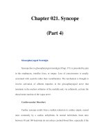
• A short HA interval results in the superimposition of P waves on the QRS, thus
obscuring P waves on the surface electrocardiogram. This may result in a pseudo
R wave in V1 or pseudo S in inferior leads (Fig. 5.10a and 5.10b).
• During tachycardia, if there is simultaneous activation of the atrium and vent-
ricles, late PVC may separate atrial and the ventricular potentials and help
identify the atrial activation sequence.
• The reentry circuit between the atrial ends of the fast and the slow pathways
includes a large atrial component. This may explain extremely rare VA block to
the atrium during AVNRT.
• Late PAC delivered to the atrium that is incorporated in the tachycardia circuit
may advance next His and reset tachycardia. PACs should be late and reach after
retrograde earliest activation by the fast pathway.
• Post-pacing interval (stimulus to next A) may be longer than the tachycardia CL
due to delay in AVN.
• Resetting with the shortest post pacing interval occurs from the posteroseptal
RA and the proximal CS.
• Retrograde activation of the atrial septum occurs through the fast pathway, the
impulse then propagates along the left-hand side of the interatrial septum to the
proximal CS, the CS ostium, and the posterior end of the slow pathway.
• During decremental RV pacing there is little decremental conduction in the lower
common pathway.
• The HA interval during tachycardia reflects retrograde conduction over the fast
pathway minus simultaneous antegrade conduction over the lower common
pathway. The HA interval during V pacing reflects retrograde conduction time
over the lower common pathway and the fast pathway.
• HA during tachycardia tends to be shorter than during RV pacing.
• To record retrograde His, RV pacing should be performed near the anterobasal
RV septum close to right bundle.
• In the left-sided variant of S/F AVNRT the slow pathway is located in the pos-
terior mitral annulus. These patients tend to have a short HA interval of less
than 15 milliseconds. This may be due to the longer lower common pathway.
Supraventricular Tachycardia 97
(a)
(b)
1
2
Fig 5.10 (a) AVNRT: spontaneously occurring PVC (2) does not reset the tachycardia. P waves (1)
are noted in the terminal portion of the QRS. (b) Simultaneous activation of the atrium and the
ventricles results in superimposition of P and QRS. P wave location is evident by simultaneous
recording of atrial electrograms.
98 Essential Cardiac Electrophysiology
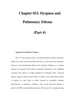
• During the onset of AVNRT, block below the AVN may occur, resulting in 2 : 1
conduction.
• Two for 1 phenomenon may occur during programmed atrial stimulation, when
there is antegrade conduction of the atrial impulse once through the fast pathway
and the same impulse conducts through the slow pathway, resulting in two
ventricular electrograms for each atrial electrogram. This may indicate absence
of retrograde penetration of the slow pathway by the antegrade fast pathway
(Fig. 5.11).
Slow/slow (S/S) AVNRT
• During this tachycardia antegrade conduction is through the slow pathway
as manifested by a long AH interval (240 milliseconds or more). Retrograde
conduction is through slow pathway with earliest atrial activation at postero-
septal RA between the tricuspid annulus and the CS ostium or within the CS.
This atrial activation may precede atrial activation in His electrogram by 30–60
milliseconds.
• The VA interval is long and may exceed 70 milliseconds.
• There may be multiple jumps in A2H2 during programmed atrial stimulation
indicative of multiple slow pathways. There may be multiple HA intervals during
tachycardia.
• Half of these patients may have S/F AVNRT.
• During ventricular pacing retrograde atrial activation may shift from the anterior
to the posterior atrial septum, indicating the shift in conduction from the fast to
the slow pathway.
• Tachycardia may be induced by programmed ventricular stimulation, resulting
in a block in the retrograde fast pathway and retrograde conduction over the
slow pathway.
I
II
aVF
V1
HRA
HBEP
HBEM
HBED
CS 9
CS 7
CS 5
CS 3
CS 1
1
2
3
1000
2000
0
Fig 5.11 PAC at a coupling interval of 440 milliseconds results in antegrade conduction through
the fast pathway (1) followed by antegrade conduction through the slow pathway (2) thus
producing two QRS complexes for a single PAC. The same impulse then conducts retrogradely
through the fast pathway (3).
Supraventricular Tachycardia 99
• S/S AVNRT has a longer lower common pathway, which is located posteriorly.
• HA during ventricular pacing tends to be longer than HA during tachycardia in
patients with S/S AVNRT.
• The HA interval represents retrograde conduction over the slow pathway minus
antegrade conduction over the lower common pathway. Because of the longer
lower common pathway the HA interval may be short or negative during S/S
AVNRT.
• Fast pathway ablation will not eliminate S/S AVNRT.
• The reoccurrence rate is higher after ablation in patients with S/S AVNRT
and may require more extensive ablation in the posteroseptal RA and the
CS ostium.
Fast/slow or uncommon type of AVNRT
• It is characterized by a short AH (30–180 milliseconds) and a long HA interval
(260 milliseconds).
• Antegrade conduction is over the fast pathway and retrograde conduction over
the slow pathway.
• Earliest retrograde atrial activation is in the posteroseptal RA or the CS ostium.
• On the surface electrocardiogram the RP interval is longer than the PR interval.
P waves are inverted in the inferior leads.
• On termination of the V pacing, where V pacing has successfully entrained
AVNRT, post-pacing interval minus tachycardia cycle length (PPI–TCL) of >120
milliseconds and Stim to A minus VA interval of >85 milliseconds suggests the
diagnosis of atypical AVNRT and excludes the diagnosis of AVRT utilizing septal
accessory pathway (Fig. 5.12).
Differential diagnosis of AVNRT (Table 5.1)
• Single late PVC is delivered 50 milliseconds after the onset of His electrogram
and advanced by 10 milliseconds without retrogradely activating His. Advancing
of atrial activation without first retrogradely activating the His will suggest the
presence of accessory pathway.
TCL = 450
StimA – VA = 130 msec
PP – TCL = 130 msec
VA = 330
100 msee
Fig 5.12 Atypical AVNRT response to ventricular pacing.
100 Essential Cardiac Electrophysiology
His Electrograms
I
aVF
II
V1
HRA
HIS-P
HIS-M
HIS-D
460 msec
460 msec
07
08
Fig 5.13 His synchronous PVC does not reset atrial electrogram.
I
aVF
II
V1
HRA
HIS-P
HIS-M
HIS-D
VA interval 180 msec
01
02
Fig 5.14 Termination of A pacing first VA is similar to subsequent VA intervals.
I
II
VI
aVF
HRA
HIS-P
HIS-M
HIS-D
RVA
VV
A
s
1
s
1
s
1
2120 22
Fig 5.15 Termination of V pacing response is VAV.
• Atrial activation sequence remains unchanged and may advance next His elec-
trogram. In AVNRT His synchronous PVC fails to advance atrial electrogram
(Fig. 5.13).
• PVCs during supraventricular tachycardia (SVT) should be delivered at the base
of right ventricular septum for posteroseptal accessory pathway (AP) and in
parahisian location for anteroseptal AP.
• On termination of atrial pacing the first VA is identical to subsequent
VA (Fig. 5.14) and on termination of V pacing the response is VAV
(Fig. 5.15). These observations make the diagnosis of atrial tachycardia (AT)
unlikely.
• Parahisian pacing is performed during sinus rhythm from the anterobasal right
ventricle, anterior and apical to His recording, where high output captures the
His, resulting in a narrow QRS and timing of His is advanced. At lower output
His RB capture is lost, resulting in a wide QRS and delay in the timing of atrial
Supraventricular Tachycardia 101
I
II
aVF
V1
HRA
HIS-P
HIS-M
HIS-D
Non capture
Capture
St-A = 130 msec
St-A=100 msec
56
57
Fig 5.16 Parahisian pacing: stimulus to A is shorter during His capture.
activation (equal to the delay in His activation) without a change in activation
sequence. This suggests conduction over the AVN (Fig. 5.16).
• With loss of HB RB capture, absence of change in the timing and sequence of
atrial activation suggests conduction over AP.
Treatment
• For immediate termination of the tachycardia, vegal maneuvers, IV adenosine;
calcium channel blockers or β-blockers can be used.
• Ablation of the slow pathway is the treatment of choice.
Ablation for AVNRT
• The preferred site of ablation for AVNRT is the slow pathway, which is located
in the triangle of Koch.
• The boundaries of the triangle of Koch are delineated by CS ostium, tendon of
Tedaro posteriorly, and septal leaflet of the tricuspid valve anteriorly and bundle
of His at the apex of the triangle.
• Mean distance from His bundle to the mid-portion of the anterior lip of the CS
ostium is 15–20 mm.
• The fast pathway is located anteriorly in close proximity to His bundle. The slow
pathway is located posteriorly near the CS ostium.
• Discrete potentials noted in the posteroseptal RA may be due to anisotropic
conduction and not due to activation of the slow pathway.
• Ablation of the slow pathway can be achieved by anatomic or electrogram
approach or a combination of both.
• Area of interest is identified on fluoroscopic examination between His bundle
electrogram and the CS ostium along the posteromedial aspect of TA. Multipolar
electrograms are recorded at this site. The AV electrogram ratio should be less
than 0.5.
• Electrogram and anatomic approaches should be combined to achieve successful
ablation.
• The majority of successful ablation sites are located between TA and CS ostium.
Other sites include within the CS ostium or inferior or the superior lip of the CS
ostium.
• Unsuccessful ablation is the result of imprecise mapping or inadequate tissue
contact and heating.
102 Essential Cardiac Electrophysiology
I
II
aVF
V1
HRA
HIS-P
HIS-M
HIS-D
ABL-D
H
H
720 msec
58
59
Fig 5.17 Junctional rhythm during RF application to the slow pathway.
• Target temperature should be 45–50
◦
C. Slow junctional rhythm that may last
for 15–20 seconds may occur (Fig. 5.17).
• If junctional rhythm does not occur within 20 seconds of achieving target
temperature RF application should be terminated.
• Complete AV block may occur in 1% of patients, undergoing slow pathway
ablation.
• Monitoring VA conduction during junctional rhythm or prolongation of the PR
interval should be performed.
• Complete AV block is unlikely to occur in the presence of intact VA conduction
during junctional rhythm. Application of RF energy should be discontinued with
the first sign of VA block, slowing of VA conduction, or prolongation of the PR
interval in conducted sinus beats.
• VA conduction during junctional rhythm may be noted even if there is no VA
conduction during ventricular pacing.
• Isorhythmic AV dissociation may mimic intact VA conduction. In the presence
of poor fast pathway conduction VA conduction during junctional rhythm may
occur and is not a reliable indicator of potential AV block.
• To monitor for intact AV conduction atrial pacing at the fastest rate associated
with 1 : 1 conduction should be performed while observing for the prolongation
of the PR interval.
• Isorhythmic AV dissociation may occur during atrial pacing and RF application
should be discontinued.
• 40–50% of patients may demonstrate residual slow pathway function, as
evidenced by single echo beats, even after successful ablation of AVNRT.
• AVNRT may reoccur in 3–5% of patients. Most of the recurrences are reported
in the first three months after ablation.
• Following a slow pathway ablation the fast pathway effective refractory period
shortens.
• RF ablation should be considered for patients with frequent episodes of sympto-
matic tachycardia. Slow pathway ablation can be performed in the presence of
prolonged PR interval. Atypical AVNRT can be successfully eliminated by slow
pathway ablation.
• The alternative to RF energy is cryothermal energy. The long-term success rate
is 90–94%.