Báo cáo y học: "Cellular transport of anti-inflammatory pro-drugs originated from a herbal formulation of Zingiber cassumunar and Nigella sativa" ppsx
Bạn đang xem bản rút gọn của tài liệu. Xem và tải ngay bản đầy đủ của tài liệu tại đây (499.02 KB, 5 trang )
BioMed Central
Open Access
Page 1 of 5
(page number not for citation purposes)
Chinese Medicine
Research
Cellular transport of anti-inflammatory pro-drugs
originated from a herbal formulation of Zingiber cassumunar and
Nigella sativa
Prasan Tangyuenyongwatana
1
, Jariya Kowapradit
2
, Praneet Opanasopit
2
and
Wandee Gritsanapan*
1
Address:
1
Department of Pharmacognosy, Faculty of Pharmacy, Mahidol University, Bangkok 10400, Thailand and
2
Department of
Pharmaceutical Technology, Faculty of Pharmacy, Silpakorn University, Nakhon Pathom 73000, Thailand
Email: Prasan Tangyuenyongwatana - ; Jariya Kowapradit - ;
Praneet Opanasopit - ; Wandee Gritsanapan* -
* Corresponding author
Abstract
Background: The rhizome of Zingiber cassumunar and the seed of Nigella sativa are two ingredients
in Thai traditional medicine to relieve dysmenorrhea and adjust the menstrual cycle. Mixture of
these two herbs produces three esters, namely (E)-4-(3,4-dimethoxyphenyl)but-3-en-1-yl linoleate
(1), (E)-4-(3,4-dimethoxyphenyl)but-3-en-1-yl oleate (2) and (E)-4-(3,4-dimethoxyphenyl)but-3-en-
1-yl palmitate (3). The aim of this study is to examine in vitro absorption of these esters and evaluate
their transport across the membrane.
Methods: In vitro transport of these three esters was observed in Caco-2 cell monolayers. The
ester compounds 1, 2 and 3 at a concentration of 10 μM were hydrolyzed by porcine liver esterase.
Results: All esters transported across the Caco-2 cell without enzymatic hydrolysis. The apparent
permeability coefficients P
app
of compound 1 at 53 μM and 106 μM were 13.94 (0.60) × 10
-6
and
14.33 (0.17) × 10
-6
cm/s respectively, while those of compound 2 were 9.45 (0.29) × 10
-6
and 10.08
(0.32) × 10
-6
cm/s, respectively. P
app
values of compound 3 were 7.48 (0.31) × 10
-6
cm/s at 53 μM
and 8.60 (0.55) × 10
-6
cm/s at 106 μM. P
app
values of the parent compound (compound D), i.e. (E)-
4-(3,4-dimethoxyphenyl)but-3-en-1-ol were 8.53 (0.83) × 10
-6
cm/s at 53 μM and 16.38 (0.61) × 10
-
6
cm/s at 106 μM. The ester hydrolysis of compounds 1, 2 and 3 by porcine liver esterase was
monitored by HPLC and the hydrolysis reactions were completed within 10 minutes.
Conclusion: Using the Caco-2 cell monolayer model, the present study finds that compounds (E)-
4-(3,4-dimethoxyphenyl)but-3-en-1-yl linoleate (1), (E)-4-(3,4-dimethoxyphenyl)but-3-en-1-yl
oleate (2) and (E)-4-(3,4-dimethoxyphenyl)but-3-en-1-yl palmitate (3) originated from Prasaplai
preparation (a Thai herbal formula) may be transported through a facilitated mechanism and serve
as pro-drugs to increase the compound D level in the blood.
Published: 25 September 2009
Chinese Medicine 2009, 4:19 doi:10.1186/1749-8546-4-19
Received: 7 December 2008
Accepted: 25 September 2009
This article is available from: />© 2009 Tangyuenyongwatana et al; licensee BioMed Central Ltd.
This is an Open Access article distributed under the terms of the Creative Commons Attribution License ( />),
which permits unrestricted use, distribution, and reproduction in any medium, provided the original work is properly cited.
Chinese Medicine 2009, 4:19 />Page 2 of 5
(page number not for citation purposes)
Background
Zingiber cassumunar (Z. cassumunar, cassumunar ginger)
and Nigella sativa (N. sativa, black cumin) are widely used
as single herbs or as components of herbal formulae in
Asian traditional medicines. One of the compounds iso-
lated from Z. cassumunar, (E)-4-(3,4-dimethoxyphe-
nyl)but-3-en-1-ol is named compound D [1,2]. In a
carrageenan-induced rat paw edema model, compound D
from a hexane extract of Z. cassumunar showed a potent
inhibitory effect on edema formation [2]. Compound D
also demonstrated a dose dependent uterine relaxant
effect in uterus of non-pregnant rat [3].
Three artificial fatty acid esters were found in the mixture
of dry powders of Z. cassumunar rhizome and N. sativa
seeds [4,5] which are main constituents of the Prasaplai
preparation, a traditional Thai herbal formula to treat dys-
menorrheal and adjusting the menstrual cycle [6]. The
three artificial fatty acid esters were identified as (E)-4-
(3,4-dimethoxy-phenyl)but-3-en-1-yl linoleate (1), (E)-4-
(3,4-dimethoxy-phenyl)but-3-en-1-yl oleate (2) and (E)-
4-(3,4-dimethoxy-phenyl)but-3-en-1-yl palmitate (3)
(Figure 1). Reaction between compound D in Z. cassumu-
nar and linoleic, oleic and palmetic acids from N. sativa
generates these three artificial compounds which are
active against Mycobacterium tuberculosis H
37
Ra. Minimal
inhibitory concentration of compounds 1 and 3 is 200 μg/
ml and that of compound 2 is 100 μg/ml. When tested for
anti-herpes simplex virus (HSV-1) activities in human
vero cell line, compound 2 was active at IC
50
of 42.6 μg/
ml without cytotoxicity whereas compound 3 was cyto-
toxic at IC
50
of 38 μg/ml [7].
We are interested in the role of these fatty acid esters in the
Prasaplai preparation. One hypothesis is that the fatty
acid esters may act as pro-drugs in order to increase the
absorption [8] of the parent compound, i.e. compound D.
The present study aims to investigate the absorption of
these fatty acid esters in an in vitro model and predict their
transport across the human intestinal membrane.
As a cell monolayer model that imitates in vivo intestinal
epithelium in human Caco-2 cell line, a human colon
adrenocarcinoma grows rapidly into confluent monolayer
that exhibits several characteristics of differentiated epi-
thelial cells. Permeation characteristic of compounds
especially drugs across Caco-2 cell monolayer correlates
with their human intestinal mucosa permeation charac-
teristics [9]. We used this cell culture model to assess the
intestinal permeability of tested compounds.
Methods
Materials
Compounds 1, 2, 3 and D were synthesized and purified
(over 95%) in our laboratory [5]. The Caco-2 cell line was
obtained from the American Type Culture Collection
(ATCC HTB-37). Dulbecco's modified Eagle's medium
(DMEM), trypsin-EDTA, L-glutamine, non-essential
amino acid, penicillin-streptomycin antibiotics and fetal
bovine serum (FBS) were obtained from GIBCO-Invitro-
gen (USA). Transwell (6-well plates) cell culture chambers
inserted with 3.0 μm pore size were purchased from Corn-
ing Life Sciences (USA). Esterase enzyme was obtained
from Sigma (USA). All other chemicals were of cell culture
and molecular biology grade from Sigma (USA).
Analytical methods
We used a high performance liquid chromatography
(HPLC) system consisting of a Knauer pump K-1001 and
a Knauer Photometer K-2600 detector (Knauer, Germany)
with detection at 254 nm. The separation was performed
on a Kromasil 5 μm 100AC
18
, 250 × 4 mm column (Phe-
nomenex, USA). Flow rate was 0.8 ml per minute and the
solvent system was a gradient elution of 1% acetic acid in
water and acetonitrile (CH
3
CN) at 85:15, 70:30, 55:45,
50:50, 30:70, 15:85, 0:100 and 0:100 at 0, 8, 25, 30, 55,
65, 80 and 110 minutes, respectively. Compounds 1, 2
and 3 were separated by non-polar stationary phase (octa-
decylsilane, ODS) eluted with 100% CH
3
CN at the last
stage of the gradient elution.
As the fatty acid esters were not stable in transported
medium, the UV spectroscopy was used to monitor the
amounts of compounds 1, 2, 3 and D. Spectrophotometry
analysis was performed on a Helios alpha UV-Vis spectro-
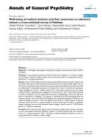
Chemical structures of compounds 1, 2, 3 and D 1: com-pound 1; 2: compound 2; 3: compound 3; 4: compound DFigure 1
Chemical structures of compounds 1, 2, 3 and D 1:
compound 1; 2: compound 2; 3: compound 3; 4: com-
pound D.
Chinese Medicine 2009, 4:19 />Page 3 of 5
(page number not for citation purposes)
photometer (Thermo Scientific, USA). The maximum
wavelength (λ
max
) was obtained at 228 nm. Standards of
each compound were freshly prepared at the concentra-
tion range of 0.62-3.73 μg/mL. Validations were per-
formed by five replicates of intra-day and three replicates
of inter-day. The linear correlation coefficients (r)
between the UV-absorption and the concentrations of all
compounds were in the range of 0.9993-0.9996 (com-
pound 1: Y = 109.67X+0.031, r = 0.9994; compound 2: Y
= 109.44X+0.028, r = 0.9995; compound 3: Y =
118.58X+0.049, r = 0.9996; compound D: Y =
169.72X+0.058, r = 0.9993).
Cell culture
Caco-2 cells were maintained in a DMEM at pH7.4, sup-
plemented with 10% fetal bovine serum, 2 mM L-
glutamine, 1% non-essential amino acid solution and
0.1% penicillin-streptomycin solution in a humidified
atmosphere (5% CO
2
, 95% air, at 37°C). Cells were
grown until 60-70% confluence. Cells from passages 20-
40 were used for all experiments. Cells were seeded on tis-
sue culture polycarbonate membrane filters (pore size: 3.0
μm) in 6-well Transwell plates (Corning, USA) at a seed-
ing density of 2 × 10
4
cells/cm
2
. Culture medium was
added to both the donor and the acceptor compartments.
Medium was changed every two days. Cells were left to
differentiate for 15-21 days after seeding with monitoring
of trans-epithelial electrical resistance (TEER) values with
a Millicell electrical resistance system (Millipore, USA)
and the value should be higher than 600Ω.cm
2
.
Chemical hydrolysis study of the compounds in transport
medium
Solution of compound 1, 2 and 3 at 53 μM and 106 μM
were prepared in Hank's balanced salt solution (HBSS) at
pH7.4. All solutions were kept at 4°C for 96 hours and
each solution was then analyzed by HPLC at 12, 24, 48,
72 and 96 hours to determine the hydrolysis product.
Transport studies
Transport experiment across the Caco-2 cell monolayers
at pH7.4 was performed. Caco-2 cell monolayers in Tran-
swell (6-well) plates were used for transport studies when
they were differentiated and the monolayer was intact, as
checked by measuring TEER. Prior to the experiment, the
cells were washed twice with phosphate buffered saline
(PBS) and pre-equilibrated for one hour with HBSS buff-
ered with 30 mM n-(2-hydroxyethyl) piperazine-n-(2-eth-
anosulfonic acid) (HEPES) at pH7.4. After medium was
removed, the cells were treated with sample solutions
(concentrations of 53 μM and 106 μM in HBSS at pH7.4)
in an apical compartment. Samples (1 mL) were taken
under sink conditions at 0, 5, 20, 40, 60, 80 and 100 min-
utes from the basolateral side and replaced with an equal
volume of fresh HBSS solution. The amount of the com-
pounds from the basolateral side was determined on a
UV-spectrophotometer (Thermo Scientific, USA) at 228
nm. Results were expressed as cumulative transport as a
function of time. Apparent permeability coefficient was
calculated according to the following equation:
where P
app
is the apparent permeability coefficient (cm/s),
dQ/dt (μg/s) is the rate of appearance of sample on the
basolateral side, A is the surface area of the monolayer
(cm
2
) and C
0
(μg/mL) is the initial drug concentration in
the donor compartment. All rate constants were obtained
from the permeation profiles of each compound. Statisti-
cal significance was evaluated with one-way analysis of
variance (one-way ANOVA). A value of P < 0.05 was con-
sidered statistically significant.
Enzyme hydrolysis study of the compounds
Compound 1 (2 mL, 10 μM solution) was pre-incubated
at 37°C and 200 μL of esterase enzyme (porcrine liver,
750 units) was added. Samples (200 μL each) were taken
at 5, 10, 20, 30 and 60 minutes and added to 200 μL of
methanol. Mixtures were vortexed to stop enzymatic activ-
ity. Samples were then centrifuged for five minutes at
14,000 × g (Lab Essentials, USA). Supernatant was
injected to the HPLC system for the determination of ester
pro-drugs and compound D. This procedure was repeated
for compounds 2 and 3.
Results
Compounds 1, 2 and 3 were prepared in transport
medium at concentrations of 53 μM and 106 μM and
stored at 4°C for 96 hours. The samples were collected
every 12 hours and analyzed on the HPLC system. Com-
pound D was detected by the HPLC after 24 hours. As the
fatty acid esters were not stable long enough in the trans-
ported medium, the HPLC analysis must be carried out in
a short period of time. A UV absorption method was used
to measure the amount of transported compounds in this
experiment. The UV analysis was designed and validated
in the range of 0.62-3.73 μg/mL and the linear correla-
tions (r) of all compounds were shown in the range of
0.9993-0.9996. All samples were analyzed and finished
within several hours.
Rate of transport of each compound was estimated from
the slope of the linear portion of a plot of cumulative
amount. The apparent permeability coefficients (P
app
) for
these compounds were calculated from the experimental
data which were determined from the apical to basolateral
side (Table 1, Figure 2).
The hydrolysis of the fatty acid esters was confirmed by an
in vitro assay with esterase enzyme from porcine liver.
PdQdt AC
app o
=×(/)(/ )1
Chinese Medicine 2009, 4:19 />Page 4 of 5
(page number not for citation purposes)
After the esters were mixed with esterase enzyme and incu-
bated at 37°C, the hydrolysis reaction was completed
within 10 minutes and compound D was detected.
The apparent permeability coefficient (P
app
) of com-
pounds 1 and 2 at 53 μM showed no significant difference
compared with those at concentration of 106 μM (com-
pound 1: P = 0.364; compound 2: P = 0.146). P
app
of com-
pound D at 106 μM was significantly different from that
at 53 μM (P = 0.001). Compound D had P
app
values close
to those of compounds 2 and 3 at 53 μM, while P
app
values
of compounds D, 1, 2 and 3 differed significantly (P =
0.001) at 106 μM.
The lipophilicity of these compounds can be estimated
with software ACD/labs version 11.03 (Advanced Chem-
istry Development, USA). The log P of compounds 1, 2,
and 3 were 10.13 (0.42), 10.65 (0.40) and 10.11 (0.39)
respectively.
Discussion
The results suggest that the absorption mechanism of
compounds 1 and 2 was not dependent on the concentra-
tions of the fatty acid esters. Facilitated transport may be
the mechanism. Compound D seemed to follow a con-
centration dependent passive diffusion mechanism.
P
app
values ranged from 7.47 × 10
-6
to 16.38 × 10
-6
cm/s in
an apical to basolateral direction. According to a previous
study [10], P
app
value in Caco-2 cells higher than 1 × 10
-
6
cm/s is associated with efficient intestinal absorption in
human. At 53 μM, compound 1 showed the highest trans-
port across the Caco-2 cell monolayers among all com-
pounds. Compound 2 demonstrated higher permeability
than that of compound D (P = 0.146). At 106 μM, com-
pound 1 showed higher transport across Caco-2 cell than
that of compounds 2 and 3 but lower transport than that
of compound D.
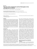
These lipophilicity values do not correlate well to the
transport results. More valid estimation of lipophilicity of
the compounds may come from analysis of retention time
of a reverse phase HPLC chromatogram [11,12] (Figure
3). The retention times (t
r
) of compounds 1, 2 and 3 were
92.4, 101.0 and 102.0 minutes, respectively. Polarity of
compound 1 was higher than that of compounds 2 and 3.
The hydrolysis of compound 1, 2 and 3 did not occur dur-
ing the transport across Caco-2 cell monolayers. This may
be due to the unusual long chain fatty acid structure of
these compounds and other factors such as the duration
of the time contact with the enzyme during transport.
However the hydrolysis of the ester linkage in these com-
pounds may occur in the blood circulation or in the liver
because these compounds can be hydrolyzed by porcine
liver esterase within in ten minutes.
Conclusion
Using the Caco-2 cell monolayer model, the present study
finds that compounds (E)-4-(3,4-dimethoxy-phenyl)but-
3-en-1-yl linoleate (1), (E)-4-(3,4-dimethoxy-phenyl)but-
3-en-1-yl oleate (2) and (E)-4-(3,4-dimethoxy-phe-
nyl)but-3-en-1-yl palmitate (3) originated from the Pras-
aplai preparation (a Thai herbal formula) may be
Table 1: Apparent permeability coefficient (P
app
) of compounds
1, 2, 3 and D (n = 3)
Compound *P
app
at 53 μM
(10
-6
cm/s)
*P
app
at 106 μM
(10
-6
cm/s)
P
Value
Compound 1 13.94 (0.60) 14.33 (0.17) 0.364
Compound 2 9.45 (0.29) 10.08 (0.32) 0.146
Compound 3 7.48 (0.31)** 8.60 (0.55)** 0.014
Compound D 8.53 (0.83)** 16.38 (0.61)** 0.001
*expressed as mean (SD), n = 3
**P < 0.05, significantly different between these two concentrations.
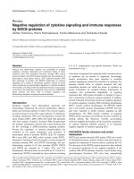
Cumulative amounts of the fatty acid esters and compound DFigure 2
Cumulative amounts of the fatty acid esters and
compound D. (A) At 53 μM in the apical to basolateral
direction, compound 1 (A1) showed highest cumulative
transport over other two fatty acid ester (A2 and A3) and
compound D. (B) At 106 μM in the apical to basolateral
direction, compound 1 (A1) showed higher cumulative trans-
port over other two fatty acid ester (A2 and A3) while com-
pound D had cumulative transport close to compound 3
(A3). A1: compound 1; A2: compound 2; A3: compound 3;
D: compound D.
Publish with BioMed Central and every
scientist can read your work free of charge
"BioMed Central will be the most significant development for
disseminating the results of biomedical research in our lifetime."
Sir Paul Nurse, Cancer Research UK
Your research papers will be:
available free of charge to the entire biomedical community
peer reviewed and published immediately upon acceptance
cited in PubMed and archived on PubMed Central
yours — you keep the copyright
Submit your manuscript here:
/>BioMedcentral
Chinese Medicine 2009, 4:19 />Page 5 of 5
(page number not for citation purposes)
transported through a facilitated mechanism and serve as
pro-drugs to increase the compound D level in the blood.
Abbreviations
HSV-1: herpes simplex virus-1; DMEM: Dulbecco's modi-
fied Eagle's medium; FBS: fetal bovine serum; TEER: trans-
epithelial electrical resistance; HBSS: Hank's balanced salt
solution; HEPES: n-(2-hydroxyethyl) piperazine-n-(2-eth-
anosulfonic acid); PBS: phosphate buffered saline; P
app
:
apparent permeability coefficient; ODS: octadecylsilane.
Competing interests
The authors declare that they have no competing interests.
Authors' contributions
PT and WG conceived the study design, synthesized the
compounds 1, 2, 3 and D, performed HPLC and UV anal-
ysis, and drafted the manuscript. JK and PO designed and
performed the Caco-2 cell experiment and helped analyze
the data. All authors read and approved the final version
of the manuscript.
References
1. Amatayakul T, Cannon JR, Dampawan P, Dechatiwong T, Giles RG,
Huntrakul C, Kusamran K, Mokkhasamit M, Raston CL, Reutrakul V,
White AH: Chemistry and crystal structures of some constit-
uents of Zingiber cassumuar. Aust J Chem 1979, 32:71-88.
2. Panthong A, Kanjanapothi D, Niwatananun V, Tuntiwachwuttikul P,
Reutrakul V: Anti-inflammatory activity of compounds iso-
lated from Zingiber cassumunar. Planta Med 1990, 56:655.
3. Kanjanapothi D, Soparat P, Panthong A, Tuntiwachwuttikul P, Reu-
trakul V: A uterine relaxant compound from Zingiber cassum-
unar. Planta Med 1987, 53:329-332.
4. Nualkaew S, Gritsanapan W, Petereit F, Nahrstedt A: New fatty
acid esters originate during storage by the interaction of
components in Prasaplai, a Thai traditional medicine. Planta
Med 2004, 70:1243-1246.
5. Tangyuenyongwatana P, Gritsanapan W: A study on artifacts for-
mation in the Thai traditional medicine Prasaplai. Planta Med
2008, 74:1403-1405.
6. Poomchusri NT: Ayurvedic Study 2nd edition. Bangkok: Promjakkan-
pimp; 1973.
7. Tangyuenyongwatana P, Gritsanapan W: Biological evaluations of
fatty acid esters originated during storage of Prasaplai, a
Thai traditional medicine. Nat Prod Res 2007, 21:990-997.
8. Hostetler KY, Parker S, Sridhar CN, Martin MJ, Li JL, Stuhmiller LM,
Vanwijk GM, van den Bosch H, Gardner MJ, Aldern KA, Richman DD:
Acyclovir diphosphate dimyristoylglycerol: a phospholipid
prodrug with activity against acyclovir-resistant herpes sim-
plex virus. Proc Natl Acad Sci 1993, 90:11835.
9. Yamashita S, Furubayashi T, Kataoka M, Sakane T, Sezaki H, Tokuda
H: Optimized conditions for prediction of intestinal drug per-
meability using Caco-2 cells. Eur J Pharm Sci 2000, 10:195-204.
10. Artursson P, Karlsson J: Correlation between oral drug absorp-
tion in humans and apparent drug permeability coefficients
in human intestinal epithelial (Caco-2) cells.
Biochem Biophys
Res Commun 1991, 175:880-885.
11. Lambert WJ: Modeling oil-water partitioning and membrane
permeation. using reversed-phase chromatography. J Chro-
matogr A 1993, 656:469-484.
12. Dorsey J, Khaledi M: Hydrophobicity estimation by reversed-
phase liquid chromatography. J Chromatogr A 1993,
656:485-499.
HPLC chromatograms of the mixture of Z. cassumunar and N. sativaFigure 3
HPLC chromatograms of the mixture of Z. cassumunar and N. sativa. 1: compound 1; 2: compound 2; 3: compound 3.