Báo cáo y học: "Antioxidant effects of ethyl acetate extract of Desmodium gangeticum root on myocardial ischemia reperfusion injury in rat hearts" pps
Bạn đang xem bản rút gọn của tài liệu. Xem và tải ngay bản đầy đủ của tài liệu tại đây (313.04 KB, 7 trang )
RESEARC H Open Access
Antioxidant effects of ethyl acetate extract of
Desmodium gangeticum root on myocardial
ischemia reperfusion injury in rat hearts
Gino A Kurian
1*
, Srilalitha Suryanarayanan
2
, Archana Raman
2
, Jose Padikkala
3
Abstract
Background: This study aims to evaluate the antioxidant potential of the ethyl acetate extract of Desmodium
gangeticum root for cardioprotection from ischemia reperfusion-induced oxidative stress.
Methods: The in vitro antioxidant potential of the extract was in terms of hydroxyl radical scavenging activity, lipid
peroxide scavenging activity, nitric oxide scavenging activity and diphenylpicryl hydrazyl radical scavenging activity.
The in vivo antioxidant potential of the extract was assessed in an isolated rat heart model.
Results: Free radicals were scavenged by the extract in a concentration-dependent manner within the range of
the given concentrations in all models. Administration of the ethyl acetate extract of Desmodium gangeticum root
(100 mg per kg body weight) before global ischemia caused a significant improvement of cardiac function and a
decrease in the release of lactate dehydrogenase in coronary effluent, as well as the level of malondialdehyde in
myocardial tissues.
Conclusion: The ethyl acetate extract of Desmodium gangeticum root protects the myocardium against ischemia-
reperfusion-induced damage in rats. The effects of the extract may be related to the inhibition of lipid
peroxidation.
Background
Many plants contain substantial amounts of antioxidants
such as vitamins C and E, carotenoids, flavonoids and
tannins that can be utilized to scavenge excess free radi-
cals from the human body [1]. The free radical scaven-
ging potential of natural antioxidants varies among
diseases and types of antioxidant [2].
Antioxidants protect the human body against free
radical attacks that may cause pathological conditions
such as ischemia reperfusion [3]. Ische mia reperfusion
causes tissue and cell damages when blood supply
returns after a period of ischemia (i.e. inadequate blood
supply) [4]. The onset of reperfusion in ischemic myo-
cardium results in the release of reactive oxygen species
[5]. The extensive production of reactive oxygen species
during ischemia reperfusion injury is deleterious to the
endogenous antioxidant defense pool. This recovery is
an effective defense mechanism during the postoperative
period of a patient.
Free radical scavengers and a ntioxidants have cardio-
protective eff ects in experimental ischemic r eperfusion
models [6]. There is growing interes t natural antioxi-
dants because of the concern over the possible carcino-
genic effects of synthetic antioxidants.
Desmodium gangeticum (Dayeshan Ludou, Fabaceae
fam ily) is found in India, China, Africa and Australia. It
is an important plant used in the indigenous Indian
medicine [7,8]ayurveda to treat various conditions such
as snakebite, ulcer and diabetes mellitus [9,10]. The ster-
ols, N,N-dimethyltryptamine, their oxides and other
derivatives have been isolated from aerial parts of the
plant; three pterocarpinoids, gangetin, gangetinin and
desmodin, are the major chemical constituents of the
root [11].
The present study investigates the use of ethyl acetate
extract of Desmodium gangeticum root to protect iso-
lated rat hearts fro m oxidative stress induced by ische-
mia reperfusion. In vitro and in vivo antioxidant models
* Correspondence:
1
School of Chemical and Biotechnology, SASTRA University,
Thirumalaisamudram, Thanjavur, Tamil Nadu, India
Kurian et al . Chinese Medicine 2010, 5:3
/>© 2010 Kurian et al; licensee BioMed Central Ltd. This is an Open Access article distributed under the terms of the Creative Commons
Attribution Licens e ( which permits unrestricted use, distribution, an d reproduction in
any medium, provided the original work is properly cited.
were used to assess the antioxidant potential of t he her-
bal extract.
Methods
Preparation of ethyl acetate extract of Desmodium
gangeticum root
The whole plant of Desmodium gangeticum was authen-
ticate d by Prof James Joseph. The voucher specimen A/
C no. 3908 was retained in our laboratory for future
reference.
The roots were dried under shade and ground to a
powder (100 g) which was extracted by ethyl acetate
(60-80°C) in a Soxh let apparatus for 72 hours. The
extract was concentrated under vacuum and dried at
room temperature. The brownish extract (8.8 g) was
resinous. Various qualitative tests [12] were performed
on the extract to confirm the chemical constituents,
namely triterpe noids, tannins, phenolic compounds
and glycosides. All chemicals used were of analytical
grade.
Experimental animals
Adult albino Wistar male rats (weighing 250-280 g)
were obtained from King Institute o f Preventive Medi-
cine, Chennai, India. They were fed on commercial rat
chow (Hindustan Lever, India) and had free access to
water. Handling of the animals was approved by the
Indian Ministry of Social Justices and Empowerment.
The experimental protocol was approved by the institu-
tional ethics committee.
Heart preparation
Isolated rat heart model was prepared according to Dör-
ing [13]. The rats were anesthetized at a dosage of 40
mg per kg b ody weight of sodium t hiopentenone. After
an intravenous inje ction of heparin (300 units), the
heart w as rapidly excised via a midsternal thoracotomy
and arrested in ice cold Krebs-Henseleit (KH) buffer
containing 118 mM/L NaCl, 4.7 mM/L KCl, 1.2 mM/L
MgSO
4
, 1.2 mM/L KH
2
PO
4
, 1.8 mM/L CaCl
2
, 25 mM/L
NaHCO
3
and 11 mM/L C
6
H
12
O
6
. The heart was
attached to a Lagendorf f apparatus via an aorta for ret-
rograde perfusion with KH buffer maintained at 37°C
and pH7.4 and saturated with a g as mixture o f 95 ml
O
2
and 5 ml CO
2
. The coronary perfusion pressure was
maintained at 80 mmHg. The left ventricular pressure
developed with t he ventricle filled with Krebs solution
was recorded with a pressure transducer, which in turn
was connected to a device amplifier and chart recorder.
This left ventricular pressure was an indication of the
mechanical performance of the heart. Coronary flow
wasmeasuredsimplybycollectingtheperfusatedrain-
ing from the heart in a graduated cylinder for a defined
time. The heart rate was measured by counting the
number of contractions (obtained from the left ventricu-
lar pressure recorder) per minute.
Experimental protocol
Rats were divided into three groups. In the normal/con-
trol group (Group 1), hearts were perfused for 90 min-
utes with KH buffer and used for the biochemical
analysis. In the reperfusion group (Group 2), the 30-
minute ischemic hearts (n = 6 in each subgroup) were
subjected to 15 minutes of reperfusion (Subgroup 2.1),
30 minutes of reperfusion (Subgroup 2.2) or 45 minutes
of reperfusion (Subgroup 2.3). All animals in the treat-
ment group (Group 3) were pretreated orally (through a
ball-tipped classic steel 15-16 gauge hypodermic needle)
with Desmodium gangeticum at a dose of 100 mg per kg
body weight for 30 days and then divided into three
subgroups . In Subgroup 3.1, rat hearts (n = 6) were per-
fused for 90 minutes with KH buffer and used for the
biochemical analysis. In Subgroup 3.2, rat hear ts (n =6)
were subjected to 30 minutes of glo bal ischemia after
equilibration, followed by 30 minutes of reperfusion. In
Subgroup 3.3, rat hearts (n = 6) were subjected to 30
minutes of global ischemia after equilibration, followed
by 45 minutes of reperfusion.
Biochemical assays
Thiobarbituric acid-reactive substances (TBARS) were
measured [14] as a marker of lipid peroxidation. The
endogenous antioxidants, superoxide dismutases (SOD)
Cu-Zn SOD and Mn SOD [15,16], catalase [17] and glu-
tathione peroxidase [18] were estimated in a UV-1601
Shimad zu spectrophotometer (Shimadzu, USA). Protein
concentration was measured with Folin phenol reagent
according to Lowry et al [19].
In vitro antioxidant activity
Determination of superoxide radical scavenging activity
Sup eroxide scavenging was determined by the nitroblue
tetrazolium reduction method [20]. The reaction mix-
ture consisted of ethylenediaminetetraacetic acid
(EDTA; 6 μM), sodium cyanide (3 μg), riboflavin (2
μM), nitroblue tetrazolium (50 μM), various concentra-
tions of Desmodium gangeticum extracts (5-50 μg/ml)
and phosphate buffer (67 mM, pH7.8) in a final volume
of 3 ml. The tubes were uniformly illuminated with an
incandescent visible light for 15 minutes, and the optical
density was measured at 530 nm before and after the
illumination. The percentage inhibition of superoxide
generation was evaluated by comparing the absorbance
values of the control and experimental tubes.
Determination of hydroxyl radical scavenging activity
The scavenging capacity for hydroxyl radical was mea-
suredaccordingtoamodifiedmethodofHalliwellet al.
[21]. Stock solutions of EDTA (1 mM), FeCl
3
(10 mM),
ascorbic acid (1 mM), H
2
O
2
(10 mM) and deoxyribose (10
mM) were prepared in distilled deionized water. The assay
was performed by adding 0.1 ml EDTA, 0.01 ml of FeCl
3
,
0.1 ml of H
2
O
2
, 0.36 ml of deoxyribose, 1.0 ml of Desmo-
dium gangeticum extract (10-100 μg/ml) dissolved in
Kurian et al . Chinese Medicine 2010, 5:3
/>Page 2 of 7
distilled water, 0.33 ml of phosphate buffer (50 mM,
pH7.4) and 0.1 ml of ascorbic acid in sequence. The mix-
ture was then incubated at 37°C for 1 hour. A 1.0 ml por-
tion of the incubated mixture was mixed with 1.0 ml of 10
g/100 g TCA and 1.0 ml of 0.5 g/100 g TBA (in 0.025 M
NaOH containing 0.025 g/100 g TBA) to develop the pink
chromogen measured at 532 nm. The hydroxyl radical
scavenging activity of the extract is reported as percentage
inhibition of deoxyribose degradation.
Lipid peroxide scavenging activity
A 5 ml reaction mixture containing rat liver homoge-
nate (0.1 ml, 25 g/100 ml) in Tris-HCl buffer (40 mM,
pH7.0), KCl (30 mM), ferrous iron (0.16 mM) and
ascorbic acid (0.06 mM) was incubated for 1 hour at 37°
C in the presence or absence of Desmodium gangeticum
extract (20-180 μg/ml). The lipid peroxidation was mea-
sured by TBARS formation [14]. Of this incubation mix-
ture, 0.4 ml was treated with sodium dodecyl sulphate
(8.1 g/100 ml, 0.2 ml), TBA (0.8 g/100 g, 1.5 ml) and
acetic acid (20 ml/100 ml, 1.5 ml, pH3.5). The total
volume was then made up to 4 ml by adding distilled
water and kept in a water bath at 100°C for 1 hour.
After cooling, 1 ml of distilled water and 5 ml of a mix-
ture of n-butanol and pyridine (15:1 v/v) was added.
The mixture was centrifuged at 5000 × g for10 minutes
and remixed. The absorbance of the organic layer was
measured at 532 nm. The percentage inhibition of lipid
peroxidation was determined by comparing results o f
the test compounds with those of controls and tubes
not treated with the extracts.
Diphenylpicrylhydrazyl radical scavenging activity
The free radic al scavenging activity of the Desmodium
gangeticum extract and butylated hydroxyl toluene was
measured with the stable radical diphenylpicrylhydrazyl
(DPPH) [22] in terms of hydrogen-donating or radical-
scavenging activity. A 0.1 mM solution of DPPH in
ethanol was prepared, and 1.0 ml of this solution was
added to 3.0 ml of extract solution in water at different
concentrations (10-100 μg/ml). After 30 minutes, the
absorbance was measured at 517 nm. Lower absorbance
of the reaction mixture indicates higher free radical
scavenging activity. The antioxidant activity of the
extract was expressed as IC
50
, which was defined as the
conce ntration (in μg/ml) of extract that inhibits the for-
mation of DPPH radicals by 50%.
Nitric oxide scavenging
Sodium nitroprusside in a queous solution at physiologi-
cal pH spontaneously generates nitric oxide (NO),
which interacts with oxygen to produce nitrite ions that
can be estimated by use of Griess reagent [23,24]. Sca-
vengers of NO compete with oxygen, leading to reduced
production of NO. Sodium nitroprusside (5 mM) in
phosphat e-buffered saline was mixed with 3.0 ml of var-
ious concentrations (10-320 μg/ml) of Desmodium gang-
eticum extract dissolved and incubated at 25°C for 150
minutes. The samples were then reacted with Greiss
reagent (1 g/100 ml sulphanilamide, 2 ml/100 ml
H
3
PO
4
, and 0.1 g/100 ml napthylethylenediamine dihy-
drochloride). The abso rbance of the chromophore
formed during the diazotization of nitrite with sulphani-
lamide and subsequent coupling with napthylethylene-
diamine was read at 546 nm and referred to the
absorbance of standard solutions of potassium nitrite
also treated with Griess reagent.
Gas chromatography-mass spectrometry (GC-MS) analysis
All GC-MS analyses were conducted with a PerkinElmer
Clarus 500 gas chromatograph (Perkin Elmer, USA).
The chromatographic conditions were as follows. Elite-1
(100 g/100 ml dimet hylpolysiloxane) column was used.
Helium was used as the carrier gas with a flow rate of 1
ml per minute. Desmodium gangeticum aqueous root
extract (1 ml) was injected into the system in splitless
modeat250°C.Thecolumnoventemperaturewas
maintained at 110°C for 2 minutes, then programmed at
75°C to 200°C for 1 minute and incre ased to 280°C by
sequential increment of 5°C per minute.
Table 1 Hemodynamic characteristics of rat hearts subjected to ischemia reperfusion
Group Left ventricular developed
pressure (mmHg)
Coronary flow
(ml/min)
Heart rate
(beats/min)
Rate pressure product ×10
3
(mmHg·beats/min)
Mean arterial pressure
(mmHg)
Normal control
1 99.21 ± 4.1 9.1 ± 1.24 340 ± 16.1 33.46 ± 4.3 121 ± 7
Ischemia reperfusion control
2.1 50.43 ± 4.0* 9.0 ± 0.19 255 ± 17.2* 12.14 ± 4.2* 97 ± 6*
2.2 52.86 ± 4.3* 9.0 ± 1.10 232 ± 18.3* 11.43 ± 5.2* 96 ± 7*
2.3 40.26 ± 4.3* 9.1 ± 1.02 235 ± 30.5* 9.55 ± 7.4* 113 ± 8
Drug treated
3.1 92.97 ± 4.9 9.2 ± 1.10 338 ± 27.8 31.24 ± 4.3 114 ± 7
3.2 75.21 ± 4.2* 9.1 ± 0.95 321 ± 30.2 22.22 ± 5.6* 104 ± 5*
3.3 84.70 ± 4.2 9.3 ± 1.05 320 ± 30.1 24.94 ± 7.4* 103 ± 6*
Values are mean ± SD in each group (n = 6). *P < 0.05, compared with control.
Kurian et al . Chinese Medicine 2010, 5:3
/>Page 3 of 7
Statistical analysis
All data are presented as m ean ± SD. Results were ana-
lyzed by one-way analysis of variance with SPSS software
12.00 (IBM, USA), followed by Duncan’s multiple range
test. P < 0.05 was considered statistically significant . Lin-
ear regression analysis was used to calculate IC
50
values.
Results
Hemodynamic changes occurred during ischemia reper-
fusion of the isolated rat heart. Reperfusing the ischemic
heart with KH buffer did not recover the mean arterial
pressure and heart rate in the early reperfusion stage of
the experiment. Because heart rate and left ventricular
developed pressure m ay recover to varying deg rees, the
rate pressure product was calculated by multiplying the
heart rate by the left ventricular developed pressu re and
is presented as a reliable left ventricular function para-
meter for the isolated heart (Table 1). No significant dif-
ference was noted between the experimental groups for
rate pressure product at the end of the 30-minute adap-
tation period before starting treatments and global
ischemia. During the 30-minute global ischemia, there
was a reduction in rate pressure product to zero, which
started to recover gradually by continued reperfusion.
Pretreatment with Desmodium gangeticum increased the
recovery of the rate pressure product in the drug group
(60% of basal value) compared with the reperfusion
group (35% of basal value) (Table 1).
Gas chromatography-mass spectrometry analysis
resulted in the identification of 38 compounds (Addi-
tional file 1). Major (71%) comprised n-hexadecanoic
acid, octadecanoic acid, 1,2-benzenedicarboxylic acid,
diisooctyl ester, phenol, 2,5-bis(1,1-dimethyl ethyl)-, 9-
octadecenoic acid(z)-methyl ester, 2,4-bis(1-pheny-
lethyl)phenol. Minor compounds such as cyclohexane,
isocyanato azulene, 1,4-dimethyl-7-(1-methyl ethyl)-, 1-
tridecanol, didodecyl phthalate, hexadecanoic acid
methyl ester, 1,2-benzenedicarboxylic acid, butyloctyl
ester, 1-hexadecanol and o leic acid were also identified.
Several c oncentrations ranging from 2 to 1000 μg/ml
of ethyl acetate extract of Desmodium gangeticum were
tested for their antioxidant a ctivity in various in vitro
models (Table 2). Free radicals were scavenged by the
test compounds in a concentration-dependent manner
within the given range of concentrations in all the mod-
els. The half maximum inhibitory concentration ( IC
50
)
in the DPPH, superoxide scavenging activity, hydroxide
scavenging activity, n itric oxide scavenging activity and
lipid peroxidation models were 36.3, 55.3, 43.7, 39.4 and
248 μg/ml respectively (Table 2 &3).
The in vivo antioxidant effect of the extract was deter-
mined by administering the rats with Desmodium gange-
ticum orally for 30 days and then sacrificing them for
reperfusion-induced ischemic injury. Lipid peroxidation
in drug treated rat hearts were reduced as compared to
ischemia reperfusion control hearts. Similarly antioxi-
dant enzymes also recovered significantly in drug treated
rat hearts (Table 4). The se observations in the present
study suggest a potent in vivo antioxidant capacity for
Desmodium gangeticum against revascularization injury.
Table 2 Free radical scavenging activities of Desmodium
gangeticum extract
Extract
concentration (μg/
ml)
Inhibition (%)
DPPH Nitric
oxide
Superoxide Hydroxyl
radical
1000 89.25 ±
2.11
87.21 ±
3.11
92.31 ± 2.63 81.27 ± 3.82
500 86.49 ±
3.46
82.28 ±
5.23
87.66 ± 3.51 78.63 ± 4.62
250 81.67 ±
2.34
77.55 ±
3.45
79.41 ± 3.65 74.41 ± 4.43
125 75.22 ±
3.74
70.39 ±
4.84
67.51 ± 2.78 65.52 ± 2.76
62 46.83 ±
2.28
46.63 ±
5.28
61.39 ± 3.51 51.62 ± 3.52
32 32.57 ±
3.38
38.68 ±
4.38
50.47 ± 2.54 30.61 ± 2.31
16 4.48 ±
2.55
19.25 ±
3.27
39.78 ± 2.89 21.42 ± 1.62
10 2.21 ±
1.52
7.52 ±
1.32
29.37 ± 1.12 4.21 ± 0.52
7 1.02 ±
0.74
4.33 ±
0.50
19.67 ± 1.44 3.34 ± 1.25
5 0.10 ±
0.03
1.31 ±
0.10
7.21 ± 1.05 1.23 ± 0.33
Ascorbic acid (100 μg) 95.11 ±
4.22
85.34 ±
4.11
87.32 ± 5.87 94.44 ± 4.71
Butylated
hydroxytoluene (20
μg)
92.27 ±
3.31
NT NT NT
Curcumin NT 91.7 ±
3.11
NT NT
IC
50
36.3 ±
1.47
39.4 ±
2.33
55.3 ± 1.29 43.7 ± 2.43
Values are mean ± SD of three replicates. NT: Not tested.
Table 3 Effects of ethyl acetate root extract of
Desmodium gangeticum on ferrous sulphate-induced lipid
peroxidation in rat liver homogenate
Extract concentration (μg/
ml)
TBARS (nmol/mg
protein)
a
Inhibition (%)
a
Control 2.32 ± 0.27
1000 0.1 ± 0.02 96.34 ± 2.7
800 0.38 ± 0.04 83.75 ± 2.6
600 0.55 ± 0.12 76.21 ± 2.1
400 0.87 ± 0.14 62.36 ± 2.5
200 1.00 ± 0.23 58.75 ± 2.4
Tocopherol (10 μmol/L) 0.07 ± 0.02 97.11 ± 3.5
a
Mean ± SD of n =6
Kurian et al . Chinese Medicine 2010, 5:3
/>Page 4 of 7
Cardiac enzymes like CK, LDH, SGOT and SGPT in the
tissue homogenate were significantly high in ischemia
reperfusion control rats (Table 5). However administration
of the DG root extract improved the level of these
enzymes and thereby mediates myocardial protection.
Discussion
Previous studies on the use of medicinal plants to treat
cardiac disorders suggested that methanol extract of
Desmodium gangeticum root renders cardioprotection
from isoproterenol-induced myocardial infarction in rats
[25,26]. The preventive effects of ethyl acetate extract of
Desmodium gangeticum root were shown in terms of
cardiac marker enzymes and antioxidants in ischemic
reperfused rat hearts. We found that ethyl acetate
extract of Desmodium gangeticum root induces myocar-
dial protection against ischemia reperfusion injury in
isolated rat hearts, as indicated by the improved
recovery of cardiac function, reduction in cardiac
enzyme release in the perfusate and reduction of tissue
necrosis.
The functional recovery of myocardium from ischemia
reperfusion-induced assault was observed through the
changes in hemodynamic parameters (Ta ble 1). Signifi-
cant recovery of left ventricular developed pressure in
drug-treated rat heart suggested the physiological recov-
ery of heart from ischemia r eperfusion injury. Similarly,
improvement of rate pressure product and mean arterial
pressure in ethyl acetate-treated rat heart explained the
recovered ionic balance for the normal physiological
functions of hearts.
The cardiac damage due to ischemia reperfusion was
monitored by the presence of cardiac marker enzymes
in the cardiac perfusate and the level of these enzymes
in myocardium. The presence of lactate d ehydrogenase
and creatine kinase in coronary perfusate of isol ated rat
Table 4 Effects of ethyl acetate root extract of Desmodium gangeticum on TBARS, catalase, superoxide dismutase
(SOD), and glutathione peroxidase (GPx) in the tissue homogenate of isolated rat hearts
Group TBARS (μM/g wet
tissue)
Catalase (μMofH
2
O
2
consumed/min/g
protein)
SOD (U/mg protein)
#
GPx (μg of GSH consumed/min/g
protein)
Mn
SOD
Cu-Zn
SOD
Normal control
1 6.1 ± 0.2 7617 ± 441 8.1 ± 0.62 50.2 ± 4.1 1859 ± 181
Ischemia reperfusion control
2.1 7.9 ± 0.6* 4 087 ± 246* 5.1 ± 0.52* 30.3 ± 3.5* 1228 ± 142*
2.2 7.5 ± 0.5* 5176 ± 372* 6.1 ± 0.54* 34.1 ± 3.2* 1117 ± 114*
2.3 7.1 ± 0.5* 5208 ± 316* 5.6 ± 0.57* 33.8 ± 3.8* 1216 ± 116*
Drug treated
3.1 5.9 ± 0.3 7856 ± 447 8.0 ± 0.71 50.1 ± 4.3 1855 ± 178
3.2 5.9 ± 0.3 7573 ± 433 8.0 ± 0.78 51.0 ± 4.9 1804 ± 183
3.3 4.8 ± 0.2* 6176 ± 455* 7.1 ± 0.62* 44.3 ± 4.1 1572 ± 176*
#
SOD unit: One unit is defined as the enzyme concentration required to inhibit the optical density (at 560 nm) produced by 50% of chromogen 50% in 1 minute.
Values are mean ± SD in each group (n = 6). Significantly differing values (from normal control group) are marked with an asterisk (P < 0.05).
Table 5 Activities of creatine kinase, lactate dehydrogenase, SGOT, and SGPT in the tissue homogenate of isolated rat
hearts
Group Creatine kinase (μmol
phosphorous liberated/min/mg
protein)
Lactate dehydrogenase (nmol
pyruvate liberated/min/mg
protein)
SGOT (nanomol pyruvate
generated/min/mg protein)
SGPT (nanomol pyruvate
generated/min/mg protein)
Normal control
1 16.1 ± 1.4 104.4 ± 8.7 35.3 ± 4.1 26.1 ± 2.5
Ischemia reperfusion control
2.1 9.2 ± 0.8* 62.4 ± 4.6* 19.8 ± 1.1* 14.6 ± 1.2*
2.2 10.1 ± 2.7* 59.5 ± 7.2* 17.1 ± 3.8* 13.3 ± 2.2*
2.3 10.4 ± 2.2* 57.1 ± 7.8* 16.6 ± 3.1* 14.9 ± 2.7*
Drug treated
3.1 15.55 ± 2.1 85.6 ± 8.1* 26.5 ± 1.3* 23.5 ± 2.1
3.2 15.56 ± 1.6 90.8 ± 6.5* 29.8 ± 2.8* 26.6 ± 1.9
3.3 15.84 ± 1.8 93.8 ± 6.9 30.5 ± 4.3 27.5 ± 2.8
Values are mean ± SD in each group (n = 6).
Values that differ significantly from normal control group are marked with an asterisk (P < 0.01).
Kurian et al . Chinese Medicine 2010, 5:3
/>Page 5 of 7
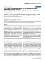
heart indicated myocardial necrosis [27]. In this study,
however, the levels of these enzymes in perfusate were
limited (Figure 1) and a subsequently increased level
was found in the myocardial tissue of rat hearts treated
with ethyl acetate extract (Table 5).
The superoxide anion scavenging activity of ethyl acet-
ate extract of Desmodium gangeticum root increased
markedly with the increase of concentrations (Table 2),
and the IC
50
of the ex tract was 55.3 μg/ml. The Desmo-
dium gangeticum extract exhibited concentration-depen-
dent scavenging activities against hydroxyl radicals
generated in a Fenton reaction system, and the IC
50
of
the extract was 43.7 μg/ml (Table 2). NO is known to
be involved in inflammation, cancer and other patholo-
gical conditions [28]. The Desmodium g angeticum
extract moderately inhibited NO in a dose-dependent
manner (Table 2), and the IC
50
was 39.4 μg/ml. The
Desmodium gangeticum extract inhibited FeSO
4
-induced
lipid peroxidation in rat liver in a dose-dependent man-
ner. The DPPH m ethod is a simple, rapid, and conveni-
ent method independent of sample polarity for
screening of many samples for radical scavenging activ-
ity[29].TheextractIC
50
value as measured by the
DPPH method was 36.3 μg/ml.
In vivo antioxidant potential of ethyl acetate extract o f
Desmodium gangeticum root was determined in isolated
rat hearts. A massive release of reactive oxygen species
was identified as one of the main causative factors for
myocardial ischemia reperfusion injury [6]. Xanthine
dehydrogenase, which normally utilizes NADH as an
electron acceptor, is converted under the conditions of
ischemia/reperfusion into xanthine oxidase, whic h uses
oxygen as a substrate [30]. Similarly, NA DPH oxidase
and mitochondrial electron transport chain complexes
were reported as the other sources of free radicals [6].
In the present study, increased myocardial TBARS indi-
cated oxidative stress induced by myocardial ischemia
reperfusion injury. However, administration of Desmo-
dium gangeticum extract not only reduced TBARS in
myocardium but also enhanced the recovery of antioxi-
dant enzymes from the assault of ischemia reperfusion
injury (Table 4).
Conclusion
The ethyl acetate extract of Desmodium gangeticum root
protects the myocardium against ischemia-reperfusion-
induced damage in rats. The effects of the extract may
be related to the inhibition of lipid peroxidation.
Additional file 1: Chemical composition of ethyl acetate extract of
Desmodium gangeticum root by gas chromatography-mass
spectrometry
Click here for file
[ />S1.DOC ]
Abbreviations
DG: Desmodium gangeticum; BHA: Butylated hydroxyanisole; BHT: Butylated
hydroxytoluene; IRI: Ischemia reperfusion injury; ROS: Reactive oxygen
species; KH: Krebs - Henseleit buffer; TBARS: Thiobarbituric acid reactive
substances; SOD: Superoxide dismutase; GPx: Glutathione peroxidase; NBT:
Nitroblue tetrazolium; DPPH: diphenylpicrylhydrazy l; MAP: Mean arterial
Figure 1 Activities of creatine kinase and lactate dehydrogenase in the perfusate of isolated rat hearts. Group 1: normal control; Group
2.1, 2.2, 2.3: ischemic reperfusion control; Group 3.1, 3.2, 3.3: drug pretreated and subjected to ischemic reperfusion. Values are mean ± SD in
each group (n = 6).
Kurian et al . Chinese Medicine 2010, 5:3
/>Page 6 of 7
pressure; HR: Heart rate; LVDP: Left ventricular developed pressure; RPP: Rate
pressure product
Acknowledgements
We would like to thank Prof James Joseph, Department of Botany, Saint
Berchman’s College, Mahatma Gandhi University, Kerala, India for his
assistance in authenticating the plant used in this study.
Author details
1
School of Chemical and Biotechnology, SASTRA University,
Thirumalaisamudram, Thanjavur, Tamil Nadu, India.
2
SASTRA University,
Thirumalaisamudram, Thanjavur, Tamil Nadu, India.
3
Department of Plant
Biotechnology, Amala Cancer Research Center, Amalanagar, Trichur, Kerala,
India.
Authors’ contributions
GAK designed the study, performed the experiment, interpreted the data
and prepared the manuscript. SS and AR performed the experiment and
revised the manuscript. JP designed the study, interpreted the data and
revised the manuscript. All authors read and approved the final version of
the manuscript.
Competing interests
The authors declare that they have no competing interests.
Received: 6 August 2009
Accepted: 22 January 2010 Published: 22 January 2010
References
1. Hasani-Ranjbar S, Larijani B, Abdollahi M: A systematic review of the
potential herbal sources of future drugs effective in oxidant-related
diseases. Inflamm Allergy Drug Targets 2009, 8(1):2-10.
2. Iannitti T, Palmieri B: Antioxidant therapy effectiveness: an up to date. Eur
Rev Med Pharmacol Sci 2009, 13(4):245-278.
3. van Rooyen J, Esterhuyse AJ, Engelbrecht AM, du Toit EF: Health benefits
of a natural carotenoid rich oil: a proposed mechanism of protection
against ischaemia-reperfusion injury. Asia Pac J Clin Nutr 2008, 17(Suppl
1):316-319.
4. Lefer DJ: Emerging role of nitrite in myocardial protection. Arch Pharm
Res 2009, 32(8):1127-1138.
5. Niccoli G, Burzotta F, Galiuto L, Crea F: Myocardial no-reflow in humans. J
Am Coll Cardiol 2009, 54(4):281-292.
6. Penna C, Mancardi D, Rastaldo R, Pagliaro P: Cardioprotection: a radical
view of free radicals in pre and postconditioning. Biochim Biophys Acta
2009, 1787(7):781-793.
7. Gosh D, Anandkumar A: Anti-inflammatory and analgesic activities of
Gangetin-A pterocarpenoid from Desmodium gangeticum. Indian J
Pharmacol 1981, 15:391-402.
8. Ghosal S, Bhattacharya SK: Desmodium alkaloids. II. Chemical and
pharmacological evaluation of Desmodium gangeticum. Planta Med 1972,
22:434-440.
9. Dharmani P, Mishra PK, Maurya R, Chauhan VS, Palit G: Desmodium
gangeticum: a potent anti-ulcer agent. Indian J Exp Biol 2001,
43(6):517-521.
10. Dharmani P, Palit G: Exploring Indian medicinal plants for antiulcer
activity. Indian J Pharmacol 2006, 38:95-99.
11. Purushothaman K, Kishore VM, Narayanaswamy V: The structure and
sterochemistry of Gangetin, a new pterocarpan from Desmodium
gangeticum (Leguminosae). J Chem Soc 1971, C:2420-2422.
12. Harbone JB: Phytochemical methods. A Guide to Modern Techniques of
plant Analysis New York: Chapman and Hall, 3 1998, 1-198.
13. Döring HJ: The isolated perfused heart according to Langendorff
technique–function–application. Physiol Bohemoslov 1990,
39(6):481-504.
14. Ohkawa H, Ohishi N, Yagi K: Assay for lipid peroxides in animal tissues by
thiobarbituric acid reaction. Anal Biochem 1979, 95:351-358.
15. Marklund S, Marklund G: Involvement of superoxide anion radical in
autoxidation of pyragallol and a convenient assay for superoxide
dismutase. Eur J Biochem 1974, 47:469-474.
16. Geller BL, Winge DR: A method for distinguishing CuZn SOD and Mn
containing superoxide dismutases. Anal Biochem 1983, 128:86-92.
17. Aebi HE: Catalase. Methods of Enzymatic Analysis Weinheim: VCH
VerlagBergmeyer HU 1984, 273-278.
18. Wendel A: Glutathione peroxidise. Methods of Enzymology San Diego:
Academic Press 1981, 325-333.
19. Lowry OH, Rosenbrough NJ, Farr AL, Randal RJ: Protein measurements
with the folin phenol reagent. J Biol Chem 1951, 193:265-275.
20. McCord JM, Fridovich I: Superoxide dismutase: an enzymatic function of
erythrocuprin. J Biol Chem 1969, 244:6049-6055.
21. Halliwell B, Gutteridge JMC, Aruoma OI: The deoxyribose method: a
simple “test tube” assay for determination of rate constants for
reactions of hydroxyl radicals. Anal Biochem 1987, 165:215-219.
22. Bandonienė D, Murkovic M: The detection of radical scavenging
compounds in crude extract of borage (Borago officinalis L.) by using an
on-line HPLC-DPPH method. J Biochem Biophys Methods 2002, 53(1-
3):45-49.
23. Green L, Wagner D, Glogowski J, Skipper PL, Wishnok JS, Tannenbaum SR:
Analysis of nitrate, nitrite and (
15
N) nitrate in biological fluids. Anal
Biochem 1982, 126:131-138.
24. Marcocci L, Maguire JJ, Droy-Lefaix MT, Packer L: The nitric oxide-
scavenging properties of Ginkgo biloba extract EGb 761. Biochem Biophys
Res Commun 1994, 201(2):748-755.
25. Kurian GA, Yagnesh N, Kishan RS, Paddikkala J: Methanol extract of
Desmodium gangeticum roots preserves mitochondrial respiratory
enzymes, protecting rat heart against oxidative stress induced by
reperfusion injury. J Pharm Pharmacol 2008, 60:523-530.
26. Kurian GA, Phillip S, Vargese S: Effect of aqueous extract of the
Desmodium gangeticum root in the severity of myocardial infarction. J
Ethnopharmacol 2005, 97:457-461.
27. De Windt LJ, Willems J, Roemen THM, Coumans WA, Reneman RS,
Vusse Van Der GJ, Van Bilsen M: Ischemic-reperfused isolated working
mouse hearts: membrane damage and type IIA phospholipase A2. Am J
Physiol Heart Circ Physio 2001, 280:2572-2580.
28. Fukumura D, Kashiwagi S, Jain RK: The role of nitric oxide in tumour
progression. Nature Rev Cancer 2006, 6:521-534.
29. Koleva II, Van Beek TA, Linssen JPH, De Groot A, Evstatieva LN: Screening of
plant extracts for antioxidant activity: a comparative study on three
testing methods. Phytochemical Analysis 2002, 13:8-17.
30. Baker JE, Su J, Fu X, Hsu A, Gross GJ, Tweddell JS, Hogg N: Nitrite confers
protection against myocardial infarction: role of xanthine
oxidoreductase, NADPH oxidase and K(ATP) channels. J Mol Cell Cardiol
2007, 43(4):437-444.
doi:10.1186/1749-8546-5-3
Cite this article as: Kurian et al.: Antioxidant effects of ethyl acetate
extract of Desmodium gangeticum root on myocardial ischemia
reperfusion injury in rat hearts. Chinese Medicine 2010 5:3.
Submit your next manuscript to BioMed Central
and take full advantage of:
• Convenient online submission
• Thorough peer review
• No space constraints or color figure charges
• Immediate publication on acceptance
• Inclusion in PubMed, CAS, Scopus and Google Scholar
• Research which is freely available for redistribution
Submit your manuscript at
www.biomedcentral.com/submit
Kurian et al . Chinese Medicine 2010, 5:3
/>Page 7 of 7