Báo cáo y học: "Altered muscular activation during prone hip extension in women with and without low back pain" ppt
Bạn đang xem bản rút gọn của tài liệu. Xem và tải ngay bản đầy đủ của tài liệu tại đây (255.93 KB, 6 trang )
RESEARCH Open Access
Altered muscular activation during prone hip
extension in women with and without low
back pain
Amir M Arab
1*
, Leila Ghamkhar
1
, Mahnaz Emami
2
and Mohammad R Nourbakhsh
3
Abstract
Background: Altered movement pattern has been associated with the development of low back pain (LBP). The
purpose of this study was to investigate the activity pattern of the ipsilat eral erector spinae (IES) and contrala teral
erectorspinae (CES), gluteus maximus (GM) and hamstring (HAM) muscles during prone hip exten sion (PHE) test in
women with and without LBP. A cross-sectional non-experimental design was used.
Methods: Convenience sample of 20 female participated in the study. Subjects were categorized into two groups:
with LBP (n = 10) and without LBP (n = 10). The electromyography (EMG) signal amplitude of the tested muscles
during PHE (normalized to maximum voluntary electrical activity (MVE )) was measured in the dominant lower
extremity in all subjects.
Results: Statistical analysis revealed greater normalized EMG signal amplitude in women with LBP compared to
non-LBP women. There was significant difference in EMG activity of the IES (P = 0.03) and CES (P = 0.03) between
two groups. However, no significant difference was found in EMG signals of the GM (P = 0.11) and HAM (P = 0.14)
among two groups.
Conclusion: The findings of this study demonstrated altered activation pattern of the lumbo-pelvic muscles during
PHE in the women with chronic LBP. This information is important for investigators using PHE as either an
evaluation tool or a rehabilitation exercise.
Keywords: Electromyography, Low back pain, Movement pattern, Prone hip extension
Background
Low back pain (LBP) is one of the most common and
costly musculoskeletal complaints in today’ssocieties,
affecting up to 70-80% of the population at least one
episode during their lifetime [1,2]. Despite its high inci-
dence and detrimental effects on individuals’ activities,
the exact causes of mechanical LBP have not yet been
fully u nderstood as any approach to diagnosis or treat-
ment has been shown to be clearly effective. However,
during the recent decades the approach in assessment
and treatment of LBP has been progressed from
strength ening of lumbo-pelvic muscles toward modifica-
tion of the motor system [3]. Balanced motor system is
resulted from coordinated activity of synergist and
antagonist muscles. According to this point of view,
repet itive movements and long-term faulty postures will
change muscle tissue characteristics and can lead to
muscle dysfunction, altered movement pattern, pain and
finally movement disorders [3]. Increased or decreased
muscle activity and delayed muscular activation can
change the normal move ment pattern [4,5]. Hence, the
main focus has been recently placed on modification of
the altered movement pattern in patients with muscu-
loskeletal pain [4,6,7].
Several studies have demonstrated altered activation
pattern of the certain lumbo-pelvic muscles during var-
ious tasks in people who suffer f rom LBP [8-11]. There
are few clinical tests that assess the altered movement
pattern in subjects with LBP. Prone hip extension (PHE)
which has been developed by Janda is a common and
* Correspondence:
1
Department of Physical Therapy, University of Social Welfare and
Rehabilitation Sciences, Evin, Tehran, Iran
Full list of author information is available at the end of the article
Arab et al . Chiropractic & Manual Therapies 2011, 19:18
/>CHIROPRACTIC & MANUAL THERAPIES
© 2011 Arab et al; licensee BioMed Central Ltd. This is an Open Access article distributed under the terms of the Creative Commons
Attribution License (http://cre ativecom mons.org/licenses/by/2.0), which permits unrestrict ed use, distribution, and reproduction in
any medium, provided the original work is properly cited.
widely accepted test for measuring the muscular activa-
tion pattern in the lumbo-pelvic area [4]. The impor-
tance of PHE is that the muscle activity pattern during
this movement has been theorized to simulate those
used during functional movement patterns such as gait
[5,6]. It is thought that changes in this pattern can
decrease the stability of lumbo-pelvic region during
walking [12]. Good reliability has been reported for PHE
in detecting deviation of lumbar spine from the midline
[13].
The timing (onset time) and amplitude of muscle acti-
vation are commonly measured to assess muscular acti-
vation patterns in musculoskeletal disorders using
electromyography (EMG) [14-17]. However, most pre-
vious studies have examined the timing of muscle activ-
ity during PHE in patients with LBP to determine the
order in which the muscles are activated during this
motor pattern [14-17].
To our knowledge, no study has investigated this
motor pattern in order to determine the amplitude of
lumbo-pelvic muscles activity in patients with chronic
LBP. The purpose of this study was to investigate the
amplitude of the activation pattern of the ipsilateral
erectors pinae (IES), contralateral erector spinae (CES),
ipsilateral gluteus maximus (GM) and ipsilateral ham-
string (HAM) muscles during PHE in women with and
without LBP and t o compare time broadness among
peak muscles activities in percent of total time of a
movement cycle between groups.
Methods
Subjects
A cross sectional study design was used to compare the
muscle activity pattern during PHE in two groups of
women: women with chronic non-specific LBP (N = 10,
average age: 33.6 (SD = 7.27) years old, average height:
163.1 (SD = 8.25) cm, average weight: 59.5 (SD = 10.34)
kg) and women with no history of LBP (N = 10, average
age: 29.8 (SD = 5.67) years old, average height: 161.2
(SD = 7.36) cm, average weight: 58.4 (SD = 5.44) kg).
The LBP patients were referred by orthopedic specialist
and physiotherapy clinics. The patients included if they
have a history of non-spe cific LBP for more than 6
weeks duration b efore the study date, or intermittent
LBP with at least three previous episodes lasting more
than one week during the year before the study [18].
The healthy subjects were recruited from university stu-
dents. The exclusion criteria in both groups were preg-
nancy, history of dyspnea, history of hip pain,
dislocation or fracture, history of lumbar spine surgeries,
history of anterior knee ligament injury or rupture, his-
tory of anterior knee pa in, recent episodes of ankle
sprain, leg length difference of more than 1 cm, inability
to perform active PHE without pain, history of lower
extremity i njury in the past 3 months, shortness of hip
flexors, those who participate in programs t o prepare
for competitive sports (exercise more than 3 days a
week), positive neurological symptoms and cardiopul-
monary disorders. Each eligible subject was enrolled
after signing an informed consent form approved by the
human subjects committ ee at the University of Social
Welfare and Rehabilitation Sciences. Ethic al approval for
this study was granted from the internal ethics commit-
tee at the University of Social Welfare and Rehabilita-
tion Sciences.
The dominant leg was chosen for investigation. The
muscle activ ity of IES, CES, GM and HAM duri ng PHE
was measured by the MIE-MT8 Telemetry EMG instru-
ment (MIE-Medical Research Ltd). A preamplifier with
a gain of (4000 ×), band pass fil tered (6-500 HZ), A-D
converted (sampling rate = 1000 HZ) was used. The
subjects were asked to lie prone with their arms at their
side and head was in mid lin e. The skin was shaved,
rubbed and cleaned with alcohol. To record muscle
activity, disposable, bipolar, self adhesive Ag/Agcl elec-
trodes were placed in pairs with distance of 1.5-2 cm
from each other and parallel to the muscle fibers [19].
Electrodes placement to collect EMG signals were as
follow: f or the ES muscles, bilaterally at least 2 cm lat-
eral to spinous process of L3 parallel t o the vertebral
column on the muscle belly; for the GM, at the mid
point o f a line running from S2 to the greater trochan-
ter; and for the HAM, laterally on the mid distance
between gluteal and popliteal fold.
The maximum voluntary electrical activity (MVE) for
each muscle was firstly calculated for normalization pro-
cedure. Test methods to calculate MVE w ere similar to
those described for manual muscle testing of the mus-
cles, as described by Kendall et al [20]. The pelvis was
secured to the bed with a sling to prevent pelvic motion
substitution only during MVE testing. For the ES mus-
cles the subject was asked to bring up her trunk against
the maximum resistance that entered bellow the scapula.
For the GM, hip joint was placed in extension position
and knee flexed to 90 degrees, resistance applied to the
distal aspect of posterior portion of thigh. The HAM
was tested while hip joint was placing in extension posi-
tion, the knee was flexed to nearly 70 degrees, and resis-
tance was applied to the distal aspect of the posterior
portion of the shank during knee flexion. Each contrac-
tion was repeated 2 times and held 5 seconds. One min-
ute rest was given between contractions. Before testing,
the subjects were familiarized with the standard position
and movement. All subjects were asked to lift the cho-
sen leg off the bed to 10 degrees whilst keeping the
knee straigh t, as soon as they heard the command “lift”.
An adjustable bar was placed at this level and the sub-
jects were asked to extend thei r hip until the calcaneous
Arab et al . Chiropractic & Manual Therapies 2011, 19:18
/>Page 2 of 6
touched the bar. The subjects were instructed o nly to
reach the adjustable bar and were not instructed to
press against the bar with the distal segment of the
lower extremity.
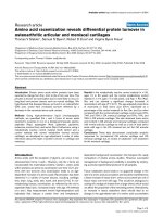
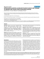
This was repeated 3 times for each individual. Figure 1
depicts an example of the raw EMG signals for tested
muscles. The raw data were processed into the root
mean square (RMS). The EMG signals collected during
hip extension were expressed as percentage of the calcu-
lated mean RMS of MVE (%MVE).
Time broadness is the time elapsed (in %) of the
motion cycle between the peak of the first muscle to
reach maximal activity and the peak of the last muscle
to reach maximal activity. Time broadness can show to
what extent the muscles are simultaneously involved i n
producing a motion during a motion cycle. Time broad-
ness provides indirect information on muscle coordina-
tion [21]. The muscle activit y pattern w as characterized
by maximal amplitude of normalized voluntary electrical
activity and by time broadness in the percent o f the
movement cycle. The pattern is different in case there is
a difference in any of the parameters above.
Data Analysis
Statistical analysis was performed using SPSS version
15.0. Independent t-test was used to compare the maxi-
mal amplitude of normalized voluntary electrical activity
of the tested muscles between women with and without
LBP. Stat istical significant was attributed to P value less
than 0.05.
Results
The demographic data for two individual groups are dis-
played in Table 1.
There was no statistically significant difference in sub-
jects’ age, height, weight and BMI among the two
groups.
The maximal amplitude of normalized electrical activ-
ity of the IES, CES, GM and HAM muscles during PHE
test in women with a nd without LBP is presented in
Table 2. There was significant difference in EMG activ-
ity of the IES (P = 0.03) and CES (P = 0.03) between
two groups. The results indicated that normalized elec-
trical activity o f the muscles during PHE is h igher in
women with LBP compared to those without LBP. How-
ever, no significant difference was found EMG signals of
theGM(P=0.11)andHAM(P=0.14)amongtwo
groups.
Discussion
The current study compared lumbo-pelvic muscle acti-
vation pattern between sub jects with and without L BP.
The results of this study showed higher maximal ampli-
tude of normalized electrical activity of the IES, CES in
patients with chronic LBP compared to those without
LBP. The normalized electrical activity of the GM and
HAM, although not statistically significant, was greater
in women with LBP than healthy subjects. These find-
ings demonstrate an altered activity pattern of the
lumbo-pelvic muscles during hip extension in patients
with chronic LBP. In this study, none of the subjects
reported that pain was a limiting factor to perform PHE
test, so, direct effects of pain can be minimized. How-
ever, nocioce ption can influence muscle activity. Bruno
et al [15] studied the PHE movement pattern difference
between subjects with and without LBP, measuring
onset time of the EMG activity in IES, CES, GM a nd
HAM. They found delayed activat ion of t he GM during
IES
CES
GM
HAM
Figure 1 Example of data recording from the tested muscles.
Arab et al . Chiropractic & Manual Therapies 2011, 19:18
/>Page 3 of 6
PHE in patients with unilateral LBP and concluded that
the movement pattern is changed in LBP [15].
In many other studies, increased signal EMG ampli-
tude of trunk muscle has been shown in patients with
LBP during functional activities such as bending the
trunk forward, back ward and gait [22-26]. In contrast,
some studies showed vague results or even reduced sig-
nal EMG amplitudes [27]. Some of these differences can
be explained by methodological problems, an important
one of them is how the data is normalized. Many factors
affect on absolute EMG amplitudes, such as thickness of
tissues overlying the muscle and skin impedance. To
obtain a net signal that is independent of these factors,
the EMG amplitude must be n ormalized to the ampli-
tudes obtained in MVE. However, this procedure may
not be appropriate for patients because they usually
unwillin g or not able to perform maximum contractions
due to pain or fear of re-creating pain. Normalization to
sub maximal contractions is not a good way because the
EMG amplitudes during these contractions will be
affected similarly to the levels during the activities to be
studied. In current study, MVE method was used
because patients had no pain during the test.
It is commonly believed that lumbo-pelvic instability is
an important component in chronic LBP. Investigators
have attributed the increased activity of trunk muscles
found in patients with LBP to functional adaptations fol-
lowing reduced spinal stability in t hese patients [ 26].
The spinal stabilizing system was primarily described by
Panjabi [28], including of 3 subsystems: the spinal col-
umn providing intrinsic stability; spinal muscles,
providing dynamic stability and neural control unit con-
trolling and determining the requirements for stability
and coordinating the muscle responses [28]. Under nor-
mal situatio ns, the three sub systems work in harmony
and provide the needed mechanical stability [29,30]. It
seems t hat the spinal instability as a result of dysfunc-
tion of spinal structures or decreased neural c ontrol is
compensated by increasing trunk muscle activity [28].
Co-contraction of ES muscles could be used to compen-
sate the loss of passive stability [22,31,32]. Muscles can
contribute to increase stability of trunk through co-con-
traction [31,33,34]. An alternative explanation might be
that in the spine, the local stabilizers muscles (e.g. Tr.A)
contract first then global stabilizer (e.g. ES), and acting
as synergist to increase the stability in times of extreme
need. With pain, injury or other pathologies an abnor-
mal stabilizer recruitment pattern can be developed
[35]. In this case, the activity of global stab ilizer muscles
will increase significantly to compensate the deep local
muscles dysfunction and decreased spinal stability.
Increased activity of ES, could cause pain in muscles
themselves, contribute to vicious circle of pain-spasm-
pain. In addition, co-contraction of trunk muscles would
increase the loads on the spine [36].
Increased GM activity, although not statistically signif-
icant, was found in subjects with LBP. According to Van
Wingerden [37], GM has an important role in sacroiliac
joint (SIJ) stability because of its perpendicular fibers to
the SIJ. Therefore, any pain and pelvic instability can
lead to increased muscle activity especially in tasks that
are required hip extension to enhance the SIJ stability.
However, about 2-20% of the patients suffering from
LBP have S IJ dysfunctions [38], while most of the
patients in this study demonstrated increased GM mus-
cle EMG activity. However, in this study we did not dif-
ferentiate the SIJ pain. More research is needed to
resolve the existing ambiguities in this area.
Increased activity of the HAM in women with LBP
may be due to high fatigability [39] and poor endurance
of the lumbar ES muscles [40,41]. As a result, increased
HAM activity is an adaptive mechanism following lum-
bar muscles fatigue and possibly weakness in those
Table 2 Electromyographic activity of the muscles during
prone hip extension in subjects with and without LBP
Muscle activity (%MVE) With no LBP With LBP P-value
Ipsilateral Erector Spinea 46.86 (25.57) 70.74 (21.80) 0.03
Contralateral Erector Spinea 50.36 (20.25) 72.11 (24.10) 0.04
Gluteus Maximus 29.81 (14.14) 42.32 (18.93) 0.11
Hamstring 52.78 (33.44) 74.06 (28.69) 0.14
Values are Mean (SD)
LBP = Low Back Pain
Table 1 Demographic data of the women in each group
Variables With no LBP (n = 10) With LBP (n = 10) P-value
Average (SD) Median Average (SD) Median
Age (years) 29.8 (5.67) 27.5 33.6 (7.27) 35 0.20
Weight (kg) 58.4 (5.44) 58.5 59.5 (10.34) 60.5 0.76
Height (cm) 161.2 (7.36) 160.5 163.1 (8.25) 161.5 0.59
BMI (kg/m
2
) 22.58 (2.88) 22.25 22.31 (3.31) 22.36 0.84
SD = Standard Deviation
LBP = Low Back Pain
BMI = Body Mass Index
Arab et al . Chiropractic & Manual Therapies 2011, 19:18
/>Page 4 of 6
muscles [42]. GM, BF, ES a nd latissimus do rsi are th e
key structures in providing SIJ stability [43]. Decrease in
endurance of ES in subjects with LBP may relax the
sacrotuberous l igament which is considered as the pri-
mary stabilizer structure in the SIJ [44]. The HAM can
affect on sacrotuberous ligament by its proximal attach-
ment to this ligament. It is thought that increased HAM
activity in patients with LBP may be a compensatory
functional mechanism resul ting from this situation [44].
Considering difference in muscle activity pattern during
PHE b etween subjects with and without LBP, PHE can
be used as either an evaluation tool or a rehabilitation
exercise for the subjects with LBP.
However, we acknowledge several important limita-
tions. One of the limitations and weakness of this study
was the sample size.
One point must be consider ed with regard to general-
izing the present results, is the sample population. In
this study, only women were recruited and men were
not included. Therefore the results of this study may be
more applicable to female subjects, who constituted the
participants and could not be extrapolated to the men.
It is suggested to perform this study in men to compare
data between men and women.
EMG measurements do not always guarantee magni-
tude of force production and therefore muscle strength,
as in some cases an inhibited muscle may be working
harder than normal to produce the required force for a
particular task. The timing of muscle activity in addition
to EMG amplitude can provide more useful information
regarding the muscular activation pattern.
Another area of concern in o ur study w as this issu e
that L BP women were not categorized as with or with-
out SIJ involvement.
Conclusions
The results of this study indicate higher maximal ampli-
tude of normalized electrical activity of the IES, CES in
patients with chronic LBP compared to those without
LBP. The normalized electrical activity of the GM and
HAM, although not statistically significant, was also
greater in women with LBP than healthy subjects. These
findings demonstrate an altered activity pattern of the
lumbo-pelvic muscles during hip extension in patients
with chronic LBP. This information is important for
investigators us ing PHE as either an evaluation tool or a
rehabilitation exercise.
List of abbreviations
LBP: Low Back Pain; IES: Ipsilateral erector spinae; CES: Contralateral erector
spinae; GM: Gluteus maximus; HAM: Hamstring; PHE: Prone hip extension;
EMG: Electromyography; MVE: Maximum voluntary electrical activity.
Author details
1
Department of Physical Therapy, University of Social Welfare and
Rehabilitation Sciences, Evin, Tehran, Iran.
2
Student Research Committee,
University of Social Welfare and Rehabilitation Sciences, Evin, Tehran, Iran.
3
Department of Physical Therapy, North Georgia College and State
University, Dahlonega, GA, USA.
Authors’ contributions
AMA contributed to conception, design, analysis, interpretation of data and
drafting the manuscript. LG carried out the data collection and involved in
interpretation of data and drafting the manuscript. ME participated in data
collection and analysis of EMG signals. MRN participated in design and
helped to draft the manuscript. All authors read and approved the final
manuscript.
Competing interests
The authors declare that they have no competing interests.
Received: 12 September 2010 Accepted: 14 August 2011
Published: 14 August 2011
References
1. Ekman M, Jonhagen S, Hunsche E, Jonsson L: Burden of illness of chronic
low back pain in Sweden: a cross-sectional, retrospective study in
primary care setting. Spine 2005, 30:1777-85.
2. Van Tulder M, Koes B: Low back pain and sciatica: chronic. Clin Evid 2002,
1032-48.
3. Sahrmann S: Diagnosis and treatment of movement impairment
syndromes. Missouri: Mosby. Inc;, 1 2002, 121-92.
4. Janda V: On the concept of postural muscles and posture in man. Aust J
Physiother 1983, 29:83-4.
5. Sahrmann S: Posture and muscle imbalance: faulty lumbar-pelvic
alignment and associated musculoskeletal pain syndromes. Orthop Div
Rev 1992, 13-20.
6. Janda V: Pain in the locomotor system-A broad approach. Aspects of
Manipulative Therapy Melbourne: Churchill Livingstone; 1985, 148-51.
7. O’Sullivan P, Phyty D, Twomey L, Allison G: Evaluation of specific
stabilizing exercise in the treatment of chronic low back pain with
radiologic diagnosis of spondylolysis or spondylolisthesis. Spine 1997,
22:2959-67.
8. Hodges P, Moseley G: Pain and motor control of the lumbopelvic region:
effect and possible mechanisms. J Electromyogr Kinesiol 2003, 13:361-70.
9. Hungerford B, Gilleard W, Hodges P: Evidence of altered lumbopelvic
muscle recruitment in the presence of sacroiliac joint pain. Spine 2003,
28:1593-600.
10. Leinonen V, Kankaanpaa M, Airaksinen O, Hanninen O: Back and hip
extensor activities during trunk flexion/extension: Effects of low back
pain and rehabilitation. Arch Phys Med Rehabil 2000, 81:32-7.
11. Newcomer K, Jacobson T, Gabriel D, Larson D, Brey R, An K: Muscle
activation patterns in subjects with and without low back pain. Arch
Phys Med Rehabil 2002, 83:816-21.
12. Vogt L, Pfeifer K, Banzer W: Neuromuscular control of walking with
chronic low-back pain. Man Ther 2003, 8:21-8.
13. Murphy D, Byfield D, Mccarthy P, Humphreys K, Gregory A, Rochon R:
Interexaminer reliability of the hip extension test for suspected impaired
motor control of the lumbar spine. J Manipulative Physiol Ther 2006,
29:374-7.
14. Lehman G, Lennon D, Tresidder B, Rayfield B, Poschar M: Muscle
recruitment patterns during the prone leg extension. BMC Musculoskelet
Disord 2004, 5:3-7.
15. Bruno P, Bagust J: An investigation into motor pattern differences used
during prone hip extension between subjects with and without low
back pain. Clin Chiro 2007, 10
:68-80.
16.
Lewis C, Sahrmann S: Muscle Activation and Movement Patterns During
Prone Hip Extension Exercise in Women. J Athl Train 2009, 44:238-48.
17. Sakamoto A, Teixeira-Salmela L, de Paula-Goulart F, de Morais Faria C,
Guimaraes C: Muscular activation patterns during active prone hip
extension exercises. J Electromyogr Kinesiol 2009, 19:105-12.
Arab et al . Chiropractic & Manual Therapies 2011, 19:18
/>Page 5 of 6
18. Nourbakhsh M, Arab A: Relationship between mechanical factors and
incidence of low back pain. J Orthop Sports Phys Ther 2002, 32:447-60.
19. Cram J, Kasman G, Holtz J: Introduction to surface EMG. Maryland: Aspen
Publishing, Gathersburg, PA);, 1 1998, 336-70.
20. Kendall F, McCreary E, Provance P: Muscles Testing and Function
Baltimore. MD: Williams & Wilkins; 1993.
21. Illyés Á, Kiss J, Kiss R: Electromyographic analysis during pull, forward
punch, elevation and overhead throw after conservative treatment or
capsular shift at patient with multidirectional shoulder joint instability. J
Electromyogr Kinesiol 2009, 19:e438-47.
22. Ambroz C, Scott A, Ambroz A, Talbott E: Chronic low back pain
assessment using surface electromyography. J Occup Environ Med 2000,
42:660-9.
23. Arendt-Nielsen L, Graven-Nielsen T, Svarrer H, Svensson P: The influence of
low back pain on muscle activity and coordination during gait: a clinical
and experimental study. Pain 1996, 64:231-40.
24. Roland M: A critical review of the evidence for a pain-spasm-pain cycle
in spinal disorders. Clin Biomech 1986, 1:102-9.
25. Ferguson S, Marras W, Burr D, Davis K, Gupta P: Differences in motor
recruitment and resulting kinematics between low back pain patients
and asymptomatic participants during lifting exertions. Clin Biomech
2004, 19:992-9.
26. Van Dieen J, Cholewicki J, Radebold A: Trunk muscle recruitment patterns
in patients with low back pain enhance the stability of the lumbar
spine. Spine 2003, 28:834-41.
27. Lund J, Donga R, Widmer C, Stohler C: The pain-adaptation model: a
discussion of the relationship between chronic musculoskeletal pain and
motor activity. Can J Physiol Pharmacol 1991, 69:683-94.
28. Panjabi M: The stabilizing system of the spine. Part I. Function,
dysfunction, adaptation, and enhancement. J Spinal Disord 1992, 5:383-9.
29. Barr K, Griggs M, Caby T: Lumbar stabilization: core concept and current
literature I. Am J Phys Med Rehabil 2005, 84:473-80.
30. Bergmark A: Stability ofthe lumbar spine. A study in mechanical
engineering. Acta Orthop Scand 1989, 230:20-4.
31. Cholewicki J, Panjabi M, Khachatryan A: Stabilizing function of trunk
flexor-extensor muscles around a neutral spine posture. Spine 1997,
22:2207-12.
32. Granata K, Marras W: Cost-benefit of muscle cocontraction in protecting
against spinal instability. Spine 2000, 25:1398-404.
33. Cholewicki J, Simons A, Radebold A: Effects of external trunk loads on
lumbar spine stability. J Biomech 2000, 33:1377-85.
34. Gardner-Morse M, Stokes I: Trunk stiffness increases with steady-state
effort. J Biomech 2001, 34:457-63.
35. Magee D, Zachazewski J, Quillen W: Scientific foundations and principles
of practice in musculoskeletal rehabilitation. WB Saunders Co; 2007.
36. Van Dieen J, De Looze M: Sensitivity of single-equivalent trunk extensor
muscle models to anatomical and functional assumptions. J Biomech
1999, 32:195-8.
37. van Wingerden J, Vleeming A, Buyruk H, Raissadat K: Stabilization of the
sacroiliac joint in vivo: verification of muscular contribution to force
closure of the pelvis. Eur Spine J 2004, 13:199-205.
38. Maigne J, Aivaliklis A, Pfefer F: Results of sacroiliac joint double block and
value of sacroiliac pain provocation tests in 54 patients with low back
pain. Spine 1996, 21:1889-92.
39. Mannion A, Connolly B, Wood K, Dolan P: The use of surface EMG power
spectral analysis in the evaluation of back muscle function. J Rehabil Res
Dev 1997, 34:427-39.
40. Biering-Sorensen F: Physical measurements as risk indicators for low-back
trouble over a one-year period. Spine 1984, 9:106-9.
41. Biering-Sorensen F: A one-year prospective study of low back trouble in
a general population. The prognostic value of low back history and
physical measurements. Dan Med Bull 1984, 31:362-75.
42. Hammill R, Beazell J, Hart J: Neuromuscular consequences of low back
pain and core dysfunction. Clin Sports Med 2008, 27:449-62.
43. Cholewicki J, Juluru K, McGill S: Intra-abdominal pressure mechanism for
stabilizing the lumbar spine. J Biomech 1999, 32:13-7.
44. Vleeming A, Snijders C, Stoeckart R, Mens J: The role of the sacroiliac
joints in coupling between spine, pelvis, legs and arms. Movement,
stability, and low back pain: The essential role of the pelvis New York:
Churchill Livingstone; 1997, 53-71.
doi:10.1186/2045-709X-19-18
Cite this article as: Arab et al.: Altered muscular activation during prone
hip extension in women with and without low back pain. Chiropractic &
Manual Therapies 2011 19:18.
Submit your next manuscript to BioMed Central
and take full advantage of:
• Convenient online submission
• Thorough peer review
• No space constraints or color figure charges
• Immediate publication on acceptance
• Inclusion in PubMed, CAS, Scopus and Google Scholar
• Research which is freely available for redistribution
Submit your manuscript at
www.biomedcentral.com/submit
Arab et al . Chiropractic & Manual Therapies 2011, 19:18
/>Page 6 of 6