Báo cáo y học: "Vaginal metastasis of a Ewing sarcoma five years after resection of the primary tumor" pdf
Bạn đang xem bản rút gọn của tài liệu. Xem và tải ngay bản đầy đủ của tài liệu tại đây (1.25 MB, 3 trang )
CAS E REP O R T Open Access
Vaginal metastasis of a Ewing sarcoma five years
after resection of the primary tumor
Noemie Vanel
1
, Victoire Vierling
1
, Jennifer Kreshak
1
, Marco Gambarotti
1
, Stefania Cocchi
1
, Cristina Tranfaglia
2
and
Daniel Vanel
1*
Abstract
A 35-year-old female presented with pain and swellin g of the distal left radius. A diagnosis of Ewing sarcoma was
made and she underwent neoadjuvant chemotherapy and surgery. Macroscopic viable areas remained on the map
of the surgical specimen; as such, she was classified as a poor responder and received high dose adjuvant
chemotherapy. She remained disease-free for five years, until age 40. A vaginal polyp was then detected during a
routine gynaecologic examination. It was removed and histopathology revealed metastatic Ewing sarcoma.
To our knowledge, this is the first reported case of a vaginal metastasis of Ewing sarcoma.
Keywords: Ewing sarcoma, vaginal metastasis
Introduction
Ewing sarcoma (ES) is a small blue round cell tumor
belonging to the Ewing Family Tumour (EFT) together
with Primary Neuroectodermal Tumour (PNET) and
ASKIN tumor (of the thoracic wall). Eight hundred and
ninety six cases have been reported in our institute
since 1982. Ewing sarcoma has a distinct predilection
for m ales and occurs in the first t wo decades of life in
more than 75 percent of cases [1,2].
Metastases are frequent [3] and are mostly pulmonary
and osseo us, but can be found in various other
locations.
We present here the first description of a vaginal
metastasis of Ewing sarcoma.
Case Report
A 35 year-old woman with no significant medical his-
tory presented with pain and swelling of the left wrist
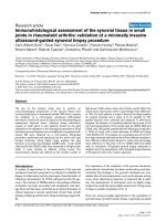
over the past year. A radiograph and computed tomo-
graphy scan revealed a lytic lesion of the distal left
radius (Figure 1), with soft tissue extension on MR
examination (Figure 2, 3).
A biopsy was performed and histological examination
revealed a typical Ewing sarcoma. The diagnosis was
confirmed by FISH analysis, which demonstrated the
translocation t (11, 22).
Staging revealed a solitary non-specific pulmonary
nodule of the inferior right lobe that did not change
with time and was not considered metastatic.
The patient underwent neoadjuvant chemotherapy and
resection and allograft of the distal radius. She was con-
sidered as poor responder as macroscopic areas
remained on the surgical specimen, but all margins were
free of disease. High dose chemotherapy was then
performed.
After completion of her treatment, it was followed up
as per protocol and remained disease-free.
Five years l ater, during a routine gynaecologic exam, a
vaginal polyp was found and removed. Histology
revealed a ES metastasis (Fig ure 4), as confirmed by the
characteristic transl ocation t(11, 22).(F igure 5). The rest
of the evaluation (CT and bone scintigraphy) was
normal.
No treatment has been undertaken.
Discussion
Ewing s arcoma (ES) represents approximately ten per-
cent[1] of primary malignant bone tumours and one
percent of soft tissue tumours. It t ends to arise in the
diaphysis or metaphyseal-diaphyseal portion of long
bones, although any bone may be involved.
Frequently, the first symptoms are pain and swelling.
* Correspondence:
1
Departement of Pathology, the Rizzoli Institute, Via del Barbiano 1/10,
40106, Bologna, Italy
Full list of author information is available at the end of the article
Vanel et al. Clinical Sarcoma Research 2011, 1:9
/>CLINICAL SARCOMA RESEARC
H
© 2011 Vanel et al; licensee BioMed Central Ltd. This is an Open Access article distributed under the terms of the Creative Commons
Attribution L icense ( which permits unrestricted use, distribution, and reproduction in
any medium, provided the original work is properly cited.
Twenty percent of ES at diagnosis hav e radiographic
evidence of metastasis. Lungs and bone are the main
metastatic locations.
Radiologically, an aggressive osteolytic lesion is com-
monly observed.
ES is characterized by a morphologically uniform
round cell proliferation with round nuclei containing
fine chromatin.
CD99 is expressed in nearly all ES and thereby is a
highly sensitive immunohistochemical marker.
Several studies have confirmed a cha racteristic 11, 22
(q24, q12) chromosomal translocation in 85 percent o f
the cases [3]; the translocation t (21, 22) and three even
rarer translocations (t (7, 22), t(2, 22) t(17, 22)) have
also been found.
Necrosis has a strong prognostic value [4]. High dose
chemotherapy is used in poor responders [5].
Figure 1 Initial evaluation. CT: lytic lesion with partial cortical
destruction.
Figure 2 Axial T1W MR image after contrast medium injection:
the soft tissue extension is well studied.
Figure 3 Sagital T1W MR image, after contrast medium
injection. Medullary and soft tissue extensions are well evaluated.
Figure 4 The metastasis is made of homogeneous small round
cells.
Vanel et al. Clinical Sarcoma Research 2011, 1:9
/>Page 2 of 3
We fo und 17 cases of primary ES involving the vagina
and/or vulva in the literature[6]. A few cases of primary
neuro ectodermal tumors (PNET) in the pelvis have also
been reported [7].
Unusual metastasic locations have been described, for
example, the breast [8], myocardial muscle [9], paranasal
sinuses [10], iris [11]), and pancreas [12]. That explains
why, even if a second primary cannot be completely
excluded in our case, the probability of a metastasis is
much higher. To our knowledge, this is the first case
ever reported of a vaginal Ewing metastasis.
Conclusion
This case exemplifies the idea that every new lesion in a
patient with Ewing sarcoma should be considered as a
possible metastasis.
Author details
1
Departement of Pathology, the Rizzoli Institute, Via del Barbiano 1/10,
40106, Bologna, Italy.
2
Departement of Nuclear Medicine, San Orsola
Hospital, Via Giuseppe Massarenti, 940138 Bologna, Italy.
Authors’ contributions
NV wrote the article, VV checked the case, JK corrected the writing (English
and scientific content) MG checked and selected the histology, SC checked
and selected the FISH, CT checked the scientific content, DV proposed the
subject and directed the article. All authors read and approved the final
manuscript
Competing interests
The authors declare that they have no competing interests.
Received: 13 April 2011 Accepted: 1 August 2011
Published: 1 August 2011
References
1. Dahin’s Bone Tumors. Lippincott ,618:249.
2. Bacci G, Ferrari S, Rosito P, Avella M, Barbieri E, Picci P, Battistini A, Brach del
Prever A: Minerva Pediatr. Ewing’s sarcoma of the bone. Anatomoclinical
study of 424 cases 1992, 44(7-8):345-59.
3. Zoubek A, Kovar H, Gadner H: Cytogenetic and molecular genetic
changes in malignant primary bone tumors] Radiologe. 1998,
38(6):467-71.
4. Picci P, Böhling T, Bacci G, Ferrari S, Sangiorgi L, Mercuri M, Ruggieri P,
Manfrini M, Ferraro A, Casadei R, Benassi MS, Mancini AF, Rosito P,
Cazzola A, Barbieri E, Tienghi A, Brach del Prever A, Comandone A,
Bacchini P, Bertoni F: Chemotherapy-induced tumor necrosis as a
prognostic factor in localized Ewing’s sarcoma of the extremities. J Clin
Oncol 1997, 15(4):1553-9.
5. Ferrari S, Sundby Hall K, Luksch R, Tienghi A, Wiebe T, Fagioli F,
Alvegard TA, Brach Del Prever A, Tamburini A, Alberghini M, Gandola L,
Mercuri M, Capanna R, Mapelli S, Prete A, Carli M, Picci P, Barbieri E, Bacci G,
Smeland S: Nonmetastatic Ewing family tumors: high-dose
chemotherapy with stem cell rescue in poor responder patients. Results
of the Italian Sarcoma Group/Scandinavian Sarcoma Group III protocol.
Ann Oncol 2010.
6. McCluggage WG, Sumathi VP, Nucci MR, Hirsch M, Dal Cin P, Wells M,
Flanagan AM, Fisher C: Ewing family of tumours involving the vulva and
vagina: report of a series of four cases. J Clin Pqthol 2007, 60(6):674-80.
7. Raney RB, Asmar L, Newton WA Jr, Bagwell C, Breneman JC, Crist W,
Gehan EA, Webber B, Wharam M, Wienes ES, Anderson JR, Maurer HM:
Ewing’s sarcoma of soft tissue in childhood: a report of the Intergroup
Rhabdomyosarcoma study,1972 to 1991. J Clin Oncol 1997, 15(2):574-82.
8. Astudillo L, Lacroix-triki M, Ferron G, Rolland F, Maisongrosse V, Chevreau C:
Bilateral breast metastases from Ewing sarcoma of the femur. Am I clin
Oncol 2005, 28(1):102-3.
9. Larbre F, Elbaz N, Verney R, Gilly J, Rousson R: Acute cardiac failure caused
by myocardial metastasis of an unrecognised Ewing sarcoma. Pediatrie
1981, 36(2):135-40.
10. Gaba RC, Cousins JP, Basil IS, Shadid H, Valyi-Nagy T, Mafee MF: Metastatic
Ewing sarcoma masquerading as olfactory neuroblastoma. Eur Arch
Otorhinolaryngol 2006, 263(10):960-2, Epub 2006 Jun 27.
11. Gündüz K, Shields JA, Shields CL, De Potter P, Wayner MJ: Ewing sarcoma
metastatic to the iris. Am J Ophthalmol 1997, 124(4) :550-2.
12. Mulligan ME, Fellows DW, Mullen SE: Pancreatic metastasis from Ewing’s
sarcoma. Clin Imaging 1997, 21(1):23-6.
doi:10.1186/2045-3329-1-9
Cite this article as: Vanel et al.: Vaginal metastasis of a Ewing sarcoma
five years after resection of the primary tumor. Clinical Sarcoma Research
2011 1:9.
Submit your next manuscript to BioMed Central
and take full advantage of:
• Convenient online submission
• Thorough peer review
• No space constraints or color figure charges
• Immediate publication on acceptance
• Inclusion in PubMed, CAS, Scopus and Google Scholar
• Research which is freely available for redistribution
Submit your manuscript at
www.biomedcentral.com/submit
Figure 5 Inter phase FISH with the EWSR1 (22q12) bre ak-apart
probe. Within a single nucleus fused red/green signals mark one
intact 22q12 region, whereas split red/green signals indicate the
presence of an EWSR1 gene rearrangement.
Vanel et al. Clinical Sarcoma Research 2011, 1:9
/>Page 3 of 3