Báo cáo y học: "Inhibition of complement C5a prevents breakdown of the blood-brain barrier and pituitary dysfunction in experimental sepsis" ppt
Bạn đang xem bản rút gọn của tài liệu. Xem và tải ngay bản đầy đủ của tài liệu tại đây (815.98 KB, 9 trang )
Open Access
Available online />Page 1 of 9
(page number not for citation purposes)
Vol 13 No 1
Research
Inhibition of complement C5a prevents breakdown of the
blood-brain barrier and pituitary dysfunction in experimental
sepsis
Michael A Flierl
1
, Philip F Stahel
1
, Daniel Rittirsch
2,5
, Markus Huber-Lang
3
,
Andreas D Niederbichler
4
, L Marco Hoesel
5
, Basel M Touban
1
, Steven J Morgan
1
, Wade R Smith
1
,
Peter A Ward
5
and Kyros Ipaktchi
1
1
Department of Orthopaedic Surgery, Denver Health Medical Center, University of Colorado School of Medicine, 777 Bannock Street, Denver, CO
80204, USA
2
Department of Trauma Surgery, University Hospital Zurich, Rämistrasse 100, 8091 Zurich, Switzerland
3
Department of Trauma, Hand-, Plastic-, and Reconstructive Surgery, University Hospital Ulm, Steinhövelstrasse 9, 89075 Ulm, Germany
4
Department of Plastic, Hand, and Reconstructive Surgery, University Medical Center Hannover (MHH), Carl-Neuberg-Strasse 1, 30625 Hannover,
Germany
5
Department of Pathology, University of Michigan Medical School, 1301 Catherine Road, Ann Arbor, MI 48109, USA
Corresponding author: Philip F Stahel,
Received: 27 Oct 2008 Revisions requested: 9 Jan 2009 Revisions received: 12 Jan 2009 Accepted: 6 Feb 2009 Published: 6 Feb 2009
Critical Care 2009, 13:R12 (doi:10.1186/cc7710)
This article is online at: />© 2009 Flierl et al.; licensee BioMed Central Ltd.
This is an open access article distributed under the terms of the Creative Commons Attribution License ( />),
which permits unrestricted use, distribution, and reproduction in any medium, provided the original work is properly cited.
Abstract
Introduction Septic encephalopathy secondary to a breakdown
of the blood-brain barrier (BBB) is a known complication of
sepsis. However, its pathophysiology remains unclear. The
present study investigated the effect of complement C5a
blockade in preventing BBB damage and pituitary dysfunction
during experimental sepsis.
Methods Using the standardised caecal ligation and puncture
(CLP) model, Sprague-Dawley rats were treated with either
neutralising anti-C5a antibody or pre-immune immunoglobulin
(Ig) G as a placebo. Sham-operated animals served as internal
controls.
Results Placebo-treated septic rats showed severe BBB
dysfunction within 24 hours, accompanied by a significant
upregulation of pituitary C5a receptor and pro-inflammatory
cytokine expression, although gene levels of growth hormone
were significantly attenuated. The pathophysiological changes
in placebo-treated septic rats were restored by administration of
neutralising anti-C5a antibody to the normal levels of BBB and
pituitary function seen in the sham-operated group.
Conclusions Collectively, the neutralisation of C5a greatly
ameliorated pathophysiological changes associated with septic
encephalopathy, implying a further rationale for the concept of
pharmacological C5a inhibition in sepsis.
Introduction
Sepsis remains a leading cause of morbidity and mortality in
the intensive care unit (ICU), and one of the top 10 causes of
death worldwide [1,2]. The underlying pathophysiological cas-
cade of sepsis is highly complex and far from fully understood
[3-5]. From an immunological standpoint, the activation of the
complement cascade, a potent arm of the innate immune
response, has been associated with fatal outcomes in septic
patients [6-9]. Particularly, the complement anaphylatoxin
C5a, a small inflammatory peptide derived from complement
activation, has been characterised as a 'key' mediator of sep-
sis and septic organ dysfunction [10-14], and was recently
labelled as 'too much of a good thing' or to reveal 'a dark side
in sepsis' [15,16].
ACTH: adrenocorticotropic hormone; BBB: blood-brain barrier; C5aR: C5a receptor; C5L2: C5a like receptor 2; CLP: caecal ligation and puncture;
CNS: central nervous system; C
T
: cycle threshold; EB: Evans Blue; ELISA: enzyme immunosorbent assay; GAPDH: glyceraldehyde 3-phosphate
dehydrogenase; GH: growth hormone; ICAM: intracellular adhesion molecule; ICU: intensive care unit; Ig: immunoglobulin; IL: interleukin; MSH:
melanocyte-stimulating hormone; PBS: phosphate buffered saline; PCR: polymerase chain reaction; POMC: proopiomelanocortin; RIPA: radio immu-
noprecipitation assay; TBST: Tris-buffered saline Tween-20; TNF: tumour necrosis factor; VCAM: vascular adhesion molecule
Critical Care Vol 13 No 1 Flierl et al.
Page 2 of 9
(page number not for citation purposes)
Although intentionally beneficial, disproportionate activation of
complement during sepsis has been found to contribute to
thymocyte and adrenomedullary apoptosis [17,18], paralysis
of innate immunity [19,20], deterioration of the coagulation/
fibrinolytic system [21] and multiple organ failure [22]. Accord-
ingly, blockade of C5a or its receptors has been shown to pre-
vent multiple organ failure and to greatly attenuate mortality
after caecal ligation and puncture (CLP)-induced sepsis
[10,11,14,19,22].
Encephalopathy syndrome is a well described complication of
sepsis in the ICU. This phenomenon is thought to represent a
consequence of inflammation-mediated dysfunction of the
blood-brain barrier (BBB), thus allowing neurotoxic mediators
to extravasate from the peripheral circulation into the sub-
arachnoid space or into the brain parenchyma. Noteworthy,
the focus of research studies have only addressed in more
depth the neuro-inflammatory and metabolic intracerebral
changes in sepsis [23-29]. The complement anaphylatoxin
C5a has been characterised as a mediator of BBB dysfunction
in a variety of central nervous system (CNS) disorders, includ-
ing traumatic brain injury and bacterial meningitis [30-32]. In
addition, the detection of the C5a receptor (C5aR) on neurons
and the observed upregulation of neuronal C5aR expression
under inflammatory conditions [31,33-35] renders the brain
more vulnerable to C5a-mediated neuropathophysiological
sequelae secondary to a disruption of the BBB [30,31,36,37].
The complement cascade has only recently been implicated in
the pathophysiology of septic encephalopathy [38].
Based on the established functions of C5a in the pathophysi-
ology of sepsis and on experimental data which imply C5a is a
potent mediator of BBB damage and neuroinflammation, we
designed the present study to investigate the effect of anti-
body-mediated C5a-blockade on preventing the development
of encephalopathy in experimental sepsis. We hypothesised
that blockade of C5a would reverse the dysfunction of the
BBB and restore the immunological and endocrinological
homeostasis in the septic brain.
Materials and methods
Experimental CLP model
All procedures were performed in accordance with the
National Institutes of Health guidelines and University Commit-
tee on Use and Care of Animals, University of Michigan
(UCUCA approval #8575, 08/11/2008). Specific pathogen-
free, adult male Sprague-Dawley rats (Harlan Inc., Indianapo-
lis, IN, USA) weighing 300 to 350 g were used in all experi-
ments. Sepsis was induced by the CLP procedure as
previously described [39]. In brief, rats were anaesthetised
with isoflurane (3%, oxygen flow 3 L/minute). After abdominal
midline incision, the caecum was exposed, ligated and punc-
tured through with a 18-gauge needle, and a small portion of
faeces was expressed to ensure potency of the punctures.
After repositioning of the bowel, the abdomen was closed in
layers using 4-0 surgical sutures (Ethicon Inc., Somerville, NJ,
USA) and metallic clips. Sham animals underwent the same
procedure except for ligation and puncture of the caecum.
Before and after the surgery, animals had unrestricted access
to food and water. Where indicated, animals intravenously
received 500 μg anti-C5a antibody or 500 μg preimmune
immunoglobulin (Ig) G in 500 μl sterile Dulbecco's PBS imme-
diately after CLP or sham procedure, as previously described
[13].
Anti-C5a antibody
The neutralising anti-rat C5a antibody used in this study was
previously characterised [10,22]. In short, rat C5a peptide cor-
responding to amino acid residues 17 to 36 was coupled to
keyhole limpet haemocyanin and used as an antigen to immu-
nise rabbits. After several immunisations, the antibody was
affinity purified from serum using the synthetic peptide cou-
pled to beads. Cross-reactivity with recombinant rat C5a was
confirmed by Western blotting.
Albumin immunohistochemistry
Rat brains were surgically removed, embedded in optimal cut-
ting temperature compound (Miles, Elkhart, IN, USA) and
stored at -80°C. Frozen sections (10 μm) were prepared from
the embedded tissue and incubated with rabbit anti-rat albu-
min antibody (Bethyl Laboratories, Montgomery, TX, USA) at a
dilution of 1/100 overnight. After washing, sections were incu-
bated with a 1/200 dilution of peroxidase-conjugated goat
anti-rabbit IgG for two hours (Jackson ImmunoResearch Lab-
oratories, West Grove, PA, USA). After thorough washing,
sections were stained using the ImmPACT DAB staining kit
(Vector Laboratories, Burlingame, CA, USA). Tissue sections
were then mounted with Crystal Mount mounting medium
(Sigma, St. Louis, MO, USA) and addition of coverslips. Stain-
ing was assessed using light microscopy (BX41, Olympus,
Center Valley, PA, USA) and digital imaging (Adobe Pho-
toshop, Adobe, San Jose, CA, USA). For each experimental
condition, three animals were used. The immunostainings dis-
played are representative for three independent experiments.
Intracerebral Evans Blue assessment
The extent of BBB dysfunction was assessed 24 hours after
induction of CLP by assessment of Evans Blue (EB) extrava-
sation in four animals per experimental condition. Briefly, 20 μl
of a 2% solution of EB in saline was injected into the tail vein
one hour before harvesting of brains (i.e. at t = 23 hours after
CLP), and allowed to circulate for 60 minutes. Subsequently,
the chest was surgically opened under anaesthesia and the
intravascular dye was removed by saline perfusion (40 to 50
ml) through the left heart ventricle. The brain/pituitary was then
removed and weighed before homogenisation and placed in 4
mL 99.5% formamide per gram of tissue in polyethylene tubes
(BD Bioscience, Rockville, MD, USA). Tubes were placed for
48 hours on a shaker (Barnstead International, Dubuque, IA,
USA) at room temperature for EB extraction. Supernatants
Available online />Page 3 of 9
(page number not for citation purposes)
were collected and the absorbance read in a plate reader
(Biotek Instruments, Winooski, VT, USA) at 620 nm and com-
pared with an EB standard curve and formamide blanks. The
result are expressed as microgram EB per milligram tissue.
Isolation of total RNA and quantitative real-time PCR
Total RNA was isolated from five to seven pituitary glands per
experimental condition using the TRIzol method (Life Technol-
ogies Inc., Gaithersburg, MD, USA) according to the manufac-
turer's instructions. Digestion of any contaminating DNA was
achieved by treatment of samples with RNase-Free DNase
(Promega Corp., Madison, WI, USA). Reverse transcription
was performed with 5 μg RNA using the Superscript II RNase
H
-
Reverse Transcriptase (GIBCO BRL; Life Technologies
Inc., Gaithersburg, MD, USA) according to the manufacturer's
protocol. Real-time PCR was then performed as previously
described [13]. Reactions were prepared in duplicates using
the iQ SYBR green Supermix reagent (Bio Rad Laboratories,
Hercules, CA, USA). An amplification plot was generated
using two-fold dilutions of the cDNA generated from a known
amount (1 μg) of mRNA. The cycle threshold (C
T
) was set
above the baseline fluorescence. Plotting the log of the dilu-
tions versus the C
T
values then generated a standard curve.
Quantitation of samples was determined using the standard
curves. Genes analysed were C5aR, the adrenocorticotropic
hormone (ACTH)-precursor proopiomelanocortin (POMC)
and growth hormone (GH).
The following primer pairs were used:
C5aR:
5' primer, 5'-TATAGTCCTGCCCTCGCTCAT-3'; 3'
primer, 5'-TCACCACTTTGAGCGTCTTGG-3'.
POMC:
5' primer, 5'-AGCCTCTGTCCAGTCCTGAG-3'; 3'
primer, 5'-CTTAGTCACTGCTCCTTAAC-3'.
GH
: 5' primer, 5'-CTCGGACCGCGTCTATGAGA-3'; and 3'
primer, 5'-TGAGGATCTGCCCAATACGG-3'.
TNF
: 5' primer, 5'-GTGATCGGTCCCAACAAGGA-3'; and 3'
primer, 5'-AGGGTCTGGGCCATGGAA-3'.
IL-6
: 5' primer, 5'-ATATGTTCTCAGGGAGATCTTGGAA-3';
and 3' primer, 5'-GTGCATCATCGCTGTTCATACA-3'.
GAPDH:
5' primer, 5'-GCCTCGTCTCATAGACAAGATG-3';
and 3' primer, 5'-CAGTAGACTCCACGACATAC-3'.
Western blot analysis of C5aR
Following decapitation, rat brains were immediately removed
surgically, the pituitary identified and excised, homogenised in
ice-cold radio immunoprecipitation assay (RIPA) buffer
(Upstate, Temecula, CA, USA) and subjected to BCA Protein
Assay analysis (Thermo Scientific, Rockford, IL, USA) for
equal protein loading. Total protein from pituitary lysates (50
μg) was separated by electrophoresis in a denaturing 12%
polyacrylamide gel and then transferred to a polyvinylidene flu-
oride membrane. Equal loading was confirmed by detection of
glyceraldehyde 3-phosphate dehydrogenase (GAPDH)
(Santa Cruz, Santa Cruz, CA, USA) as a 'housekeeping' pro-
tein. Non-specific binding sites were blocked with Tris-buff-
ered saline Tween-20 (TBST) plus 5% nonfat dry milk for one
hour at room temperature. Following washing in TBST, the
membrane was incubated with rabbit anti-rat C5aR antibody
(kindly provided by P.A. Ward, University of Michigan
[14,40,41]) at a 1/500 dilution overnight. After three washes
in TBST, the membrane was incubated in a 1/10,000 dilution
of horseradish peroxidase-conjugated donkey anti-rabbit IgG
as the secondary antibody (Amersham, Piscataway, NJ, USA).
The membrane was developed by enhanced chemilumines-
cence technique according to the manufacturer's protocol
(Amersham, Piscataway, NJ, USA). Pituitary tissue was har-
vested from five animals and assessed by Western blotting.
Due to the lane restrictions (n = 10) of a typical Western mini
gel, two (sham groups) or three (CLP groups) samples were
compared, in order to be able to evaluate samples directly
'side-by-side'. The blot depicted is representative of three
independent experiments.
ELISA of pituitary hormone levels
Rat whole blood was collected into syringes containing antico-
agulant citrate dextrose (Baxter, Deerfield, IL, USA) in a 9:1
ratio by puncture of the inferior vena cava 24 hours after CLP
or sham surgery. Samples were centrifuged (2500 rpm for 10
minutes at 4°C), plasma was obtained and immediately stored
at -80°C. Levels of GH and corticosterone were determined
using commercially available ELISA kits (both Diagnostic Sys-
tems Laboratories, Webster, TX, USA) according to the man-
ufacturer's instructions. For plasma measurements of GH and
corticosterone, five to seven samples were assessed for each
experimental group.
Statistical analysis
All values are expressed as mean ± standard deviation. Data
were analysed with a one-way analysis of variance, and individ-
ual group means were then compared with the Tukey multiple
comparison test. Differences were considered statistically sig-
nificant at p < 0.05.
Results
Anti-C5a prevents BBB breakdown during experimental
sepsis
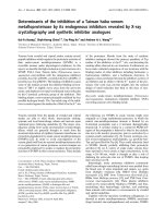
As depicted in Figure 1, as a negative control, primary anti-
body was omitted in a section obtained from a preimmune
IgG-treated animal (panel a) and little straining of brain tissue
for albumin was noted. Sham animals treated with either pre-
immune IgG (panel b) or anti-C5a (panel c) displayed compa-
rable levels of baseline immunostaining for albumin. However,
there was a significant increase in diffuse cerebral albumin
accumulation 24 hours after CLP in animals treated with pre-
Critical Care Vol 13 No 1 Flierl et al.
Page 4 of 9
(page number not for citation purposes)
immune IgG (panel d). In contrast, when rats received anti-
C5a immediately after the CLP procedure, cerebral albumin
build-up was dramatically reduced to sham levels (panels b
and e).
Similar results were found when breakdown of the BBB was
analysed by EB extravasation 24 hours after CLP. Animals
treated with preimmune IgG displayed robust EB extravasa-
tion in the cerebral and pituitary areas (panel f), while anti-C5a-
treated littermates exhibited far less buildup of EB (panel g).
Panels h and i show quantified EB extravasation 24 hours after
CLP as mg EB/mg tissue ratio and confirm the observations
made in panels f and g on a quantitative level.
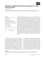
C5aR is upregulated in the pituitary gland of septic rats
Rat pituitary glands were surgically removed, total RNA was
isolated and analysed by quantitative real-time PCR. There
was a significant increase of pituitary C5aR expression 24
hours after CLP in animals receiving preimmune IgG, while
anti-C5a-treated littermates displayed expression levels com-
parable with sham animals (Figure 2a). Similar results were
found when pituitaries were homogenised and obtained pro-
teins were subjected to Western blot analysis. Protein expres-
sion of C5aR in the pituitary was markedly increased in IgG-
treated animals 24 hours after CLP, while rats that were
administered anti-C5a revealed C5aR protein expression sim-
ilar to sham controls (Figure 2b). Such patterns of increased
C5aR expression in CLP mice have been described in lung,
liver, kidney and heart [12].
C5a-blockade partially reverses cytokine upregulation in
the pituitary gland
Following isolation of pituitary total RNA, quantitative real-time
PCR was performed for TNF and IL-6. Sham animals treated
with either preimmune IgG or anti-C5a displayed similar
expression of mRNA for both proinflammatory mediators.
However, 24 hours after the CLP procedure, mRNA expres-
sion for TNF and IL-6 was substantially increased in IgG-
treated rats (Figure 3). Elevated mRNA levels were partially
reduced to sham levels when animals were administered anti-
C5a immediately after CLP.
Figure 1
Anti-C5a ameliorates impairment of the blood-brain barrier after caecal ligation and puncture (CLP)Anti-C5a ameliorates impairment of the blood-brain barrier after caecal ligation and puncture (CLP). (a-e) Brains were surgically removed, snap-fro-
zen and tissue sections (10 μm) were obtained. Cerebral extravasation of rat albumin was assessed by immunohistochemistry 24 hours after CLP or
sham procedure, three samples per experimental condition. Stains displayed are representative of three independent experiments. (f, g) Comparison
of Evans Blue extravasation into the cerebellum and pituitary in IgG-treated or anti-C5a treated rats 24 hours post CLP. Displayed depictions are
representative of four animals. (h, i) Quantification of Evans Blue extravasation into the brain or pituitary by determination of mg Evans Blue/mg tis-
sue ratio in different groups at indicated time-points, four for each experimental condition. # p < 0.05 between sham and 24 hours CLP animals; * p
< 0.05 between IgG-treated and anti-C5a-treated rats.
Available online />Page 5 of 9
(page number not for citation purposes)
Pituitary dysfunction is reversed by C5a blockade
Pituitary glands were surgically removed, total RNA was iso-
lated and analysed for POMC and GH by quantitative real-time
PCR. mRNA expression for both, POMC and GH was dramat-
ically reduced 24 hours after CLP in IgG-treated rats (Figures
4a,b). Anti-C5a administration completely reversed these
changes, resulting in POMC and GH mRNA expression levels
equivalent to sham groups. Plasma samples were obtained 24
hours after CLP or sham procedure and subjected to ELISA
analysis for GH and corticosterone, the main glucocorticoid of
rodents. When compared with sham animals, IgG-treated rats
had with significantly increased plasma levels of GH and cor-
ticosterone 24 hours after CLP. Again, these changes were
reversed by administration of anti-C5a immediately after CLP
(Figures 4c,d).
Discussion
Under physiological conditions, the BBB maintains the cere-
bral micro-environment by tightly regulating the passage of
molecules into and out of the brain to protect the brain from
microorganisms and neurotoxic substances. During sepsis,
however, blood-borne proinflammatory mediators are
released, coincidental with diffuse endothelial activation and
subsequent upregulation of vascular adhesion molecule-1
(VCAM-1), intracellular adhesion molecule-1 (ICAM-1), E-
selectin on cerebral endothelia [42-45], facilitating adhesion
and transmigration of neutrophils and monocytes into the brain
tissue. Especially the anaphylatoxin C5a is known to be a
strong inducer of ICAM-1, VCAM-1 and various selectins [46-
50]. In addition, cerebral endothelia produce IL-1β, TNF and
IL-6 [51-53], all of which have been shown to directly induce
a disruption of the BBB in vitro [54]. Thus, these mediators
interact with surrounding brain cells, relaying into the brain
inflammatory response and jeopardising the functional integ-
rity of the BBB [55-57]. In the present study, we found that,
during experimental sepsis, the antibody-mediated blockade
of anaphylatoxin C5a prevented breakdown of the blood-brain
barrier, reducing cerebral and pituitary edema formation, as
assessed by extravasation of albumin and EB (Figure 1).
Although the CNS itself was traditionally thought to be immu-
nologically privileged, recent studies have demonstrated that
the CNS is a rich source of inflammatory mediators, such as
cytokines, chemokines and complement components [58-64],
and has therefore been termed both 'culprit' and 'victim' during
sepsis [57]. Thus, during sepsis, the BBB is exposed to harm-
ful proinflammatory mediators deriving from two different com-
partments, the brain as well as the systemic circulation. This
results in an extrinsic, as well as intrinsic, attack on the BBB,
accelerating the deterioration of its barrier function. As
described above, we demonstrate significant upregulation of
pituitary expression of TNF and IL-6 mRNA following CLP (Fig-
ure 3), indicating that the pituitary might be an additional
source of cerebral proinflammatory mediators. Blood-derived
proinflammatory mediators reach the hypophyseal circulation
via the anterior hypophyseal arteries and cytokines can diffuse
into the pituitary because these areas are free from CNS BBB
[65]. Therefore, we decided that mRNA analysis for TNF and
IL-6 might shed a more accurate light on the origin of these
proinflammatory mediators.
Resident cells of the brain, such as neurons, astrocytes and
microglia, are capable of synthesising essentially all comple-
ment proteins, complement regulatory molecules and comple-
ment receptors [31,66-70]. It has been reported that the
pituitary expresses the complement receptors C3aR, C5aR
and C5a-like receptor 2 (C5L2), and that these molecules may
contribute to regulation of the immune response [71,72]. We
found upregulation of C5aR in the pituitary based on mRNA
and protein levels following CLP and reversal of these
changes by administration of anti-C5a (Figure 2). Upregulation
Figure 2
Pituitary expression of C5a receptor (R) during experimental sepsis in IgG or anti-C5a IgG treated sham animals and septic littermates 24 hours after caecal ligation and puncture (CLP) procedurePituitary expression of C5a receptor (R) during experimental sepsis in
IgG or anti-C5a IgG treated sham animals and septic littermates 24
hours after caecal ligation and puncture (CLP) procedure. (a) Follow-
ing total RNA isolation from pituitary tissue, pituitary C5aR mRNA
expression was assessed by real-time PCR. For each bar, sample size
was five to seven. (b) Five pituitary tissue samples were removed at
indicated time-points, homogenised and C5aR protein expression was
analysed by Western blotting. For sham groups, two samples were
taken; for CLP groups, three samples were taken. Blot is representative
for three independent experiments. GAPDH = glyceraldehyde 3-phos-
phate dehydrogenase. # p < 0.05 between sham and 24 hours CLP
animals; * p < 0.05 between IgG-treated and anti-C5a-treated rats.
Critical Care Vol 13 No 1 Flierl et al.
Page 6 of 9
(page number not for citation purposes)
of C5aR during sepsis has been described in various organs,
such as lung, liver, kidney and heart [12]. Such upregulation
infers that these tissues may develop highly undesirable out-
comes after encounters with C5a.
Sepsis is known to induce an abnormal pituitary response
[73]. Hormonal changes in cortisol, mineralocorticoids, thyroid
hormones, GH and vasopressin have all been described dur-
ing sepsis. Although the acute phase of sepsis is character-
ised by high levels of GH, GH insufficiency is reported in the
late stage of sepsis [73,74]. In line with these findings, we
have found elevated levels of GH protein 24 hours after CLP,
while pituitary mRNA expression is significantly reduced, most
likely because of a negative feedback mechanism.
Hypercortisolism during the early stages of sepsis is usually
followed by cortisol insufficiency in 60% of septic patients
[73]. Significantly increased levels of corticosterone occurred
in rats following CLP (Figure 4). In the current study, pituitary
POMC mRNA expression was completely abolished during
experimental sepsis (Figure 4). POMC is a polypeptide precur-
sor of multiple molecules, including ACTH and melanocyte-
stimulating hormones (MSH) α, β and γ [75]. Interestingly,
MSH-α has recently emerged as a molecule with potent anti-
inflammatory effects, which orchestrates descending neuro-
genic anti-inflammatory pathways and ameliorates the
inflammatory response of immune cells [76]. At a molecular
level, MSH-α decreases the intracellular production of proin-
flammatory cytokines and chemokines by inhibiting nuclear
factor-κB activation and reduces cellular expression of VCAM-
1, ICAM-1 and E-selectin [76]. During human sepsis, plasma
concentrations of MSH-α have been found to be significantly
decreased during the early course of the disease and gradu-
ally recovered as a function of time [77]. More importantly, its
concentrations negatively correlated with plasma concentra-
tions of TNF and IL-1β [77]. Thus, it is tempting to speculate,
whether the observed pituitary upregulation of TNF and IL-6
mRNA (Figure 3) is related to the complete absence of POMC
expression (Figure 4), which will result in a lack of pituitary
MSH-α production with uninhibited proinflammatory activation
of pituitary cells.
It remains to be determined if our findings can be extrapolated
into humans. Several reports have stressed the disconnection
between rodent and human sepsis [78-80], making it difficult
to draw definitive conclusions from an experimental study for
the clinical setting. Moreover, in the current study, the C5a-
neutralising antibody was administered immediately after the
CLP procedure. Follow-up studies need to address the effects
of delayed anti-C5a treatment, because diagnosis of the sep-
sis syndrome in patients might be delayed due to several co-
morbidities. Thus, it is imperative to address if anti-C5a treat-
ment also reverses BBB and pituitary dysfunction after onset
of CLP, which would greatly enhance the therapeutic impact
of a potential C5a-blockade in humans.
Conclusions
We describe amelioration of BBB breakdown and partial
reversal of pituitary dysfunction by neutralisation of C5a during
experimental sepsis. Similarly, blockade of C5aR has recently
been described to reduce the LPS-induced activation in the
paraventricular nucleus and the central amygdala [81]. Thus,
as we are beginning to gain novel insights into the crucial role
of C5a in the development of sepsis-induced BBB dysfunc-
tion, we might be able to immunomodulate its detrimental
effects and improve the outcome of septic encephalopathy.
Figure 3
Expression of inflammatory mediators in the pituitaryExpression of inflammatory mediators in the pituitary. Pituitary tissue samples were surgically removed, snap-frozen, homogenised and total RNA was
extracted. Samples were then analysed by quantitative real-time PCR analysis. Expression of (a) TNF and (b) IL-6 mRNA in the pituitary 24 hours
after sham procedure or caecal ligation and puncture (CLP). Five to seven samples were taken per experimental condition. GAPDH = glyceralde-
hyde 3-phosphate dehydrogenase. # p < 0.05 between sham and 24 hours CLP animals; * p < 0.05 between IgG-treated and anti-C5a-treated rats.
Available online />Page 7 of 9
(page number not for citation purposes)
Competing interests
The authors declare that they have no competing interests.
Authors' contributions
MAF, PFS, MHL, PAW and KI designed the study and super-
vised the experiments. MAF, DR, AND, LMH and BMT per-
formed the experiments. MAF, PFS, SJM, WRS and KI
analysed the data and drafted the manuscript. All authors
revised the manuscript for important scientific content, read
and approved the final manuscript.
Key messages
• Anti-C5a prevents break-down of the BBB during
experimental sepsis.
• The C5aR is robustly upregulated in the pituitary gland
during CLP-induced sepsis.
• Pituitary mRNA expression of proinflammatory TNF and
IL-6 is upregulated during experimental sepsis.
• Experimental sepsis induces pituitary dysfunction which
is ameliorated by a neutralising anti-C5a antibody.
Figure 4
Evaluation of pituitary function after caecal ligation and puncture (CLP)Evaluation of pituitary function after caecal ligation and puncture (CLP). Pituitary tissue samples were removed from four to five mice, snap-frozen,
homogenised in Trizol and total RNA was extracted. Assessment of mRNA expression of (a) proopiomelanocortin (POMC) and (b) growth hormone
(GH) 24 hours after CLP or sham operation by real-time PCR. Whole blood samples were drawn at given time-points by puncture of the inferior vena
cava, plasma was obtained by centrifugation and subjected to ELISA analysis. Samples were assessed for (c) GH or (d) corticosterone under iden-
tical conditions. For all graphs, there were five to seven samples per experimental condition. GAPDH = glyceraldehyde 3-phosphate dehydrogenase.
# p < 0.05 between sham and 24 hours CLP animals; * p < 0.05 between IgG-treated and anti-C5a-treated rats.
Critical Care Vol 13 No 1 Flierl et al.
Page 8 of 9
(page number not for citation purposes)
References
1. Dombrovskiy VY, Martin AA, Sunderram J, Paz HL: Rapid increase
in hospitalization and mortality rates for severe sepsis in the
United States: a trend analysis from 1993 to 2003. Crit Care
Med 2007, 35:1244-1250.
2. Kung HC, Hoyert DL, Xu J, Murphy SL: Deaths: final data for
2005. Natl Vital Stat Rep 2008, 56:1-120.
3. Rittirsch D, Flierl MA, Ward PA: Harmful molecular mechanisms
in sepsis. Nat Rev Immunol 2008, 8:776-787.
4. Levy MM, Fink MP, Marshall JC, Abraham E, Angus D, Cook D,
Cohen J, Opal SM, Vincent JL, Ramsay G: 2001 SCCM/ESICM/
ACCP/ATS/SIS International Sepsis Definitions Conference.
Crit Care Med 2003, 31:1250-1256.
5. Annane D, Bellissant E, Cavaillon JM: Septic shock. Lancet 2005,
365:63-78.
6. Nakae H, Endo S, Inada K, Takakuwa T, Kasai T, Yoshida M:
Serum complement levels and severity of sepsis. Res Com-
mun Chem Pathol Pharmacol 1994, 84:189-195.
7. Nakae H, Endo S, Inada K, Yoshida M: Chronological changes in
the complement system in sepsis. Surg Today 1996,
26:225-229.
8. Stove S, Welte T, Wagner TO, Kola A, Klos A, Bautsch W, Kohl J:
Circulating complement proteins in patients with sepsis or
systemic inflammatory response syndrome. Clin Diagn Lab
Immunol 1996, 3:175-183.
9. Hack CE, Nuijens JH, Felt-Bersma RJ, Schreuder WO, Eerenberg-
Belmer AJ, Paardekooper J, Bronsveld W, Thijs LG: Elevated
plasma levels of the anaphylatoxins C3a and C4a are associ-
ated with a fatal outcome in sepsis. Am J Med 1989, 86:20-26.
10. Czermak BJ, Sarma V, Pierson CL, Warner RL, Huber-Lang M,
Bless NM, Schmal H, Friedl HP, Ward PA: Protective effects of
C5a blockade in sepsis. Nat Med 1999, 5:788-792.
11. Huber-Lang MS, Sarma JV, McGuire SR, Lu KT, Guo RF,
Padgaonkar VA, Younkin EM, Laudes IJ, Riedemann NC, Younger
JG, Ward PA: Protective effects of anti-C5a peptide antibodies
in experimental sepsis. Faseb J 2001, 15:568-570.
12. Riedemann NC, Guo RF, Neff TA, Laudes IJ, Keller KA, Sarma VJ,
Markiewski MM, Mastellos D, Strey CW, Pierson CL, Lambris JD,
Zetoune FS, Ward PA: Increased C5a receptor expression in
sepsis. J Clin Invest 2002, 110:101-108.
13. Niederbichler AD, Hoesel LM, Westfall MV, Gao H, Ipaktchi KR,
Sun L, Zetoune FS, Su GL, Arbabi S, Sarma JV, Wang SC, Hem-
mila MR, Ward PA: An essential role for complement C5a in the
pathogenesis of septic cardiac dysfunction. J Exp Med 2006,
203:53-61.
14. Rittirsch D, Flierl MA, Nadeau BA, Day DE, Huber-Lang M, Mackay
CR, Zetoune FS, Gerard NP, Cianflone K, Kohl J, Gerard C, Sarma
JV, Ward PA: Functional roles for C5a receptors in sepsis. Nat
Med 2008, 14:551-557.
15. Gerard C: Complement C5a in the sepsis syndrome – too
much of a good thing? N Engl J Med 2003, 348:167-169.
16. Ward PA: The dark side of C5a in sepsis. Nat Rev Immunol
2004, 4:133-142.
17. Guo RF, Huber-Lang M, Wang X, Sarma V, Padgaonkar VA, Craig
RA, Riedemann NC, McClintock SD, Hlaing T, Shi MM, Ward PA:
Protective effects of anti-C5a in sepsis-induced thymocyte
apoptosis. J Clin Invest 2000, 106:1271-1280.
18. Flierl MA, Rittirsch D, Chen AJ, Nadeau BA, Day DE, Sarma JV,
Huber-Lang MS, Ward PA: The complement anaphylatoxin C5a
induces apoptosis in adrenomedullary cells during experi-
mental sepsis. PLoS ONE 2008, 3:e2560.
19. Huber-Lang MS, Riedeman NC, Sarma JV, Younkin EM, McGuire
SR, Laudes IJ, Lu KT, Guo RF, Neff TA, Padgaonkar VA, Lambris
JD, Spruce L, Mastellos D, Zetoune FS, Ward PA: Protection of
innate immunity by C5aR antagonist in septic mice. Faseb J
2002, 16:1567-1574.
20. Huber-Lang MS, Younkin EM, Sarma JV, McGuire SR, Lu KT, Guo
RF, Padgaonkar VA, Curnutte JT, Erickson R, Ward PA: Comple-
ment-induced impairment of innate immunity during sepsis. J
Immunol 2002, 169:3223-3231.
21. Laudes IJ, Chu JC, Sikranth S, Huber-Lang M, Guo RF, Riedemann
N, Sarma JV, Schmaier AH, Ward PA: Anti-c5a ameliorates
coagulation/fibrinolytic protein changes in a rat model of sep-
sis. Am J Pathol 2002, 160:1867-1875.
22. Huber-Lang M, Sarma VJ, Lu KT, McGuire SR, Padgaonkar VA,
Guo RF, Younkin EM, Kunkel RG, Ding J, Erickson R, Curnutte JT,
Ward PA:
Role of C5a in multiorgan failure during sepsis. J
Immunol 2001, 166:1193-1199.
23. Semmler A, Hermann S, Mormann F, Weberpals M, Paxian SA,
Okulla T, Schafers M, Kummer MP, Klockgether T, Heneka MT:
Sepsis causes neuroinflammation and concomitant decrease
of cerebral metabolism. J Neuroinflammation 2008, 5:38.
24. Piazza O, Russo E, Cotena S, Esposito G, Tufano R: Elevated
S100B levels do not correlate with the severity of encephalop-
athy during sepsis. Br J Anaesth 2007, 99:518-521.
25. Alexander JJ, Jacob A, Cunningham P, Hensley L, Quigg RJ: TNF
is a key mediator of septic encephalopathy acting through its
receptor, TNF receptor-1. Neurochem Int 2008, 52:447-456.
26. Wratten ML: Therapeutic approaches to reduce systemic
inflammation in septic-associated neurologic complications.
Eur J Anaesthesiol Suppl 2008, 42:1-7.
27. Handa O, Stephen J, Cepinskas G: Role of eNOS-derived nitric
oxide (NO) in activation and dysfunction of cerebrovascular
endothelial cells during early onsets of sepsis. Am J Physiol
Heart Circ Physiol 2008, 295:H1712-1719.
28. Toklu HZ, Uysal MK, Kabasakal L, Sirvanci S, Ercan F, Kaya M: The
effects of riluzole on neurological, brain biochemical, and his-
tological changes in early and late term of sepsis in rats. J
Surg Res 2008.
29. Hofer S, Bopp C, Hoerner C, Plaschke K, Faden RM, Martin E,
Bardenheuer HJ, Weigand MA: Injury of the blood brain barrier
and up-regulation of icam-1 in polymicrobial sepsis. J Surg
Res 2008, 146:276-281.
30. Faustmann PM, Krause D, Dux R, Dermietzel R: Morphological
study in the early stages of complement C5a fragment-
induced experimental meningitis: activation of macrophages
and astrocytes. Acta Neuropathol 1995, 89:239-247.
31. Nataf S, Stahel PF, Davoust N, Barnum SR: Complement ana-
phylatoxin receptors on neurons: new tricks for old receptors?
Trends Neurosci 1999, 22:397-402.
32. Stahel PF, Barnum SR: The role of the complement system in
CNS inflammatory diseases. Expert Rev Clin Immunol 2006,
2:445-456.
33. Stahel PF, Frei K, Eugster HP, Fontana A, Hummel KM, Wetsel RA,
Ames RS, Barnum SR: TNF-alpha-mediated expression of the
receptor for anaphylatoxin C5a on neurons in experimental
Listeria meningoencephalitis.
J Immunol 1997, 159:861-869.
34. Osaka H, McGinty A, Hoepken UE, Lu B, Gerard C, Pasinetti GM:
Expression of C5a receptor in mouse brain: role in signal
transduction and neurodegeneration. Neuroscience 1999,
88:1073-1082.
35. O'Barr SA, Caguioa J, Gruol D, Perkins G, Ember JA, Hugli T,
Cooper NR: Neuronal expression of a functional receptor for
the C5a complement activation fragment. J Immunol 2001,
166:4154-4162.
36. Ernst JD, Hartiala KT, Goldstein IM, Sande MA: Complement
(C5)-derived chemotactic activity accounts for accumulation
of polymorphonuclear leukocytes in cerebrospinal fluid of rab-
bits with pneumococcal meningitis. Infect Immun 1984,
46:81-86.
37. Williams CA, Schupf N, Hugli TE: Anaphylatoxin C5a modulation
of an alpha-adrenergic receptor system in the rat hypothala-
mus. J Neuroimmunol 1985, 9:29-40.
38. Jacob A, Hensley LK, Safratowich BD, Quigg RJ, Alexander JJ: The
role of the complement cascade in endotoxin-induced septic
encephalopathy. Lab Invest 2007, 87:1186-1194.
39. Baker CC, Chaudry IH, Gaines HO, Baue AE: Evaluation of fac-
tors affecting mortality rate after sepsis in a murine cecal liga-
tion and puncture model. Surgery 1983, 94:331-335.
40. Laudes IJ, Chu JC, Huber-Lang M, Guo RF, Riedemann NC, Sarma
JV, Mahdi F, Murphy HS, Speyer C, Lu KT, Lambris JD, Zetoune
FS, Ward PA: Expression and function of C5a receptor in
mouse microvascular endothelial cells. J Immunol 2002,
169:5962-5970.
41. Riedemann NC, Neff TA, Guo RF, Bernacki KD, Laudes IJ, Sarma
JV, Lambris JD, Ward PA: Protective effects of IL-6 blockade in
sepsis are linked to reduced C5a receptor expression. J Immu-
nol 2003, 170:503-507.
42. Wong D, Dorovini-Zis K: Expression of vascular cell adhesion
molecule-1 (VCAM-1) by human brain microvessel endothelial
cells in primary culture. Microvasc Res 1995, 49:325-339.
Available online />Page 9 of 9
(page number not for citation purposes)
43. Hess DC, Thompson Y, Sprinkle A, Carroll J, Smith J: E-selectin
expression on human brain microvascular endothelial cells.
Neurosci Lett 1996, 213:37-40.
44. Hess DC, Bhutwala T, Sheppard JC, Zhao W, Smith J: ICAM-1
expression on human brain microvascular endothelial cells.
Neurosci Lett 1994, 168:201-204.
45. Rieckmann P, Michel U, Albrecht M, Bruck W, Wockel L, Felgen-
hauer K: Cerebral endothelial cells are a major source for sol-
uble intercellular adhesion molecule-1 in the human central
nervous system. Neurosci Lett 1995, 186:61-64.
46. Mulligan MS, Schmid E, Till GO, Hugli TE, Friedl HP, Roth RA,
Ward PA: C5a-dependent up-regulation in vivo of lung vascu-
lar P-selectin. J Immunol 1997, 158:1857-1861.
47. Foreman KE, Vaporciyan AA, Bonish BK, Jones ML, Johnson KJ,
Glovsky MM, Eddy SM, Ward PA: C5a-induced expression of P-
selectin in endothelial cells. J Clin Invest 1994, 94:1147-1155.
48. Albrecht EA, Chinnaiyan AM, Varambally S, Kumar-Sinha C, Bar-
rette TR, Sarma JV, Ward PA: C5a-induced gene expression in
human umbilical vein endothelial cells. Am J Pathol 2004,
164:849-859.
49. Mulligan MS, Schmid E, Beck-Schimmer B, Till GO, Friedl HP,
Brauer RB, Hugli TE, Miyasaka M, Warner RL, Johnson KJ, Ward
PA: Requirement and role of C5a in acute lung inflammatory
injury in rats. J Clin Invest 1996, 98:503-512.
50. Schmid E, Piccolo MT, Friedl HP, Warner RL, Mulligan MS, Hugli
TE, Till GO, Ward PA: Requirement for C5a in lung vascular
injury following thermal trauma to rat skin. Shock 1997,
8:119-124.
51. Breder CD, Hazuka C, Ghayur T, Klug C, Huginin M, Yasuda K,
Teng M, Saper CB: Regional induction of tumor necrosis factor
alpha expression in the mouse brain after systemic lipopoly-
saccharide administration. Proc Natl Acad Sci USA 1994,
91:11393-11397.
52. Fabry Z, Fitzsimmons KM, Herlein JA, Moninger TO, Dobbs MB,
Hart MN: Production of the cytokines interleukin 1 and 6 by
murine brain microvessel endothelium and smooth muscle
pericytes. J Neuroimmunol 1993, 47:23-34.
53. Reyes TM, Fabry Z, Coe CL: Brain endothelial cell production of
a neuroprotective cytokine, interleukin-6, in response to nox-
ious stimuli. Brain Res 1999, 851:215-220.
54. de Vries HE, Blom-Roosemalen MC, van Oosten M, de Boer AG,
van Berkel TJ, Breimer DD, Kuiper J: The influence of cytokines
on the integrity of the blood-brain barrier in vitro. J Neuroim-
munol 1996, 64:37-43.
55. Papadopoulos MC, Davies DC, Moss RF, Tighe D, Bennett ED:
Pathophysiology of septic encephalopathy: a review. Crit Care
Med 2000, 28:3019-3024.
56. Sharshar T, Carlier R, Bernard F, Guidoux C, Brouland JP, Nardi O,
de la Grandmaison GL, Aboab J, Gray F, Menon D, Annane D:
Brain lesions in septic shock: a magnetic resonance imaging
study. Intensive Care Med 2007, 33:798-806.
57. Sharshar T, Hopkinson NS, Orlikowski D, Annane D: Science
review: The brain in sepsis – culprit and victim. Crit Care 2005,
9:37-44.
58. Hanisch UK: Microglia as a source and target of cytokines. Glia
2002, 40:140-155.
59. Ghirnikar RS, Lee YL, Eng LF: Inflammation in traumatic brain
injury: role of cytokines and chemokines. Neurochem Res
1998, 23:329-340.
60. Ransohoff RM, Tani M: Do chemokines mediate leukocyte
recruitment in post-traumatic CNS inflammation? Trends Neu-
rosci 1998, 21:154-159.
61. Stahel PF, Kariya K, Shohami E, Barnum SR, Eugster H, Trentz O,
Kossmann T, Morganti-Kossmann MC: Intracerebral comple-
ment C5a receptor (CD88) expression is regulated by TNF and
lymphotoxin-alpha following closed head injury in mice. J
Neuroimmunol 2000, 109:164-172.
62. Gasque P, Dean YD, McGreal EP, VanBeek J, Morgan BP: Com-
plement components of the innate immune system in health
and disease in the CNS. Immunopharmacology 2000,
49:171-186.
63. Boos L, Campbell IL, Ames R, Wetsel RA, Barnum SR: Deletion
of the complement anaphylatoxin C3a receptor attenuates,
whereas ectopic expression of C3a in the brain exacerbates,
experimental autoimmune encephalomyelitis.
J Immunol
2004, 173:4708-4714.
64. Lynch NJ, Willis CL, Nolan CC, Roscher S, Fowler MJ, Weihe E,
Ray DE, Schwaeble WJ: Microglial activation and increased
synthesis of complement component C1q precedes blood-
brain barrier dysfunction in rats. Mol Immunol 2004,
40:709-716.
65. Porter JC, Sissom JF, Arita J, Reymond MJ: Hypothalamic-hypo-
physial vasculature and its relationship to secretory cells of
the hypothalamus and pituitary gland. Vitam Horm 1983,
40:145-174.
66. Rancan M, Morganti-Kossmann MC, Barnum SR, Saft S, Schmidt
OI, Ertel W, Stahel PF: Central nervous system-targeted com-
plement inhibition mediates neuroprotection after closed
head injury in transgenic mice. J Cereb Blood Flow Metab
2003, 23:1070-1074.
67. Stahel PF, Kossmann T, Morganti-Kossmann MC, Hans VH, Bar-
num SR: Experimental diffuse axonal injury induces enhanced
neuronal C5a receptor mRNA expression in rats. Brain Res
Mol Brain Res 1997, 50:205-212.
68. Gasque P, Singhrao SK, Neal JW, Gotze O, Morgan BP: Expres-
sion of the receptor for complement C5a (CD88) is up-regu-
lated on reactive astrocytes, microglia, and endothelial cells in
the inflamed human central nervous system. Am J Pathol
1997, 150:31-41.
69. Francis K, van Beek J, Canova C, Neal JW, Gasque P: Innate
immunity and brain inflammation: the key role of complement.
Expert Rev Mol Med 2003, 5:1-19.
70. Davoust N, Jones J, Stahel PF, Ames RS, Barnum SR: Receptor
for the C3a anaphylatoxin is expressed by neurons and glial
cells. Glia 1999, 26:201-211.
71. Francis K, Lewis BM, Akatsu H, Monk PN, Cain SA, Scanlon MF,
Morgan BP, Ham J, Gasque P: Complement C3a receptors in
the pituitary gland: a novel pathway by which an innate
immune molecule releases hormones involved in the control
of inflammation. Faseb J 2003, 17:2266-2268.
72. Lewis BM, Francis K, Monk P, Scanlon MF, Ham J: Complement
C5a inhibits the secretion of macrophage migration inhibitory
factor in anterior pituitary cell lines. Endocrine Abstracts 2006,
11:P602.
73. Maxime V, Siami S, Annane D: Metabolism modulators in sep-
sis: the abnormal pituitary response. Crit Care Med 2007,
35:S596-601.
74. Ross R, Miell J, Freeman E, Jones J, Matthews D, Preece M, Bucha-
nan C: Critically ill patients have high basal growth hormone
levels with attenuated oscillatory activity associated with low
levels of insulin-like growth factor-I. Clin Endocrinol (Oxf)
1991, 35:47-54.
75. Raffin-Sanson ML, de Keyzer Y, Bertagna X: Proopiomelanocor-
tin, a polypeptide precursor with multiple functions: from
physiology to pathological conditions. Eur J Endocrinol 2003,
149:79-90.
76. Brzoska T, Luger TA, Maaser C, Abels C, Bohm M: Alpha-
melanocyte-stimulating hormone and related tripeptides: bio-
chemistry, antiinflammatory and protective effects in vitro and
in vivo, and future perspectives for the treatment of immune-
mediated inflammatory diseases. Endocr Rev 2008,
29:581-602.
77. Catania A, Cutuli M, Garofalo L, Airaghi L, Valenza F, Lipton JM,
Gattinoni L: Plasma concentrations and anti-L-cytokine effects
of alpha-melanocyte stimulating hormone in septic patients.
Crit Care Med 2000, 28:1403-1407.
78. Rittirsch D, Hoesel LM, Ward PA: The disconnect between ani-
mal models of sepsis and human sepsis. J Leukoc Biol 2007,
81:137-143.
79. Esmon CT: Why do animal models (sometimes) fail to mimic
human sepsis? Crit Care Med 2004, 32:S219-222.
80. Poli-de-Figueiredo LF, Garrido AG, Nakagawa N, Sannomiya P:
Experimental models of sepsis and their clinical relevance.
Shock 2008, 30(Suppl 1):53-59.
81. Crane JW, Buller KM: Systemic blockade of complement C5a
receptors reduces lipopolysacharride-induced responses in
the paraventricular nucleus and the central amygdala. Neuro-
sci Lett 2007, 424:10-15.