Báo cáo y học: "Comparative modeling of DNA and RNA polymerases from Moniliophthora perniciosa mitochondrial plasmid" doc
Bạn đang xem bản rút gọn của tài liệu. Xem và tải ngay bản đầy đủ của tài liệu tại đây (2.06 MB, 6 trang )
BioMed Central
Page 1 of 6
(page number not for citation purposes)
Theoretical Biology and Medical
Modelling
Open Access
Research
Comparative modeling of DNA and RNA polymerases from
Moniliophthora perniciosa mitochondrial plasmid
Bruno S Andrade*
†1
, Alex G Taranto
†2
, Aristóteles Góes-Neto
†1
and
Angelo A Duarte
†3
Address:
1
Departamento de Ciências Biológicas, Universidade Estadual de Feira de Santana, Feira de Santana, Brazil,
2
Departamento de Saúde,
Universidade Estadual de Feira de Santana, Feira de Santana, Brazil and
3
Departamento de Tecnologia, Universidade Estadual de Feira de Santana,
Feira de Santana, Brazil
Email: Bruno S Andrade* - ; Alex G Taranto - ; Aristóteles Góes-
Neto - ; Angelo A Duarte -
* Corresponding author †Equal contributors
Abstract
Background: The filamentous fungus Moniliophthora perniciosa (Stahel) Aime & Phillips-Mora is a
hemibiotrophic Basidiomycota that causes witches' broom disease of cocoa (Theobroma cacao L.).
This disease has resulted in a severe decrease in Brazilian cocoa production, which changed the
position of Brazil in the market from the second largest cocoa exporter to a cocoa importer. Fungal
mitochondrial plasmids are usually invertrons encoding DNA and RNA polymerases. Plasmid
insertions into host mitochondrial genomes are probably associated with modifications in host
generation time, which can be involved in fungal aging. This association suggests activity of
polymerases, and these can be used as new targets for drugs against mitochondrial activity of fungi,
more specifically against witches' broom disease. Sequencing and modeling: DNA and RNA
polymerases of M. perniciosa mitochondrial plasmid were completely sequenced and their models
were carried out by Comparative Homology approach. The sequences of DNA and RNA
polymerase showed 25% of identity to 1XHX and 1ARO (pdb code) using BLASTp, which were
used as templates. The models were constructed using Swiss PDB-Viewer and refined with a set
of Molecular Mechanics (MM) and Molecular Dynamics (MD) in water carried out with AMBER 8.0,
both working under the ff99 force fields, respectively. Ramachandran plots were generated by
Procheck 3.0 and exhibited models with 97% and 98% for DNA and RNA polymerases,
respectively. MD simulations in water showed models with thermodynamic stability after 2000 ps
and 300 K of simulation.
Conclusion: This work contributes to the development of new alternatives for controlling the
fungal agent of witches' broom disease.
Background
The filamentous fungus Moniliophthora perniciosa (Stahel)
Aime & Phillips-Mora is a hemibiotrophic Basidiomycota
(Agaricales, Tricholomataceae) that causes witches'
broom disease of cocoa (Theobroma cacao L.). It has been
claimed as one of the most important phytopathological
problems that has afflicted the Southern Hemisphere in
recent decades. In Brazil, this phytopathogen is endemic
Published: 10 September 2009
Theoretical Biology and Medical Modelling 2009, 6:22 doi:10.1186/1742-4682-6-22
Received: 20 March 2009
Accepted: 10 September 2009
This article is available from: />© 2009 Andrade et al; licensee BioMed Central Ltd.
This is an Open Access article distributed under the terms of the Creative Commons Attribution License ( />),
which permits unrestricted use, distribution, and reproduction in any medium, provided the original work is properly cited.
Theoretical Biology and Medical Modelling 2009, 6:22 />Page 2 of 6
(page number not for citation purposes)
in the Amazon region [1]. However, since 1989, this fun-
gus has been found in the cultivated regions in the state of
Bahia, the largest production area in the country. The fun-
gus caused a severe decrease in the Brazilian cocoa pro-
duction reducing Brazil from the second largest cocoa
exporter to a cocoa importer in just few years [2].
Plasmids are extragenomic DNA or RNA molecules that
can independently reproduce in live cells. Their structure
can be circular or linear, and include complete protein
coding genes, pseudogenes, non-protein coding genes
and inverted repetitive elements. The probable plasmid
function in their fungal hosts is related to the change of
aging time. Fungal linear mitochondrial plasmids present
the same basic structure as in other organisms, but they
also carry viral-like DNA and RNA polymerase (DPO and
RPO, respectively) ORFs and have 3' and 5' inverted ter-
minal repeats, also a 5' binding protein. This protein can
be involved in both replication and integration processes
of these plasmids in the mitochondrial genomes [3,4].
Interestingly, a linear mitochondrial plasmid with the
same typical characteristics carried by the other mitochon-
drial plasmids was found to be completely integrated in
the M. perniciosa mitochondrial genome, by the Witches'
Broom Genome Project />vassoura/[5].
The Φ29 DNA polymerase is in the group α-DNA-
polymerases due to its sensitivity to aphidicolin and spe-
cific inhibitors, nucleotides similar to BuAaATP and BuP-
dGTP [6]. This polymerase is the main replication enzyme
of double-strand-DNA viruses from bacteria and eucaryo-
tes. It is a 66 KDa enzyme included in the eucaryotic rep-
licase family [7], able to use a protein as primer in the
replication process [8,9]. The T7 RNA polymerase is a 99
KDa single chain viral enzyme that executes a specific-pro-
moter transcription process in vivo and in vitro and is in
the single-chain RNA polymerase family. The transcrip-
tion mechanism carried out by this enzyme shares several
similarities with other multichain RNA polymerases [9].
It is generally accepted that the water molecules in the
hydration environment around a protein play an impor-
tant role in its biological activity [10], and contribute to
stabilizing the native state of the protein [11]. In addition,
this interaction has long been recognized as a major deter-
minant of chain folding, conformational stability, and
internal dynamics of many proteins, and as important to
the interactions related to substrate binding, enzyme
catalysis, and supramolecular recognition and assembly
[12]. Standard Molecular Dynamics approaches measure
the conformational space of a protein using atomic inter-
actions from several force fields and include explicitly
treated water to reproduce solvent effects [13].
The aim of this work to carry out homology modeling of
both DNA and RNA polymerases from the linear mito-
chondrial plasmid of M. perniciosa. With the accomplish-
ment of this work, these models can be used as new
molecular targets to find drugs against witches' broom
disease by de novo design methods [10].
Methods
After the release of the primary sequences of DNA and
RNA polymerases from M. perniciosa mitochondrial plas-
mid, they are available in the Witches' broom project
database (LGE). 3D models were built by Comparative
Modeling approach. Initially, both DNA and RNA
polymerase sequences were subjected to the BLASTp algo-
rithm [14] restricted to the Protein Data Bank (PDB). The
templates found were aligned with the protein sequences
of both DNA and RNA polymerases by TCOFFEE [15] to
find conserved regions and motifs. The 3D models were
constructed using SwissPdb Viewer 3.7 [16] following a
standard protocol: (I) load template pdb file; (II) align
primary target sequence with template; (III) submit mod-
eling request to Swiss Model Server. Then, the initial mod-
els constructed by SwissPdb Viewer were prepared using
LEAP and submitted to SANDER for structure refinement.
The model structures were fully minimized with 100 steps
of steepest descent followed by 100 more steps of conju-
gate gradient to an RMS gradient of 0.01 kcal/2.71Å in
vacuum, and then in water for 200 steps of steepest
descent followed by 200 more steps of conjugate gradient
to an RMS gradient of 0.01 kcal/2.71Å. Next, MD simula-
tions of the refined structures were performed in water
using f99 force field at 300 K for 2000 ps. All MD simula-
tions were carried out without constrain methods. The
cutoff value of 14 Å was used for minimization of geome-
try and MD simulations. LEAP and SANDER are utilities
of AMBER 9.0 [17,18]. Additionally, all calculations were
performed without restraints. Time averaged structures
were generated by time averaging of simulations from the
point of a stable trajectory, which was obtained through
the end of simulation. The Visual Molecular Dynamics
(VMD) software [19] was used to visualize trajectory
results produced by the SANDER module. Finally, PRO-
CHECK 3.4 [20] and Atomic Non-Local Environment
Assessment (ANOLEA) [21,22] were used to evaluate both
DNA and RNA polymerases using a Ramachandran plot
[23] and energy calculations on a protein chain of each
heavy atom in the molecule, respectively [24]. Graphics of
RMS × Time were generated by VMD 1.8.6 [25]
Results and Discussion
Blastp results for both DNA and RNA polymerases of the
M. perniciosa linear mitochondrial plasmid showed just
one reliable template to each enzyme (Table 1). 1XHX
[26] and 1ARO [27] were used as template DPO and RPO
respectively. Although both of them showed low identity
Theoretical Biology and Medical Modelling 2009, 6:22 />Page 3 of 6
(page number not for citation purposes)
with the targets, it is possible to build useful models for
docking studies [10]. The root-mean-squared deviations
(RMSD) for Cα between DPO-1XHX and RPO-1ARO are
2.40 Å and 1.84 Å respectively. These values show some
differences between models and crystal structures, as one
might expect, principally in relation to the number of res-
idues. The models have 543 and 766 residues in DPO and
RPO, while the crystal structures have 575 and 883 resi-
dues for 1XHX and 1ARO, respectively.
In addition, these results address the hypothesis of several
authors correlating plasmid sequences to DNA and RNA
polymerases of adenovirus and retrovirus sequences
[3,27].
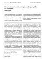
Using 1HXH as a template, the 3D structure of the DNA
polymerase was built from the linear mitochondrial plas-
mid of M. perniciosa. This polymerase was classified
within the B family of DNA polymerases, which can be
found in viruses and cellular organelles. Figure 1 shows
that the DPO model has transferase features with alpha-
beta secondary structure.
This model shows 17 alpha-helices, 36 beta-strands, 57
turns, and 315 hydrogen bonds can be observed in the
whole structure. As well as other polymerases from that
family, this polymerase showed the three standard
domains of the group: Palm, Fingers, and Thumb.
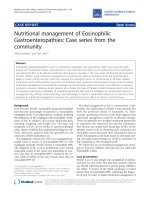
The active site of the DNA polymerase of M. perniciosa
(Figure 2) carries the conserved motif B represented by
Lys380, Leu381, Leu382, Leu383, Asn384, Ser385,
Leu386, Tyr387, Gly388, and it is involved in dNTP selec-
tion and template DNA binding activity as described by
Truniger et al. [6] in the homologous Φ29 DNA polymer-
ase. These amino acids are distributed among three
domains: Palm, Fingers and Thumb. Other motifs
involved with DNA polymerization were found in this
polymerase, such as Dx2SLYP (Asp247, Val248, Asn249,
Ser250, Leu251, Tyr252, Pro253), YxDTDS (Tyr455,
Ser456, Asp457, Thr458, Asp459), Tx2A/GR (Thr309,
Asp310, Lys311, Gly312, Tyr313, Arg314) and KxY
(Lys494, Met495, Tyr496), which have been reported in
several studies [6,8,9,28-31].
The active site of the RNA polymerase (Figure 3) from M.
perniciosa plasmid is formed by amino acids from two
domains: Palm (Asp457 and Asp695) and Fingers
(Tyr537 and Lys529) (Figure 4). In comparison to the
template structure, these amino acids perform an align-
ment in the region of the active site, with the amino acids
Asp537 and Asp812 (Palm), and Tyr639 and Lys631 (Fin-
gers) of the template. The presence of these residues (Asp,
Tyr, and Lys) in this region is a sign in this group of
polymerases that they are involved with transcriptional
processes [10,32,33].
Both the DNA and RNA polymerases, after refinement by
optimization of geometry and MD simulations, had their
structures validated by PROCHECK and ANOLEA (Figure
5). The Ramachandran plot showed that 97% and 98% of
residues are within the allowed regions for DPO and RPO,
respectively. Almost all residues show negative values of
energy (green), whereas few amino acids obtained posi-
tive values of energy (red). This means that most residues
are in a favourable energy environment. In other words,
the quality of both main chain and side chain was evalu-
ated showing that the models had appropriate stereo-
chemical and thermodynamic values. As a result,
although the target and template proteins showed a low
Table 1: Selected templates obtained by Blastp algorithm
Template Identity E-value Organism RMS (Å)
DPO 1XHX 32% 8e-06 Phage Φ29 2,40
RPO 1ARO 25% 1e-33 Phage T7 1.84
The 3D structure of the DNA polymerase from the M. perni-ciosa mitochondrial plasmidFigure 1
The 3D structure of the DNA polymerase from the
M. perniciosa mitochondrial plasmid. Magenta: helices;
yellow: strands; blue: turns.
Theoretical Biology and Medical Modelling 2009, 6:22 />Page 4 of 6
(page number not for citation purposes)
homology identity, the tertiary structure obtained had the
same sign of family.
Conclusion
The great challenge of genome projects is to elucidate new
molecular targets, mainly proteins and enzymes. Func-
tional characterization of proteins is one of the most fre-
quent problems in biology. While sequences provide
valuable information, the identification of relevant resi-
dues inside them is frequently impossible because of their
high plasticity, suggesting a need to construct 3D models.
In the case of enzymes, a similar function can be assumed
between two proteins if their sequence identity is above
40%. In addition, polymerases are suitable targets for
antiviral drugs [34], which have nucleoside analogs as
substrates. These inhibitors can be developed by rational
design. Thus, our findings address the use of fungi
polymerases as starting points for drug design against
witches' broom disease, following methodologies similar
to those used for the development of inhibitors of
polymerases of virus. Our models are suitable for compu-
ter aided-drug design approaches, such as docking, virtual
screening, and QM/MM in order to search a new lead
compound against witches' broom disease.
Competing interests
The authors declare that they have no competing interests.
Authors' contributions
BA carried out the templates searching, alignment of tar-
get sequences with templates sequences, built the initial
models, performed molecular dynamics of the initial
Active site of the DNA polymerase from the M. perniciosa mitochondrial plasmid presenting the conserved motif BFigure 2
Active site of the DNA polymerase from the M. per-
niciosa mitochondrial plasmid presenting the con-
served motif B.
The 3D structure of the RNA polymerase from the M. perni-ciosa mitochondrial plasmidFigure 3
The 3D structure of the RNA polymerase from the
M. perniciosa mitochondrial plasmid. Magenta: helices;
yellow: strands; blue: turn.
Active site of the RNA polymerase from M. perniciosa mito-chondrial plasmid formed by two domains: Palm (green) and Fingers (red)Figure 4
Active site of the RNA polymerase from M. pernici-
osa mitochondrial plasmid formed by two domains:
Palm (green) and Fingers (red).
Theoretical Biology and Medical Modelling 2009, 6:22 />Page 5 of 6
(page number not for citation purposes)
models and drafted the manuscript. AT participated in the
construction of the initial models, participated in the
implementation of molecular dynamics and participated
in its design and coordination. AGN participated in the
alignment of the sequences of templates with the targets
and participated in its design and coordination. AD par-
ticipated in the implementation of molecular dynamics
and participated in its design and coordination. All
authors read and approved the final manuscript.
Acknowledgements
State University of Feira de Santana (UEFS); and the scholarship and finan-
cial support by FAPESB.
References
1. Aime MC, Phillips-Mora W: The causal agents of witches' broom
and frosty pod rot of cacao (chocolate, Theobroma cacao)
form a new lineage of Marasmiaceae. Mycologia 2005,
97(5):1012-1022.
2. Lopes MA: Estudo molecular de quitinases de Crinipellis per-
niciosa (Stahel) Singer. In Master Thesis State University of Santa
Cruz, Ilhéus, Bahia, Brazil; 2005.
3. Griffiths AJF: Natural Plasmids of Filamentous Fungi. Microbiol
Rev 1995, 59(4):673-685.
4. Jack Kennell and lab co-workers at Saint Louis University
[ />]
5. Formighieri E, Tiburcio RA, Armas ED, Medrano FJ, Shimo H, Carels
N, Góes-Neto A, Cotomacci C, Carazzolle MF, Sardinha-Pinto N,
Thomazella DP, Rincones J, Digiampietri L, Carraro DM, Azeredo-
Espin AM, Reis SF, Deckmann AC, Gramacho K, Gonçalves MS,
Moura Neto JP, Barbosa LV, Meinhardt LW, Cascardo JC, Pereira GA:
The mitochondrial genome of the phytopathogenic basidio-
mycete Moniliophthora perniciosa is 109 kb in size and con-
tains a stably integrated linear plasmid. Mycol Res 2008, 112(Pt
10):1136-52.
6. Truniger V, Lázaro JM, Vega M, Blanco L, Salas M: Φ29 DNA
Polymerase Residue Leu384, Highly Conserved in Motif B of
Eukaryiotic Type DNA Replicases, Is Involved in Nucleotide
Insertion Fidelity. J Biol Chem 2003, 278(35):33482-33491.
7. Koonin EV, Senkevich TG, Dolja VV: The ancient Virus World
and evolution of cells. Biol Direct 2006, 1:29.
8. Blasco MA, Lazaro JM, Blanco L, Salas M: Φ29 DNA polymerase
active site. Residue Asp249 of conserved amino acid motif
Dx2SLYP is critical for synthetic activities. J Biol Chem 1993,
268(32):24106-24113.
9. Sousa R, Chung YJ, Rose JP, Wang BC: Structure of bacteri-
ophage T7 RNA polymerase at 3.3 A resolution. Nature 1993,
364(6438):593-599.
10. Holtje HD, Sippl W, Rognan D, Folkers G: Molecular Modeling: Basic
principles and applications WILEY-VCH; 2003.
11. Balasubramanian S, Bandyopadhyay S, Pal S, Bagchi B: Dynamics of
water at the interface of a small protein, enterotoxin. Curr Sci
2003, 85(11):1571-1578.
12. Denisov VP, Halle B: Protein Hydration Dynamics in Aqueous
Solution: A Comparison of Bovine Pancreatic Trypsin Inhib-
itor and Ubiquitin by Oxygen-17 Spin Relaxation Dispersion.
J Mol Biol 1995, 245(5):682-697.
13. Zhou1 L, Siegelbaum SA: Effects of surface water on protein
dynamics studied by a novel coarse-grained normal mode
approach. Biophys J 2008, 94(9):3461-3474.
14. Altschul SF, Madden TL, Schäffer AA, Zhang J, Zhang Z, Miller W, Lip-
man DJ: Gapped BLAST and PSI-BLAST: a new generation of
protein database search programs. Nucleic Acids Res 1997,
25(17):3389-402.
15. Notredame C, Higgins DG, Heringa J: T-Coffee: A novel method
for multiple sequence alignments. J M Biol 2000, 302(1):205-17.
16. Guex N, Diemand A, Peitsch MC: Protein modeling for all. Trends
Biochem Sci 1999, 24(9):364-7.
17. Weiner SJ, Kollman PA, Case DA, Singh UC, Ghio C, Alagona G, Pro-
feta S, Weiner P: A New Force Field for Molecular Mechanical
Simulation of Nucleic Acids and Proteins. J Am Chem Soc 1984,
106(3):765-784.
18. Weiner SJ, Kollman PA, Nguyen DT, Case DA: An A11 Atom
Force Field for Simulations of Proteins and Nucleic Acids. J
Comput Chem 1986:230-252.
19. Humphrey W, Dalke A, Schulten K: "VMD - Visual Molecular
Dynamics". J Molec Graph 1996:33-38.
20. Laskowski RA, MacArthurm MW, Smith DK, Jones DT, Hutchinson
EG, Morris AL, Naylor D, Moss DS, Thornton JM: PROCHECK
v.3.0 - Program to check the stereochemistry quality of pro-
tein structures - Operating instructions. 1994.
21. Melo F, Feytmans E: Assessing Protein Structures with a Non-
local Atomic Interaction Energy. J M Biol 1998, 277(5):1141-52.
22. Melo F, Feytmans E: Novel knowledge-based mean force poten-
tial at atomic level. J M Biol 1997, 267(1):207-22.
23. Ramachandran GN, Ramakrishnan C, Sasisekharan V: Stereochem-
istry of polypeptide chain configurations. J Mol Biol 1963,
7:95-9.
ANOLEA validation of the built modelFigure 5
ANOLEA validation of the built model. A) DPO; B)
RPO. Green and red mean negative and positive values of
energy.
Publish with BioMed Central and every
scientist can read your work free of charge
"BioMed Central will be the most significant development for
disseminating the results of biomedical research in our lifetime."
Sir Paul Nurse, Cancer Research UK
Your research papers will be:
available free of charge to the entire biomedical community
peer reviewed and published immediately upon acceptance
cited in PubMed and archived on PubMed Central
yours — you keep the copyright
Submit your manuscript here:
/>BioMedcentral
Theoretical Biology and Medical Modelling 2009, 6:22 />Page 6 of 6
(page number not for citation purposes)
24. Melo F, Devos D, Depiereux E, Feytmans E: ANOLEA: a www
server to assess protein structures. Proc Int Conf Intell Syst Mol
Biol 1997, 5:187-90.
25. Humphrey W, Dalke A, Schulten K: VMD: visual molecular
dynamics. J Mol Graph 1996:33-38.
26. Kamtekar S, Berman AJ, Wang J, Lázaro JM, de Vega M, Blanco L, Salas
M, Steitz TA: Insights into strand displacement and processiv-
ity from the crystal structure of the protein-primed DNA
polymerase of bacteriophage phi29. Mol Cell 2004,
16(4):609-18.
27. Jeruzalmi D, Steitz TA: Structure of T7 RNA polymerase com-
plexed to the transcriptional inhibitor T7 lysozyme. EMBO J
1998, 17(14):4101-13.
28. Truniger V, Lázaro JM, Salas M, Blanco L: Φ29 DNA polymerase
requires the N-terminal domain to bind terminal protein
and DNA primer substrates. J M Biol 1998, 278:741-755.
29. Esteban JA, Salas M, Blanco L: Fidelity of Φ29 DNA Polymerase:
Comparison Between Protein-Primed Initiation and DNA
Polymerization. J Biol Chem 1993, 268:2719-2726.
30. Garmendia C, Bernard A, Esteban JA, Blanco L, Salas M: The Bacte-
riophage Φ29 DNA Polymerase, a Proofreading Enzyme. J
Biol Chem 1992, 267:2594-2599.
31. Eisenbrandt R, Lázaro JM, Salas M, Vega M: Φ29 DNA Polymerase
residues Tyr59, His61 and Phe69 of the high conserved ExoII
motif are essential for interaction with the terminal protein.
Nuc Acid Res 2002, 30(6):1379-1386.
32. Bonner G, Patra D, Lafer EM, Sousa R: Mutations in T7 RNA
polymerase that support the proposal for a common
polymerase active site structure. EMBO J 1992,
11(10):3767-3775.
33. Cheetham GMT, Jeruzalmi D, Steitz T: Structural basis for initia-
tion of transcription from an RNA polymerase-promoter
complex. Nature 1999, 399(6731):80-3.
34. Öberg B:
Rational design of polymerase inhibitors as antiviral
drugs. Antiviral Research 2006, 71:90-95.