Báo cáo y học: " mathematical model of brain glucose homeostasis" doc
Bạn đang xem bản rút gọn của tài liệu. Xem và tải ngay bản đầy đủ của tài liệu tại đây (691.55 KB, 24 trang )
BioMed Central
Page 1 of 24
(page number not for citation purposes)
Theoretical Biology and Medical
Modelling
Open Access
Research
A mathematical model of brain glucose homeostasis
Lu Gaohua* and Hidenori Kimura
Address: Brain Science Institute, the Institute of Physical and Chemical Research (RIKEN) 2271-130 Anagahora, Shimoshidami, Moriyama-ku,
Nagoya, 463-0003, Japan
Email: Lu Gaohua* - ; Hidenori Kimura -
* Corresponding author
Abstract
Background: The physiological fact that a stable level of brain glucose is more important than that
of blood glucose suggests that the ultimate goal of the glucose-insulin-glucagon (GIG) regulatory
system may be homeostasis of glucose concentration in the brain rather than in the circulation.
Methods: In order to demonstrate the relationship between brain glucose homeostasis and blood
hyperglycemia in diabetes, a brain-oriented mathematical model was developed by considering the
brain as the controlled object while the remaining body as the actuator. After approximating the
body compartmentally, the concentration dynamics of glucose, as well as those of insulin and
glucagon, are described in each compartment. The brain-endocrine crosstalk, which regulates
blood glucose level for brain glucose homeostasis together with the peripheral interactions among
glucose, insulin and glucagon, is modeled as a proportional feedback control of brain glucose.
Correlated to the brain, long-term effects of psychological stress and effects of blood-brain-barrier
(BBB) adaptation to dysglycemia on the generation of hyperglycemia are also taken into account in
the model.
Results: It is shown that simulation profiles obtained from the model are qualitatively or partially
quantitatively consistent with clinical data, concerning the GIG regulatory system responses to
bolus glucose, stepwise and continuous glucose infusion. Simulations also revealed that both stress
and BBB adaptation contribute to the generation of hyperglycemia.
Conclusion: Simulations of the model of a healthy person under long-term severe stress
demonstrated that feedback control of brain glucose concentration results in elevation of blood
glucose level. In this paper, we try to suggest that hyperglycemia in diabetes may be a normal
outcome of brain glucose homeostasis.
Background
The concentration of blood glucose is controlled continu-
ously through regulatory hormones, mainly insulin and
glucagon. An increase in glucose concentration in the
blood, for example after meals or under stress, increases
insulin secretion and depresses glucagon secretion from
the pancreas. The balanced counteraction of insulin and
glucagon regulates glucose production from the liver and
glucose conversion into fat and maintains blood glucose
level within a relatively narrow range. Diabetes is gener-
ally considered as a peripheral disease characteristic of
dysfunction of such peripheral glucose-insulin-glucagon
(GIG) interactions.
Published: 27 November 2009
Theoretical Biology and Medical Modelling 2009, 6:26 doi:10.1186/1742-4682-6-26
Received: 12 June 2009
Accepted: 27 November 2009
This article is available from: />© 2009 Gaohua and Kimura; licensee BioMed Central Ltd.
This is an Open Access article distributed under the terms of the Creative Commons Attribution License ( />),
which permits unrestricted use, distribution, and reproduction in any medium, provided the original work is properly cited.
Theoretical Biology and Medical Modelling 2009, 6:26 />Page 2 of 24
(page number not for citation purposes)
In addition to peripheral GIG interactions, the recently
recognized central brain-endocrine crosstalk also plays a
critical role in glucose homeostasis [1]. The brain, on one
hand, possesses its own glucose sensing machinery that
protects itself from hypoglycemic injury by triggering a
rapid secretion of counterregulatory hormones in
response to low extracellular glucose levels [2]. On the
other hand, repressive adaptation of glucose transport
across the blood-brain-barrier (BBB) occurs in response to
chronic hyperglycemia to prevent a rise in brain glucose
content [3]. The physiological fact that maintenance of a
constant brain glucose level is more important than that
of blood glucose level suggests that the ultimate goal of
the GIG regulatory system, which consists of peripheral
GIG interactions and central brain-endocrine crosstalk, is
homeostasis of glucose concentration in the brain rather
than in the blood.
Correlated to the brain, psychological stress is also consid-
ered to have major effects on metabolic activity since
energy mobilization is the primary result of fight-or-flight
response. Stress stimulates the release of various hor-
mones through the hypothalamus-pituitary-adrenal
(HPA) axis and results in elevated blood glucose levels.
Due to the same mechanism, stress may be a potential
contributor to chronic hyperglycemia in diabetes. In par-
ticular, the disease and its medical treatments add further
stress due to restriction on life style of diabetes. Although
human studies on the role of stress in the onset and devel-
opment of diabetes are few, a large body of animal
research supports the notion that stress enhances the state
of hyperglycemia in this disease [4].
Over the last 50 years, peripheral GIG interactions have
been studied theoretically. Various mathematical models
of glucose-insulin interaction have been developed after
the first analogue model proposed by Bolie [5]. For exam-
ple, Bergman and colleagues developed the so-called min-
imal model, which has been the model most applied in
the current research on diabetes due to the small number
of identifiable parameters used in the model [6]. Sturis et
al. developed a mathematical model that uses three differ-
ential equations and one implicit time-delay to explore
the physiological mechanism underlying ultradian oscil-
lations in blood glucose and insulin concentrations [7]. Li
et al. modified Sturis' model by introducing two explicit
time-delays [8]. Excellent overviews of the mathematical
models dealing with many aspects of diabetes are availa-
ble, e.g., [9,10].
Although these models are useful either theoretically or
practically, they lack key physiological aspects of the GIG
regulatory system. That is, the roles of brain and stress are
not included in any of these models. One of the major rea-
sons is the general consideration that blood insulin is the
main player in glucose homeostasis, while the brain and
stress, which are participants in glucose homeostasis, are
completely ignored in either physiology or clinical medi-
cine. In fact, insulin is used as the sole drug in clinical
practice to deal with blood hyperglycemia in diabetes.
Another reason is related to the difficulty to represent
stress quantitatively. To the best of our knowledge, quan-
titative measure of psychological stress has not yet been
established.
The major purpose of this paper is to demonstrate the
relationship between brain glucose homeostasis and
peripheral blood hyperglycemia in diabetes. At the same
time, this paper describes a theoretical model of the GIG
regulatory system, in which the brain plays a major role,
to provide a framework for quantitative discussion of the
roles of brain and stress in glucose homeostasis both in
normoglycemia and hyperglycemia.
For this purpose, the body is approximated compartmen-
tally, while considering the brain as the controlled object
and the body, with the exception of the brain, as the actu-
ator. The concentration dynamics of glucose, as well as
those of insulin and glucagon, are described in each com-
partment based on mass conservation law. The brain-
endocrine crosstalk is modeled as a proportional feedback
control that regulates hepatic glucose production and
pancreatic hormonal secretion, together with the periph-
eral GIG interactions. Psychological stress, which is quan-
titatively represented by an abstract parameter in the
model, introduces modification to the feedback control. A
transfer function characteristic of gain and time constant
is used to describe BBB adaptation to dysglycemia as a
dynamic process.
The model is verified through comparison of its simula-
tion profiles with the clinical data reported independently
in various original studies. The model is subsequently
used to simulate a healthy person under long-term severe
stress with and without fast BBB adaptation. The relation-
ship between brain glucose homeostasis and blood hyper-
glycemia are demonstrated by extensive simulations.
Finally, theoretical discussion opens up the door for novel
strategies for diabetes control.
Hypothesis of brain glucose homeostasis
Glucose level in the brain mass is about 20% of that in the
blood [11]. Control of brain glucose concentration is of
supreme importance for human beings. Very low glucose
concentrations can immediately induce seizures, loss of
consciousness, and death, while chronic hyperglycemia
would induce changes in hippocampal gene expression
and function [12]. The range of brain glucose fluctuation
is much smaller than that of blood glucose during euglyc-
emia [13]. In rats, an increase of 50 mg/dl in blood glu-
Theoretical Biology and Medical Modelling 2009, 6:26 />Page 3 of 24
(page number not for citation purposes)
cose level from baseline value only caused 10 mg/dl
increase in brain glucose level [14]. These physiological
facts imply that the ultimate goal of the GIG regulatory
system in the body, no matter whether it is healthy or not,
may be not the homeostasis of glucose concentration in
the blood, but the homeostasis of glucose concentration
in the brain.
From the viewpoint of systems control, a one-to-one cor-
respondence is established between the control of brain
glucose homeostasis and a servo-mechanical control sys-
tem, as shown in Fig. 1. In this framework of glucose
homeostasis, the brain is the controlled object and the
brain glucose concentration is the controlled variable,
while the rest of the body is considered as the actuator,
where peripheral GIG interactions function under the
influence of brain-endocrine crosstalk.
Such a framework corresponding to feedback control of
brain glucose homeostasis is supported by anatomical evi-
dences. Various glucosensing neurons are located in an
interconnected network distributed throughout the brain,
which also receives afferent neural input from glucosen-
sors in the liver, carotid body, and small intestines [15].
Central insulin is also a hormonal signal that provides
negative feedback to the brain for the regulation of glu-
cose homeostasis [16]. After receiving information (cen-
tral and peripheral glucose, central insulin) from afferent
nerves, the hypothalamus sends signals, by stimulating
the autonomic nerves or by releasing hormones from the
pituitary gland, to the peripheral organs, including the
liver and pancreas, to maintain homeostasis [17].
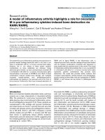
As shown in Fig. 1, stress can be viewed as a disturbance
input to the controller. Therefore, various efferent signals
from the controller to the actuator are affected by stress
before regulating hepatic glucose production and pancre-
atic hormonal secretion.
Methods
Model development
Assumptions
In order to establish a mathematical model of brain glu-
cose homeostasis, some assumptions are unavoidable.
The main assumptions include,
(1) The human body is composed of various segments,
each of which consists of homogeneous mass and/or
blood compartments;
(2) All parameters are time-invariant, such as constant
blood flow into the blood compartment of each segment,
constant distribution volumes for glucose, insulin and
glucagon in each compartment;
(3) Hepatic glucose production and pancreatic hormonal
secretion are regulatory methods of blood glucose. There
are no limitations in these processes;
(4) Glucose is utilized in tissue mass compartment or red
blood cells, while insulin and glucagon are cleared in tis-
sue mass compartments only;
(5) Both hepatic glucose production and pancreatic hor-
monal secretion depend on local state of the liver mass
and pancreas mass. The same is true for glucose utilization
or hormone removal in the tissue mass compartment.
(6) The GIG regulatory system is independent of other
physiological functions.
Feedback control for brain glucose homeostasisFigure 1
Feedback control for brain glucose homeostasis.
Central glucose sensor
Peripheral glucose sensor
Central insulin sensor
Hypothalamus Brain
Body (excluding brain,
including peripheral
GIG interactions)
Brain glucose
concentration
Normal brain
glucose
concentration
Stress
Arterial
blood glucose
concentration
Liver blood glucose
concentration
Brain insulin
concentration
Theoretical Biology and Medical Modelling 2009, 6:26 />Page 4 of 24
(page number not for citation purposes)
Other less important assumptions are made in the text
when necessary.
Model structure
In an integrative model developed by the authors previ-
ously for systems medicine in the intensive care unit, the
body is approximated by 6 segments (cranial, cardiocircu-
latory, lungs, muscle, visceral and others) or 13 compart-
ments, and various parameters are determined mainly
from the literature [18-21].
The compartmental structure and its parameters are
applied in this study. Since the visceral segment in the
original model represented a set of visceral tissues, includ-
ing the liver, kidneys, gut, and pancreas, it is necessary to
describe this visceral segment in detail in order to take
account of hepatic glucose production, pancreatic hormo-
nal secretion, gastrointestinal glucose absorption and glu-
cose loss via urine in case of hyperglycemia.
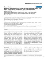
As shown in Fig. 2, the extended model consists of 9 seg-
ments or 19 compartments. The cranial segment consists
of 3 compartments, corresponding to the brain mass,
blood and cerebrospinal fluid (CSF). The cardiocircula-
tory segment is composed of arterial and venous compart-
ments. Each of the other 7 extracranial segments
comprises 2 compartments, that is, one is the mass and
the other is the blood.
Based on the anatomy of the hepatic portal vein, blood
flow from the pancreas segment and that from the gut seg-
ment enter the blood compartment of the liver segment
and then join the systemic circulation, together with the
hepatic arterial blood flow from the arterial part of the car-
diocirculatory segment.
Glucose, insulin and glucagon in the blood circulate
through various blood compartments, which transact
with their adjacent mass compartments. Glucose is pro-
duced endogenously in the liver mass compartment, or
given exogenously into the gut mass or venous blood
compartment. Insulin is produced in the pancreas mass or
infused into the muscle mass or venous blood compart-
ment. By contrast, glucagon is generated only in the pan-
creas mass compartment.
Compartment model of brain glucose homeostasisFigure 2
Compartment model of brain glucose homeostasis.
Venous bloodArterial blood
Liver blood
Liver mass
Brain mass
CSF
Brain blood
Lung mass
Lung blood
Muscle mass
Muscle blood
Other mass
Other blood
Kidney mass
Kidney blood
Gut mass
Gut blood
Pancreas mass
Pancreas blood
Hypothalamus
Hepatic glucose
production
Controlled
object
Pancreatic hormonal
secretion
Stress
Actuator
Controller
Theoretical Biology and Medical Modelling 2009, 6:26 />Page 5 of 24
(page number not for citation purposes)
Glucose is utilized in all tissue mass compartments and in
the arterial compartment by erythrocytes, while insulin
and glucagon are cleared in tissue mass compartments
only.
One of the main differences between the current extended
model and the original model is that the former considers
the cranial segment as the controlled object, the rest of the
body as the actuator, and arterial blood glucose concen-
tration as the actuating signal.
Another difference is the introduction of a feedback con-
trol loop of brain glucose in the extended model. Neuro-
nal and hormonal signals are generated based on the
brain glucose state and modified by stress before they reg-
ulate hepatic glucose production and pancreatic hormo-
nal secretion. Such feedback loop of brain-endocrine
crosstalk contributes to the control of brain glucose
homeostasis, together with the peripheral GIG interac-
tions occurring in the actuator.
Mathematical descriptions of the controlled object, the
actuator and the controller are given separately during
modeling.
Model of controlled object
Governing equations
Applying the mass conservation law, the dynamics of glu-
cose in the cranial segment is described mathematically as
follows,
where V denotes the diagonal matrix (3 × 3) of distribu-
tion volume, G vector (3 × 1) of glucose concentration,
G
art
is the glucose concentration in the arterial blood flow-
ing into the brain blood compartment of the cranial seg-
ment. The left hand side represents the storage rate of
glucose. Various matrices and vectors are given as follows,
The superscript T denotes transposition of the vector. All
symbols,
subscripts
and
superscripts
are summarized in the Glos-
sary. From the viewpoint of systems control, is con-
sidered as the controlled variable (output) and G
art
as the
controlling input.
Detailed description of the nonlinear term F in Equation (1)
Metabolism
Brain cells use glucose without the intermediation of insu-
lin [22]. Such insulin-independent glucose utilization in
the brain mass, M
brain
, is assumed a function of brain glu-
cose concentration, as described by the following Michae-
lis-Menten equation,
where m
1
and m
2
are estimable parameters. However, nei-
ther m
1
nor m
2
is currently available from reported data by
the authors, as it is based on glucose concentration in the
brain mass, but not in the blood. For simulation purpose,
it is assumed in this model that and
, on the basis of brain glucose concentra-
tion at the steady-state . Then equation (2) is mod-
ified to,
Facilitated transport through BBB/BCB
The blood-brain-barrier (BBB) and blood-CSF barrier
(BCB) are the interfaces between the brain blood and
brain mass. Physiologically, the BBB and BCB help to
maintain brain glucose homeostasis by regulating the
facilitated saturable transport of glucose with their semi-
impermeability, as shown in Fig. 3.
According to Rapoport [23], glucose transport across the
BBB, , is described by the Michaelis-Menten
equation with two parameters, the maximal transport rate
(T
G0
) and Michaelis constant (F
G0
), as follows,
V
G
KG W F
d
dt
G
art
=+ +
(1)
V =
⎡
⎣
⎤
⎦
diagVVV
mass
brain
csf
brain
blood
brain
,, ;
G =
⎡
⎣
⎤
⎦
GGG
mass
brain
csf
brain
blood
brain
T
;
K =
−
−
−− −−
−− −
kk
kk
G csf mass
brain
G csf mass
brain
G csf mass
brain
Gcs
0
ffmass
brain
brain
w
−
−
⎡
⎣
⎢
⎢
⎢
⎢
⎤
⎦
⎥
⎥
⎥
⎥
0
00
;
W =
⎡
⎣
⎤
⎦
00w
brain
T
;
F =− −
−− −− −−
fMff
G blood mass
brain brain
G blood csf
brain
G blood mass
bbrain
G blood csf
brain
T
f−
⎡
⎣
⎤
⎦
−−
.
G
mass
brain
M
mG
mass
brain
mG
mass
brain
brain
=
+
1
2
(2)
mM
brain
10
13= .
mG
mass
brain
20
03= .
G
mass
brain
0
M
G
mass
brain
G
mass
brain
G
mass
brain
M
brain brain
=
+
13
03
0
0
.
.
(2A)
f
G blood mass
brain
−−
f
T
G
G
blood
brain
K
G
G
blood
brain
G blood mass
brain
−−
=
+
0
0
(3)
Theoretical Biology and Medical Modelling 2009, 6:26 />Page 6 of 24
(page number not for citation purposes)
In this model, K
G0
= 0.9 [mg/ml] (Rapoport 1976) and
then .
The facilitated infusion of glucose across the BCB,
, is described similarly, although it is minor.
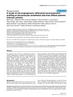
Dynamic BBB/BCB adaptation
Various clinical observations suggest that the dynamics of
glucose transport across BBB/BCB is influenced by the
adaptive nature of the barriers. For example, experimen-
tally-induced chronic hypoglycemia in rats elicited over-
expression of glucose transporter-1 (GLUT-1) and
redistribution of GLUT-1 at the BBB [24]. Overexpression
of GLUT-1 is viewed to have a positive effect on the max-
imal transport rate, T
G0
, without altering the Michaelis
constant, K
G0
[25]. In rats with chronic hyperglycemia, the
maximum glucose transport capacity of the BBB decreased
from 400 to 290 micromoles per 100 grams per minute,
and the glucose transport rate in the brain decreased to 20
percent below normal when plasma glucose was lowered
to normal values [3].
This mechanism, termed BBB adaptation to chronic
hyperglycemia in Fig. 3, represents a dynamic process
with a long time constant, since brain glucose transport is
not altered following short episodes of recurrent hypogly-
cemia in healthy human volunteers [25]. The adaptation
must be inactive within the euglycemic range, since fre-
quent variations, known as ultradian oscillations, occur in
blood glucose.
Therefore, a first-order dynamics of two parameters,
namely, gain and time constant, is introduced into this
model to modify the maximal glucose transport rate T
G0
with respect to blood dysglycemia as follows,
where ΔT
G
is the response of maximal glucose transport
rate T
G
with respect to hyperglycemia
( , is the maximum
value of glucose concentration in the brain blood com-
partment at the steady state) or hypoglycemia
( , is the minimum
value of glucose concentration in the brain blood com-
partment at the steady state).
κ
G
and
τ
G
denote gain and
time constant, respectively. s in equation (5) is LapLace
operator.
Equation (3) describing the facilitated infusion through
BBB/BCB is thus modified to,
where T
G
is adaptable according to equations (4) and (5)
with respect to dysglycemia in the brain blood.
Model of the actuator characteristic of peripheral GIG
interactions
Governing equations
The dynamics of glucose concentration in each extracra-
nial segment (with the exception of the cardiocirculatory
segment) is described as follows,
where V denotes glucose distribution volume and G glu-
cose concentration. The superscript x represents the seg-
ment while the subscript mass or blood represents the mass
T
G
blood
brain
G
blood
brain
f
G G blood mass
brain
00
09
0
0
=
+
−−
.
.
f
G blood csf
brain
−−
T
T G within euglycemic range
TTG
G
G blood
brain
G G blood
b
=
−
0
0
()
(
Δ
rrain
without euglycemic range )
⎧
⎨
⎪
⎩
⎪
(4)
Δ
Δ
T
G
s
Gs
G
G
s
()
()
=
+
κ
τ
1
(5)
ΔGG G
blood
brain
blood
brain
=− >
0
0
max
G
blood
brain
0max
ΔGG G
blood
brain
blood
brain
=− <
0
0
min
G
blood
brain
0min
f
T
G
G
blood
brain
K
G
G
blood
brain
G blood mass
brain
−−
=
+
0
(3A)
V
dG
mass
x
dt
fpu
mass
x
G blood mass
x
G
x
G
x
=+−
−−
(6)
V
dG
blood
x
dt
fwGG
blood
x
G blood mass
xx
art blood
x
=− + −
−−
()
(7)
Facilitated glucose transport across BBBFigure 3
Facilitated glucose transport across BBB.
Glucose flux across BBB
Plasma glucose (mg/dl)
After chronic hyperglycemia
Normal
BBB
adaptation
Theoretical Biology and Medical Modelling 2009, 6:26 />Page 7 of 24
(page number not for citation purposes)
or blood compartment, respectively. denotes
glucose diffusion from the blood compartment into its
adjacent mass compartment in segment x. and
denote glucose production and utilization in the mass
compartment of segment x. w
x
is blood flow. G
art
is glucose
concentration in the arterial blood. The last term on the
right hand side of equation (7) represents net glucose
delivery by blood flow into the blood compartment of
segment x.
The term p in equation (6) only appears in the mass com-
partment of the liver segment due to the endogenous
hepatic glucose production or in the mass compartment
of the gut segment due to the exogenous gastrointestinal
glucose absorption. The term u in equation (6) consists of
the two parts, namely, insulin-independent utilization
and insulin-dependent utilization. The former corre-
sponds to glucose discard from the kidney mass compart-
ment through the urine in hyperglycemia, while the latter
is mostly due to glucose metabolism in the muscular mass
and various visceral mass compartments.
In the arterial and venous compartments of the cardiocir-
culatory segment, the dynamics of glucose concentrations
are given as follows,
where V
art
and V
ven
denote the distribution volumes of glu-
cose in the arterial and venous compartments of the cardi-
ocirculatory segment. G
art
and G
ven
are the glucose
concentrations in these two compartments. w is total car-
diac output, while W
y
represents blood flow from the
blood compartment of segment y (including the cranial,
liver, kidneys, muscle and the other segment), particu-
larly, . is the blood glucose concentra-
tion in segment y, in particular, denotes glucose
concentration in the blood compartment of the lung seg-
ment. The term in equation (8) describes the insulin-
independent glucose utilization by erythrocytes, which is
assumed to occur only in the arterial compartment for
simplification, although the glucose is physiologically uti-
lized by erythrocytes in all blood compartments. The term
of in equation (9) denotes exogenous glucose infu-
sion into the venous blood.
Explanation of various terms in equations (6)-(9)
Hepatic glucose production
Conversion of glucose into glycogen, as well as glycoge-
nolysis and/or gluconeogenesis, in the liver is one of the
primary strategies involved in the regulation of blood glu-
cose concentration. High levels of either glucose or insu-
lin serve to reduce glucose production by the liver, while
glucagon stimulates hepatic glucose production.
The action of insulin on glucose production is a reflection
of insulin concentration in the extracellular space, rather
than in blood [26]. Therefore, hepatic glucose production
depends not on the concentrations of glucose, insulin and
glucagon in the blood compartment, but rather on their
concentrations in the mass compartment of the liver seg-
ment.
The following linear equation is introduced to describe
hepatic glucose production phenomenologically,
where denotes net hepatic glucose production and
is its steady-state value. Positive means net glu-
cose is produced by the liver while negative means net
glucose is stored or degraded in the liver. , and
are local concentrations of glucose, insulin and glu-
cagon in the hepatic mass compartment, respectively.
, and are their respective steady-state
values. k
1
, k
2
and k
3
are positive parameters to be esti-
mated. The four terms in the brackets correspond to basal
production, contribution of hepatic glucose state, that of
hepatic insulin state and that of hepatic glucagon state,
respectively, to hepatic glucose production based on their
steady-states. The signs of addition and subtraction are
based on physiological functions concerning the effects of
glucose, insulin and glucagon on hepatic glucose produc-
tion.
f
G blood mass
x
−−
p
G
x
u
G
x
V
dG
art
dt
wG G u
art
blood
lung
art G
red
=−−()
(8)
V
dG
ven
dt
wG wG p
ven
y
blood
y
ven
G
inf
=−+
∑
(9)
ww
y
=
∑
G
blood
y
G
blood
lung
u
G
red
p
G
inf
pk
G
mass
liv
G
mass
liv
G
mass
liv
k
I
mass
liv
I
mass
li
G
liv
=−
−
−
−
(1
0
0
0
1
2
vv
I
mass
liv
k
E
mass
liv
E
mass
liv
E
mass
liv
p
G
liv
0
0
0
30
+
−
⋅)
(10)
p
G
liv
p
G
liv
0
p
G
liv
p
G
liv
G
mass
liv
I
mass
liv
E
mass
liv
G
mass
liv
0
I
mass
liv
0
E
mass
liv
0
Theoretical Biology and Medical Modelling 2009, 6:26 />Page 8 of 24
(page number not for citation purposes)
Equation (10), which is based on the concentrations of
glucose, insulin and glucagon in the hepatic mass com-
partment, is completely different from mathematical
descriptions based on their concentrations in the blood,
which are commonly used in the currently existing theo-
retical models. Therefore, it is difficult to determine the
numerical values for parameters k
1
, k
2
and k
3
from the lit-
erature. The values of these parameters are chosen based
on trial-and-error during model verification and improve-
ment.
Utilization by peripheral tissue
The action of insulin necessary to stimulate peripheral
glucose utilization is also determined by its concentration
in interstitial fluid that bathes insulin-sensitive cells [27].
The uptake of glucose peripherally (primarily the muscle,
gut, lungs, liver, pancreas, kidneys and the other mass)
depends not only on local glucose concentration but also
on local insulin concentration. A suitable form of this uti-
lization in the peripheral segment x is given as follows,
where denotes glucose utilization by the mass com-
partment in segment x, is its steady-state value. k
4
and
k
5
are estimable positive parameters. The two brackets on
the right hand describe the contributions of local glucose
state and local insulin state to glucose utilization. Since no
data are available to the authors from the literature, both
the value of parameter k
4
and that of k
5
are estimated by
trial-and-error.
Utilization by erythrocytes
Similar to brain glucose metabolism, glucose utilization
by erythrocytes, u
red
in equation (8), is independent of
insulin (and glucagon) concentration, but dependent on
glucose concentration. It is a function of arterial blood
glucose concentration with saturation. Following this per-
spective, the form of this contribution in equation (8) is
described by the following equation (12).
where is glucose utilization by erythrocytes, is
its steady-state value. G
art
denotes glucose concentration
in the arterial blood, and G
art0
is its steady-state value.
Loss through urine
Glucose uptake by the kidneys consists of the following
two types. First, insulin-glucose-dependent metabolism
occurs in the kidney mass compartment, as given by equa-
tion (11). Second, increased blood glucose concentration
leads to loss of glucose through urine. Such glucose loss is
independent of insulin and glucagon. On account of the
physiological fact that glucose appears in the urine when
the blood glucose concentration is over 1.8 mg/ml [22], it
is assumed that, when glucose concentration in the kidney
mass compartment is below a threshold level (for simpli-
fication, 1.8 mg/ml in the model), urinary glucose loss is
zero. In contrast, the rate of urinary glucose loss increases
linearly with increasing concentration in the kidney mass
compartment, once the glucose concentration in the kid-
ney mass compartment exceeds the threshold level. Math-
ematically, this is described by the equation,
where is the urinary glucose loss, w
urine
is urinary
flow, at an average of 2 liters per day (0.023 ml/s),
is the glucose concentration in the mass compartment of
the kidney segment.
Permeability via capillary bed
The transcapillary delivery of glucose between the blood
compartment and its adjacent mass compartment
depends on the permeability coefficient and the concen-
tration difference between the two compartments.
Assuming that the relation between transcapillary delivery
of glucose and the concentration difference is linear, the
term in equations (6) and (7) is described by,
where is the permeability coefficient of glucose
between the mass and blood compartments.
At steady-state, metabolic utilization of glucose in each
mass compartment of the extracranial segments should be
equal to the net glucose transport from its adjacent blood
compartment. Accordingly, it is possible to estimate the
permeability coefficient from the metabolic glucose
utilization ( ), steady-state glucose concentrations in
the mass compartment and the blood compartment, as
given by the following equation (15).
uk
G
mass
x
G
mass
x
G
mass
x
k
I
mass
x
I
mass
x
I
mass
x
G
x
=+
−
⋅+
−
()
(
1
0
0
1
0
0
4
5
)) ⋅ u
G
x
0
(11)
u
G
x
u
G
x
0
u
G
art
G
art
G
art
u
G
red
G
red
=
+
13
03
0
0
.
.
(12)
u
G
red
u
G
red
0
u
G
wG G
G
urine
mass
kid
urine
mass
kid
mass
kid
=
≤
−>
⎧
018
18 18
(.)
(.)(.)
⎨⎨
⎪
⎩
⎪
(13)
u
G
urine
G
mass
kid
f
G blood mass
x
−−
fhGG
G blood mass
x
G
x
blood
x
mass
x
−−
=−()
(14)
h
G
x
h
G
x
u
G
x
0
Theoretical Biology and Medical Modelling 2009, 6:26 />Page 9 of 24
(page number not for citation purposes)
However, the transcapillary delivery of glucose is under
the influence of blood insulin [28]. Therefore, the glucose
diffusion in equation (14) is modified as fol-
lows,
where the contribution of local insulin state to transcapil-
lary glucose delivery is taken into account by introducing
the last brackets.
Model of insulin and glucagon dynamics
Insulin dynamics
The concentration dynamics of insulin in segment x, rep-
resentatively consisting of mass and blood compartments,
is described by dynamic mass balance as follows,
where and are distribution volumes of insu-
lin, and are insulin concentration in the mass
and blood compartments of segment x, respectively,
is insulin transport from the blood compart-
ment to its adjacent mass compartment in segment x, I
art
denotes the arterial insulin concentration, is the pro-
duction rate of insulin, is insulin removal from the
mass compartment in segment x, and w
x
is blood flow to
segment x. The last term on the right hand side of equa-
tion (17) represents net insulin delivery through blood
flow into the blood compartment of segment x.
Endogenous insulin is secreted from beta-cells in the pan-
creatic mass. Both elevated blood glucose and glucagon
stimulate insulin secretion [22]. It is reasonable to con-
sider that the concentrations of glucose and glucagon in
the pancreatic mass determine the level of endogenous
insulin production. The following equation mathemati-
cally describes pancreatic insulin secretion,
where is insulin secretion within the pancreatic mass
compartment, is its steady-state value, and k
6
and k
7
are estimable positive parameters. Negative calculated
is set to zero because of its physiological meaning-
less. The three terms in the brackets correspond to basal
secretion, contribution of pancreatic glucose state and
that of pancreatic glucagon state, respectively, to pancre-
atic insulin secretion based on their steady-states. The plus
signs are based on the physiological functions concerning
effects of glucose and glucagon on pancreatic insulin
secretion. The values for parameters k
6
and k
7
are unavail-
able in the literature and given by the authors based on
trial-and-error.
Since insulin is cleared by all insulin-sensitive tissues,
is dependent on the local concentration of insulin in each
of the extracranial mass compartments. Therefore,
where denotes rate of insulin removal from the mass
compartment of segment x, is its steady-state value,
and k
8
is estimable positive parameter. The bracket on the
right hand describes the contribution of local insulin state
to insulin removal. Value of parameter k
8
is also not avail-
able in the literature.
As no insulin is produced in the brain, intracranial insulin
concentrations depend on BBB/BCB transport of periph-
eral insulin. However, such transport is characterized by
saturation [16]. Furthermore, hyperglycemia abolishes
insulin transport across BBB [16]. Therefore, it is reasona-
ble in this model to consider that BBB/BCB insulin trans-
port also adapts with respect to dysglycemia.
Like that of glucose, insulin transport from brain blood to
brain mass or to CSF also follows the Michaelis-Menten
h
u
mass
x
G
blood
x
G
mass
x
G
x
=
−
0
0
0
(15)
f
G blood mass
x
−−
fhGG
I
blood
x
I
blood
x
I
bloo
G blood mass
x
G
x
mass
x
blood
x
−−
=−+
−
()(1
0
dd
x
0
)
(14A)
V
dI
mass
x
dt
fpu
mass
x
I blood mass
x
I
x
I
x
=+−
−−
(16)
V
dI
blood
x
dt
fwII
blood
x
I blood mass
xx
art blood
x
=− + −
−−
()
(17)
V
mass
x
V
blood
x
I
mass
x
I
blood
x
f
I blood mass
x
−−
p
I
x
u
I
x
pk
G
mass
pan
G
mass
pan
G
mass
pan
k
E
mass
pan
E
mass
pa
I
pan
=+
−
+
−
(1
0
0
0
6
7
nn
E
mass
pan
p
I
pan
0
0
) ⋅
(18)
p
I
pan
p
I
pan
0
p
I
pan
u
I
x
uk
I
mass
x
I
mass
x
I
mass
x
u
I
x
I
x
=+
−
⋅()1
0
0
80
(19)
u
I
x
u
I
x
0
Theoretical Biology and Medical Modelling 2009, 6:26 />Page 10 of 24
(page number not for citation purposes)
equation mathematically, on account of the analogous
blood-brain barrier transport systems existing for glucose,
amino acids, plasma proteins, as well as the circulating
insulin [29]. Altogether, a formula similar to equation (3)
is introduced to describe the facilitated transport across
BBB/BCB of insulin as follows,
where K
I0
is the Michaelis constant for the facilitated insu-
lin diffusion across BBB/BCB, and T
I
is the maximal trans-
port rate of insulin across BBB/BCB, which is glucose
dependent, as given by equations (4) and (5), as follows,
where T
I0
is the steady-state value of T
I
. ΔT
I
is the response
of maximal insulin transport rate T
I
with respect to hyper-
glycemia ( , is the
maximum value of glucose concentration in the brain
blood compartment at the steady state) or hypoglycemia
( , is the minimum
value of glucose concentration in the brain blood com-
partment at the steady state). Similar to glucose transport
into brain mass and CSF compartments,
κ
I
and
τ
I
are gain
and time constant, respectively. The time constant
τ
I
should be some days in the rat and some years in human.
Both gain
κ
I
and time constant
τ
I
are individual depend-
ent.
In the extracranial segments, the transcapillary delivery of
insulin from the blood compartment to its adjacent mass
compartment is mediated through passive diffusion [28].
Thus,
where is permeability coefficient of insulin between
the mass and blood compartments.
Since the metabolic removal of insulin in all insulin-sen-
sitive mass compartments of the extracranial segments is
equal to insulin diffusion from their adjacent blood com-
partments, could be determined from metabolic insu-
lin removal, insulin concentrations in the mass
compartment and blood compartment at steady-state, as
follows,
The dynamics of insulin concentrations in the cranial seg-
ment are represented mathematically similar to equation
(1), while the dynamics of insulin concentrations in the
arterial and venous compartments of cardiocirculatory
segment are described by using mathematical equations
similar to equations (8) and (9).
Glucagon dynamics
Similar to that of glucose and insulin, the concentration
dynamics of glucagon in each segment x consisting of the
mass and blood compartments is described as follows,
where and are the distribution volumes of
glucagon, and are the glucagon concentra-
tions of the mass and blood compartments in segment x,
respectively, is glucagon transport from the
blood compartment to its adjacent mass compartment in
segment x, E
art
is the arterial glucagon concentration,
and denote glucagon production and removal from
the mass compartment in segment x, respectively, and w
x
is the blood flow to segment x. The last term on the right
hand side of equation (26) represents net glucagon deliv-
ery through blood flow into the blood compartment
within segment x.
In case of glucagon dynamics, the term corresponds to
glucagon production from alpha-cells in the pancreatic
mass. It depends on the concentrations of glucose and
insulin in the pancreatic mass. In other words, either ele-
f
T
I
I
blood
brain
K
I
I
blood
brain
I blood mass
brain
−−
=
+
0
(20)
T
T G within euglycemic range
TTG
I
I blood
brain
I I blood
b
=
−
0
0
()
(
Δ
rrain
without euglycemic range )
⎧
⎨
⎪
⎩
⎪
(21)
Δ
Δ
T
I
s
Gs
I
I
s
()
()
=
+
κ
τ
1
(22)
ΔGG G
blood
brain
blood
brain
=− >
0
0
max
G
blood
brain
0max
ΔGG G
blood
brain
blood
brain
=− <
0
0
min
G
blood
brain
0min
fhIG
I blood mass
x
I
x
blood
x
mass
x
−−
=−()
(23)
h
I
x
h
I
x
h
u
mass
x
I
blood
x
I
mass
x
I
x
=
−
0
0
0
(24)
V
dE
mass
x
dt
fpu
mass
x
E blood mass
x
E
x
E
x
=+−
−−
(25)
V
dE
blood
x
dt
fwEE
blood
x
E blood mass
xx
art blood
x
=− + −
−−
()
(26)
V
mass
x
V
blood
x
E
mass
x
E
blood
x
f
E blood mass
x
−−
p
E
x
u
E
x
p
E
x
Theoretical Biology and Medical Modelling 2009, 6:26 />Page 11 of 24
(page number not for citation purposes)
vated level of glucose or that of insulin depresses pancre-
atic glucagon secretion. Its mathematical description is
given by:
where is glucagon secretion in the pancreatic mass
compartment, is its steady-state value, and k
9
and k
10
are estimable positive parameters. Similar to that of ,
negative calculated is set to zero. Three terms in the
brackets correspond to the basal secretion, contribution of
pancreatic glucose state and that of pancreatic insulin
state, respectively, to pancreatic glucagon secretion based
on their steady-states. The minus signs are based on the
physiological functions describing the effects of glucose
and insulin on pancreatic glucagon secretion. Values of
parameter k
9
and k
10
are not available in literature.
Glucagon is degraded in all extracranial mass compart-
ments, mainly by the kidney and the liver. Compared to
extracranial glucose metabolism and insulin removal,
is assumed to be independent of ambient glucose or insu-
lin concentrations. That is, the term is a function of
local glucagon concentration only, as given by,
where v
E
denotes degradation constant, which is deter-
mined from the steady-state values of glucagon concentra-
tion ( ) and glucagon removal ( ) in the mass
compartment,
The dynamics of glucagon concentrations in the arterial
and venous compartments of the cardiocirculatory seg-
ment are described by using mathematical descriptions
similar to equations (8) and (9).
Brain-endocrine crosstalk
As mentioned above, the GIG regulatory system com-
prises the peripheral GIG interactions and the central
brain-endocrine crosstalk. The former is described mathe-
matically in detail during modeling the actuator in equa-
tions (10), (11), (18) and (19). The latter, mainly
consisting of the glucosensor-hypothalamus-liver-pan-
creas link [17,30], modifies the peripheral GIG interac-
tions in order to control brain glucose homeostasis, as
shown in Fig. 1.
Various glucosensing neurons are distributed throughout
the brain, which also receives afferent neural inputs from
glucosensors in the liver, carotid body, and small intes-
tines [15]. It is considered that glucose concentration in
the brain mass is the major signal to the hypothalamus for
the regulation of brain glucose homeostasis, while central
insulin is a hormonal signal that provides negative feed-
back to the brain. For example, an increase in insulin sig-
nal in the hypothalamus elicits responses that reduce
hepatic glucose production [31]. The anatomy of the
brain-endocrine crosstalk is described in detail by Uyama
and colleagues [17].
However, the current knowledge regarding neuronal and
hormonal signals for hepatic glucose production and pan-
creatic hormonal production are not quantitative but
rather qualitative in nature. For simplification of theoret-
ical discussion, it is assumed that a proportional feedback
control of brain glucose occurs, mainly based on differ-
ences between brain glucose concentration and baseline.
As shown in Fig. 1, peripheral glucose and central insulin
act as auxiliary input to the central hypothalamic control-
ler.
The controlled error is given as follows,
where a
cg
, a
ci
and a
pg
are parameters with appropriate unit.
They are adjustable in the model to reflect varying impor-
tance of various glucose and insulin sensors in monitor-
ing the glucose state in the body. The three brackets on the
right hand describe the contributions of central glucose
state, central insulin state and peripheral glucose state.
The two plus signs (+) in equation (30) are based on the
physiological fact that glucose-sensing neurons in the
brain serve as integrators of various metabolic signals
[13].
Based on the assumption of proportional feedback con-
trol of brain glucose, three efferent signals are generated
by the hypothalamus, that is,
pk
G
mass
pan
G
mass
pan
G
mass
pan
k
I
mass
pan
I
mass
p
E
pan
=−
−
−
−
(1
0
0
0
9
10
aan
I
mass
pan
p
E
pan
0
0
) ⋅
(27)
p
E
pan
p
E
pan
0
p
I
pan
p
E
pan
u
E
x
u
E
x
uvE
E
x
Emass
x
=
(28)
E
mass
x
0
u
E
x
0
v
u
E
x
E
mass
x
E
=
0
0
(29)
GaG G
aI I
error cg mass
brain
mass
brain
ci mass
brain
mass
br
=−
+−
()
(
0
0
aain
pg blood
liv
blood
liv
aG G)( )+−
0
(30)
γβα
γβα
===kG kG kG
error error error
,,
(31)
Theoretical Biology and Medical Modelling 2009, 6:26 />Page 12 of 24
(page number not for citation purposes)
where
α
,
β
and
γ
denote signals regulating hepatic glucose
production, pancreatic secretion of insulin and glucagon,
respectively, k
γ
, k
β
and k
α
are parameters with appropriate
units. They are all adjustable in the model to reflect vary-
ing importance of various mechanisms involved in glu-
cose homeostasis. Since insulin depresses both hepatic
glucose production and pancreatic glucagon secretion, k
γ
> 0 and k
a
> 0, while k
β
> 0.
Each of these signals acts to modify hepatic glucose pro-
duction, pancreatic secretion of insulin and glucagon.
Therefore, equations (10), (18) and (27) are changed to,
Where , and denote hepatic glucose produc-
tion, pancreatic insulin secretion and pancreatic glucagon
secretion, respectively. , and are steady-
state values. The first bracket on the right hand of each
equation describes the effect of brain-endocrine crosstalk.
Stress input to the central controller
To take into consideration the effect of psychological
stress, it is necessary to quantify it. To the best of our
knowledge, a quantitative measure of stress has not been
established. Therefore, a rather abstract variable of posi-
tive value varying between 0 and 1 is introduced to
describe mild to severe stress, respectively. Various dura-
tions of stress, namely, short-term, repeated, long-term,
are also used to describe the stress encountered in daily
life.
It is well documented that stress causes a direct increase in
pancreatic glucagon production through catecholamines,
which play a critical role in these fight-or-flight circum-
stances [32]. The increase in glucagon levels, stimulated
by increased catecholamine, drives increased glycogenol-
ysis and gluconeogenesis in liver. In contrast, insulin is
decreased during times of stress [32,33]. The fight-or-
flight response to stress characteristically increases hepatic
glucose production.
Taking account of the systemic effects of stress on the
hepatic glucose production and pancreatic hormonal
secretion, various coefficients can be introduced into
equations (10'), (18') and (27') to describe the total
effects of stress. That is,
where s denotes the severity of stress. The first bracket on
the right hand of each equation describes the effect of
stress.
Parameters
Various physiological parameters, such as distribution
volume, blood flow and metabolic allocation, are com-
mon to the current model and the integrated model devel-
oped previously for systems medicine in the intensive care
[18-21]. In addition to the original data applicable in this
model, values of special parameters for the GIG regulatory
system are added mainly based on literature.
pk
G
mass
liv
G
mass
liv
G
mass
liv
k
I
mass
liv
I
ma
G
liv
=+ −
−
−
−
()(11
0
0
1
2
γ
sss
liv
I
mass
liv
k
E
mass
liv
E
mass
liv
E
mass
liv
p
G
liv
0
0
0
0
30
+
−
⋅)
(10A)
pk
G
mass
pan
G
mass
pan
G
mass
pan
k
E
mass
pan
E
ma
I
pan
=+ +
−
+
−
()(11
0
0
6
7
β
sss
pan
E
mass
pan
p
I
pan
0
0
0
) ⋅
(18A)
pk
G
mass
pan
G
mass
pan
G
mass
pan
k
I
mass
pan
I
m
E
pan
=+ −
−
−
−
()(11
0
0
9
10
α
aass
pan
I
mass
pan
p
E
pan
0
0
0
) ⋅
(27A)
p
G
liv
p
I
pan
p
E
pan
p
G
liv
0
p
I
pan
0
p
E
pan
0
ps k
G
mass
liv
G
mass
liv
G
mass
liv
k
I
mass
li
G
liv
=+ + −
−
−
()( )(11 1
0
0
1
2
γ
vv
I
mass
liv
I
mass
liv
k
E
mass
liv
E
mass
liv
E
mass
liv
p
G
li
−
+
−
⋅
0
0
0
0
30
)
vv
(10B)
ps k
G
mass
pan
G
mass
pan
G
mass
pan
k
E
mass
pa
I
pan
=− + +
−
+
()( )(11 1
0
0
6
7
β
nn
E
mass
pan
E
mass
pan
p
I
pan
−
⋅
0
0
0
)
(18B)
ps k
G
mass
pan
G
mass
pan
G
mass
pan
k
I
mass
p
E
pan
=+ + −
−
−
()( )(11 1
0
0
9
10
α
aan
I
mass
pan
I
mass
pan
p
E
pan
−
⋅
0
0
0
)
(27B)
Theoretical Biology and Medical Modelling 2009, 6:26 />Page 13 of 24
(page number not for citation purposes)
Production or secretion
Glucose input into the circulation is normally approxi-
mately 2 mg/min/kg of body weight [34]. At normal fast-
ing level of blood glucose, the rate of insulin secretion is
in the order of 25 ng/min/kg of body weight [22]. The
secretion rate of glucagon could be estimated based on
glucagon removal under steady-state conditions. Such
estimation yields a value of approximately 1400 pg/min/
kg of body weight in humans [35].
Utilization or removal
The main contributors to glucose disappearance in the
fasting, resting state are brain, lean tissues, adipose tissue
and red blood cells [34]. In the model, total glucose input
of 2.24 mg/s is assumed to be utilized in various mass
compartments and the arterial blood compartment (brain
0.65, lung 0.08, pancreas 0.01, gut 0.25, liver 0.12, kidney
0.02, muscle 0.67, the residual 0.22, and the arterial com-
partment 0.22 mg/s). The allocation of glucose utilization
is completely assumed while taking account of the weight
of various mass compartments.
Under normal physiological state, insulin secreted by the
pancreas is cleared in the liver, kidney, muscle, adipose
tissue and other tissues [32]. As in case of glucose utiliza-
tion, total insulin secretion of 28 ng/s is considered to be
cleared in various tissues (brain 0.27, lung 3.70, gut 0.26,
liver 21.43, kidney 0.79, muscle 0.74, and the residual
0.81 ng/s).
Both the kidney and liver also remove glucagon from the
circulation, accounting for 30% and 20% of disposal,
respectively [32]. The total glucagon secretion of 1569.73
pg/s is cleared in various mass compartments (brain
19.42, lung 12.23, gut 39.80, liver 470.62, kidney 313.75,
muscle 390.91, and residual 323.0 pg/s).
Concentration
In normal subjects, glucose concentration in the brain is
about 20 mg/dl, CSF 60 mg/dl, jugular venous blood 90
mg/dl and carotid arterial blood 100 mg/dl [11]. In the
present model, they are assumed to be 18.42, 58.42,
88.42, and 96.75 mg/dl, respectively. The glucose concen-
trations in various mass compartments are assumed on a
mass-to-blood glucose level of about 60% [36]. Particu-
larly, it is possible to calculate the concentration in vari-
ous blood compartments according the steady-state
values of glucose concentration and glucose utilization in
a mass compartment without glucose production as fol-
lows,
Insulin concentration in the brain is less than that in the
blood [37]. Insulin concentrations in CSF, brain mass and
arterial blood are assumed to be 2.83, 2.41 and 43.56 ng/
dl, respectively, in the model, while considering that the
insulin permeation across BBB/BCB into the CSF and
brain mass is only removed from the brain mass. Insulin
concentrations in various extracranial mass compart-
ments are assumed on account of mass-to-blood insulin
level of about 25% [27].
In the model, the arterial blood glucagon concentration is
0.81 pg/dl, which is consistent with the physiological
range of blood glucagon concentration of between 0.25
and 1.5 pg/dl after 12 h fast [35]. Glucagon concentra-
tions in various mass and blood compartments are
assumed on account of mass-to-blood glucose level of
about 40%, taking account of the physiological fact that
the plasma protein concentration in the interstitial space
is normally about 40% of that in the plasma [38].
Furthermore, the concentrations of insulin and glucagon
in various blood compartments could be calculated as in
case of glucose.
Permeability coefficient
The permeability coefficient at the capillary bed could be
estimated by equations (15), (24) and (29), based on the
steady-state value of local concentrations and metabolic
utilization or removal. The estimated values are con-
firmed by a few available clinical parameters. For exam-
ple, the permeability coefficient in muscle is 1.7-6.0 ml/
min/100 g for glucose and 0.5 ml/min/100 g for insulin
[28].
As assumed above, all these parameters are time-invariant.
Various physical and physiological parameters used for
simulations are summarized in Table 1, Table 2 and Table
3.
Suggested values for some unavailable parameters
Values of some parameters are assumed during model ver-
ification and model improvement, since no data are avail-
able in the literature, as mentioned above. Calculations of
these parameters are also based on the assumption that
hepatic glucose production, pancreatic hormonal secre-
tion, glucose utilization and hormonal removal depend
on local concentrations of glucose, insulin and glucagon
in the mass compartments, as described in equations
(10), (11), (12), (13), (18), (19), (27) and (28), in the
present model. Such assumption is acceptable in a math-
ematical approach because the shapes of the functions,
instead of their mathematical forms, are more important
[8]. Particularly, the following values of appropriate units
are assumed (Table 4) in the model for simulation.
G
w
x
G
art
u
mass
x
w
x
blood
x
=
−
(32)
Theoretical Biology and Medical Modelling 2009, 6:26 />Page 14 of 24
(page number not for citation purposes)
Table 1: Parameters characterizing the GIG regulatory system: Concentration
Segment Compartment Volume (ml)* Glucose concentration
(mg/ml)
&
Insulin concentration
(ng/ml)
&
Glucagon
concentration (pg/ml)
&
Brain CSF 150 0.584 0.028 8.02
mass 1374 0.184 0.024 8
blood 6.0 0.884 0.402 78.614
Lung mass 1669 0.591 0.080 16.001
blood 70 0.971 0.436 81.120
Pancreas mass 70 0.662 150.672 27985.9
blood 0.29 0.962 27.115 1575.13
Gastrointestinal mass 3041 0.639 0.087 15.995
blood 12.4 0.939 0.407 76.705
Liver mass 1541 1.304 0.182 18.027
blood 6.28 1.104 0.902 159.225
Kidney mass 253 0.668 0.052 11.554
blood 1.03 0.968 0.372 55.701
Muscle mass 22023 0.611 0.061 16.767
blood 101 0.951 0.377 57.982
Residual mass 27668 0.847 0.081 20.747
blood 125 0.914 0.377 49.921
Cardiocirculatory arterial 1129 0.968 0.436 81.104
venous 3609 0.973 0.497 81.324
*: [21]
&: [32]. Estimated on base of compartmental mass.
Table 2: Parameters characterizing the GIG regulatory system. Consumption
Segment Compartment Flow (ml/s)* Glucose consumption
(mg/s)
&
Insulin consumption
(ng/s)
&
Glucagon consumption
(pg/s)
&
Brain CSF 0
mass 0 0.65 0.27 19.42
blood 13
Lung mass 0 0.08 3.70 12.23
blood 100
Pancreas mass 0 0.01 0.0 0.0
blood 1.75
Gastrointestinal mass 0 0.25 0.26 39.80
blood 15.75
Liver mass 0 0.12 21.43 470.62
blood 22.523
Kidney mass 0 0.02 0.79 313.75
blood 20.5
Muscle mass 0 0.67 0.74 390.91
blood 21
Residual mass 0 0.22 0.81 323.0
blood 23
Cardiocirculatory arterial 100 0.22
venous 100
*: [21]
&: [32]. Estimated on base of compartmental mass.
Theoretical Biology and Medical Modelling 2009, 6:26 />Page 15 of 24
(page number not for citation purposes)
Table 3: Parameters characterizing the GIG regulatory system: Permeability
Segment Compartment Glucose permeability (ml/s)
&
Insulin permeability (ml/s)
&
Glucagon permeability (ml/s)
&
Brain CSF 0.022 (csf-mass) 1.734 (csf-mass) 9.711 (csf-mass)
mass (K = 0.9 mg/ml) (K = 10.59 ng/ml) (K = 2118 pg/ml)
blood
Lung mass 0.061 0.341 0.191
blood
Pancreas mass 0.006 0.227 0.063
blood
Gastrointestinal mass 0.253 1.111 0.622
blood
Liver mass 11.013 19.454 3.735
blood
Kidney mass 0.021 21.885 7.353
blood
Muscle mass 2.501 11.852 6.637
blood
Residual mass 0.747 9.016 5.049
blood
Cardiocirculatory arterial
venous
&: Estimated on base of equations (15), (24) and (29).
Table 4: Other assumed parameters
Symbol Description Assumed value
k
1
Positive parameter concerning contribution of hepatic glucose state to hepatic glucose production in Equation 10 1.0
k
2
Positive parameter concerning contribution of hepatic insulin state to hepatic glucose production in Equation 10 0.005
k
3
Positive parameter concerning contribution of hepatic glucagon state to hepatic glucose production in Equation 10 0.25
k
4
Positive parameter concerning contribution of local glucose state to glucose utilization by peripheral tissue in Equation 11 0.001
k
5
Positive parameter concerning contribution of local insulin state to glucose utilization by peripheral tissue in Equation 11 0.001
k
6
Positive parameter concerning contribution of local glucose state to insulin secretion within the pancreatic mass
compartment in Equation 18
1.0
k
7
Positive parameter concerning contribution of local glucagon state to insulin secretion within the pancreatic mass
compartment in Equation 18
0.01
k
8
Positive parameter concerning contribution of local insulin state to insulin removal from insulin-sensitive tissue in
Equation 19
0.03
k
9
Positive parameter concerning contribution of local glucose state to glucagon secretion within the pancreatic mass
compartment in Equation 27
0.01
k
10
Positive parameter concerning contribution of local insulin state to glucagon secretion within the pancreatic mass
compartment in Equation 27
0.1
Maximum value of glucose concentration in the brain blood compartment at the steady state in equations 4 and 21 1.0
Minimum value of glucose concentration in the brain blood compartment at the steady state in equations 4 and 21 0.8
κ
G
Gain in Equation 5 0.3
κ
I
Gain in Equation 22 0.1
τ
G
Time constant in Equation 5 1200
τ
I
Time constant in Equation 22 1200
a
cg
Positive parameter concerning contribution of central glucose state to the controlled error in Equation 30 4.0
a
ci
Positive parameter concerning contribution of central insulin state to the controlled error in Equation 30 0.004
a
pg
Positive parameter concerning contribution of peripheral glucose state to the controlled error in Equation 30 0.02
k
γ
Positive parameter concerning signal regulating hepatic glucose production in Equation 31 0.0001
k
β
Negative parameter concerning signal regulating insulin secretion in Equation 31 -0.1
k
α
Positive parameter concerning signal regulating glucagon secretion in Equation 31 100.0
G
blood
brain
0min
G
blood
brain
0max
Theoretical Biology and Medical Modelling 2009, 6:26 />Page 16 of 24
(page number not for citation purposes)
For hepatic glucose generation and peripheral glucose utiliza-
tion
For pancreatic insulin secretion and insulin removal
For pancreatic glucagon secretion
For BBB/BCB adaptation
For brain feedback regulation
The aforementioned values are assumed for simulation
only. They do not have any physiological meaning and/or
are not based on strong evidence.
Model verification
Simulations are conducted using the model to compute
the GIG regulatory responses to bolus, stepwise or contin-
uous intravenous glucose infusion. Simulation profiles
are compared with clinical data, quantitatively and/or
qualitatively, to verify the model.
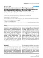
Response to bolus intravenous glucose infusion
Fishman observed in five normal adult subjects that the
CSF glucose level changes in parallel with changes of
blood glucose level following intravenous infusion of a
bolus of glucose [11]. A similar dose (0.75 mg/kg in 90
seconds) was assumed for the model and the glucose con-
centration in the arterial blood compartment and that in
the CSF compartment were simulated. The results are
shown in Fig. 4. The clinical data of Fishman are also
shown for comparison [11].
The model-estimated blood glucose concentrations at 0.5,
1, 2, 3 and 4 h were 210.0, 140.0, 116.1, 105.8 and 98.9
mg/dl, respectively (Fig. 4a). The concentration of glucose
in the blood increased rapidly during intravenous glucose
induction. Following the completion of bolus administra-
tion, the concentration decreased rapidly. A plateau con-
centration was reached at about 4 h.
As shown in Fig. 4b, the model-estimated CSF glucose
concentrations increased at first and then decreased dur-
ing the 6 h simulation. The concentration of glucose in the
CSF increased more slowly during induction than it did in
the blood and the decrease was also slower than that in
the blood. The estimated CSF glucose concentrations at
0.5, 1, 2, 3 and 4 h were 83.0, 89.8, 83.0, 73.6 and 66.5
mg/dl, respectively. Glucose concentration reached a pla-
teau after 5 h. These results demonstrate that the CSF glu-
cose concentration correlates with, and is much lower
than blood glucose concentration.
Visual inspection of the data displayed in Fig. 4 shows that
both arterial and CSF glucose concentrations estimated by
the model following bolus intravenous glucose are com-
parable with the clinical observations by Fishman [11].
Particularly, a variable time about 4-6 h was required in
vivo before the CSF glucose level reached its steady-state
equilibrium with the blood glucose. CSF glucose level in
the experimental subjects would not reach a peak level for
about 2 hours after rapid intravenous glucose injection,
kk k k k
12 3 4 5
1 0 0 05 0 25 0 001 0 001== = = =.; .; .; . ; .
kk k
67 8
10 001 003== =.; . ; .
kk
910
001 01==.; .
GG
blood
brain
blood
brain
00
10 08
max min
.; .==
κκ
GI
==03 01.; .
ττ
GI
hour hour==1200 0 1200 0.; .
aa a
cg ci pg
== =40 0004 002.; . ; .
kkk
γβα
==−=0 0001 0 1 100 0.; .; .
Response to bolus intravenous glucose infusionFigure 4
Response to bolus intravenous glucose infusion. (a)
blood glucose concentration. (b) CSF glucose concentration.
Time (hours)
Glucose concentration (mg/dl)
Simulated blood glucose concentration
Clinical data
(a)
Time (hours)
Glucose concentration (mg/dl)
Simulated CSF glucose concentration
Clinical data
(b)
Theoretical Biology and Medical Modelling 2009, 6:26 />Page 17 of 24
(page number not for citation purposes)
and did not reach its equilibrium for about 4 hours. CSF
glucose was normally about 65% of blood glucose. The
current model simulated the peak CSF glucose concentra-
tion at about 1 h and the CSF glucose level continued to
decrease for 5 h. A similar CSF-blood glucose ratio of less
than 1.0 was also simulated in the current model.
Altogether, the blood and CSF glucose concentrations pre-
dicted by the model are compatible to the clinical data
concerning the GIG regulatory system response to a bolus
intravenous glucose infusion.
Response to stepwise intravenous glucose injection
Tillil and colleagues measured blood glucose concentra-
tions following intravenous glucose administration in 7
normal subjects, each of whom received 20, 50 and 100 g
of glucose intravenously over 3 h [39]. Similar doses were
used in the model; thus, 12% of the total glucose dose was
assumed to be infused into the venous compartment of
the model in the first 30 min, 48 and 32% over the next 2
h, respectively, and 8% over the final 30 min. The simula-
tion results of arterial blood glucose are shown in Fig. 5.
Glucose concentration in the blood increased immedi-
ately in response to intravenous glucose infusion. As
shown by the first half of the simulation profiles, the
resultant blood glucose concentrations, as well as their
changes, depended on the infusion rate. The incremental
and total areas under the glucose concentration curves
increased significantly in response to increasing doses. In
each simulation profile, the peak glucose concentration
occurred at 1.5 h, i.e., the end of infusion at maximum
rate.
These simulation results demonstrated that blood insulin
concentration also increased with respect to intravenous
glucose infusion. The incremental and total areas under
the blood insulin concentration curves increased in paral-
lel with that of blood glucose curves in response to
increasing glucose dose (data not shown). The peak insu-
lin concentration occurred after the peak glucose concen-
tration. With increasing doses of intravenous glucose, the
blood insulin concentrations were elevated. Even when
glucose concentration returned to its basal value at the
end of the simulation period, the concentration of insulin
in blood was still higher than the basal value.
Tillil and coworkers noted that incremental glucose areas
after intravenous glucose injection increased as a function
of the glucose dose [39]. The same was true for the incre-
mental and total areas under the insulin concentration
curves. The elevated insulin concentration returned to its
base value later than the elevated glucose concentration. It
was also demonstrated in their normal subjects that with
increasing doses of intravenous glucose there was an
increase in peripheral blood glucose response as well as in
the insulin secretory response.
Considered together, the responses depicted by the model
with respect to stepwise intravenous glucose infusion are
consistent with the clinical observations.
Response to continuous intravenous glucose injection
Changes in blood concentrations of glucose, insulin and
glucagon following continuous intravenous glucose infu-
sion (at 0.01 mg/s) were simulated in the model. As
shown in Fig. 6, oscillations were observed in each of the
simulation profiles of blood glucose, insulin and gluca-
gon. All periods of oscillation were about 120 min., while
their amplitudes were about 20 mg/dl for glucose, 15 ng/
dl for insulin and 110 pg/dl for glucagon. It was demon-
strated that both glucose and glucagon reached peak levels
before that of insulin. The estimated time difference
between glucose and insulin peaks was about 30 min.
Blood glucose concentration response to stepwise intrave-nous glucose infusionFigure 5
Blood glucose concentration response to stepwise
intravenous glucose infusion.
Time (hours)
Glucose concentration (mg/dl)
Total glucose 100 g
Total glucose 50 g
Total glucose 25 g
Response to continuous intravenous glucose infusionFigure 6
Response to continuous intravenous glucose infusion.
Time (hours)
Glucose concentration (mg/dl)
Insulin concentration (ng/dl)
Glucagon concentration (pg/dl)
continuous infusion at 0.01 mg/s
glucoseglucagon
insulin
Theoretical Biology and Medical Modelling 2009, 6:26 />Page 18 of 24
(page number not for citation purposes)
The model simulation results are consistent with those of
currently existing theoretical models, such as the models
of Sturis et al. and Li et al. [7,8], both of which were devel-
oped to mimic experimental findings of ultradian oscilla-
tions during constant glucose infusion. Sturis' model
simulated the amplitude of glucose oscillation of 20 mg/
dl, as simulated in the current model. Li's model demon-
strated that both the oscillation amplitude of blood glu-
cose and that of blood insulin depended on the glucose
infusion rate. It was confirmed that the simulated oscilla-
tion amplitudes of blood glucose and insulin in the cur-
rent model were within the amplitude ranges predicted by
Li's model.
Both Sturis' and Li's models simulated earlier glucose
oscillation than insulin oscillation. For example, Li's
model predicted the time difference between glucose and
insulin peak levels to be about 20 min, which also
depended on the glucose infusion rate. The simulation
results in our model are in agreement with these reported
results.
With respect to the effect of glucose infusion rate on ultra-
dian oscillation, it was demonstrated in Li's model that
there existed a bifurcation point for the glucose infusion
rate (about 1.25 mg/dl/min or 2 mg/s if the volume of
glucose space is 10 liters [7]. Therefore, the oscillations in
blood glucose concentrations were sustained when the
rate of infused glucose was somewhat less than the bifur-
cation point. Particularly, ultradian oscillation would be
sustained theoretically by a little glucose infusion. When
the rate of infused glucose was larger than the bifurcation
point, no oscillation would be sustained. It should be
pointed out that the oscillations arise even without glu-
cose infusion in the Li's model, majorly because the oscil-
lation is induced by two explicit time delays.
The current developed model was used to simulate how
changes in glucose infusion rate affect the ultradian oscil-
lation. Fig. 7 shows that oscillation would occur even
when the rate of intravenous glucose was 0.0001 mg/s
while it would disappear when the constant glucose infu-
sion rate was 1.0 mg/s. Furthermore, the bifurcation point
for the glucose infusion rate in this model is estimated to
be about 0.4374 mg/s. Although this identified bifurca-
tion point is somewhat different from that of Li's model,
the current model is comparable with Li's model in simu-
lating the existence of Hopf bifurcation point for glucose
infusion rate.
Taken together, our model is consistent with the currently
existing models in describing ultradian oscillations, based
on their quantitative and qualitative agreements.
Other qualitative verifications
Response to bolus intravenous insulin infusion
Simulated infusion of insulin (375 ng/kg over 90 sec-
onds) into the venous compartment resulted in increases
in both blood and CSF insulin levels, while blood glucose
level was maintained between 90 and 120 mg/dl. The sim-
ulation results demonstrated that insulin concentration
dynamics in the blood and that in the CSF are characteris-
tic of two- or three-compartmental model, and are con-
sistent with the results reported by Schwartz and
coworkers [40], where dynamics of CSF insulin was
described by a three-compartment model.
Response to continuous intracranial glucose infusion
To examine the central effects of glucose on systemic glu-
cose homeostasis, Lam and colleagues infused glucose
directly into the third cerebral ventricle of conscious rats
[2]. The results showed that such infusion lowered blood
glucose levels. In our model, simulated glucose infusion
into CSF compartment decreased blood glucose concen-
tration. However, the estimated level of blood glucose was
dependent on the infusion rate; a small rate (0.05 mg/s)
resulted in ultradian oscillations in blood glucose, while a
large infusion rate (0.25 mg/s) resulted in the disappear-
ance of such oscillations.
Response to continuous intracranial insulin infusion
Woods and colleagues suggested that insulin signaling in
the hypothalamus initiates a signal via the vagus nerve to
the liver to reduce glucose synthesis and secretion into the
blood [30]. Hence, insulin in the brain acts by reducing
peripheral blood glucose levels. In our model, simulated
insulin infusion at rates between 20 and 30 ng/s into CSF
compartment decreased blood glucose levels. The result-
ant blood glucose concentration was dependent on the
infusion rate. That is, the higher the infusion rate, the
lower the resultant blood glucose level.
Dependence of ultradian oscillation on glucose infusion rateFigure 7
Dependence of ultradian oscillation on glucose infu-
sion rate.
Time (hours)
Glucose concentration (mg/dl)
Constant infusion rate 1.0 mg/s
Constant infusion rate 0.0001 mg/s
Constant infusion rate 0.01 mg/s
Theoretical Biology and Medical Modelling 2009, 6:26 />Page 19 of 24
(page number not for citation purposes)
Altogether, the results of simulation studies demonstrate
that intracranial infusion of glucose or insulin results in
reduction of peripheral blood glucose levels.
The above simulation results (which are summarized in
Table 5) provide solid support and verification of our
model.
Results
To determine the roles of stress and BBB adaptation in the
generation of hyperglycemia, which generally accompa-
nies hyperinsulinemia and hyperglucagonemia in diabe-
tes, simulated stress input of varying strength and
duration was introduced in the model, together with fast
BBB adaptation. The use of various simulation paradigms
allowed clarification of the relationship between brain
glucose homeostasis and blood hyperglycemia in diabe-
tes.
Role of stress
As described during modeling, stress, which is described
abstractly by parameter s in eq. (10"), (18") and (27"), is
a disturbance input to the central controller. It affects the
efferent signals that regulate hepatic glucose production
and pancreatic hormonal secretion in this model. The
parameter s varies from 0 to 1, which describes the severity
of stress from mild to severe. Furthermore, the duration of
stress (short-term or long-term) can also affect glucose
production. Time constant of BBB adaptation in this sim-
ulation is of 1200 h.
The results of simulated stress are shown in Figs. 8 and 9.
Stress, independent of its duration and strength, caused
transient or persistent peripheral hyperglycemia in all
simulations.
With regard to the severity/duration of stress, a long-term
mild stress, corresponding to s = 0.1, resulted in a steady-
state peripheral hyperglycemia characteristic of ultradian
oscillation, in the GIG regulatory system (Fig. 8). In con-
trast, the ultradian oscillation disappeared upon simula-
tion of mid- (s = 0.5) to severe-stress (s = 0.9). The Hopf
bifurcation point for stress is estimated to be about 0.24.
That is to say, if stress is smaller than 0.24 (s < 0.24), the
oscillations are sustained and if stress is larger than 0.24 (s
> 0.24), no oscillation will be sustained.
The level of resultant hyperglycemia was dependent on
the severity of stress, i.e., the worst the stress, the higher
the resultant blood glucose level. Continuous application
of severe stress (s = 0.9) resulted in persistent hyperglyc-
emia and blood glucose never returned to its basal level,
rather, a new steady-state hyperglycemia (> 120 mg/dl)
was established. On the other hand, blood glucose level
was still within the euglycemic range (< 120 mg/dl) fol-
lowing imposition of mid-level stress (s = 0.5). Consid-
ered together, mild-to-mid level stress seems to induce
little disturbance of the GIG regulatory system, while
severe stress has the worst influence.
With regard to the duration of stress, short-term severe
stress (s = 0.9) applied in the first 1 h caused a transient
increase in blood glucose level (Fig. 9a), and decreases in
blood insulin (Fig. 9b) and blood glucagon levels (data
not shown). These transient responses disappeared after
removal of stress during the simulated 48 hours. That is to
say, there is no bifurcation point for the length of time
over which the short-term stress is applied. Short-term
stress resulted in a steady-state, characteristic of ultradian
oscillation, together with elevated blood insulin and glu-
cagon.
Recursive stress of 4-hour period (2 h active, as shown by
the shaded areas in Fig. 8, and then 2 h inactive) was
applied. The simulation results showed a continuous
increase in maximum blood glucose during the applica-
tion of such stress. The insulin concentration was
depressed during each active period but rebounded dur-
ing each inactive period. The peak insulin concentrations
were elevated during stress application, with a time-delay
compared to peak glucose concentration. Such increase
and time delay are probably due to the inertia of the GIG
regulatory system. Similar to that of short-term stress,
responses of blood glucose to repeated stress also con-
verged to a similar steady-state following termination of
stress.
Table 5: Compatibility of results of model simulation with clinical data.
Item Input of model Reference
CSF glucose dynamics Bolus intravenous glucose infusion [11]
Blood glucose dynamics and blood insulin dynamics Stepwise intravenous glucose infusion [39]
Ultradian oscillation Continuous intravenous glucose infusion [7,8]
Bifurcation point of ultradian oscillation Continuous intravenous glucose infusion [8]
CSF insulin dynamics Bolus intravenous insulin infusion [40]
Blood glucose dynamics Continuous intracranial glucose infusion [2]
Blood glucose dynamics Continuous intracranial insulin infusion [30]
Theoretical Biology and Medical Modelling 2009, 6:26 />Page 20 of 24
(page number not for citation purposes)
Although human studies on the role of stress on the onset
and course of diabetes are limited, a large body of evi-
dence based on animal studies supports the notion that
stress reliably induces hyperglycemia in diabetes [4].
Importantly, the results of simulation tests using the
present model compare well with clinical knowledge.
Therefore, the model is partially verified in modeling the
effects of stress on the generation of hyperglycemia.
Quantification of severity of stress using parameter s vary-
ing from 0 to 1 seems a reasonable approach.
Role of BBB adaptation
In order to simulate the roles of BBB adaptation in the
generation of hyperglycemia, the time constants in equa-
tions (5) and (22) were assumed to be 4 h in the model,
although these time constants may be some years for an
adapting BBB in human. Such an assumption of short
time constant is due to the time limitation of computer
simulation. Therefore, the simulation results (solid line in
Fig. 10) should be considered as fast-forward of the
responses (broken line). Long-term severe stress (s = 0.9)
is assumed in the simulation.
As shown in Fig. 10a, with a fast BBB adaptation, a hyper-
glycemic steady-state of blood glucose was generated in
Response to long-term stress of variable severityFigure 8
Response to long-term stress of variable severity.
Time (hours)
Glucose concentration (mg/dl)
Severe stress (s=0.9)
Mid stress (s=0.5)
Mild stress (s=0.1)
Response to severe stress of variable durationFigure 9
Response to severe stress of variable duration. (a)
blood glucose concentration. (b) blood insulin concentration.
Time (hours)
Glucose concentration (mg/dl)
Long-term stress
Periodical stress
Short-term stress
(a)
Time (hours)
Insulin concentration (ng/dl)
Long-term stress
Periodical stress
Short-term stress
(b)
Response to long-term severe stress with BBB adaptationFigure 10
Response to long-term severe stress with BBB adap-
tation. (a) blood concentrations of glucose and insulin. (b)
brain glucose concentration.
Time (hours)
Glucose concentration (mg/dl)
Insulin concentration (ng/dl)
Adaptation (τ
ττ
τ
G
=τ
ττ
τ
I
=1200h)
Fast adaptation (τ
ττ
τ
G
=τ
ττ
τ
I
= 4h)
glucose
insulin
(a)
Time (hours)
Brain glucose concentration (mg/dl)
Adaptation (τ
ττ
τ
G
=τ
ττ
τ
I
=1200h)
Fast adaptation (τ
ττ
τ
G
=τ
ττ
τ
I
= 4h)
(b)
Theoretical Biology and Medical Modelling 2009, 6:26 />Page 21 of 24
(page number not for citation purposes)
the present model, while brain glucose was maintained
within the euglycemic range. At the same time, both the
concentrations of blood insulin and blood glucagon were
higher than their basal values, while the ratio of glucagon
to insulin was elevated. The present model simulated an
abnormal state, which is similar to diabetes/hyperglyc-
emia, hyperinsulinemia and hyperglucagonemia [32,41],
of the GIG regulatory system under long-term stress,
together with a fast BBB adaptation.
The simulation results are consistent with clinical facts.
That is, as reviewed by Jiang and Zhang [42], the absolute
levels of glucagon or the ratios of glucagon to insulin are
often elevated in various forms of diabetes in both animal
and human subjects. The same authors also pointed out
that chronic hyperglucagonemia correlated with and was
in part responsible for increased hepatic glucose produc-
tion and hyperglycemia in type 2 diabetes. Based on the
physiological fact that stress increases glucagon secretion
through the HPA axis, it is reasonable to consider stress as
one of the causes of hyperglycemia in diabetes. As shown
in Fig. 10b, brain glucose homeostasis was achieved dur-
ing peripheral hyperglycemia. The brain glucose is at eug-
lycemic level while blood glucose is at hyperglycemic
level. The simulation result should motivate clinical veri-
fication whether chronic hyperglycemia in diabetic
patients does not alter brain glucose concentrations, com-
pared with the healthy subjects.
In order to simulate the effect of gain of BBB adaptation
on the resultant hyperglycemia,
κ
G
in equation (5) was
varied from 0 to 0.5. The simulation results showed that
the resultant peripheral hyperglycemia was dependent on
κ
G
. That is, the larger the gain
κ
G
, the worse the resultant
hyperglycemia (data not shown). If
κ
G
= 0, which corre-
sponds to an inadaptive BBB, a state of elevated glucose
levels in brain and blood within the euglycemic range
would be inevitable. However, the elevated brain glucose
would never return to its basal level.
These simulation results demonstrated that BBB adapta-
tion contributes to the generation of hyperglycemia and
homeostasis of brain glucose level under stress. Both the
gain and time constant of BBB adaptation to dysglycemia
are important. Based on their dependence on the individ-
ual, it is reasonable to speculate that a person with a large
gain and short time constant would be sensitive to stress-
induced hyperglycemia and ultimately develops diabetes.
Discussion
Importance of brain glucose homeostasis
The brain is one of the major organs that require continu-
ous energy supply by glucose. Maintenance of constant
glucose concentration in the brain is of supreme impor-
tance mainly due to the fact that the brain is uniquely
dependent on the availability of glucose and that dysglyc-
emia, either hypoglycemia or hyperglycemia, would
induce brain dysfunction. Not only in healthy individuals
but also in diabetics, brain glucose concentration is main-
tained at constant level. Physiological evidence suggests
that the maintenance of constant glucose level in the brain
is more important than that in the blood.
The present study was based on the theme that the ulti-
mate goal of glucose-insulin-glucagon (GIG) regulatory
system is not blood glucose homeostasis, but rather brain
glucose homeostasis. Anatomically, different parts of the
brain, particularly the hypothalamus, are important cent-
ers involved in the regulation of brain glucose homeosta-
sis. Physiologically, changes in brain glucose levels elicit a
complex neuroendocrine response that rapidly corrects
dysglycemia in the brain.
Whereas the relations among the brain, glucose homeos-
tasis and diabetes have been qualitatively recognized in
experimental animals and clinically in patients, no theo-
retical analysis is available on the relationship between
brain glucose homeostasis and hyperglycemia in diabetes.
To the best of our knowledge, this is the first paper that
targets the control of brain glucose homeostasis and pro-
vides a theoretical framework for the role of the brain in
the generation of hyperglycemia in diabetes.
Role of stress
Stress has long been shown to have major effects on met-
abolic activity as energy mobilization is a primary result of
the fight-or-flight response. Stress stimulates the release of
various hormones, which can increase blood glucose lev-
els. Based on the same mechanism, stress could also
potentially induce a state of chronic hyperglycemia in dia-
betes. Although human studies on the role of stress on the
onset and course of diabetes are limited, the notion that
stress reliably produces hyperglycemia in disease has been
supported by animal studies [4].
Since no quantitative measure of stress is available at
present, an abstract variable of positive value varying
between 0 and 1 was introduced in the model to describe
the severity of stress from mild to severe. The stress-simu-
lation results compared well with clinical knowledge con-
cerning the stress-induced hyperglycemia in diabetes [4].
Various simulations of the severity and duration of stress
conducted in the present study emphasized the roles of
stress in the appearance of hyperglycemia, not only qual-
itatively but also quantitatively. As a corollary to these
simulation results, some patients with diabetes may not
require glucose-lowering agents at all, rather, they may
benefit from a less obvious treatment directed against
stress, such as relaxation.
Theoretical Biology and Medical Modelling 2009, 6:26 />Page 22 of 24
(page number not for citation purposes)
BBB adaptation as a main cause of diabetes
To maintain brain glucose homeostasis, glucose transport
from the blood to the brain should be regulated accu-
rately, according to the short-term and long-term states of
blood glucose. In fact, BBB helps govern the brain glucose
homeostasis through temporary and permanent mecha-
nisms. The former is through the BBB semi-permeability
to glucose, which is a physical filter with respect to sharp
swings in blood glucose levels, while the latter involves
BBB adaptation, which is a physiological change with
respect to chronic hypoglycemia or hyperglycemia.
As shown by the simulation results, BBB adaptation for
brain glucose homeostasis, together with long-term severe
stress, contributes to hyperglycemia. A vicious cycle of
hyperglycemia and BBB adaptation would exist in diabe-
tes. However, such a feedback loop should be considered
as a self-protective mechanism of the brain, through
which brain glucose homeostasis is maintained. In other
words, brain glucose homeostasis requires increasing
blood glucose levels if glucose transport from the blood to
the brain is depressed due to BBB adaptation. If not, the
brain would be short of glucose, as seen in "insulin
shock", where the blood glucose is controlled euglycemi-
cally by insulin while the glucose transport across the
adapted BBB is not improved.
Theoretical analysis suggests a novel hypothesis, namely,
hyperglycemia observed in diabetes is one of the control-
led results for brain glucose homeostasis through a per-
manent adaptation mechanism. Also, as a suggestion, re-
modification of BBB adaptation in diabetes, with pharma-
cokinetic or genetic measures, could be a potentially suit-
able strategy for clinical control of hyperglycemia in
diabetes.
Modeling of ultradian oscillation
Although our understanding of diabetes has progressed
very rapidly in the past decades, diabetes is still a disease
that has no cure. Every effort, both in vivo and in vitro,
should be made to prevent and treat this disease. In this
regard, theoretical or mathematical models constitute
interesting tools, because they can provide insights,
improve intuitions, clarify assumptions of formal theory,
allow for estimating parameters, determining sensitivities,
simulating simple and complex phenomena and provid-
ing future predictions for diabetes control.
The current model was particularly successful in providing
a novel interpretation of the mechanism underlying ultra-
dian oscillation. The hypothesis that ultradian insulin
secretion represents an instability in peripheral GIG inter-
actions has been the subject of a number of studies,
including the mathematical model originally proposed by
Sturis et al. and lately modified by Li et al. [7,8], where
time-delay is included implicitly or explicitly to produce
ultradian oscillation.
The present model has similar characteristics of the mod-
els of Sturis et al. and Li et al. [7,8], in simulating ultradian
oscillation, without including any time-delay. It is consid-
ered that oscillation is generated in the model mainly due
to feedback control via peripheral GIG interactions and
the central brain-endocrine crosstalk, but not due to time-
delay. The model does not exclude the possible role of
time-delay in generating ultradian oscillation, as permea-
bility at the blood-mass interface may introduce some
implicit time-delay and partially contribute to the genera-
tion of ultradian oscillation in the model.
Limitations of the model
No model ever reproduces all facets of the original system.
As pointed by Gatewood and colleagues [43], no matter
how small pieces one chooses, there always seems to be
smaller ones; no matter how many details one models,
there are always others. The same applies to modeling the
GIG regulatory system. In this paper, the brain glucose
homeostasis is targeted and modeled as the principle of
GIG regulatory system. The relationship between brain, as
well as stress, and hyperglycemia in diabetes is simulated
in the model. Although the simulation results are highly
suggestive, modeling is based on some assumptions.
One of the important limitations of the model is the need
of a set of physical and physiological parameters that is
difficult or impossible to estimate within a single individ-
ual. Although various values are given for most of these
parameters based on the literature, more than 20 parame-
ters have to be given arbitrary values selected by the
authors. Verifications given in the section of model verifi-
cation provide greater assurance that predictions made by
the model are correct. However, the model only simulates
an "average subject". A formal sensitivity analysis is left
for future research.
Further testing of the model is also required to determine
whether it is suitable for individual patient parameteriza-
tion which is a key requirement for clinical use. Further
refinements might be appropriate after tuning various
parameters to fit pertinent clinical data. However, the
model in its current form clearly has a role as a patient
simulator. In this respect, it provides theoretical frame-
work with which to analyze the relationship between
brain glucose homeostasis and blood hyperglycemia in
diabetes and to plan therapeutic strategies for diabetes.
Conclusion
In this paper, the control of brain glucose homeostasis
was considered as the ultimate goal of the glucose-insulin-
glucagon (GIG) regulatory system. In order to demon-
Theoretical Biology and Medical Modelling 2009, 6:26 />Page 23 of 24
(page number not for citation purposes)
strate theoretically the relationship between brain glucose
homeostasis and hyperglycemia in diabetes, a brain-cen-
tered compartment model of GIG regulatory system is
developed from the viewpoint of systems control. The
model consists of both peripheral GIG interactions and
central brain-endocrine crosstalk, while taking account of
the effects of stress and BBB adaptation to dysglycemia.
The results of various simulations highlighted the unique
features of the GIG regulatory system. In particular, an
abnormal state similar to diabetes, characterized by blood
hyperglycemia but brain glucose homeostasis, was simu-
lated in the model of a person with a fast BBB adaptation
and under long-term severe stress. Based on the simula-
tion results, we conclude that, (i) both long-term severe
stress and BBB adaptation contribute to hyperglycemia;
(ii) blood hyperglycemia may be an outcome of control of
brain glucose homeostasis in diabetes. Based on these the-
oretical results, in vivo experiments are welcome to pro-
vide direct evidence for the relationship between brain
glucose homeostasis and hyperglycemia in diabetes.
Abbreviations
E: glucagon concentration; E: vector of glucagon concen-
tration; G: glucose concentration; G: vector of glucose
concentration; I: insulin concentration; I: vector of insulin
concentration; K: Michaelis constant of Michaelis-Menten
equation; M: metabolism; T: maximal transport rate of
Michaelis-Menten equation; V: distribution volume; V:
matrix of distribution volume; W: vector of blood flow; a:
estimable parameter in equation (30); f: transfer rate
among the adjacent compartments; h: permeability coeffi-
cient; k: estimable parameters in equations (10), (11),
(18), (19), (27) and (31); m: estimable parameters in
equation (2); p: production rate; s: strength of stress; t:
time; u: utilization rate; v: degradation constant of gluca-
gon; w: blood flow or urine flow;
α
: regulatory signal of
glucagon secretion;
β
: regulatory signal of insulin secre-
tion;
γ
: regulatory signal of hepatic glucose production;
κ
:
gain;
τ
: time constant.
Subscript
0: value at the steady-state; E: glucagon; G: glucose; I: insu-
lin; art: arterial blood or arterial part of cardiocirculatory
segment; blood: blood compartment; cg: central informa-
tion of glucose; ci: central information of insulin; csf: cer-
ebrospinal fluid (CSF) compartment; error: controlled
error; mass: mass compartment; pg: peripheral informa-
tion of glucose; ven: venous blood or venous part of cardi-
ocirculatory segment; E-blood-mass: glucagon transport
from the blood compartment to the mass compartment;
G-blood-csf: glucose transport from the blood compart-
ment to the CSF compartment; G-csf-mass: glucose trans-
port from the CSF compartment to the brain mass
compartment; G-blood-mass: glucose transport from the
blood compartment to the mass compartment; I-blood-
mass: insulin transport from the blood compartment to
the mass compartment.
Superscript
brain: brain (cranial) segment; inf: exogenous infusion;
lung: lung segment; liv: liver segment; pan: pancreas seg-
ment; red: red blood cells; urine: urine.
Competing interests
The authors declare that they have no competing interests.
Authors' contributions
The authors contributed equally to this work. All authors
read and approved the final manuscript.
Acknowledgements
The preparation of this manuscript was supported by a Grant-in-aid for
Young Scientists (B) from The Ministry of Education, Culture, Sports, Sci-
ence and Technology of Japan (No. 18700448) to Lu Gaohua.
References
1. Sandoval D, Cota D, Seeley RJ: The integrative role of CNS fuel-
sensing mechanisms in energy balance and glucose regula-
tion. Annu Rev Physiol 2008, 70:513-535.
2. Lam TK, Gutierrez-Juarez R, Pocai A, Rossetti L: Regulation of
blood glucose by hypothalamic pyruvate metabolism. Science
2005, 309:943-947.
3. Gjedde A, Crone C: Blood-brain glucose transfer: repression in
chronic hyperglycemia. Science 1981, 214:456-457.
4. Surwit RS, Schneider MS, Feinglos MN: Stress and diabetes melli-
tus. Diabetes Care 1992, 15:1413-1422.
5. Bolie VW: Coefficients of normal blood glucose regulation. J
Appl Physiol 1961, 16:783-788.
6. Bergman RN, Ider YZ, Bowden CR, Cobelli C: Quantitative esti-
mation of insulin sensitivity. Am J Physiol 1979, 236:E667-677.
7. Sturis J, Polonsky KS, Mosekilde E, Van Cauter E: Computer model
for mechanisms underlying ultradian oscillations of insulin
and glucose. Am J Physiol 1991, 260:E801-809.
8. Li J, Kuang Y, Mason CC: Modeling the glucose-insulin regula-
tory system and ultradian insulin secretory oscillations with
two explicit time delays. J Theor Biol 2006, 242:722-735.
9. Boutayeb A, Chetouani A: A critical review of mathematical
models and data used in diabetology. Biomed Eng Online 2006,
5:43.
10. Makroglou A, Li J, Kuang Y: Mathematical models and software
tools for the glucose-insulin regulatory system and diabetes:
an overview. Appl Numer Math 2006, 56:559-573.
11. Fishman RA: Cerebrospinal fluid in diseases of the nervous system W.B.
Saunders Company, Pennsylvania; 1992.
12. Aszalós Z: [Cerebral complications of diabetes mellitus]
[Article in Hungarian]. Orv Hetil 2007, 148:2371-2376.
13. Routh VH: Glucose-sensing neurons: are they physiologically
relevant? Physiol Behav 2002, 76:403-413.
14. Silver IA, Erecinska M: Glucose-induced intracellular ion
changes in sugar-sensitive hypothalamic neurons. J Neurophys-
iol 1998, 79:1733-1745.
15. Levin BE, Kang L, Sanders NM, Dunn-Meynell AA: Role of Neuronal
Glucosensing in the Regulation of Energy Homeostasis. Dia-
betes 2006, 55:S122-130.
16. Banks WA: The source of cerebral insulin. Eur J Pharmacol 2004,
490:5-12.
17. Uyama N, Geerts A, Reynaert H: Neural connections between
the hypothalamus and the liver. Anat Rec A Discov Mol Cell Evol
Biol 2004, 280:808-820.
18. Gaohua L, Kimura H: A mathematical model of intracranial
pressure dynamics for brain hypothermia treatment. J Theor
Biol 2006, 238:882-900.
Publish with Bio Med Central and every
scientist can read your work free of charge
"BioMed Central will be the most significant development for
disseminating the results of biomedical research in our lifetime."
Sir Paul Nurse, Cancer Research UK
Your research papers will be:
available free of charge to the entire biomedical community
peer reviewed and published immediately upon acceptance
cited in PubMed and archived on PubMed Central
yours — you keep the copyright
Submit your manuscript here:
/>BioMedcentral
Theoretical Biology and Medical Modelling 2009, 6:26 />Page 24 of 24
(page number not for citation purposes)
19. Gaohua L, Kimura H: Simulation of propofol anaesthesia for
intracranial decompression using brain hypothermia treat-
ment. Theor Biol Med Model 2007, 4:46.
20. Gaohua L, Kimura H: A mathematical model of respiratory and
biothermal dynamics in brain hypothermia treatment. IEEE
Trans Biomed Eng 2008, 55:1266-1278.
21. Gaohua L, Maekawa T, Kimura H: An integrated model of ther-
modynamic-hemodynamic-pharmacokinetic system and its
application on decoupling control of intracranial tempera-
ture and pressure in brain hypothermia treatment. J Theor
Biol 2006, 242:16-31.
22. Guyton AC, Hall JE: Textbook of Medical Physiology W.B. Saunders
Company, Pennsylvania; 2001.
23. Rapoport SI: Blood-Brain Barrier in Physiology and Medicine Raven Press,
New York; 1976.
24. Simpson IA, Appel NM, Hokari M, Oki J, Holman GD, Maher F, Koeh-
ler-Stec EM, Vannucci SJ, Smith QR: Blood-brain barrier glucose
transporter: effects of hypo- and hyperglycemia revisited. J
Neurochem 1999, 72:238-247.
25. Criego AB, Tkac I, Kumar A, Thomas W, Gruetter R, Seaquist ER:
Brain glucose concentrations in healthy humans subjected to
recurrent hypoglycemia. J Neurosci Res 2005, 82:525-530.
26. Cerasi E: An analogue computer model for the insulin
response to glucose infusion. Simulation 1971, 16:243-255.
27. Bodenlenz M, Schaupp LA, Druml T, Sommer R, Wutte A, Schaller
HC, Sinner F, Wach P, Pieber TR: Measurement of interstitial
insulin in human adipose and muscle tissue under moderate
hyperinsulinemia by means of direct interstitial access. Am J
Physiol 2005, 289:E296-300.
28. Gudbjörnsdóttir S, Sjöstrand M, Strindberg L, Wahren J, Lönnroth P:
Direct measurements of the permeability surface area for
insulin and glucose in human skeletal muscle. J Clin Endocrinol
Metab 2003, 88:4559-4564.
29. Baura GD, Foster DM, Porte D Jr, Kahn SE, Bergman RN, Cobelli C,
Schwartz MW: Saturable transport of insulin from plasma into
the central nervous system of dogs in vivo. A mechanism for
regulated insulin delivery to the brain. J Clin Invest 1993,
92:1824-1830.
30. Woods SC, Benoit SC, Clegg DJ:
The Brain-Gut-Islet Connec-
tion. Diabetes 2006, 55:S114-121.
31. Puglianiello A, Cianfarani S: Central control of glucose homeos-
tasis. Rev Diabet Stud 2006, 3:54-60.
32. Kahn CR, Weir GC, King GL, Jacobson AM, Moses AC, Smith RJ: Jos-
lin's Diabetes Mellitus Lippincott Williams & Wilkins, Philadelphia;
2005.
33. Woods SC, Porte D Jr, Bobbioni E, Ionescu E, Sauter JF, Rohner-Jean-
renaud F, Jeanrenaud B: Insulin: its relationship to the central
nervous system and to the control of food intake and body
weight. Am J Clin Nutr 1985, 42:1063-1071.
34. Sonksen P, Sonksen J: Insulin: understanding its action in health
and disease. Br J Anaesth 2000, 85:69-79.
35. Fisher M, Sherwin RS, Hendler R, Felig P: Kinetics of glucagon in
man: effects of starvation. Proc Natl Acad Sci USA 1976,
73:1735-1739.
36. Regittnig W, Ellmerer M, Fauler G, Sendlhofer G, Trajanoski Z, Leis
HJ, Schaupp L, Wach P, Pieber TR: Assessment of transcapillary
glucose exchange in human skeletal muscle and adipose tis-
sue. Am J Physiol Endocrinol Metab 2003, 285:E241-251.
37. Eng J, Yalow RS: Evidence against extrapancreatic insulin syn-
thesis. Proc Natl Acad Sci USA 1981, 78:4576-4578.
38. Curry FR: Atrial natriuretic peptide: an essential physiological
regulator of transvascular fluid, protein transport, and
plasma volume. J Clin Invest 2005, 115:1458-1461.
39. Tillil H, Shapiro ET, Miller MA, Karrison T, Frank BH, Galloway JA,
Rubenstein AH, Polonsky KS: Dose-dependent effects of oral
and intravenous glucose on insulin secretion and clearance in
normal humans. Am J Physiol 1988, 254:E349-357.
40. Schwartz MW, Bergman RN, Kahn SE, Taborsky GJ, Fisher LD, Sipols
AJ, Woods SC, Steil GM, Porte D: Evidence for entry of plasma
insulin into cerebrospinal fluid through an intermediate
compartment in dogs. Quantitative aspects and implications
for transport. J Clin Invest 1991, 88:1272-1281.
41. Reaven GM, Chen YD, Golay A, Swislocki AL, Jaspan JB: Documen-
tation of hyperglucagonemia throughout the day in non-
obese and obese patients with noninsulin-dependent
diabetes mellitus. J Clin Endocrinol Metab 1987, 64:106-110.
42. Jiang G, Zhang BB: Glucagon and regulation of glucose metab-
olism. Am J Physiol 2003, 284:E671-678.
43. Gatewood LC, Ackerman E, Rosevear JW, Molnar GD: Modeling
blood glucose dynamics. Behav Sci 1970, 15:72-87.