Báo cáo y học: "Markers of collagen synthesis and degradation are increased in serum in severe sepsis: a longitudinal study of 44 patients" pdf
Bạn đang xem bản rút gọn của tài liệu. Xem và tải ngay bản đầy đủ của tài liệu tại đây (538.82 KB, 10 trang )
Open Access
Available online />Page 1 of 10
(page number not for citation purposes)
Vol 13 No 2
Research
Markers of collagen synthesis and degradation are increased in
serum in severe sepsis: a longitudinal study of 44 patients
Fiia Gäddnäs
1
, Marjo Koskela
1,2
, Vesa Koivukangas
2
, Juha Risteli
3
, Aarne Oikarinen
4
,
Jouko Laurila
1
, Juha Saarnio
2
and Tero Ala-Kokko
1
1
Department of Anesthesiology, Division of Intensive Care, Oulu University Hospital, FI-90029, Oulu, Finland
2
Department of Surgery, Oulu University Hospital, FI-90029, Oulu, Finland
3
Department of Clinical Chemistry, Oulu University Hospital, FI-90029, Oulu, Finland
4
Department of Dermatology, Oulu University Hospital, FI-90029, Oulu, Finland
Corresponding author: Tero Ala-Kokko,
Received: 4 Jan 2009 Revisions requested: 18 Feb 2009 Revisions received: 18 Mar 2009 Accepted: 9 Apr 2009 Published: 9 Apr 2009
Critical Care 2009, 13:R53 (doi:10.1186/cc7780)
This article is online at: />© 2009 Gäddnäs et al.; licensee BioMed Central Ltd.
This is an open access article distributed under the terms of the Creative Commons Attribution License ( />),
which permits unrestricted use, distribution, and reproduction in any medium, provided the original work is properly cited.
Abstract
Introduction Sepsis-related multiple organ dysfunction is a
common cause of death in the intensive care unit. The effect of
sepsis on markers of tissue repair is only partly understood. The
aim of this study was to measure markers of collagen synthesis
and degradation during sepsis and investigate the association
with disease severity and outcome.
Methods Forty-four patients with severe sepsis participated in
the study and 15 volunteers acted as controls. Blood samples
were collected for 10 days after the first sepsis-induced organ
dysfunction and after three and six months. Procollagen type I
and III aminoterminal propeptides (PINP and PIIINP) and cross-
linked telopeptides of type I collagen (ICTP) were measured.
Results The PIIINP concentration was elevated in the septic
patients (8.8 ug/L, 25th to 75th percentile = 6.8 to 26.0) when
compared with controls (3.0 ug/L, 25th to 75th percentile = 2.7
to 3.3; P < 0.001) on day one. Maximum serum PIIINP
concentrations during sepsis were higher in non-survivors
compared with survivors (26.1 ug/L, 25th to 75th percentile =
18.7 to 84.3; vs. 15.1 ug/L, 25th to 75th percentile = 9.6 to
25.5; P = 0.033) and in multiple organ failure (MOF) compared
with multiple organ dysfunction syndrome (MODS) (24.2 ug/L,
25th to 75th percentile = 13.4 to 48.2; vs. 8.9 ug/L, 25th to
75th percentile = 7.4 to 19.4; P = 0.002). Although the PINP
values of the septic patients remained within the laboratory
reference values, patients with MOF had higher values than
patients with MODS (79.8, 25th to 75th percentile = 44.1 to
150.0; vs.40.4, 25th to 75th percentile = 23.6 to 99.3; P =
0.007). Day one ICTP levels were elevated in septic patients
compared with the controls (19.4 ug/L, 25th to 75th percentile
= 12.0 to 29.8; vs. 4.1 ug/L, 25th to 75th percentile = 3.4 to
5.0; P < 0.001).
Conclusions Markers of collagen metabolism are increased in
patients with severe sepsis and can be investigated further as
markers of disease severity and outcome.
Introduction
The host response in sepsis is a dynamic process activating
the pathways of coagulation, inflammation and tissue repair.
When the response becomes overwhelming, it leads to multi-
ple organ failure (MOF) and death [1-3]. Disturbed connective
tissue metabolism is the key element in complications of
inflammatory disease, so it was of interest to determine
whether high systemic inflammation in sepsis has any effect
and whether the level of connective tissue metabolism reflects
disease severity and outcome.
Fibroblasts synthesise a wide array of extracellular matrix pro-
teins, predominantly type I and III collagens, which provide
structural support to the organs [4,5]. The aim of this process
APACHE II: acute physiology and chronic health evaluation II; ARDS: adult respiratory distress syndrome; AUC: area under the curve; CI: confidence
interval; CV: coefficients of variation; ICTP: collagen I cross-linked telopeptide; ICU: Intensive care unit; IL: interleukin; MMP: matrix metalloproteinase;
MODS: multiple organ dysfunction syndrome; MOF: multiple organ failure; PINP: procollagen I aminoterminal propeptide; PIIINP: procollagen III ami-
noterminal propeptide; ROC: receiving operating characteristics; SOFA: sequential organ failure assessment; TGF: transforming growth factor; TNF:
tumour necrosis factor.
Critical Care Vol 13 No 2 Gäddnäs et al.
Page 2 of 10
(page number not for citation purposes)
is to maintain tissue integrity in a steady state and restore the
integrity of the organ after injury. Prolonged inflammatory
response may lead to persistent or progressive fibrosis impair-
ing the function of an organ. Collagen synthesis has been
shown to be pathologically increased, not only in wound kel-
oids and wound infections, but also in acute respiratory dis-
tress syndrome (ARDS), chronic liver diseases, myocardial
infarction and kidney diseases [4,6-10]. Indeed, it has been
suggested that progressive fibrosis is a central mechanism of
organ failure, which is related to the host's inflammatory
responses and subsequent fibroblast response [11].
In the course of collagen biosynthesis, procollagen-derived
peptides are deposited in the extracellular matrix and released
into the circulation. Aminoterminal propeptides are cleaved
from procollagens in a one to one proportion and thus reflect
the synthesis of collagen. Increased serum levels of procolla-
gen type III propeptide have been found in severely injured
patients and have been associated with MOF and death [12].
Additionally, procollagen III propeptide levels in plasma and
bronchoalveolar lavage fluid from patients with ARDS are
increased in early phases and are related to disease progres-
sion, multiple organ dysfunction and death [7,13].
Cross-linked type I collagen telopeptides (ICTP) were
assessed as markers of collagen I degradation. Previously,
Wenisch and colleagues have reported elevated ICTP levels
in Gram-negative septicaemia [14].
As fibrosing activity, measured by synthesis and degradation
of collagen, seems to have an important role in inflammatory
processes, we hypothesised that procollagen propeptide
serum levels have a prognostic value in MOF and death sub-
sequent to severe sepsis. The collagen metabolism through
the period of sepsis in humans has not been profoundly stud-
ied before.
Materials and methods
Patients
The study was conducted in Oulu University Hospital, Finland,
which is a 900-bed tertiary-level teaching hospital. In this pro-
spective observational study all the patients admitted to the
12-bed mixed-type adult intensive care unit (ICU) during the
period from May 2005 to December 2006 were screened for
eligibility for the study. The study protocol was approved by
the hospital's ethics committee and all the patients or their next
of kin gave written consent for inclusion in the study. The
patients were treated according to the normal ICU protocol
and severe sepsis guidelines [15], including steroid supple-
mentation in septic shock. Severe sepsis and septic shock
were defined according to the American College of Chest
Physicians/Society of Critical Care Medicine criteria [3].
Exclusion criteria included age under 18 years, a bleeding dis-
order, immunosuppressant therapy, surgery not related to sep-
sis, surgery during the preceding six months, malignancy,
chronic hepatic failure, chronic renal failure and steroid ther-
apy not related to sepsis.
A patient entered the study when the diagnosis of severe sep-
sis had been confirmed and the patient or his or her next of kin
had given informed consent for the study. If the time window
of 48 hours from the fulfilment of the first organ dysfunction cri-
terion was exceeded, a patient was no longer considered to be
an eligible candidate for the study. Sampling was started
immediately on study admission. Fifteen healthy Caucasian
sex- and age-matched volunteers were used as controls
(seven male and eight female). This is a part of a larger study
investigating wound healing and collagen metabolism in sep-
sis.
Measurements
The following information was collected from all the study
patients: age, sex, type of ICU admission (medical or surgical),
reason for ICU admission, focus of infection, severity of the
disease on admission as assessed by acute physiology and
chronic health evaluation (APACHE) II, evolution of daily organ
dysfunctions assessed by daily sequential organ failure
assessment (SOFA) scores and presence of chronic underly-
ing diseases. The length of the ICU and hospital stays, as well
as the ICU, hospital and 30-day mortalities, were recorded.
Organ dysfunction was defined on a daily basis as an organ
specific SOFA score of one to two and organ failure as a
SOFA score of three to four. A patient was defined to have
MOF if the daily SOFA scores of two or more organ systems
were three to four on one or more days during the study
period. Additively, multiple organ dysfunction syndrome
(MODS) was defined as daily SOFA scores of one to two in
two or more organ systems on one or more days [16,17].
Blood samples and collagen analysis
First blood samples for procollagen types I and III aminotermi-
nal propeptides (PINP, PIIINP) and ICTP were obtained imme-
diately after study admission. The blood samples were
collected at six-hour intervals up to 48 hours and thereafter
once a day for 10 days. If a patient died or was discharged
from the hospital, the follow-up was discontinued earlier.
Blood samples were collected once in the control group. After
centrifugation, the serum was stored at -70°C. PINP, PIIINP
and ICTP were analysed using radioimmunological assays
(Orion diagnostica, Espoo, Finland). Reference values are
published elsewhere [18]. The coefficients of variation (CV) of
the ICTP method were between 3 and 8% for a wide range of
concentrations. For serum intact PINP assay, the inter- and
intra-assay of CVs were 3.1 to 9.3% for values within the ref-
erence intervals. For serum PIIINP assay, inter- and intra-assay
of CVs were 3.0 to 7.2% for values ranging from 2.7 to 12.2
μg/L.
Available online />Page 3 of 10
(page number not for citation purposes)
Statistical analysis
The data was analysed with SPSS (version 15.0, SPSS Inc.,
Chigaco, IL, USA). Unless otherwise stated, summary statis-
tics are expressed as medians with 25th to 75th percentiles.
Categorical variables were analysed with Pearson's chi-
squared test or Fisher's exact test. The Mann-Whitney U test
was used to analyse the differences between two groups. The
PIIINP, PINP and ICTP values of the septic patients and con-
trols on day one were compared. In addition, the maximum PII-
INP, PINP and ICTP values of the surgical and medical,
survivors and non-survivors, and MODS and MOF patients
during the first 10 days of the study were compared. Also,
patients that received cortisone therapy were compared with
those who did not. The predictive value of PIIINP, PINP and
ICTP on the organ failures was measured by using the receiv-
ing operating characteristics (ROC) curve analysis. The ROC
curve analysis measures post-test probability. The area under
ROC curves of one has the best discriminative value and the
area less than 0.5 has no discriminative value. The correlations
were tested with Spearman's rank order. Two-tailed P values
are reported and differences were considered significant at P
< 0.05. Readers should treat the P values with caution,
because several comparisons are made, and no P value cor-
rection coefficient method is used. Power analysis for the
study could not be conducted before the study because of a
lack of previous studies on collagen turnover in severe sepsis.
Results
Patients
A total of 1361 patients admitted to the ICU over a period of
1.5 years were screened for eligibility. Of those, 238 admitted
adult patients met the inclusion criteria and 172 of them were
excluded. Of the remaining patients consent was obtained
from 44 patients (29 male, 15 female) and 22 patients or their
next of kin refused consent or could not be reached within 48
hours. The control group consisted of seven females and eight
males. The median age of the controls was 60 years (25th to
75th percentile = 56 to 68).
There were no major differences in the patient characteristics
between the surgical and medical admissions (Table 1). The
median age of the whole study population was 63 years (25th
to 75th percentile = 56 to 71). The median APACHE II score
at the time of admission was 26 (25th to 75th percentile = 22
to 30). The most common location of infection in the surgical
group was intra-abdominal (15 of 25). In the medical group,
infections in the lungs were most abundant (13 of 19).
The blood culture was positive in 13 cases and pathogens
included Escherichia coli (n = 3), Streptococcus pneumoniae
(n = 1), Klebsiella pneumoniae (n = 1), Klebsiella oxytoca (n
= 2), Staphylococcus aureus (n = 1), Bacteroideus fragilis (n
= 3), Clostridium paraputrificum (n = 1), Haemophilus influ-
enzae (n = 1) and Proteus mirabilis (n = 1).
Sixty-eight percent of cases developed MOF. Mortality over 30
days was 25%. The majority of patients (73%) received hydro-
cortisone treatment for septic shock refractory to noradrena-
line. Noradrenaline support was needed in 86% of cases and
the medium of maximum rate was 0.42 μg/kg/minute (25th to
75th percentile = 0.19 to 1.0). Need for noradrenaline support
lasted for a median time of 62 hours (25th to 75th percentile
= 27 to 147). One of the patients needed adrenaline for septic
shock. Vasopressin or its analogues were used in six patients
and activated protein C in six patients.
Markers of collagen synthesis
PIIINP
On the first day, median PIIINP concentration was higher in
septic patients (8.8 μg/L, 25th to 75th percentile = 6.8 to
26.0) compared with controls (3.0 μg/L, 25th to 75th percen-
tile = 2.7 to 3.3; P < 0.001). Furthermore, the median of min-
imum PIIINP values of the patients over the 10-day period after
the first organ failure exceeded the median PIIINP value of con-
trols (7.2 μg/L, 25th to 75th percentile = 4.9 to 10.9; vs. 3.0
μg/L, 25th to 75th percentile = 2.7 to 3.3; P < 0.001). There
was no significant difference in the maximum PIIINP values
between the surgical sepsis patients compared with the med-
ical patients (21.1 μg/L, 25th to 75th percentile = 13.0 to
48.2; vs. 15.1 μg/L, 25th to 75th percentile = 8.1 to 25.8; P
= 0.159). The maximum serum PIIINP concentrations were
significantly higher in nonsurvivors compared with survivors
(26.1 μg/L, 25th to 75th percentile = 18.7 to 84.3; vs. 15.1
μg/L, 25th to 75th percentile = 9.6 to 25.5; P = 0.033), as
well as in MOF compared with MODS (24.2 μg/L, 25th to
75th percentile = 13.4 to 48.2; vs. 8.9 μg/L; 25th to 75th per-
centile = 7.4 to 19.4; P = 0.002). At three and six months the
surviving patients still had slightly elevated values when com-
pared with laboratory reference values (Figure 1).
PINP
PINP concentrations did not differ between septic patients
(38.2 μg/L, 25th to 75th percentile = 20.5 to 83.7) and con-
trols (46.1 μg/L, 25th to 75th percentile = 34.7 to 58.4; P =
0.513) at the beginning of the follow-up. The maximum PINP
value of the septic patients over the whole 10-day study period
tended to be higher than the control value (64.0 μg/L, 25th to
75th percentile = 39.3 to 119.7; P = 0.054), whereas the min-
imum was lower (24.1 μg/L, 25th to 75th percentile = 18.5 to
39.1; P = 0.004). Within the first four days of the study, there
was a reduction in PINP values in all septic patients. Thereafter
an increase was seen, and it was more pronounced in the sur-
gical group (Figure 2). There was no difference in the maxi-
mum PINP values between surgical and medical patients
(64.9 μg/L, 25th to 75th percentile = 38.5 to 120.9; vs. 50.5
μg/L, 25th to 75th percentile = 39.8 to 114.5; P = 0.54) or
between non-survivors and survivors (118.5 μg/L, 25th to
75th percentile = 52.4 to 190.5; vs.52.4 μg/L, 25th to 75th
percentile = 38.5 to 109.7; P = 0.065). Although the PINP val-
ues of the septic patients remained within the laboratory refer-
Critical Care Vol 13 No 2 Gäddnäs et al.
Page 4 of 10
(page number not for citation purposes)
ence values, the patients with MOF had higher values than
patients with MODS (79.8, 25th to 75th percentile = 44.1 to
150.0; vs.40.4, 25th to 75th percentile = 23.6 to 99.3; P =
0.007).
Hydrocortisone therapy
Twelve patients in the study population did not receive hydro-
cortisone therapy. The maximum values of markers of collagen
synthesis and degradation did not differ between those receiv-
ing steroid treatment and those who did not (PIIINP: 19.9 μg/
L, 25th to 75th percentile = 9.1 to 49.3; vs. 14.4 μg/L, 25th to
75th percentile = 11.5 to 22.1; P = 0.368; PINP: 64.0 μg/L,
25th to 75th percentile = 39.3 to 146.1; vs. 60.0 μg/L, 25th
to 75th percentile = 40.0 to 146.1; P = 0.630; ICTP: 35.5 μg/
L, 25th to 75th percentile = 20.8 to 57.9; vs. 26.7 μg/L, 25th
to 75th percentile = 16.4 to 39.8; P = 0.358). After corticos-
Table 1
Characteristics of the study patients according to type of ICU admission
Surgical patients (25) Medical patients (19) P value All (n = 44)
Age, years 63 (57 to 68) 63 (56 to 74) 0.9 63 (56 to 71)
Body mass index, kg/m
2
26 (24 to 32) 26 (24 to 30) 0.8 26 (24 to 32)
Male sex 15 14 0.3 29 (66%)
Chronic diseases
- Ischaemic heart disease 2 7 9 (20%)
- Arteriosclerosis obliterans 2 2 4 (9%)
- Diabetes 5 5 10 (23%)
- Chronic obstructive pulmonary disease 1 4 5 (11%)
- Asthma 2 2 4 (9%)
- Inflammatory disease
(inflammatory bowel disease, vasculitis or rheumatoid diseases.)
00 0
Focus of infection
-Lungs 5 13 18
-Intra-abdominal 14 2 16
-Urinary 1 0 1
-Primary blood 2 1 3
-Other 3 3 6
Positive blood culture 8 4 0.8 13
Hydrocortisone therapy 19 13 0.6 32 (73%)
Noradrenaline 22 16 1.0 38 (86%)
-maximum rate, μg/kg/minute 0.44 (0.29 to 1) 0.24(0.12 to 1.14) 0.3 0.42(0.19 to 1)
Adrenaline 0 1 0.4 1 (2%)
Vasopressin and analogues 3 3 1.0 6 (14%)
Activated protein C 2 4 0.4 6 (14%)
Length of stay at the intensive care unit 7 (4 to 12) 5 (4 to 12) 0.7 7 (4 to 12)
APACHE II 24 (23 to 29) 26 (22 to 30) 0.7 26 (22 to 30)
Organ failure 18 12 0.5 30 (68%)
SOFA 8 (6 to 11) 8 (7 to 11) 0.9 8 (6 to 12)
Lung spesific SOFA score even once three to four 16 10 0.4 26
Mortality during the stay at the intensive care unit 6 3 0.5 9 (21%)
Mortality (30-days) 7 4 0.6 11(25%)
Data is expressed as medians and 25th to 75th percentile or with frequencies and percentages.
APACHE II = acute physiology and chronic health evaluation II; SOFA = sequential organ failure assessment.
Available online />Page 5 of 10
(page number not for citation purposes)
teroid therapy, which most often continued for seven days, the
serum concentration of collagen propeptides increased. The
levels of PINP in hydrocortisone-treated patients were lower
than in the controls or in those not receiving hydrocortisone for
up to six days, and after that the PINP levels of all the patients
increased above those of the controls. PIIINP levels were not
down-regulated with hydrocortisone to the same extent (Fig-
ure 3). The patients who received corticosteroid medication
were more severely ill: The median for maximal total SOFA
score was 10.5 (25th to 75th percentile = 5.5 to 8.0) for the
group receiving hydrocortisone and 7.5 (25th to 75th percen-
tile = 8.0 to 10.0) for those who did not (P = 0.003).
Correlations
PIIINP and PINP maximal concentrations as well as day one
levels were analysed for correlations with 30-day mortality,
maximal SOFA scores and maximal lactate levels. A positive
correlation was found between day one PIIINP (P = 0.007)
and maximal PIIINP (P < 0.004) concentration and 30-day
mortality. Maximum PINP concentration correlated with 30-day
mortality (P = 0.02), whereas day one PINP did not (P =
0.157). Day one and maximum levels of both markers of colla-
gen synthesis correlated positively with the maximal total
SOFA scores (P < 0.001 for both correlations). Positive cor-
relation was also found between day one on the maximum
PINP and PIIINP and maximum lactate level (P < 0.001 for
both PINP and PIIINP) and PINP and PIIINP and liver and kid-
ney-specific SOFA scores. ROC curve analysis of maximum
PIIINP for liver failure shows an area under the curve (AUC) of
0.737 (95% confidence interval (CI) = 0.518 to 0.956; P =
0.065) and for renal failure an AUC of 0.545 (95% CI = 0.233
to 0.856; P = 0.798). ROC curve analysis of maximum PINP
for liver failure shows an AUC of 0.750 (95% CI = 0.564 to
0.936; P = 0.051) and for renal failure an AUC of 0.520 (95%
CI = 0.163 to 0.877; P = 0.907).
ICTP, marker of collagen degradation
ICTP levels were higher in the septic patients compared with
the controls (19.4 μg/L, 25th to 75th percentile = 12.0 to
29.8; vs. 4.1 μg/L, 25th to 75th percentile = 3.4 to 5.0; P <
0.001) on the first day. The maximum and minimum values over
the 10-day period were clearly higher in comparison with the
control value (31.3 μg/L, 25th to 75th percentile = 18.3 to
49.0; P < 0.001; 16.0 μg/L, 25th to 75th percentile = 10.5 to
26.5; P < 0.001). The surgical patients had maximum ICTP
values similar to those of the medical patients (35.3 μg/L, 25th
to 75th percentile = 25.3 to 56.2; vs. 27.0 μg/L, 25th to 75th
percentile = 15.2 to 41.5; P = 0.115). Non-survivors had
higher concentrations than survivors (39.0 μg/L, 25th to 75th
percentile = 30.6 to 73.7; vs. 27.9 μg/L, 25th to 75th percen-
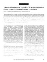
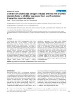
Figure 1
Serum procollagen I and III aminoterminal propeptide concentrations in surviving and non-survived sepsis patients during the 10-day follow up and at three and six monthsSerum procollagen I and III aminoterminal propeptide concentrations in surviving and non-survived sepsis patients during the 10-day follow up and at
three and six months. The symbols mark the median values and the vertical lines stand for ranges from 25th to 75th percentile. The laboratory refer-
ence values are presented as a solid grey area in the background. Reference values for serum procollagen III aminoterminal propeptide (PIIINP) are
the same for both males and females (1.7 to 4.2 μg/L). For serum procollagen I aminoterminal propeptide (PINP) the reference area for females (19
to 84 μg/L) is slightly broader than for males (20 to 76 μg/L) and is presented in darker grey.
Critical Care Vol 13 No 2 Gäddnäs et al.
Page 6 of 10
(page number not for citation purposes)
tile = 16.0 to 44.4; P = 0.038), and the difference tended to
increase with time (Figure 4). The same trend was found in the
MOF group compared with patients with MODS (37.8 μg/L,
25th to 75th percentile = 26.3 to 66.1; vs. 6.7 μg/L, 25th to
75th percentile = 11.4 to 34.3; P = 0.004). The patients that
received hydrocortisone therapy had no statistically significant
difference in maximum ICTP value compared with those who
did not receive supplementation therapy (35.5 μg/L, 25th to
75th percentile = 20.8 to 57.9; vs. 26.7 μg/L, 25th to 75th
percentile = 16.4 to 39.8; P = 0.343).
The maximum ICTP value correlated positively with the maxi-
mum total SOFA score and maximum lactate levels (P =
0.000; P = 0.011). Also ICTP level on day one correlated with
maximum total SOFA score (P < 0.001) and maximum lactate
levels (P = 0.013). ROC curve analysis of maximum ICTP for
liver failure shows an AUC of 0.610 (25th to 75th percentile =
0.450 to 0.769; P = 0.393) and for renal failure an AUC of
0.472 (25th to 75th percentile = 0.252 to 0.691; P = 0.871).
Neither maximum nor day one ICTP correlated with 30-day
mortality.
Discussion
This is the first longitudinal study reporting serum procollagen
propeptide levels in human severe sepsis. Previous studies
have focused on collagen metabolism in severe trauma, ARDS
or Gram-negative sepsis [7,12,14]. Increasing collagen
propeptide levels (PIIINP throughout the disease process and
PINP in the late phase) were associated with the development
of MOF and death and they correlated with maximum lactate
concentrations. All the values in survivors had returned to the
normal range and were lower at three and six months than they
were at the beginning of the study.
Of the different organs, collagen synthesis in lungs has been
most profoundly studied in critical illness. ARDS is the most
severe manifestation of acute lung injury and is also one of the
most common organ failures in severe sepsis. The collagen I
and III propeptides have been showed to be elevated in
plasma and bronchoalveolar lavage fluid in patients with
ARDS during the first days of disease and are associated with
increased risk of death [7,13,19]. In our data the patients with
lung specific SOFA scores of three to four had only slightly
pronounced PINP, PIIINP and ICTP values (day one and the
maximal values over the study period) compared with patients
with less severe scores. The difference did not reach statistical
significance (data not shown). Hence the increased procolla-
gen propeptide levels observed in this study seem to be only
partly due to increased synthesis and degradation of collagen
in the lungs.
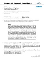
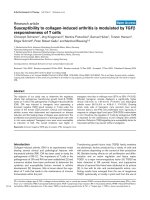
Figure 2
Serum procollagen I and III aminoterminal propeptide concentrations in surgical and medical groups of sepsis patients during the 10-day follow up and at three and six monthsSerum procollagen I and III aminoterminal propeptide concentrations in surgical and medical groups of sepsis patients during the 10-day follow up
and at three and six months. The symbols mark the median values and the vertical lines stand for ranges from 25th to 75th percentile. The laboratory
reference values are presented as a solid grey area in the background. Reference values for serum procollagen III aminoterminal propeptide (PIIINP)
are the same for both males and females (1.7 to 4.2 μg/L). For serum procollagen I aminoterminal propeptide (PINP) the reference area for females
(19 to 84 μg/L) is slightly broader than for males (20 to 76 μg/L) and is presented in darker grey.
Available online />Page 7 of 10
(page number not for citation purposes)
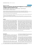
Figure 3
Serum procollagen I and III aminoterminal propeptide concentrations in sepsis patients during the 10-day follow up and at three and six months according to whether they received hydrocortisone supplementation or notSerum procollagen I and III aminoterminal propeptide concentrations in sepsis patients during the 10-day follow up and at three and six months
according to whether they received hydrocortisone supplementation or not. The symbols mark the median values and the vertical lines stand for
ranges from 25th to 75th percentile. The laboratory reference values are presented as a solid grey area in the background. Reference values for
serum procollagen III aminoterminal propeptide (PIIINP) are the same for both males and females (1.7 to 4.2 μg/L). For serum procollagen I aminot-
erminal propeptide (PINP) the reference area for females (19 to 84 μg/L) is slightly broader than for males (20 to 76 μg/L) and is presented in darker
grey.
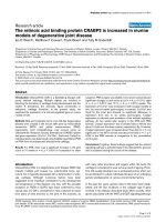
Figure 4
Concentrations of type I collagen cross-linked telopeptides in groups of survived and non-survived sepsis patients during the 10-day follow up and at three and six monthsConcentrations of type I collagen cross-linked telopeptides in groups of survived and non-survived sepsis patients during the 10-day follow up and
at three and six months. The laboratory reference values (1.6 to 4.6 μg/l) are presented as a solid grey area in the background. The symbols mark the
median values and the vertical lines stand for ranges from 25th to 75th percentile. ICTP = type I collagen cross-linked telopeptides.
Critical Care Vol 13 No 2 Gäddnäs et al.
Page 8 of 10
(page number not for citation purposes)
Waydhas and colleagues reported increased PIIINP serum
concentrations in severely injured patients [12]. Similar to our
findings in septic patients, serum concentrations were ele-
vated in severely injured non-survivors and in those who devel-
oped MOF. It was noted in the study by Waydhas and
colleagues that PIIINP levels correlated with increasing
bilirubin levels. The procollagen propeptides are eliminated by
the liver, thus the increased serum levels may result from
increased synthesis or decreased uptake by liver cells [20].
The study by Waydhas and colleagues did not determine
whether the increased concentrations were due to excess syn-
thesis or diminished elimination [12].
On the other hand, in alcoholic liver fibrosis it has been shown
that elevated PIIINP concentrations are caused by increased
histologically confirmed fibrogenesis [21]. In our study, PINP
and PIIINP did correlate with liver function implying that either
synthesis or elimination by the liver in sepsis is affected.
Because PINP and PIIINP are eliminated via the same liver
endothelial cell receptor, the serum levels of both propeptides
should have increased if the increased concentrations were
solely a result of decreased elimination.
A correlation with kidney function was also observed. Small
fractions of PIIINP are excreted by the kidneys [22,23]. Inter-
estingly, increased PIIINP levels have been reported in acute
renal disease, exemplifying the influence of systemic disease
on collagen metabolism. Keller and colleagues reported that,
compared with values in chronic renal failure, the values of PII-
INP were even higher in patients with acute renal failure and
MOF [8]. Furthermore, experimental data have shown that
renal damage increases the release of a collagen synthesis-
stimulating factor [24]. Previous data thus suggests that acute
renal failure is associated with increased synthesis of type III
collagen. In the present study, maximum PINP, PIIINP and
ICTP levels did not have statistically significant prognostic val-
ues for liver and renal failure in the ROC analysis.
It is tempting to speculate that increased collagen propeptide
levels found are at least partly due to increased synthesis and
are likely to be a summation of collagen synthesis from differ-
ent organs. To find out the contribution of the different organs
affected, further studies are required.
ICTP is a marker of collagen degradation and is eliminated by
the kidneys [20]. In a small study Wenisch and colleagues
reported elevated ICTP levels in Gram-negative sepsis on day
0 and day 28 [14]. We found that serum ICTP, but not PINP,
was increased in severe sepsis. Thus, the increased ICTP lev-
els most likely indicate increased degradation of collagen type
I. As type I collagen is most abundant in bone, it could be
speculated that high levels of ICTP could partly be a result of
immobilisation. However, increased ICTP levels most likely
mirror high systemic inflammation, because the levels were
highest in patients with the most severe forms of the disease.
Collagens are degraded by specific matrix metalloproteinases
(MMPs) produced by fibroblasts, other connective tissue cells
and inflammatory cells. MMPs are induced by proinflammatory
cytokines (e.g. IL-1, IL-6 and TNF). In vitro it has been shown
that, following exposure to S. aureus, fibroblasts have
increased MMP expression, which is associated with degrada-
tion of collagen [25].
Our study suggests that collagen turnover may be increased
in severe sepsis. Over the past years, our understanding on
the complexity of the host healing response in sepsis has
grown: Phases of coagulation, inflammation and fibroprolifera-
tion overlap and exert regulatory control on one another. The
collagen synthesis in fibroblasts is regulated by coagulation
cascade proteases, proinflammatory cytokines and growth
factors. Coagulation protease thrombin seems to act as
fibroblast chemoattractant [26], stimulator of procollagen pro-
duction [27], promoter of myofibroblast formation [28] and
MMP activator [29]. Recently, a similar role of the upstream
coagulation protease Xa has been acknowledged. It seems to
enhance the expression of tranforming growth factor beta
(TGF-β), fibroblast proliferation and differentiation to myofi-
broblasts, migration and fibronectin production [30]. Thus the
activated coagulation in sepsis is one factor promoting the
fibrogenetic response.
Of the proinflammatory cytokines TNF-α has a pivotal effect on
collagen synthesis. In addition to stimulating fibroblast growth
and collagen synthesis, it has been shown that TNF-α in high
concentrations inhibits collagen and fibronectin production
and induces collagenase synthesis [31]. Among the growth
factors TGF-β deserves special attention. It is a multifunctional
growth factor that regulates proliferation, differentiation of
cells, protein synthesis and angiogenesis. TGF-β has been
reported to act as an inducer, as well as an inhibitor, of fibrob-
last growth [32]. Increased fibrosis is mediated by TGF-β1 in
various disease states, and progressive fibrosis has been sug-
gested to be a common pathway to organ failure [11]. Accord-
ingly, in ARDS it has been demonstrated that bronchoalveolar
lavage fluid obtained from patients is capable of activating a
human procollagen 1 promoter by means of TGF-β1 present
in the bronchoalveolar lavage fluid. Furthermore, in ARDS
TGF-β1 levels have been shown to be higher in non-survivors,
although the result is not statistically significant [33]. Higher
levels have also been reported in trauma patients developing
sepsis [34]. Indeed, sepsis could be called a systemic wound
with activated coagulation, inflammation and fibrogenetic
response.
Other factors that can affect collagen metabolism in severe
sepsis include surgery, hydrocortisone treatment and tissue
hypoxia. Surgery and trauma induce the healing process and
thus account for the fibroproliferative response. In a previous
study, it was shown that surgery itself (and wound infection
especially) increases serum procollagen concentrations [35].
Available online />Page 9 of 10
(page number not for citation purposes)
In our study no differences could be found between the surgi-
cal and medical groups. The surgical group consisted of
patients with trauma or those who underwent major surgical
procedure requiring general anaesthesia. Minor standard ICU
procedures such as tracheostomy, drainage or cannulations
were also performed in the medical group and could partly
have contributed to the controversial result of our study.
It is known that corticosteroid therapy reduces collagen depo-
sition [7,36]. In our material, treatment of sepsis with steroids
decreased serum PINP levels, indicating that type I collagen
synthesis is decreased in the early phase (up to six days) of
sepsis in patients treated with hydrocortisone. After hydrocor-
tisone therapy, which most often lasted seven days, the PINP
values were upregulated as in the group not treated with
hydrocortisone. Hypoxia is a fibrotic stimulus associated with
enhanced collagen synthesis and it has been shown to aug-
ment collagen prolyl 4-hydroxylase activity in vitro [37]. Tissue
hypoxia and activation of the coagulation and inflammatory
cascades play a key role in the pathogenesis of MODS.
Although adequate initial resuscitation usually restores oxygen
delivery at the systemic level, regional hypoxia at the organ
level is a well-documented phenomenon. The mechanisms are
considered to include microcirculatory disturbances, that
block the oxygen supply, and mitochondrial malfunction that
results in inadequate use of oxygen at the cellular level.
Increased circulating lactate levels are suggestive of tissue
hypoxia and are associated with a poor outcome. In our study
PINP, PIIINP and ICTP correlated with maximum lactate levels.
The importance of tissue hypoxia in the stimulation of collagen
synthesis is also suggested by the results in patients with
chronic heart failure in which relative collagen deposition in the
intestinal wall was the highest in advanced cases of heart fail-
ure [38]. Furthermore, in a rat model, sepsis has been shown
to induce significant increases in collagen content in hepatic
and ileal interstitial tissues, which were prevented with a leu-
cotriene antagonist [39]. Yet there is also evidence to the con-
trary. In a mice model of lipopolysaccharide-stimulated ARDS,
hypoxia suppressed inflammation in lungs via adenosine A
2A
-
receptor-mediated pathway and resulted in lower lung injury
score and thickening of the alveocapillary membrane [40].
This study is limited by the fact that our study population was
relatively small because this was a one-centre study and a con-
siderable number of patients were excluded because of under-
lying diseases affecting collagen metabolism. Second, the
controls were healthy volunteers and thus could not be
matched for chronic diseases, of which arteriosclerosis, diabe-
tes and pulmonary diseases may have altered collagen metab-
olism. Third, the serum markers of inflammation were not
measured. The septic response is individual and patients may
have entered the study in different phases of inflammation,
although all of them entered within 48 hours of the first organ
failure. Further studies are needed to connect the levels of col-
lagen turnover to timely development of coagulation and
inflammatory responses. Nonetheless, this study provides new
in vivo measured information on connective tissue metabolism
and its timely development in sepsis.
Conclusions
Serum levels of PIIINP and ICTP are significantly increased in
patients with severe sepsis and can be investigated further as
markers of disease severity and outcome. These results imply
that fibrosis may be a central mechanism in the pathogenesis
of multiple organ dysfunction.
Competing interests
The authors declare that they have no competing interests.
Authors' contributions
All authors participated in the study design. FG participated in
collecting the data, performed statistical analysis and drafted
the manuscript with TA. MK participated in collecting the data.
VK conceived the study and helped to draft the manuscript.
AO provided the equipment for the suction blister method and
helped to draft the manuscript. JR provided collagen propep-
tide analyses. JL helped to draft the manuscript. JS conceived
the study with VK. TA performed the statistical analysis and
drafted the manuscript with FG. All authors read and approved
the final manuscript.
Acknowledgements
The skillful help of study nurses Sinikka Sälkiö and Tarja Lamberg in
screening the patients and obtaining serum samples is highly appreci-
ated. The excellent technical assistance of Mirja Mäkelä is acknowl-
edged. The help of M.Sc Pasi Ohtonen in statistical analysis is
appreciated. The study was supported by grants from the Instrumentar-
ium Foundation and Oulu University Hospital, Finland.
References
1. Bone RC, Balk RA, Cerra FB, Dellinger RP, Fein AM, Knaus WA,
Schein RM, Sibbald WJ: Definitions for sepsis and organ failure
and guidelines for the use of innovative therapies in sepsis.
The ACCP/SCCM Consensus Conference Committee. Ameri-
can College of Chest Physicians/Society of Critical Care Med-
icine. Chest 1992, 101:1644-1655.
2. Hotchkiss RS, Karl IE: The pathophysiology and treatment of
sepsis. N Engl J Med 2003, 348:138-150.
3. Levy MM, Fink MP, Marshall JC, Abraham E, Angus D, Cook D,
Cohen J, Opal SM, Vincent JL, Ramsay G, SCCM/ESICM/ACCP/
ATS/SIS: 2001 SCCM/ESICM/ACCP/ATS/SIS International
Sepsis Definitions Conference. Crit Care Med 2003,
31:1250-1256.
Key messages
• Serum levels of PIIINP and ICTP are significantly
increased in patients with severe sepsis and can be
investigated further as markers of disease severity and
outcome.
• PIIINP and ICTP values in survivors returned to the nor-
mal range and were lower at three and six months than
they were at the beginning of the study.
Critical Care Vol 13 No 2 Gäddnäs et al.
Page 10 of 10
(page number not for citation purposes)
4. Haukipuro K, Risteli L, Kairaluoma MI, Risteli J: Aminoterminal
propeptide of type III procollagen in healing wound in humans.
Ann Surg 1987, 206:752-756.
5. Petri JB, Konig S, Haupt B, Haustein UF, Herrmann K: Molecular
analysis of different phases in human wound healing. Exp Der-
matol 1997, 6:133-139.
6. Jensen LT, Horslev-Petersen K, Toft P, Bentsen KD, Grande P,
Simonsen EE, Lorenzen I: Serum aminoterminal type III procol-
lagen peptide reflects repair after acute myocardial infarction.
Circulation 1990, 81:52-57.
7. Meduri GU, Tolley EA, Chinn A, Stentz F, Postlethwaite A: Procol-
lagen types I and III aminoterminal propeptide levels during
acute respiratory distress syndrome and in response to meth-
ylprednisolone treatment. Am J Respir Crit Care Med 1998,
158:1432-1441.
8. Keller F, Rehbein C, Schwarz A, Fleck M, Hayasaka A, Schuppan
D, Offermann G, Hahn EG: Increased procollagen III production
in patients with kidney disease. Nephron 1988, 50:332-337.
9. Moller S, Hansen M, Hillingso J, Jensen JE, Henriksen JH: Ele-
vated carboxy terminal cross linked telopeptide of type I colla-
gen in alcoholic cirrhosis: relation to liver and kidney function
and bone metabolism. Gut 1999, 44:417-423.
10. Zhang K, Garner W, Cohen L, Rodriguez J, Phan S: Increased
types I and III collagen and transforming growth factor-beta 1
mRNA and protein in hypertrophic burn scar. J Invest Dermatol
1995, 104:750-754.
11. Weber KT: Fibrosis, a common pathway to organ failure: angi-
otensin II and tissue repair. Semin Nephrol 1997, 17:467-491.
12. Waydhas C, Nast-Kolb D, Trupka A, Lenk S, Duswald KH, Sch-
weiberer L, Jochum M: Increased serum concentrations of pro-
collagen type III peptide in severely injured patients: an
indicator of fibrosing activity? Crit Care Med 1993,
21:240-247.
13. Clark JG, Milberg JA, Steinberg KP, Hudson LD: Type III procol-
lagen peptide in the adult respiratory distress syndrome.
Association of increased peptide levels in bronchoalveolar
lavage fluid with increased risk for death. Ann Intern Med
1995, 122:17-23.
14. Wenisch C, Graninger W, Schonthal E, Rumpold H: Increased
serum concentrations of the carboxy-terminal cross-linked
telopeptide of collagen type I in patients with Gram-negative
septicaemia.
Eur J Clin Invest 1996, 26:237-239.
15. Dellinger RP, Carlet JM, Masur H, Gerlach H, Calandra T, Cohen
J, Gea-Banacloche J, Keh D, Marshall JC, Parker MM, Ramsay G,
Zimmerman JL, Vincent JL, Levy MM, Surviving Sepsis Campaign
Management Guidelines, Committee: Surviving Sepsis Cam-
paign guidelines for management of severe sepsis and septic
shock. Crit Care Med 2004, 32:858-873.
16. Vincent JL, de Mendonca A, Cantraine F, Moreno R, Takala J, Suter
PM, Sprung CL, Colardyn F, Blecher S: Use of the SOFA score
to assess the incidence of organ dysfunction/failure in inten-
sive care units: results of a multicenter, prospective study.
Working group on "sepsis-related problems" of the European
Society of Intensive Care Medicine. Crit Care Med 1998,
26:1793-1800.
17. Vincent JL, Moreno R, Takala J, Willatts S, De Mendonca A, Bruin-
ing H, Reinhart CK, Suter PM, Thijs LG: The SOFA (Sepsis-
related Organ Failure Assessment) score to describe organ
dysfunction/failure. On behalf of the Working Group on Sep-
sis-Related Problems of the European Society of Intensive
Care Medicine. Intensive Care Med 1996, 22:707-710.
18. Risteli Juha RL: Extracellular matrix metabolites in body fluids.
In In Connective tissue and its heritable disorders: molecular,
genetic and medical aspects 2nd edition. Edited by: Royce PM,
Steinmann B. New York: Wiley-liss Inc; 2002:1141-1160.
19. Marshall RP, Bellingan G, Webb S, Puddicombe A, Goldsack N,
McAnulty RJ, Laurent GJ: Fibroproliferation occurs early in the
acute respiratory distress syndrome and impacts on outcome.
Am J Respir Crit Care Med 2000, 162:1783-1788.
20. Risteli J, Risteli L: Analysing connective tissue metabolites in
human serum. Biochemical, physiological and methodological
aspects. J Hepatol 1995, 22:77-81.
21. Nøjgaard C, Johansen JS, Christensen E, Skovgaard LT, Price PA,
Becker U: Serum levels of YKL-40 and PIIINP as prognostic
markers in patients with alcoholic liver disease. J Hepatol
2003, 39:179-186.
22. Bentsen KD, Boesby S, Kirkegaard P, Hansen CP, Jensen SL,
Horslev-Petersen K, Lorenzen I: Is the aminoterminal propeptide
of type III procollagen degraded in the liver? A study of type III
procollagen peptide in serum during liver transplantation in
pigs. J Hepatol 1988, 6:144-150.
23. Jensen LT, Blaehr H, Andersen CB, Risteli J, Lorenzen I: Metabo-
lism of the aminoterminal propeptide of type III procollagen in
cultures of human proximal tubular cells. Scand J Clin Lab
Invest 1992, 52:
1-8.
24. Ohyama K, Seyer JM, Raghow R, Kang AH: A factor from dam-
aged rat kidney stimulates collagen biosynthesis by mesang-
ial cells. Biochim Biophys Acta 1990, 1053:173-178.
25. Kanangat S, Postlethwaite A, Hasty K, Kang A, Smeltzer M,
Appling W, Schaberg D: Induction of multiple matrix metallo-
proteinases in human dermal and synovial fibroblasts by Sta-
phylococcus aureus: implications in the pathogenesis of
septic arthritis and other soft tissue infections. Arthritis Res
Ther 2006, 8:R176.
26. Dawes KE, Gray AJ, Laurent GJ: Thrombin stimulates fibroblast
chemotaxis and replication. Eur J Cell Biol 1993, 61:126-130.
27. Chambers RC, Dabbagh K, McAnulty RJ, Gray AJ, Blanc-Brude
OP, Laurent GJ: Thrombin stimulates fibroblast procollagen
production via proteolytic activation of protease-activated
receptor 1. Biochem J 1998, 333:121-127.
28. Bogatkevich GS, Tourkina E, Silver RM, Ludwicka-Bradley A:
Thrombin differentiates normal lung fibroblasts to a myofi-
broblast phenotype via the proteolytically activated receptor-1
and a protein kinase C-dependent pathway. J Biol Chem 2001,
276:45184-45192.
29. Duhamel-Clerin E, Orvain C, Lanza F, Cazenave JP, Klein-Soyer C:
Thrombin receptor-mediated increase of two matrix metallo-
proteinases, MMP-1 and MMP-3, in human endothelial cells.
Arterioscler Thromb Vasc Biol 1997, 17:1931-1938.
30. Borensztajn K, Stiekema J, Nijmeijer S, Reitsma PH, Peppelen-
bosch MP, Spek CA: Factor Xa stimulates proinflammatory and
profibrotic responses in fibroblasts via protease-activated
receptor-2 activation. Am J Pathol 2008, 172:309-320.
31. Maish GO 3rd, Shumate ML, Ehrlich HP, Cooney RN: Tumor
necrosis factor binding protein improves incisional wound
healing in sepsis. J Surg Res 1998, 78:108-117.
32. Thornton SC, Por SB, Walsh BJ, Penny R, Breit SN: Interaction
of immune and connective tissue cells: I. The effect of lym-
phokines and monokines on fibroblast growth. J Leukoc Biol
1990, 47:312-320.
33. Budinger GR, Chandel NS, Donnelly HK, Eisenbart J, Oberoi M,
Jain M: Active transforming growth factor-beta1 activates the
procollagen I promoter in patients with acute lung injury.
Intensive Care Med 2005, 31:121-128.
34. Laun RA, Schroder O, Schoppnies M, Roher HD, Ekkernkamp A,
Schulte KM: Transforming growth factor-beta1 and major
trauma: time-dependent association with hepatic and renal
insufficiency. Shock 2003, 19:16-23.
35. Haukipuro K, Risteli L, Kairaluoma MI, Risteli J: Aminoterminal
propeptide of type III procollagen in serum during wound heal-
ing in human beings. Surgery 1990, 107:381-388.
36. Oikarinen A, Autio P, Vuori J, Vaananen K, Risteli L, Kiistala U, Ris-
teli J: Systemic glucocorticoid treatment decreases serum
concentrations of carboxyterminal propeptide of type I procol-
lagen and aminoterminal propeptide of type III procollagen. Br
J Dermatol 1992, 126:172-178.
37. Fahling M, Mrowka R, Steege A, Nebrich G, Perlewitz A, Persson
PB, Thiele BJ: Translational control of collagen prolyl 4-hydrox-
ylase-alpha(I) gene expression under hypoxia. J Biol Chem
2006, 281:26089-26101.
38. Arutyunov GP, Kostyukevich OI, Serov RA, Rylova NV, Bylova NA:
Collagen accumulation and dysfunctional mucosal barrier of
the small intestine in patients with chronic heart failure. Int J
Cardiol 2008, 125:240-245.
39. Sener G, Sehirli O, Cetinel S, Ercan F, Yuksel M, Gedik N, Yegen
BC: Amelioration of sepsis-induced hepatic and ileal injury in
rats by the leukotriene receptor blocker montelukast. Prostag-
landins Leukot Essent Fatty Acids 2005, 73:453-462.
40. Thiel M, Chouker A, Ohta A, Jackson E, Caldwell C, Smith P, Luka-
shev D, Bittmann I, Sitkovsky MV: Oxygenation inhibits the phys-
iological tissue-protecting mechanism and thereby
exacerbates acute inflammatory lung injury. PLoS Biol 2005,
3:e174.