Báo cáo y học: "Inclusion of the glucocorticoid receptor in a hypothalamic pituitary adrenal axis model reveals bistability" ppt
Bạn đang xem bản rút gọn của tài liệu. Xem và tải ngay bản đầy đủ của tài liệu tại đây (760.58 KB, 12 trang )
BioMed Central
Page 1 of 12
(page number not for citation purposes)
Theoretical Biology and Medical
Modelling
Open Access
Research
Inclusion of the glucocorticoid receptor in a hypothalamic pituitary
adrenal axis model reveals bistability
Shakti Gupta, Eric Aslakson*, Brian M Gurbaxani and Suzanne D Vernon
Address: Division of Viral and Rickettsial Diseases, National Center for Zoonotic, Vector-Borne, and Enteric Diseases, Centers for Disease Control
and Prevention, 600 Clifton Rd, MS-A15, Atlanta, Georgia 30333, USA
Email: Shakti Gupta - ; Eric Aslakson* - ; Brian M Gurbaxani - ;
Suzanne D Vernon -
* Corresponding author
Abstract
Background: The body's primary stress management system is the hypothalamic pituitary adrenal
(HPA) axis. The HPA axis responds to physical and mental challenge to maintain homeostasis in
part by controlling the body's cortisol level. Dysregulation of the HPA axis is implicated in
numerous stress-related diseases.
Results: We developed a structured model of the HPA axis that includes the glucocorticoid
receptor (GR). This model incorporates nonlinear kinetics of pituitary GR synthesis. The nonlinear
effect arises from the fact that GR homodimerizes after cortisol activation and induces its own
synthesis in the pituitary. This homodimerization makes possible two stable steady states (low and
high) and one unstable state of cortisol production resulting in bistability of the HPA axis. In this
model, low GR concentration represents the normal steady state, and high GR concentration
represents a dysregulated steady state. A short stress in the normal steady state produces a small
perturbation in the GR concentration that quickly returns to normal levels. Long, repeated stress
produces persistent and high GR concentration that does not return to baseline forcing the HPA
axis to an alternate steady state. One consequence of increased steady state GR is reduced steady
state cortisol, which has been observed in some stress related disorders such as Chronic Fatigue
Syndrome (CFS).
Conclusion: Inclusion of pituitary GR expression resulted in a biologically plausible model of HPA
axis bistability and hypocortisolism. High GR concentration enhanced cortisol negative feedback on
the hypothalamus and forced the HPA axis into an alternative, low cortisol state. This model can
be used to explore mechanisms underlying disorders of the HPA axis.
Background
The hypothalamic pituitary adrenal (HPA) axis represents
a self-regulated dynamic feedback neuroendocrine system
that is essential for maintaining body homeostasis in
response to various stresses. Stress can be physical (e.g.
infection, thermal exposure, dehydration) and psycholog-
ical (e.g. fear, anticipation). Both physical and psycholog-
ical stressors activate the hypothalamus to release
corticotropin releasing hormone (CRH). The CRH is
released into the closed hypophyseal portal circulation,
stimulating the pituitary to secrete adrenocorticotropic
hormone (ACTH). ACTH is released into the blood where
Published: 14 February 2007
Theoretical Biology and Medical Modelling 2007, 4:8 doi:10.1186/1742-4682-4-8
Received: 27 August 2006
Accepted: 14 February 2007
This article is available from: />© 2007 Gupta et al; licensee BioMed Central Ltd.
This is an Open Access article distributed under the terms of the Creative Commons Attribution License ( />),
which permits unrestricted use, distribution, and reproduction in any medium, provided the original work is properly cited.
Theoretical Biology and Medical Modelling 2007, 4:8 />Page 2 of 12
(page number not for citation purposes)
it travels to the adrenals, inducing the synthesis and secre-
tion of cortisol from the adrenal cortex. Cortisol has a neg-
ative feedback effect on the hypothalamus and pituitary
that further dampens CRH and ACTH secretion [1].
Cortisol affects a number of cellular and physiological
functions to maintain body homeostasis and health. Cor-
tisol suppresses inflammation and certain immune reac-
tions, inhibits the secretion of several hormones and
neuropeptides and induces lymphocyte apoptosis [1,2].
These widespread and potent effects of cortisol demand
that the feed forward and feedback loops of the HPA axis
are tightly regulated. Disruption of HPA axis regulation is
known to contribute to a number of stress-related disor-
ders. For example, increased cortisol (hypercortisolism)
has been shown in patients with major depressive disor-
der (MDD) [3,4], and decreased cortisol (hypocortiso-
lism) has been observed in people with post-traumatic
stress disorder (PTSD), Gulf War illness, post infection
fatigue and chronic fatigue syndrome (CFS) [5-9]. While
it is not clear if dysregulation of the HPA axis is a primary
or secondary effect of these disorders, there is evidence
that stress-related disorders are influenced by early life
adverse experiences that affect the neural architecture and
gene expression in the brain [10]. Childhood events such
as severe infection, malnutrition, physical, sexual and
emotional abuse are associated with many chronic ill-
nesses later in life [11].
Definitive research on HPA axis function in chronic dis-
eases has been hampered by the complexity of the numer-
ous systems affected by the HPA axis, such as the immune
and neuroendocrine systems, the lack of known or acces-
sible brain lesions and the correlative nature of much of
the existing data. Since the organization of the HPA axis
has been characterized to detail the feedback and feed for-
ward signalling that regulates HPA axis function [12], it is
a system that is amenable to modelling. Models of the
HPA axis have been constructed using deterministic cou-
pled ordinary differential equations [13-17]. These mod-
els were successful in capturing features such as negative
feedback control and diurnal cycling of the HPA axis. Our
goal was to understand the dynamic effects of CRH, ACTH
and cortisol with a mathematically parsimonious model
to gain insight into HPA axis regulation. This model is
novel in that it incorporates expression of the glucocorti-
coid receptor (GR) in the pituitary and demonstrates that
repeated stress and GR expression reveals the bistability
inherent in the HPA axis given the enhanced model.
Model
The HPA axis has three compartments representing the
hypothalamus, pituitary and adrenals regulated by sim-
ple, linear mass action kinetics for the production and
degradation of the primary chemical product of each com-
partment. In this model, stress to the HPA axis (F) stimu-
lates the hypothalamus to secrete CRH (C). CRH (C)
signals the induction of ACTH synthesis (A) in the pitui-
tary. ACTH (A) signals to the adrenal gland and activates
the synthesis and release of cortisol (O). Cortisol (O) reg-
ulates its own synthesis via inhibiting the synthesis of
CRH (C) in the hypothalamus, and ACTH (A) in the pitu-
itary. The equation for the hypothalamus can be written
as:
In this equation, -K
cd
C models a constant degradation rate
of CRH in the blood of the portal vein. The term (K
c
+
F)* models a circadian production term K
c
and a
stress term F, both reduced by a linear inhibition term rep-
resented by . For small , we may write (K
c
+
F) * ≈ . The latter form, , corre-
sponds to standard linear inhibition of (K
c
+ F) with inhi-
bition constant K
i1
. This form also guarantees positive
ACTH concentrations. We write for the hypothalamus:
For the pituitary:
Equation 3 models a constant degradation rate of ACTH
by the term -K
ad
A and an ACTH production term,
, with a cortisol inhibition factor similar to (2).
For the adrenal:
dC
dT
KF
O
K
KC
c
i
cd
=+∗− −
()
()( )11
1
()1
1
−
O
K
i
()1
1
−
O
K
i
O
K
i1
()1
1
−
O
K
i
KF
O
K
c
i
+
+1
1
KF
O
K
c
i
+
+1
1
dC
dT
KF
O
K
KC
c
i
cd
=
+
+
−
()
1
2
1
dA
dT
KC
O
K
KA
a
i
ad
=
+
−
()
1
3
2
KC
O
K
a
i
1
2
+
dO
dT
KA K O
ood
=−
()
4
Theoretical Biology and Medical Modelling 2007, 4:8 />Page 3 of 12
(page number not for citation purposes)
Equation 4 models a constant degradation rate of cortisol
-K
od
O and a cortisol production rate K
o
A linearly depend-
ent on ACTH.
We have augmented this model by including synthesis
and regulation of the glucocorticoid receptor (R) in the
pituitary [18,19]. In the pituitary, cortisol enters the cell
and binds the glucocorticoid receptor in the cytoplasm,
causing the receptor to dimerize. This dimerization causes
the complex to translocate to the nucleus (dimerization,
translocation, and transcription factor binding are not
modelled, but assumed to be fast), where it up regulates
glucocorticoid receptor (R) synthesis and down regulates
production of ACTH (A).
The following are the differential equations written for the
HPA axis model that includes glucocorticoid receptor syn-
thesis and regulation in the pituitary (Figure 1).
For the hypothalamus:
For the pituitary:
dC
dT
KF
O
K
KC
c
i
cd
=
+
+
−
()
1
5
1
dA
dT
KC
OR
K
KA
a
i
ad
=
+
−
()
1
6
2
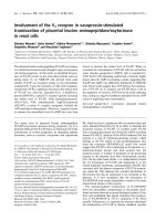
F is an external stress that triggers the hypothalamus to release CRH (C) that signals to the pituitary to release ACTH (A) stimulating the synthesis and release of cortisol (O) from the adrenalsFigure 1
F is an external stress that triggers the hypothalamus to release CRH (C) that signals to the pituitary to release ACTH (A)
stimulating the synthesis and release of cortisol (O) from the adrenals. Release of cortisol negatively regulates CRH and ACTH
after binding to the glucocorticoid receptor (R) in the pituitary. Here, GR and cortisol regulate further GR synthesis.
Theoretical Biology and Medical Modelling 2007, 4:8 />Page 4 of 12
(page number not for citation purposes)
For the adrenal:
Equation (7) describes the production of GR in the pitui-
tary. The term in equation 7 is in Michaelis-
Menten form since we assume the bound glucocorticoid
receptor (OR) dimerizes with fast kinetics, so that the
amount of dimer is in constant quasi-equilibrium,
depending on the abundance of OR and the equilibrium
binding affinity (K). The model further assumes that cor-
tisol (O) and the glucocorticoid receptor (R) bind to each
other with very fast kinetics compared to the rate of
change of the 4 state variables (A, C, O, and R), so that OR
stays in quasi-equilibrium as well. These are reasonable
assumptions, given that high affinity receptor-ligand
kinetics are often much faster than enzyme kinetics (as is
assumed in the standard Michaelis-Menten equation) or
than steps requiring transcription and/or translation for
protein synthesis. Equation (7) also models a linear pro-
duction term K
cr
and a degradation term -K
rd
R for pituitary
GR production. Equation (6) reflects the inhibition
dependence of glucocorticoid receptor (R) and cortisol
(O) with an inhibition constant K
i2
.
Scaling of the equations (5) – (8) has been done to reduce
the parameters used in simulations. The scaled variables
are defined as;
The scaled equations thereby obtained are;
These scaled equations were used in the simulations. The
advantage of scaling is that it obviates the need for knowl-
edge of unknown parameter values such as the synthesis
rate of CRH in the hypothalamus and ACTH and GR in
the pituitary. The parameter values that can be measured
are the degradation rates of CRH, ACTH, and cortisol. The
scaled parameter values used in simulation were, k
cd
= 1,
k
ad
= 10, k
rd
= 0.9, k
cr
= 0.05, k = 0.001, k
i1
= 0.1, and k
i2
=
0.1. Further, these simulated results for CRH, ACTH and
cortisol are converted back to their commonly used
dimensions and values obtained in experiments. The sim-
ulated time course plots ignore the circadian input to the
hypothalamus.
Models were programmed in Matlab (The Mathworks,
Natick, MA). The meta-modeling of bi-stability used the
CONTENT freeware package. All Matlab code will be pro-
vided upon request. Dr. Leslie Crofford provided the
human subject serum cortisol data [9].
Results
To determine if these equations could predict the general
features of cortisol production, the experimental data was
compared to a cortisol curve generated using equation 4.
As shown in Figure 2, equation 4 predicts a fit that is very
similar to the actual cortisol production in this healthy
human subject. Experimental fitting of ACTH is not possi-
ble since hypothalamic derived CRH cannot be measured.
Steady States
Equations (9)–(12) permit one or three positive steady
states depending upon the parameter values. The three
positive steady states exist because of homodimerization
of the GR with cortisol. Figure 3 shows the variation of GR
and cortisol steady state with respect to parameter k
rd
. Var-
iations in k
rd
from person to person may be expected due
to genetic differences in the details of GR production and
degradation. For a high value of k
rd
, there exists only a low
GR concentration steady state. As the value of k
rd
decreases, these equations produce two more steady
states, one stable and another unstable in GR concentra-
tion. As k
rd
decreases further, a low GR concentration state
disappears and only a high GR concentration state exists
dR
dT
KOR
KOR
KKR
r
cr rd
=
+
+−
()
()
()
2
2
7
dO
dT
KA K O
ood
=−
()
8
KOR
KOR
r
()
()
2
2
+
tKTc
KC
K
a
KA
KK
o
KO
KKK
od
od
c
od
ca
od
cao
== = =,, ,
23
r
KR
K
k
K
K
k
K
K
k
K
K
od
r
cd
cd
od
ad
ad
od
rd
rd
od
====,, ,
dc
dt
f
o
k
kc
i
cd
=
+
+
−
()
1
1
9
1
da
dt
c
or
k
ka
i
ad
=
+
−
()
1
10
2
dr
dt
or
kor
kkr
cr rd
=
+
+−
()
()
()
2
2
11
do
dt
ao=−
()
12
Theoretical Biology and Medical Modelling 2007, 4:8 />Page 5 of 12
(page number not for citation purposes)
(Figure 3a). In this model, we postulate that the low GR
concentration represents the normal steady state, and
high GR concentration denotes a dysregulated HPA axis
steady state as it results in persistent low cortisol levels
(hypocortisolism) (Figure 3b). Hypocortisolism results
from the negative feedback between GR (i.e. the symbol
"R" in Figure 1) and ACTH (A), and hence cortisol (O)
produced downstream of it, as shown in Figure 1 and
reflected by the inverse relationship between cortisol and
GR in Figure 3. Thus individuals with very large values of
k
rd
would be constitutively healthy in this model, i.e.
impervious to a dysregulated HPA-axis no matter how
much they are stressed, and those with very low values of
k
rd
would be constitutively unhealthy.
Normal stress response
The response of the normal HPA axis to small perturba-
tions is essential to the survival of an organism. Stress acti-
vates the HPA axis to regulate various body functions; first
by increasing ACTH synthesis followed by increased corti-
Experimental ACTH and cortisol from a human subject shown in blue and red in top and bottom panels respectivelyFigure 2
Experimental ACTH and cortisol from a human subject shown in blue and red in top and bottom panels respectively. Modelled
cortisol using equation 4 displayed with solid black line in lower panel.
Theoretical Biology and Medical Modelling 2007, 4:8 />Page 6 of 12
(page number not for citation purposes)
sol production and then returning to the original state.
Figure 4 shows a simulation of the response of the HPA
axis to a short stress. The initial condition of the HPA axis
was set to a normal steady state and at T = 0, a stress was
given for 0<T<1. The HPA axis responded to this distur-
bance by secreting CRH. The synthesis of CRH induced
the synthesis of ACTH and cortisol (Figures 4a and 4b).
The synthesis of CRH stopped once the stress ended, and
the concentration of CRH quickly decreased due to CRH
degradation (Figure 4c). CRH returned to steady state
meanwhile stimulating the release of ACTH that also
peaked shortly after the short stress ended (Figure 4b).
Synthesis of cortisol followed the peak ACTH secretion
(Figure 4a). The concentration of GR was only slightly ele-
vated following the short stress and then returned to base-
line (Figure 4d).
Adaptation of HPA axis
The robustness of the system was illustrated by the fact
that short stress produced small transients that returned to
the original, normal steady state. To simulate adaptation
of the HPA axis to repeated stress, recursive stress was
applied at T = 0, 8 and 16 hours for 2 hour periods. The
simulation results showed the continuous decrease in
maximum ACTH and cortisol concentration after every
stress (Figure 5a and 5b) while CRH is relatively unaf-
fected (Figure 5c). The decrease in secretion of ACTH and
cortisol occurred because of an increase in pituitary GR
concentration and the fact that the system was pulsed with
the stresses before it had time to fully recover (Figure 5d).
Chronic stress response
To simulate the response to chronic stress, a long stress
was given for 0<T<10 hours to perturb the normal steady
state of the HPA axis. Simulation results show the bistabil-
ity in the HPA axis; a long stress forces the HPA axis to an
alternate steady state (Figure 6). The HPA axis secreted
cortisol in response to stress. The increased concentration
of cortisol induced the synthesis of GR and the inhibition
of pituitary ACTH. When stress was applied for long peri-
ods, GR synthesis continued and crossed the threshold
middle unstable steady state of GR (Figure 3a). At this
point, the HPA axis reached the basin of attraction of the
second stable steady state and remained there even after
the removal of stress. The higher concentration of GR trig-
gered further pituitary ACTH inhibition, resulting in a
lower basal level ACTH and cortisol production (Figures
6a and 6b).
HPA axis challenge
Psychologic stress, CRH and dexamethasone (DEX) tests
are used to assess HPA axis function. The model was used
to simulate these various HPA axis function tests. To sim-
ulate a psychologic stress experiment, the same stress was
given with two different initial conditions: normal steady
state (low GR concentration) that would occur in a con-
trol group, and low cortisol state (high GR concentration)
Variations of steady state (a) GR and (b) cortisol with k
rd
Figure 3
Variations of steady state (a) GR and (b) cortisol with k
rd
. Solid and dashed lines denote the stable and unstable steady states,
respectively. If k
rd
for a given patient is in the region where GR and cortisol are multivalued, then the given patient can be
pushed from one value of steady state GR or cortisol to equally valid altered steady state levels by the application of an
extreme stress.
Theoretical Biology and Medical Modelling 2007, 4:8 />Page 7 of 12
(page number not for citation purposes)
that would occur in a hypocortisolemic patient group.
Because the high concentration GR inhibited ACTH syn-
thesis, the patient group exhibited continued low cortisol
and ACTH responses compared to the control (Figures 7a
and 7b). To simulate the CRH test, e.g., one that requires
exogenous CRH administration, CRH concentration was
increased by a constant amount. This resulted in increased
pituitary and adrenal gland synthesis of ACTH and corti-
sol respectively. The high concentration of pituitary GR in
the patient group blunted both responses compared to the
control (Figures 8a and 8b) Both Figures 7 and 8 demon-
strate that the model behaves in a qualitatively similar
fashion to observed experimental results.
Discussion
Previous models of the HPA axis have not demonstrated
bistability in steady state cortisol or ACTH. We believe this
is because none of the previous models have explicitly
accounted for nonlinear kinetics, such as the homodimer-
ization of GR after cortisol activation [18,19]. This is
essential for the negative feedback control of the HPA axis.
This homodimerization engenders the existence of two
stable steady states and one unstable steady state in GR
The response of the HPA axis following a short stressFigure 4
The response of the HPA axis following a short stress. Short time stress as indicated by the shaded larea was given for 0<T<1
hr.
Theoretical Biology and Medical Modelling 2007, 4:8 />Page 8 of 12
(page number not for citation purposes)
expression in the pituitary. While increased cortisol fol-
lowing a short period of stress produces a small perturba-
tion in GR concentration, long and repeated periods of
stress resulting in elevated cortisol levels produce a large
perturbation in GR concentration that force the HPA axis
into an alternate steady state. Because of the existence of
two stable steady states in this model, a small increase GR
concentration can be regulated, but a large perturbation in
GR concentration is sustained even after the removal of
the long duration stress. A higher concentration of GR
increases the concentration of cortisol-GR complexes that
in turn enhance the inhibition of ACTH synthesis in the
pituitary. Since ACTH stimulates the production of corti-
sol, less ACTH results in lower cortisol secretion and a
decrease HPA axis activity.
GR is found in cells throughout the human brain and
body. However, GR synthesis and regulation is tissue and
organ specific. For example, while corticosterone injection
in rats inhibits the synthesis of GR-mRNA in lymphocyte,
hypothalamic and hippocampal cells [20,21], it induces
the synthesis of GR-mRNA and increases the sensitivity in
the anterior pituitary [22,23]. Our model incorporates the
increased synthesis of GR in the anterior pituitary.
Transient responses of HPA axis to recursive stressesFigure 5
Transient responses of HPA axis to recursive stresses. Initially HPA axis was at a lower GR steady state and stress was given at
T = 0, 8 and 16 for 2 hours. Repeated stresses are shown by shaded areas.
Theoretical Biology and Medical Modelling 2007, 4:8 />Page 9 of 12
(page number not for citation purposes)
Increased GR makes anterior pituitary cells more sensitive
to cortisol and enhances the negative feedback effect of
cortisol on ACTH production. Enhanced negative feed-
back control of ACTH production in the anterior pituitary
may produce a hypocortisol state.
We were also able to demonstrate that these simulation
results are qualitatively similar to cortisol levels measured
in a human subject (Figure 2). A large number of studies
have investigated alterations of the HPA axis in CFS,
including both studies of basal HPA axis activity as well as
studies of HPA axis responsiveness to challenge (for
review see [24]). A hypocortisol steady state, such as was
demonstrated in this modelling and simulation study, is
in keeping with many of these studies
There may be other physiologically plausible mechanisms
that produce bi-stability other than the anterior pituitary
GR homodimerization mechanism investigated here. The
point of this investigation is not to conclusively prove that
pituitary GR dimerization is the cause of hypocortisolism,
but rather to demonstrate that there are physiologically
plausible mechanisms for producing bistability in the
HPA-axis that are stress modulated. Further mining of the
Transient responses of HPA axis to chronic stressFigure 6
Transient responses of HPA axis to chronic stress. Extended length stress was given for 0<T<10. Stress is indicated with shad-
ing.
Theoretical Biology and Medical Modelling 2007, 4:8 />Page 10 of 12
(page number not for citation purposes)
experimental literature together with mathematical mod-
elling will reveal additional plausible mechanisms.
Conclusion
Moderate, short-lived stress responses that result in tran-
sient increases in cortisol are important and necessary for
maintaining body homeostasis and health. Strong and
prolonged stress can force the HPA axis into an altered
steady state. We demonstrate bistability in the HPA axis
due to pituitary GR synthesis. This altered steady state,
characterized by hypocortisolism, is observed in a number
of stress-related illnesses. The elucidation of bistability in
this model of the HPA axis through the action of pituitary
GR effects may lead to targeted treatments of stress-related
illness where hypocortisolism is the primary clinical man-
ifestation.
Authors' contributions
SG was responsible for programming the differential
equation models, producing the mathematics for the
Transient responses of HPA axis a simulated stress experimentFigure 7
Transient responses of HPA axis a simulated stress experiment. The same stress was given with two different initial conditions;
normal steady state (low GR concentration) that would occur in a control group, and low cortisol state (high GR concentra-
tion) that would occur in a patient group. Stress was given for 0<Time<1 hr. Dash and solid lines indicate the normal and dys-
regulated HPA axis responses respectively and stress is indicated with shading.
Theoretical Biology and Medical Modelling 2007, 4:8 />Page 11 of 12
(page number not for citation purposes)
meta-analysis on stress response and bistability, and writ-
ing of the manuscript. EA and SDV were responsible for
the concept, the design of this study and preparation, val-
idation, writing, and critical review of the manuscript.
BMG provided assistance on the mathematical analysis
and was responsible for critical review and editing of the
manuscript.
Disclaimer
The findings and conclusions in this report are those of
the author(s) and do not necessarily represent the views of
the funding agency.
Declaration of competing interests
The author(s) declare that they have no competing inter-
ests.
Acknowledgements
The funding for this project was made possible by funding from DARPA
MIPR number 05-U357. We would also like to acknowledge the Dr. Leslie
Crofford and the University of Michigan (GCRC M01-RR00042 and R01-
AR43148) for providing experimental data.
References
1. Munck A, Guyre PM, Holbrook NJ: Physiological functions of glu-
cocorticoids in stress and their relation to pharmacological
actions. Endocr Rev 1984, 5:25-44.
Transient responses of HPA axis to CRH testFigure 8
Transient responses of HPA axis to CRH test. The exogenous CRH was injected at T = 0. Dashed and solid lines indicate the
normal and dysregulated HPA axis responses respectively.
Publish with BioMed Central and every
scientist can read your work free of charge
"BioMed Central will be the most significant development for
disseminating the results of biomedical research in our lifetime."
Sir Paul Nurse, Cancer Research UK
Your research papers will be:
available free of charge to the entire biomedical community
peer reviewed and published immediately upon acceptance
cited in PubMed and archived on PubMed Central
yours — you keep the copyright
Submit your manuscript here:
/>BioMedcentral
Theoretical Biology and Medical Modelling 2007, 4:8 />Page 12 of 12
(page number not for citation purposes)
2. Tuckermann JP, Kleiman A, McPherson KG, Reichardt HM: Molecu-
lar mechanisms of glucocorticoids in the control of inflam-
mation and lymphocyte apoptosis. Crit Rev Clin Lab Sci 2005,
42:71-104.
3. Juruena MF, Cleare AJ, Pariante CM: The hypothalamic pituitary
adrenal axis, glucocorticoid receptor function and relevance
to depression. Rev Bras Psiquiatr 2004, 26:189-201.
4. Gold PW, Chrousos GP: Organization of the stress system and
its dysregulation in melancholic and atypical depression:
high vs low CRH/NE states. Mol Psychiatry 2002, 7:254-75.
5. Rohleder N, Joksimovic L, Wolf JM, Kirschbaum C: Hypocortiso-
lism and increased glucocorticoid sensitivity of pro-inflam-
matory cytokine production in Bosnian war refugees with
posttraumatic stress disorder. Biol Psychiatry 2004, 55:745-751.
6. Demitrack MA, Dale JK, Straus SE, Laue L, Listwak SJ, Kruesi MJ, et al.:
Evidence for impaired activation of the hypothalamic-pitui-
tary-adrenal axis in patients with chronic fatigue syndrome.
J Clin Endocrinol Metab 1991, 73:1224-1234.
7. Di GA, Hudson M, Jerjes W, Cleare AJ: 24-hour pituitary and
adrenal hormone profiles in chronic fatigue syndrome. Psy-
chosom Med 2005, 67:433-440.
8. Jerjes WK, Peters TJ, Taylor NF, Wood PJ, Wessely S, Cleare AJ:
Diurnal excretion of urinary cortisol, cortisone, and cortisol
metabolites in chronic fatigue syndrome. J Psychosom Res 2006,
60:145-153.
9. Crofford LJ, Young EA, Engleberg NC, Korszun A, Brucksch CB,
McClure LA, Brown MB, Demitrack MA: Basal circadian and pul-
satile ACTH and cortisol secretion in patients with fibromy-
algia and/or chronic fatigue syndrome. Brain Behav Immun 2004,
18:314-25.
10. National Scientific Council on the Developing Child, Early
Exposure to Toxic Substances Damages Brain Architecture
Working Paper No. 4 2006 [ />reports.shtml]. Retrieved July 14, 006
11. Turner-Cobb JM: Psychological and stress hormone correlates
in early life: a key to HPA-axis dysregulation and normalisa-
tion. Stress 2005, 8:47-57.
12. Jacobson L: Hypothalamic-pituitary-adrenocortical axis regu-
lation. Endocrinol Metab Clin North Am 2005, 34:271-92.
13. Gonzalez-Heydrich J, Steingard RJ, Kohane I: A computer simula-
tion of the hypothalamic-pituitary-adrenal axis. Proc Annu
Symp Comput Appl Med Care 1994:1010.
14. Dempsher DP, Gann DS, Phair RD: A mechanistic model of
ACTH-stimulated cortisol secretion. Am J Physiol 1984,
246:R587-R596.
15. Sharma DC, Gabrilove JL: A study of the adrenocortical disor-
ders related to the biosynthesis and regulation of steroid
hormones and their computer simulation. Mt Sinai J Med 1975,
42:S2-S39.
16. Savic D: A mathematical model of the hypothalamo-pitui-
tary-adrenocortical system and its stability analysis. Chaos,
solitons, and fractals 2005, 26:427-436.
17. Lenbury Y, Pornsawad P: A delay-differential equation model of
the feedback-controlled hypothalamus-pituitary-adrenal
axis in humans. Math Med Biol 2005, 22:15-33.
18. Drouin J, Sun YL, Tremblay S, Lavender P, Schmidt TJ, de LA, et al.:
Homodimer formation is rate-limiting for high affinity DNA
binding by glucocorticoid receptor. Mol Endocrinol 1992,
6:1299-1309.
19. Tsai SY, Carlstedt-Duke J, Weigel NL, Dahlman K, Gustafsson JA, Tsai
MJ, et al.: Molecular interactions of steroid hormone receptor
with its enhancer element: evidence for receptor dimer for-
mation. Cell 1988, 55:361-369.
20. Makino S, Smith MA, Gold PW: Increased expression of cortico-
tropin-releasing hormone and vasopressin messenger ribo-
nucleic acid (mRNA) in the hypothalamic paraventricular
nucleus during repeated stress: association with reduction in
glucocorticoid receptor mRNA levels. Endocrinology
1995,
136:3299-3309.
21. Nishimura K, Makino S, Tanaka Y, Kaneda T, Hashimoto K: Altered
expression of p53 mRNA in the brain and pituitary during
repeated immobilization stress: negative correlation with
glucocorticoid receptor mRNA levels. J Neuroendocrinol 2004,
16:84-91.
22. Hugin-Flores ME, Steimer T, Aubert ML, Schulz P: Mineralo- and
glucocorticoid receptor mRNAs are differently regulated by
corticosterone in the rat hippocampus and anterior pitui-
tary. Neuroendocrinology 2004, 79:174-184.
23. Dayanithi G, Antoni FA: Rapid as well as delayed inhibitory
effects of glucocorticoid hormones on pituitary adrenocorti-
cotropic hormone release are mediated by type II glucocor-
ticoid receptors and require newly synthesized messenger
ribonucleic acid as well as protein. Endocrinology 1989,
125:308-31.
24. Cleare AJ: The HPA axis and the genesis of chronic fatigue
syndrome. Trends Endocrinol Metab 2004, 15:55-9.