Báo cáo y học: " Lens stem cells may reside outside the lens capsule: an hypothesis" pot
Bạn đang xem bản rút gọn của tài liệu. Xem và tải ngay bản đầy đủ của tài liệu tại đây (428.45 KB, 7 trang )
BioMed Central
Page 1 of 7
(page number not for citation purposes)
Theoretical Biology and Medical
Modelling
Open Access
Commentary
Lens stem cells may reside outside the lens capsule: an hypothesis
Susann G Remington*
1
and Rita A Meyer
2
Address:
1
Ophthalmology Research, HealthPartners Medical Group and Research Foundation, Regions Hospital, 640 Jackson Street, St. Paul, MN
55101, USA and
2
Department of Biomedical Sciences, Creighton University, Criss I, Room 217, 2500 California Plaza, Omaha, NE 68178, USA
Email: Susann G Remington* - ; Rita A Meyer -
* Corresponding author
Abstract
In this paper, we consider the ocular lens in the context of contemporary developments in
biological ideas. We attempt to reconcile lens biology with stem cell concepts and a dearth of lens
tumors.
Historically, the lens has been viewed as a closed system, in which cells at the periphery of the lens
epithelium differentiate into fiber cells. Theoretical considerations led us to question whether the
intracapsular lens is indeed self-contained. Since stem cells generate tumors and the lens does not
naturally develop tumors, we reasoned that lens stem cells may not be present within the capsule.
We hypothesize that lens stem cells reside outside the lens capsule, in the nearby ciliary body. Our
ideas challenge the existing lens biology paradigm.
We begin our discussion with lens background information, in order to describe our lens stem cell
hypothesis in the context of published data. Then we present the ciliary body as a possible source
for lens stem cells, and conclude by comparing the ocular lens with the corneal epithelium.
Background
Lens background
The vertebrate lens is a transparent cellular structure, spe-
cialized to focus and transmit light. The lens is composed
of two cell types – epithelial cells that form a single cuboi-
dal layer on the anterior surface, and elongated fiber cells
that form the posterior bulk of the lens (Figure 1). A cap-
sule of extracellular matrix components encompasses the
lens.
The lens grows slowly throughout life, primarily via cell
division in the germinative zone. The germinative zone is
a narrow cellular region that rings the lens epithelium
toward the periphery of the anterior lens surface. Newly
formed cells within the germinative zone elongate and
migrate along the inner capsular surface toward the lens
equator, forming new lens fiber cells as they continue to
elongate and migrate posteriorly beyond the equator.
These new fiber cells add to the periphery of the existing
fiber cell mass, displacing older fiber cells toward the inte-
rior of the expanding lens [1-3]. Central fiber cells are
retained for life. Historically, the adult lens has been
viewed as a closed system, in which all lens precursor cells
or stem cells reside within the capsular confines.
Lens stem cells
We use the following definition of lens stem cells – cells
with prolonged self-renewing capacity, that produce one
or more differentiated cell types with limited proliferative
capabilities [4,5]. In general, stem cells are small, undiffer-
entiated cells that reside in contact with a basement mem-
brane in a protected location known as a stem cell niche.
Published: 8 June 2007
Theoretical Biology and Medical Modelling 2007, 4:22 doi:10.1186/1742-4682-4-22
Received: 18 December 2006
Accepted: 8 June 2007
This article is available from: />© 2007 Remington and Meyer; licensee BioMed Central Ltd.
This is an Open Access article distributed under the terms of the Creative Commons Attribution License ( />),
which permits unrestricted use, distribution, and reproduction in any medium, provided the original work is properly cited.
Theoretical Biology and Medical Modelling 2007, 4:22 />Page 2 of 7
(page number not for citation purposes)
Infrequent stem cell divisions result in one of two cell out-
comes. The new cell either remains in its niche as a stem
cell, or leaves as a progenitor cell that migrates from the
niche to participate in cell differentiation events. Progeni-
tor cells destined for differentiation increase in number
through multiple, finite cell divisions as transit amplify-
ing cells [5-7].
A lifetime of cell division in the lens implies the existence
of a lens stem cell population. Typically stem cells reside
in a protected niche, which for surface or exposed epithe-
lia is a pigment protected and well vascularized location
[8,9]. The lens lacks both pigment and a vascular system.
An additional point is that tumors often arise from stem
cells [10,11], yet the lens does not develop tumors
[12,13].
How might these incongruities be reconciled? We hypoth-
esize that the lens is not a closed system. Specifically, lens
stem cells may reside outside the lens capsule. If the adult
lens does not contain its own stem cell population, we
asked where lens stem cells could exist. The pigmented,
vascularized ciliary body lies in close proximity to the lens
germinative zone, located outside of the lens capsule [14-
17]. We propose that the ciliary body could serve as a
potential source of stem cells for the lens. We will discuss
the ciliary body in more detail below.
Discussion
Cell proliferation in the lens
Cells in the lens germinative zone divide throughout life,
albeit less frequently with advancing age [14]. Newly
divided cells differentiate into fiber cells and add to the
periphery of the posterior fiber cell mass. Anterior epithe-
lial cells, if they replicate under normal circumstances, do
so infrequently [1-3].
Several observations have supported the idea that lens is a
self-contained developmental system. The lens is physi-
cally separate from other ocular tissues, and surrounded
by a thick capsule of extracellular matrix. The lens is sus-
pended in the eye orbit from the ciliary body by zonular
fibrils anchored in the lens capsule. Only two cell types,
lens epithelial cells and lens fiber cells, are found within
the intact lens. There are no nerves, no blood vessels, and
no immune cells within the lens capsule [18,19].
DNA-labeling studies demonstrated that most new lens
cells arise in the germinative zone, with a few new cells
scattered in the anterior epithelium [14,20-22]. If the lens
is a closed system, lens stem cells must reside either in the
anterior epithelium or in the more peripheral germinative
zone, the only two lens regions with cells that synthesize
DNA. As the most rapidly proliferating region and the
immediate source of differentiating fiber cells, the germi-
native zone was often assumed to harbor stem cells for the
lens [23]. In support of this argument, the cells in the ger-
minative zone are protected from direct UV radiation by
the pigmented iris.
In contrast, cells of the central lens epithelium are exposed
to UV radiation that traverses the cornea and aqueous
humor. Only a small amount of UVB (the principal DNA
damaging wavelengths) reportedly reaches the anterior
lens [24], however damage sustained by lens cells could
be cumulative [25-27]. A recent long term DNA-labeling
study [22] identified the central lens epithelium as the site
of the slowest cycling cells in the lens (discussed in more
detail below).
Regardless of the actual lens stem cell location, short term
labeling studies indicate that the transit amplifying popu-
lation for the lens resides in the germinative zone. Many
transit amplifying cell progeny migrate toward the equa-
tor and ultimately differentiate into fiber cells [14,15,21].
Do some transit amplifying cells also migrate centripetally
and provide new lens epithelial cells?
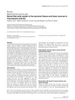
Lens and anterior eyeFigure 1
Lens and anterior eye. Cross sectional diagram of the ante-
rior portion of a developing vertebrate eye, based on a 13-
day embryonic chicken eye section (photomicrograph of San-
dra Ackerley, University of Guelph).
Theoretical Biology and Medical Modelling 2007, 4:22 />Page 3 of 7
(page number not for citation purposes)
Lens cell lineage
If cell migration occurs within the anterior portions of the
lens epithelium, the direction of this migration has not
been conclusively determined. There is some circumstan-
tial support (enumerated below) for transit amplifying
cells of the germinative zone to supply precursors of new
epithelial cells, as well as fiber cells. 1) As organisms age,
the volume of the lens increases through new fiber cell
addition at the lens equator. The growing lens maintains
an epithelial cell monolayer over its expanding anterior
surface area. While individual lens epithelial cells increase
in average size with advancing age, some epithelial cell
division is required to maintain the observed cell coverage
[23]. New cells are needed in particular toward the periph-
ery of the anterior epithelial region. Transit amplifying
cells of the germinative zone are well positioned to fill this
need. 2) Apoptosis of lens epithelial cells has been
observed in normal and cataractous lenses [28,29].
Extrapolation of estimated apoptosis rates and cell divi-
sion rates in the central epithelium suggests that replace-
ment epithelial cells originate toward the lens epithelial
periphery and migrate centripetally. 3) Injury of cells in
the central lens epithelium resulted in increased DNA syn-
thesis within 24 hours in the lens germinative zone. At
later time points (four days), DNA synthesis was also
observed in more central epithelial cells surrounding the
wound [30]. One possible interpretation of these central
epithelium wounding studies is that cells from the germi-
native zone may routinely migrate centripetally to replace
damaged epithelial cells. By analogy, limbal cells are the
recognized source of new corneal epithelial cells, and cen-
tral corneal wounding was demonstrated to stimulate lim-
bal cell proliferation [31-33]. 4) In vitro lens cell
migration studies performed in an electric field provided
indirect support for centripetal migration of lens epithe-
lial cells in vivo [34]. 5) Several other researchers have
proposed centripetal migration of lens epithelial cells
based on their own diverse experimental observations
[35-38].
If transit amplifying cells in the germinative zone provide
replacement cells for the anterior epithelium, then cells of
the germinative zone would possess differentiation poten-
tial for two different lens cell types – epithelial cells and
fiber cells. Individual cells may have the potential to dif-
ferentiate either as epithelial or fiber cells. Alternatively,
two distinct precursor cell populations may reside within
the lens germinative zone.
Lens stem cell hypothesis
While circumstantial evidence implicates the germinative
zone as the source of new cells for lens epithelium as well
as for fiber cells, results from a recent study seem to con-
tradict these ideas. Long term DNA-labeling experiments
demonstrated that central lens epithelial cells retained
label longer than cells in the lens germinative zone [22].
By analogy with stem cell studies in other adult tissues,
the lens cells that retained label for the longest time peri-
ods should include the lens stem cell population. If the
lens is a closed system, then this experimental evidence
suggests that lens stem cells reside in the central epithe-
lium. However, the central lens epithelium lies in the path
of UV radiation, an exposed position for a stem cell pop-
ulation from the standpoint of potential DNA damage.
We propose another possible interpretation for long term
labeling of cells in the central lens epithelium. If lens stem
cells reside outside the capsule, putative lens stem cells
would not have been included in the analyses. The heavily
labeled central epithelial cells could simply represent cells
that had not divided during the course of the experiment,
supporting the view that lens epithelial cells divide very
infrequently [14,29,39,40]. (Mature fiber cells, which are
maintained for life, lose their cell nuclei and hence are not
labeled in long term studies.) Since no heavily labeled
cells in the lens germinative zone were observed after 12
weeks, one can infer that slow cycling lens stem cells do
not reside in the germinative zone. We hypothesize that
lens stem cells reside outside the capsule.
Ciliary body, a possible source of lens stem cells
If the encapsulated lens does not contain its own stem cell
population, we asked where lens stem cells could reside.
The ciliary body is a pigmented and vascularized tissue,
that lies physically close to the lens germinative zone [14-
16,41]. The ciliary body represents the anterior extension
of the choroid, and is situated between the choroid and
the iris. The epithelium of the ciliary body consists of two
cell layers, an inner non-pigmented epithelium, and an
outer pigmented epithelium in intimate contact with cap-
illaries [16]. The ciliary epithelial layers represent anterior
extensions of the inner non-pigmented neural retina and
the outer pigmented retinal epithelium, respectively. (The
terms 'inner' and 'outer' are used in reference to the ocular
globe interior.) A recognized stem cell population – the
retinal stem cells – resides in the ciliary body [42-44].
At early stages of eye development, the presumptive ciliary
body abuts the lens capsule overlying the germinative
zone [41,45,46]. As the eye matures, the ciliary body elab-
orates radial processes, each consisting of the double lay-
ered epithelium surrounding a central capillary.
Extracellular zonular fibrils extend from the posterior cil-
iary body and the valley walls and floor of the ciliary proc-
esses to the equatorial lens capsule, suspending the lens in
the eye orbit. The anterior zonular fibrils insert in the lens
capsule in a ring near the lens germinative zone [47]. In
the primate adult, the inward extensions or 'hills' of the
convoluted ciliary body processes lie within one or two
millimeters of the lens capsular surface overlying the lens
Theoretical Biology and Medical Modelling 2007, 4:22 />Page 4 of 7
(page number not for citation purposes)
germinative zone [16,48]. During accomodation, the cili-
ary process 'hills' can contact the lens capsule [48,49].
If the ciliary body harbors lens stem cells, then cells within
the ciliary body must satisfy two criteria (discussed in
more detail below). 1) Some cells must have the potential
to differentiate into lens fiber cells, and 2) ciliary body cell
progeny must migrate to the lens as lens progenitor cells.
We use the term 'lens progenitor cells' to denote stem cell
progeny that will differentiate into lens epithelial or fiber
cells.
1) In support of lens fiber cell differentiation potential,
ciliary body and other pigmented tissues of the eye have
the capacity to develop lentoids in culture [50-54]. Lento-
ids are groups of cells that express lens fiber cell proteins,
such as crystallins, and exhibit lens fiber cell features, such
as enlarged transparent cytoplasm. We surmise that the
'retinal' stem cell population could include stem cells with
the potential to differentiate into lens.
Another phenomenon – lens regeneration in the newt –
also supports the concept of an extracapsular or extralen-
ticular source of lens progenitor cells. Within a few days
after loss of the ocular lens in adult urodeles, a new lens
begins to emerge from the pigmented iris [55-57]. In both
lentoid formation and lens regeneration, the mechanism
has been attributed to transdifferentiation of pigmented
epithelial cells [56,58]. While we favor ciliary body stem
cells as a potential source of lens progenitor cells, transdif-
ferentiation would be a compatible mechanism.
2) The second criterion for the existence of extracapsular
lens stem cells involves cell migration. During develop-
ment, the presumptive ciliary body abuts the lens capsule
[41,45,46]. Early migrating lens progenitor cells would
have to exit the ciliary body and traverse the immature
lens capsule overlying the lens germinative zone. In the
adult eye, migration of cells from the ciliary body to the
lens would require committed lens progenitor cells to
traverse a short acellular distance of aqueous humor
between the ciliary body and the lens, as well as traverse
the extracellular matrix of the capsule.
Cell migration is an integral part of developmental sys-
tems. In the corneal epithelium for example, limbal stem
cell progeny migrate centripetally to populate the corneal
surface [59-61]. In the case of the lens, extracapsular lens
progenitor cells would need to traverse the aqueous
humor in the vicinity of the zonular fibrils. If prospective
migrating cells require a physical scaffold for migration,
support could be provided by the zonular fibrils, which
reach the lens from the valleys of the convoluted ciliary
processes [47,62,63]. (For example, cell migration occurs
along extracellular matrix fibrils during cardiac develop-
ment [64]).
The lens capsule itself may provide a formidable cell
migration barrier along much of its surface area, however,
entry to the lens capsular interior would need to occur
only in a limited area near the germinative zone. The lens
capsule is not uniform. It differs in thickness and compo-
sition between the anterior and posterior surfaces [65-68].
Additional compositional differences near the germina-
tive zone can be inferred from lectin labeling studies [66].
Zonular fibrils interdigitate into the lens capsule structure
in the vicinity of the germinative zone [62,69,70]. Zonular
fibril tracks might provide lens capsule entry points, as
well as a cell migration substrate. We speculate that extral-
enticular cells could have access to the lens capsule inte-
rior via zonular fibril tracks. We are not aware of
experimental data to support cell migration into a lens
possessing an intact capsule.
Posterior capsule opacification
If the continuity of the lens capsule is breached, however,
extralenticular cell migration into the area delimited by
the lens capsule likely occurs. Cataract extraction disrupts
the lens capsule. Subsequent cell growth and migration
on the remaining capsule lead to complications in 25% of
adult patients (and nearly 100% of pediatric patients) that
again compromise vision [71-73]. These complications,
known as after-cataract or posterior capsule opacification,
are believed to primarily involve proliferation and migra-
tion of lens epithelial cells left behind during cataract sur-
gery [74-77]. There is also evidence that cells originating
in non-lens ocular tissues participate in cell aggregates
within the remaining capsule [78-80].
In posterior capsule opacification, the majority of aber-
rant cell growth is attributed to lens cells originating
within the capsule. However, if our hypothesis is correct
that lens stem cells normally reside outside the lens cap-
sule, then much of this aberrant growth may actually arise
from lens progenitor cells that migrate to the capsule after
the cataract surgery.
Analogies to corneal epithelium
If our lens literature summary seems contrived to explain
an improbable lens stem cell hypothesis, consider the cor-
neal epithelium. Like the lens, the corneal epithelium is a
transparent, avascular ocular tissue, specialized to focus
and transmit light [81]. One major difference between
cornea and lens is that the cornea also provides a protec-
tive surface for the eye. In its protective role at the environ-
ment interface, the corneal epithelium has well developed
tissue replacement capabilities to repair normal wear and
minor injuries [82,83]. In contrast, lens cell division
occurs on a more limited scale.
Theoretical Biology and Medical Modelling 2007, 4:22 />Page 5 of 7
(page number not for citation purposes)
Corneal epithelial stem cells reside in the limbus, a pig-
mented and vascularized tissue that inhabits the periph-
eral boundary of the cornea at its junction with the
conjunctiva [31,33,84-86]. The intact limbus forms a bar-
rier to the migration of cells from the adjacent conjuncti-
val epithelium [87,88]. Committed corneal epithelial
cells originate in the limbus and migrate centripetally
along the basal lamina to populate the basal layer of the
corneal epithelium [61]. In the cornea, basal cells repre-
sent transit amplifying cells, which continue to divide
providing renewed layers of differentiated corneal epithe-
lium [6,89]. Basal corneal epithelial cells rarely, if ever,
beget tumors, despite their ability to replicate their DNA
and divide. Most tumors observed in the cornea originate
in the limbus and grow to impinge on adjacent corneal
tissue [11,90,91].
There are many similarities between the established biol-
ogy of the corneal epithelium, and our hypothesized
source of lens stem cells. Analogous to the cornea, we pro-
pose that lens stem cells reside in a protected location –
the pigmented, vascularized ciliary body. Non-lens cells
do not indiscriminately migrate through the lens capsule.
However, committed lens progenitor cells would need to
migrate to the lens inner capsular surface (basal lamina)
to populate the germinative zone. Transit amplifying cells
in the lens germinative zone would subsequently differen-
tiate and migrate as new fiber cells along the inner surface
of the equatorial capsule. (Other transit amplifying cells
could follow an alternative differentiation pathway and
migrate centripetally as new lens epithelial cells along the
inner surface of the anterior capsule.) Analogous to com-
mitted cells in the basal layer of the corneal epithelium,
cells in the lens germinative zone continue to replicate
their DNA, yet maintain their commitment to lens cell dif-
ferentiation. Lens cells do not naturally develop tumors.
Conclusion
In light of concepts that have evolved in stem cell litera-
ture in recent years, we re-examine the ocular lens in the
context of features common to other biological tissues.
Since the lens grows throughout life and does not natu-
rally develop tumors, we ask whether lens stem cells could
reside in a more typical stem cell niche, one that is pig-
mented and vascularized. We hypothesize that lens stem
cells reside outside the lens capsule in nearby pigmented
ocular tissue, the ciliary body. Here, we present our review
of the lens literature from this novel perspective.
We conclude that a postulated extracapsular source of
ocular lens stem cells is consistent with a large body of lit-
erature. Future experiments on lens development, stem
cell biology, cell migration, and ocular oncology may
shed light on the robustness of these concepts. In the
meantime, we hope that our provocative ideas will stimu-
late discussion in the fields of lens and ocular biology,
and encourage the consideration of experimental results
from multiple perspectives.
Competing interests
The author(s) declare that they have no competing inter-
ests.
Authors' contributions
SGR conceived the hypothesis, researched the literature,
and drafted the manuscript. RAM participated in literature
research and interpretation, refined the ideas, and helped
prepare the manuscript. Both authors read and approved
the final manuscript.
Acknowledgements
J. Daniel Nelson, M.D., for his commitment to scientific inquiry.
References
1. Papaconstantinou J: Molecular aspects of lens cell differentia-
tion. Science 1967, 156:338-346.
2. Maisel H, Harding CV, Alcalá JR, Kuszak JR, Bradley R: The mor-
phology of the lens. In Molecular and Cellular Biology of the Eye Lens
Edited by: Bloemendal H. New York, J Wiley and Sons; 1981:49-84.
3. Piatigorsky J: Lens differentiation in vertebrates. A review of
cellular and molecular features. Differentiation 1981, 19:134-153.
4. Potten CS, Schofield R, Lajtha LG: A comparison of cell replace-
ment in bone marrow, testis and three regions of surface
epithelium. Biochim Biophys Acta 1979, 560:281-299.
5. Watt FM, Hogan BLM: Out of Eden: stem cells and their niches.
Science 2000, 287:1427-1430.
6. Miller SJ, Lavker RM, Sun TT: Keratinocyte stem cells of cornea,
skin and hair follicle: common and distinguishing features.
Sem Dev Biol 1993, 4:217-240.
7. Fuchs E, Tumbar T, Guasch G: Socializing with the neighbors:
stem cells and their niche. Cell 2004, 116:769-778.
8. Cotsarelis G, Sun TT, Lavker RM: Label-retaining cells reside in
the bulge area of pilsebaceous unit: implications for follicular
stem cells, hair cycle, and skin carcinogenesis. Cell 1990,
61:1329-1337.
9. Lavker RM, Sun TT: Epidermal stem cells: properties, markers,
and location. Proc Nat Acad Sci USA 2000, 97:13473-13475.
10. Reya T, Morrison SJ, Clarke MF, Weissman IL: Stem cells, cancer,
and cancer stem cells. Nature 2001, 414:105-111.
11. Miller SJ, Lavker RM, Sun TT: Interpreting epithelial cancer biol-
ogy in the context of stem cells: tumor properties and ther-
apeutic implications. Biochim Biophys Acta 2005, 1756:25-52.
12. Sachs E, Larsen RL: Cancer and the lens. Am J Ophthalmol 1948,
31:
561-564.
13. Seigel GM, Kummer A: The enigma of lenticular oncology. Dig-
ital Journal of Ophthalmology 2002, 7:1-6 [ />].
14. Hanna C, O'Brien JE: Cell production and migration in the epi-
thelial layer of the lens. Arch Ophthalmol 1961, 66:103-107.
15. Mikulicich AG, Young RW: Cell proliferation and displacement
in the lens epithelium of young rats injected with tritiated
thymidine. Invest Ophthalmol 1963, 2:344-354.
16. Streeten BW: Ciliary body. In Ocular Anatomy, Embryology, and Ter-
atology Edited by: Jakobiec FA. Philadelphia, Harper & Row, Publishers;
1982:303-330.
17. Zelenka PS, Gao CY, Rampalli A, Arora J, Chauthaiwale V, He HY:
Cell cycle regulation in the lens: proliferation, quiescence,
apoptosis and differentiation. Prog Retin Eye Res 1997,
16:303-322.
18. Hogan MJ, Alvarado JA, Weddell JE: Lens. In Histology of the Human
Eye: An Atlas and Textbook Philadelphia, W B Saunders; 1971:638-677.
19. Worgul BV: Lens. In Ocular Anatomy, Embryology, and Teratology
Edited by: Jakobiec FA. Philadelphia: Harper & Row; 1982:355-389.
Theoretical Biology and Medical Modelling 2007, 4:22 />Page 6 of 7
(page number not for citation purposes)
20. Pearsons BJ, Modak SP: The pattern of DNA synthesis in the
lens epithelium and the annular pad during development and
growth of the chick lens. Exp Eye Res 1970, 9:144-151.
21. Rafferty NS, Rafferty KA: Cell population kinetics of the mouse
lens epithelium. J Cell Physiol 1981, 107:309-315.
22. Zhou M, Lieberman J, Xu J, Lavker RM: A hierarchy of prolifera-
tive cells exists in mouse lens epithelium: implications for
lens maintenance. Invest Ophthalmol Vis Sci 2006, 47:2997-3003.
23. Kuszak JR: A re-examination of primate lens epithelial cell
size, density and structure as a function of development,
growth and age. Nova Acta Leopoldina 1997, 75:45-66.
24. Sliney DH: How light reaches the eye and its components. Int
J Toxicol 2002, 21:501-509.
25. Taylor HR, West SK, Rosenthal FS, Muñoz B, Newland HS, Abbey H,
Emmett EA: Effect of ultraviolet radiation on cataract forma-
tion. N Engl J Med 1988, 319:1429-1433.
26. Michael R, Vrensen GFJM, van Marle J, Löfgren S, Söderberg PG:
Repair in the rat lens after threshold ultraviolet radiation
injury. Invest Ophthalmol Vis Sci 2000, 41:204-212.
27. Bhat SP: The ocular lens epithelium. Biosci Rep 2001, 21:537-563.
28. Ishizaki Y, Voyvodic JT, Burne JF, Raff MC: Control of lens epithe-
lial cell survival. J Cell Biol 1993, 121:899-908.
29. Li WC, Kuszak JR, Dunn K, Wang RR, Ma W, Wang GM, Spector A,
Leib M, Cotliar AM, Weiss M, Espy J, Howard G, Farris RL, Auran J,
Donn A, Hofeldt A, Mackay C, Merriam J, Mittl R, Smith TR: Lens
epithelial cell apoptosis appears to be a common cellular
basis for non-congenital cataract development in humans
and animals. J Cell Biol 1995, 130:169-181.
30. Rakic JM, Galand A, Vrensen GFJM: Separation of fibres from the
capsule enhances mitotic activity of human lens epithelium.
Exp Eye Res 1997, 64:67-72.
31. Cotsarelis G, Cheng SZ, Dong G, Sun TT, Lavker RM: Existence of
slow-cycling limbal epithelial basal cells that can be preferen-
tially stimulated to proliferate: implications on epithelial
stem cells. Cell 1989, 57:201-209.
32. Tseng SCG: Regulation and clinical implications of corneal epi-
thelial stem cells. Mol Biol Rep 1996, 23:47-58.
33. Lehrer MS, Sun TT, Lavker RM: Strategies of epithelial repair:
modulation of stem cell and transit amplifying cell prolifera-
tion. J Cell Sci 1998, 111:2867-2875.
34. Wang E, Zhao M, Forrester JV, McCaig CD: Bi-directional migra-
tion of lens epithelial cells in a physiological electrical field.
Exp Eye Res 2003, 76:29-37.
35. Coulombre JL, Coulombre AJ: Lens development: fiber elonga-
tion and lens orientation. Science 1963, 142:1489-1490.
36. Philpott GW, Coulombre AJ: Lens development. II. The differ-
entiation of embryonic chick lens epithelial cells in vitro and
in vivo. Exp Cell Res 1965, 38:635-644.
37. Genis-Galvez JM, Santos-Gutierrez L, Rios-Gonzales A: Causal fac-
tors in corneal development: an experimental analysis in the
chick embryo. Exp Eye Res 1967, 6:48-56.
38. Kuszak JR, Costello MJ: The structure of the vertebrate lens. In
Development of the ocular lens Edited by: Lovicu FJ and Robinson ML.
Cambridge, UK, Cambridge University Press; 2004:71-118.
39. Hanna C, Bicknell DS, O'Brien JE: Cell turnover in the adult
human eye. Arch Ophthalmol 1961, 65:695-698.
40. Harocopos GJ, Alvares KM, Kolker AE, Beebe DC: Human age-
related cataract and lens epithelial cell death. Invest Ophthal-
mol Vis Sci 1998, 39:2696-2706.
41. Napier HRL, Kidson SH: Proliferation and cell shape changes
during ciliary body morphogenesis in the mouse. Dev Dyn
2005, 233:213-223.
42. Ahmad I, Tang L, Pham H:
Identification of neural progenitors in
the adult mammalian eye. Biochem Biophys Res Comm 2000,
270:517-521.
43. Fischer AJ, Reh TA: Identification of a proliferating marginal
zone of retinal progenitors in postnatal chickens. Dev Biol
2000, 220:197-210.
44. Tropepe V, Coles BL, Chiasson BJ, Horsford DJ, Elia AJ, McInnes RR,
van der Kooy D: Retinal stem cells in the adult mammalian
eye. Science 2000, 287:2032-2036.
45. Coulombre AJ, Coulombre JL: The role of intraocular pressure
in the development of the chick eye: III. Ciliary body. Am J
Ophthalmol 1957, 44:85-93.
46. Beebe DC: Development of the ciliary body: a brief review.
Trans Ophthalmol Soc U K 1986, 105:123-130.
47. Streeten BW: Zonular apparatus. In Ocular Anatomy, Embryology,
and Teratology Edited by: Jakobiec FA. Philadelphia, Harper & Row,
Publishers; 1982:331-353.
48. Croft MA, Kaufman PL, Crawford KS, Neider MW, Glasser A, Bito
LZ: Accommodation dynamics in aging rhesus monkeys. Am
J Physiol 1998, 275:R1885-1897.
49. Croft MA, Glasser A, Heatley G, McDonald J, Ebbert T, Dahl DB,
Nadkarni NV, Kaufman PL: Accomodative ciliary body and lens
function in rhesus monkeys, I: normal lens, zonule and ciliary
body process configuration in the iridectomized eye. Invest
Ophthalmol Vis Sci 2006, 47:1076-1086.
50. Moscona A: Formation of lentoids by dissociated retinal cells
of the chick embryo. Science 1957, 125:598-599.
51. Eguchi G, Okada TS: Differentiation of lens tissue from the
progeny of chick retinal pigment cells cultured in vitro: a
demonstration of a switch of cell types in clonal cell culture.
Proc Nat Acad Sci USA 1973, 70:1495-1499.
52. Eguchi G, Abe SI, Watanabe K: Differentiation of lens-like struc-
tures from newt iris epithelial cells in vitro. Proc Nat Acad Sci
USA 1974,
71:5052-5056.
53. Okada TS, Itoh Y, Watanabe K, Eguchi G: Differentiation of lens
in cultures of neural retinal cells of chick embryos. Dev Biol
1975, 45:318-329.
54. Yasuda K, Okada TS, Eguchi G, Hayashi M: A demonstration of a
switch of cell type in human fetal eye tissues in vitro: pig-
mented cells of the iris or the retina can transdifferentiate
into lens. Exp Eye Res 1978, 26:591-595.
55. Stone LS: The regeneration of the crystalline lens. Invest Oph-
thalmol 1965, 4:420-432.
56. Tsonis PA, Rio-Tsonis KD: Lens and retina regeneration:
transdifferentiation, stem cells and clinical applications. Exp
Eye Res 2004, 78:161-172.
57. Grogg MW, Call MK, Okamoto M, Vergara MN, Rio-Tsonis KD,
Tsonis PA: BMP inhibition-driven regulation of six-3 underlies
induction of newt lens regeneration. Nature 2005, 438:858-862.
58. Eguchi G, Kodama R: Transdifferentiation. Curr Opin Cell Biol 1993,
5:1023-1028.
59. Kinoshita S, Friend J, Thoft RA: Sex chromatin of donor corneal
epithelium in rabbits. Invest Ophthalmol Vis Sci 1981, 21:434-441.
60. Buck RC: Measurement of centripetal migration of normal
corneal epithelial cells in the mouse. Invest Ophthalmol Vis Sci
1985, 26:1296-1299.
61. Collinson JM, Morris L, Reid AI, Ramaesh T, Keighren MA, Flockhart
JH, Hill RE, Tan SS, Ramaesh K, Dhillon B, West JD: Clonal analysis
of patterns of growth, stem cell activity, and cell movement
during the development and maintenance of the murine cor-
neal epithelium. Dev Dyn 2002, 224:432-440.
62. Raviola G: The fine structure of the ciliary zonule and ciliary
epithelium with special regard to the organization and inser-
tion of the zonular fibrils. Invest Ophthalmol 1971, 10:851-869.
63. Rohen JW: Scanning electron microscope studies of the zonu-
lar apparatus in human and monkey eyes. Invest Ophthalmol Vis
Sci 1979, 18:133-144.
64. Markwald RR, Fitzharris TP, Bolender DL, Bernanke DH: Structural
analysis of cell:matrix association during the morphogenesis
of atrioventricular cushion tissue. Dev Biol 1979, 69:634-654.
65. Fukushi S, Spiro RG: The lens Capsule: sugar and amino acid
composition. J Biol Chem 1969, 244:2041-2048.
66. Yao R, Alcala J, Maisel H: Developmental changes in glycoconju-
gate composition during chick lens morphogenesis. Exp Eye
Res 1996, 62:419-431.
67. Ziebarth NM, Manns F, Uhlhorn SR, Venkatraman AS, Parel JM: Non-
contact optical measurement of lens capsule thickness in
human, monkey, and rabbit postmortem eyes. Invest Ophthal-
mol Vis Sci 2005, 46:1690-1697.
68. Barraquer RI, Michael R, Abreu R, Lamarca J, Tresserra F: Human
lens capsule thickness as a function of age and location along
the sagittal lens perimeter. Invest Ophthalmol Vis Sci 2006,
47:2053-2060.
69. Cohen AI: The electron microscopy of the normal human
lens. Invest Ophthalmol 1965, 4:433-446.
70. Streeten BW: The zonular insertion: a scanning electron
microscopic study. Invest Ophthalmol Vis Sci 1977, 16:364-375.
71. Schaumberg DA, Dana MR, Christen WG, Glynn RJ: A systematic
overview of the incidence of posterior capsule opacification.
Ophthalmology 1998, 105:1213-1221.
Publish with BioMed Central and every
scientist can read your work free of charge
"BioMed Central will be the most significant development for
disseminating the results of biomedical researc h in our lifetime."
Sir Paul Nurse, Cancer Research UK
Your research papers will be:
available free of charge to the entire biomedical community
peer reviewed and published immediately upon acceptance
cited in PubMed and archived on PubMed Central
yours — you keep the copyright
Submit your manuscript here:
/>BioMedcentral
Theoretical Biology and Medical Modelling 2007, 4:22 />Page 7 of 7
(page number not for citation purposes)
72. Sharma N, Pushker N, Dada T, Vajpayee RB, Dada VK: Complica-
tions of pediatric cataract surgery and intraocular lens
implantation. J Cataract Refract Surg 1999, 25:1585-1588.
73. Robb RM: Congenital and childhood cataracts. In Principles and
Practice of Ophthalmology Volume 5. 2nd edition. Edited by: Albert DM
and Jakobiec FA. Philadelphia, W B Saunders; 2000:4399-4405.
74. McDonnell PJ, Zarbin MA, Green R: Posterior capsule opacifica-
tion in pseudophakic eyes. Ophthalmology 1983, 90:1548-1553.
75. Apple DJ, Solomon KD, Tetz MR, Assia EI, Holland EY, Legler UFC,
Tsai JC, Casteneda VE, Hoggatt JP, Kostick AMP: Posterior capsule
opacification. Surv Ophthalmol 1992, 37:73-116.
76. Marcantonio JM, Rakic JM, Vrensen GFJM, Duncan G: Lens cell pop-
ulations studied in human donor capsular bags with
implanted intraocular lenses. Invest Ophthalmol Vis Sci 2000,
41:1130-1141.
77. Wormstone M: Posterior capsule opacification: a cell biologi-
cal perspective. Exp Eye Res 2002, 74:337-347.
78. Odrich MG, Hall SJ, Worgul BV, Trokel SL, Rini FJ: Posterior cap-
sule opacification: experimental analyses. Ophthalmic Res 1985,
17:75-84.
79. Kappelhof JP, Vrensen GF: The pathology of after-cataract: a
minireview. Acta Ophthalmol Suppl 1992:13-24.
80. Rafferty NS, Rafferty KA: Lens cytoskeleton and after-cataract.
Acta Ophthalmol Suppl 1992:34-45.
81. Hogan MJ, Alvarado JA, Weddell JE: The cornea. In Histology of the
Human Eye: An Atlas and Textbook Philadelphia, W B Saunders;
1971:55-111.
82. Dua HS, Gomes JAP, Singh A: Corneal epithelial wound healing.
Br J Opthalmol 1994, 78(5):401-8.
83. Lu L, Reinach PS, Kao WW: Corneal epithelial wound healing.
Exp Biol Med (Maywood) 2001, 226:653-664.
84. Schermer A, Galvin S, Sun TT: Differentiation-related expres-
sion of a major 64K corneal keratin in vivo and in culture sug-
gests limbal location of corneal epithelial stem cells. J Cell Biol
1986,
103:49-62.
85. Kenyon KR, Tseng SCG: Limbal autograft transplantation for
ocular surface disorders. Ophthalmology 1989, 96:709-723.
86. Lavker RM, Tseng SCG, Sun TT: Corneal epithelial stem cells at
the limbus: looking at some old problems from a new angle.
Exp Eye Res 2004, 78:433-446.
87. Thoft RA, Wiley LA, Sundarraj N: The multipotential cells of the
limbus. Eye 1989, 3:109-113.
88. Kruse FE, Chen JJY, Tsai RJF, Tseng SCG: Conjunctival transdiffer-
entiation is due to the incomplete removal of limbal basal
epithelium. Invest Ophthalmol Vis Sci 1990, 31:1903-1913.
89. German MJ, Pollock HM, Zhao B, Tobin MJ, Hammiche A, Bentley A,
Cooper LJ, Martin FL, Fullwood NJ: Characterization of putative
stem cell populations in the cornea using synchrotron infra-
red microspectroscopy. Invest Ophthalmol Vis Sci 2006,
47:2417-2422.
90. Waring GO III, Roth AM, Ekins MB: Clinical and pathologic
description of 17 cases of corneal intraepithelial neoplasia.
Am J Ophthalmol 1984, 97:547-559.
91. Farah S, Baum TD, Conlon MR, Alfonso EC, Starck T, Albert DM:
Tumors of the cornea and conjunctiva. In Principles and Practice
of Ophthalmology Volume 2. 2nd edition. Edited by: Albert DM and
Jakobiec FA. Philadelphia, W B Saunders; 2000:1002-1019.