HANDBOOK OF CARDIAC PACING – PART 9 ppsx
Bạn đang xem bản rút gọn của tài liệu. Xem và tải ngay bản đầy đủ của tài liệu tại đây (475.78 KB, 16 trang )
120 Handbook of Cardiac Pacing
13
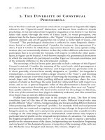
Fig. 13.11. Near and far field signals. The first group of complexes are recorded between the distal shock
coil and the defibrillator case. These are referred to as “far field” because they are distant to the intraven-
tricular sensing electrodes. The second group of complexes are recorded from the sensing electrodes within
the right ventricle. These are referred to as “near field”.
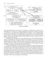
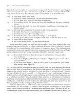
Fig. 13.12. Atrial and ventricular intracardiac signals. This is a tracing from a patient in ventricular tachy-
cardia. The top tracing is an intracardiac atrial electrogram with a sinus rate of around 105 bpm. The
bottom trace is an intracardiac ventricular electrogram showing a rate of 200 bpm. This is classic AV
dissociation, with the ventricle going faster than the atrium and is diagnostic of ventricular tachycardia. It
rules out atrial fibrillation, atrial flutter or supraventricular tachycardia as the cause for the rapid ven-
tricular rate.
121Basic Concepts of Implantable Cardioverter Defibrillators
13
MAGNET RESPONSE OF THE ICD
ICDs have a very different response to magnet application than do pacemak-
ers. In addition, the effect of sustained magnet application may differ from one
device to another and may even be programmable as to the effect of the magnet.
There is one feature that is constant across all ICDs with magnet application which
is the suspension of tachycardia therapy. If a patient is receiving inappropriate
shocks or if for any reason suspending therapy becomes necessary, placing a ring
type pacemaker magnet over the ICD will immediately prevent further therapy
from being delivered. Some devices can be programmed to the “off” setting by
keeping the magnet in place for 30 seconds. The latter type of device can be turned
back “on” by placing the magnet over it for 30 seconds as well. Others will imme-
diately resume detection and therapy when the magnet is removed no matter how
long the magnet has been applied. This is a useful feature when a programmer is
not available. Though the tachycardia therapy is disabled by the magnet, the brady-
cardia backup pacing remains active. Thus, if the patient has no heart rhythm
without the backup pacing of the defibrillator, there will still be rhythm support
during magnet application. To make things a bit more confusing, some ICDs have
a programmable magnet feature. This allows the magnet effect to be ignored by
the device.
RECOMMENDED REPLACEMENT TIME
Unlike pacemakers where the magnet rate signals the need for device replace-
ment there is no simple method to determine battery life on an ICD. At least two
manufacturers allow the programming of a beeping tone that sounds regularly
when the battery gets low. For most devices the routine follow-up in the clinic
provides telemetry that indicates the battery voltage and the time it takes to charge
the capacitors to their full voltage. When either one of these gets to a specified
value, then it is time to replace the device. Most ICDs can now be expected to last
between 4 to 7 years in normal use.
122 Handbook of Cardiac Pacing
14
Handbook of Cardiac Pacing, by Charles J. Love. © 1998 Landes Bioscience
Indications for Implantable
Cardioverter Defibrillators
Class I: General Agreement that an ICD Is Indicated 122
Class II: Some Disagreement as to the Necessity for Implant 123
Class III 123
Additional Issues for ICD Insertion 123
Implantable defibrillators represent a quantum leap in our ability to prevent
recurrent sudden cardiac death due to ventricular dysrhythmias. These devices
recognize rapid heart rates and are capable of delivering overdrive pacing,
cardioversion, or defibrillation therapy. The original implants were performed
using a thoracotomy. Newer systems are placed in a transvenous manner allow-
ing patients to leave the hospital after 24 hours. The effectiveness of these devices
exceeds 95% over several years. In general, an ICD should be used when a patient
is at high risk for recurrent life threatening arrhythmias when no other effective
therapy is available, reliable, or tolerated to prevent a sudden death event. Situa-
tions that are preventable such as digitalis toxicity with hypokalemia do not jus-
tify ICD insertion. The current guidelines as published by the American College
of Cardiology and the American Heart Association are as follows:
CLASS I: GENERAL AGREEMENT THAT AN ICD IS INDICATED
Primary VT or VF not due to a transient or reversible cause (drug toxicity,
acute myocardial infarction, electrolyte disturbance, etc.)
Spontaneous sustained VT
Syncope of uncertain cause with inducible poorly tolerated sustained ven-
tricular tachycardia or ventricular fibrillation (clinically relevant) at EP
study in a patient whom no effective, tolerated, or preferred drug is found
during testing.
Spontaneous nonsustained VT in a patient post myocardial infarction when
inducible VT or VF is found that is not suppressed by a class-1 antiar-
rhythmic drug
123Indications for Implantable Cardioverter Defibrillators
14
CLASS II: SOME DISAGREEMENT AS TO THE NECESSITY
FOR IMPLANT
A: WEIGHT OF EVIDENCE IN FAVOR OF EFFICACY
None
B: EFFICACY LESS WELL ESTABLISHED BY WEIGHT OF EVIDENCE
Cardiac arrest due to VF when electrophysiologic testing is not possible
due to medical reasons
Symptomatic VT or VF in a patient awaiting cardiac transplantation
Familial or inherited conditions that place the patient at high risk (e.g. long
QT syndrome or hypertrophic myopathy)
Spontaneous nonsustained VT in a patient post myocardial infarction when
inducible VT or VF is found during electrophysiology study
Recurrent syncope of uncertain cause in the presence of LV dysfunction,
inducible VT or VF at electophysiology study, and no other cause of
syncope is found
CLASS III
Recurrent syncope of uncertain cause in a patient without inducible ven-
tricular tachycardia or ventricular fibrillation
Incessant ventricular tachycardia or ventricular fibrillation
Ve ntricular tachycardia or ventricular fibrillation due to a reversible cause
such as drug, metabolic or ischemic conditions
VT due to an arrhythmia amenable to catheter ablation or surgical therapy
Significant psychiatric illness that may prevent proper follow-up of the
device, or which may be adversely affected by a device implant
Te rminal illness or life expectancy less than 6 months
Patients with LV dysfunction, prolonged QRS, absence of spontaneous or
inducible VT or VF, who are undergoing coronary bypass surgery
NYHA class IV in patients that are not candidates for heart transplantation
ADDITIONAL ISSUES FOR ICD INSERTION
Patient life expectancy should be greater than 6 months
The patient should be emotionally stable
The patient should be willing and able to cooperate in follow-up
Heart failure greater than NYHA Class III should contraindicate an ICD
implant unless it is being used as a “bridge” to heart transplantation.
124 Handbook of Cardiac Pacing
14
Recently the results of a multicenter trial known as MADIT were published.
This was the Multicenter Automatic Defibrillator Implant Trial. The trial was
based on the knowledge that patients with recent myocardial infarction, residual
ejection fraction of 35% or less, and spontaneous non-sustained ventricular ta-
chycardia were at high risk for sudden arrhythmic death. The patients were
evaluated by electrophysiolgy study. Those that had inducible but non suppressed
ventricular tachycardia had a significantly better survival if they were treated pro-
phylactically with an ICD as opposed to medical therapy. This prophylactic rea-
son for implantation of an ICD has recently been adapted as an indication. The
evidence is quite strong that this well defined group of patients will benefit from
having an ICD implanted.
125Preop to Postop Considerations for ICDs
15
Handbook of Cardiac Pacing, by Charles J. Love. © 1998 Landes Bioscience
Preoperative, Operative
and Postoperative Considerations
for Implantable Cardioverter
Defibrillators
Introduction 125
Preoperative Patient Issues 125
Surgical Considerations 126
Predischarge Questions and Issues 127
Emergency Care of Patients with an ICD 128
INTRODUCTION
Implantion of ICDs has become much easier with the advent of the smaller,
active can, biphasic waveform devices combined with efficient defibrillation lead
systems. As previously noted, ICD implants originally required an open chest pro-
cedure or at the least a subxiphoid approach to enter the pericardium and place
patches and screw-in leads directly onto the myocardium. The preoperative and
postoperative issues for this type of procedure were far more involved as was the
recovery time for the patient. Mortality and morbidity rates were significant from
the longer and more invasive approach. As greater than 99% of ICDs are implanted
in a subcutaneous manner with transvenous leads, the following discussion will
be focused on this less invasive approach.
PREOPERATIVE PATIENT ISSUES
We approach the preoperative management of the patient who is about to
receive an ICD in much the same way as we do the patient who is going to receive
a pacemaker (see chapter 10). The patient is first given information about the ICD
and the reason the device was recommended. It is very important to stress that the
ICD will NOT prevent the tachycardia. This is a common patient misconception.
It must be clear that the device is meant only to correct the arrhythmia once it
occurs. Though many patients may be able to reduce or discontinue their
antiarrhythmia medication, some patients may still require drug therapy to re-
duce the frequency of tachycardia. The patient should be allowed to see and hold
a model of the ICD. This helps provide a perspective of what the incision and
126 Handbook of Cardiac Pacing
15
physical appearance of the implant area will look like. A description of the lead
system and the operation is provided. The fact that the patient will have the ICD
tested several times during the procedure should be discussed. The possibility that
death may occur should be raised with an emphasis on the benefits of the proce-
dure relative to the risks. Due to the size of the ICD and the lack of true surgical
training of some physicians who implant them, infections are more frequent than
with pacemakers. The larger size of the device may compromise blood flow to the
adjacent tissues creating local ischemia. This may result in an erosion or predispo-
sition to infection.
The preparation of the patient is identical to that for a permanent pacemaker.
However, some physicians prefer the use of general anesthesia for ICD implant.
This may be a preference for all patients or only for selected cases where the pa-
tient may require more aggressive sedation. In some cases the patient may be at
very high risk due to compromised cardiovascular or pulmonary disease. Cases
such as these should be done with prior consultation of an anesthesiologist. It is
important to be sure that this consultant understands the nature of the proce-
dure. We have had cases where lidocaine was administered by the anesthetist when
recurrent ventricular ectopy was observed during our attempts to induce ven-
tricular fibrillation. It is somewhat difficult for an anesthesiologist who spends his
time trying to keep the patient out of harm’s way to accept the intentional acts of
the electrophysiologist who wants to fibrillate the ventricle.
SURGICAL CONSIDERATIONS
The implant has been greatly simplified with the advent of active can, biphasic,
small defibrillators. Patch placement directly on the myocardium or on the peri-
cardium via median sternotomy, left lateral thoracotomy, subxiphoid or subcostal
approaches has been virtually eliminated. Single transvenous lead implants are
successful 95-98% of the time without the use of subcutaneous patches, subcuta-
neous arrays or additional transvenous leads.
In the operating room the following sequence of events takes place. First all of
the equipment is checked for proper operation. The patient is sedated and the
lead is implanted. The lead is tested for routine pacing capture and sensing thresh-
olds. A pocket is made for the ICD which is attached to the lead. A low energy
shock is delivered through the high voltage ends to verify the electrical connec-
tions. The patient is then further sedated if necessary and ventricular fibrillation
is induced. A simple method commonly used is to choose a moderate shock en-
ergy level around 15 to 20 joules. If the patient can be converted twice with this
amount of energy then the incision is closed. Other physicians prefer to deter-
mine the actual defibrillation threshold (DFT). This may be done by repeatedly
fibrillating the patient and reducing the shock energy to a level that no longer
defibrillates the patient. The patient is then rescued internally or externally. Un-
127Preop to Postop Considerations for ICDs
15
less we are doing an investigational lead or ICD that requires testing of this type,
we prefer the rapid method. The patient may be returned to a telemetry unit and
may be discharged the following day if the postoperative evaluation and chest
x-ray are acceptable.
Additional postoperative patient care will be dictated by the surgical approach.
In some cases, where a thoracotomy is performed a chest tube will be present. The
cardiovascular status should be monitored closely, especially in patients who have
undergone extensive DFT testing. Pulmonary care, early mobilization and emo-
tional support may also be necessary.
PREDISCHARGE QUESTIONS AND ISSUES
Virtually all of the postoperative issues addressed in the pacing section apply
to patients with an ICD implant. However there is one subject that will consis-
tently cause the most anxiety for both the patient and the physician: driving. There
are several issues that must be addressed regarding the operation of motor ve-
hicles. The first consideration is what the law requires. This is very confusing and
will vary significantly from state to state. Some states have no requirements while
others may have mandatory license suspensions for up to two years following an
arrhythmic event resulting in a loss of consciousness. In some states the physician
is obligated to notify the Bureau of Motor Vehicles, and in others the physician is
forbidden from doing so to prevent an invasion of the patient’s right to privacy.
The approach that I use is practical when the law is unclear or does not cover
these situations. If the patient has had an arrhythmic event where loss of control
or syncope has occurred they are to abstain from driving for 6 months. If no fur-
ther events occur in this period of time they may drive again unless they are deemed
to be at excessive risk. Otherwise I permit them to resume driving in 4 weeks.
These cautions also apply to the use of machinery, lawn mowing equipment and
working on heights. Some patients will ask if they can “just drive around town but
stay off of the highway.” My answer to that is that my children play in the front
yard. The patient is more likely to injure someone else or have a property damage
accident in the city. The abstinence from driving is consistently the most difficult
part of the patient’s situation with which the physician or nurse must deal. In our
clinic we make a point of telling the patient that it is not the ICD that is preventing
them from driving. It is the fact that they have a cardiac arrhythmia. This removes
inappropriate anger directed at the ICD and allows the patient to accept the de-
vice more easily. It is often useful to compare the arrhythmia patient to the patient
with epilepsy. Seizure patients also have to deal with periodic loss of conscious-
ness and driving restrictions related to their disease.
128 Handbook of Cardiac Pacing
15
EMERGENCY CARE OF PATIENTS WITH AN ICD
Standard emergency procedures should always be initiated for ICD patients in
ventricular tachycardia or fibrillation. This includes the initiation of standard Basic
Cardiac Life Support and Advance Cardiac Life Support procedures as indicated.
If initial anterior and lateral external electrical countershock is not successful, re-
positioning the paddles to the anterior and posterior position may be helpful. If
in physical contact with patient when the ICD discharges one may feel a slight
tingle. Though this may be felt by the rescuer it will not be harmful. If rubber or
latex gloves are worn then no electrical current will be felt by the rescuer.
129Evaluation of Defibrillator Malfunction
16
Handbook of Cardiac Pacing, by Charles J. Love. © 1998 Landes Bioscience
Evaluation of Defibrillator
Malfunction
Evalution of Defibrillator Malfunction 129
Failure to Shock or Deliver Anti-Tachycardia Pacing 130
Failure to Convert Arrhythmia 133
Inappropriate Delivery of Therapy 135
Conclusion 136
EVALUTION OF DEFIBRILLATOR MALFUNCTION
When an ICD malfunctions or repeated shocks are delivered to the patient, a
life threatening or very uncomfortable situation may occur. Failure of an ICD to
deliver a shock when it should can result in death or prolonged episodes of ven-
tricular tachycardia. Delivering inappropriate shocks to the patient is very painful
and can possibly induce an arrhythmia. As clinicians we most often see patients
due to inappropriate shocks. This is an obvious problem for the patient, and they
will call the clinic or come to the emergency department. If a device fails to shock
we would at best see the patient due to a sustained arrhythmia. The worst scenario
would be failure to shock ventricular fibrillation with subsequent death of the
patient. In the latter situation we might not even know of the death until the
patient fails to show for the next clinic visit.
Since virtually all of the ICDs available in the market now have anti-bradycar-
dia pacing capability they are subject to virtually all of the same problems as pace-
makers. Please refer to the chapter on evaluation of pacemaker malfunction for a
review of these issues. The same leads that provide pacing and sensing functions
for the bradycardia section of the ICD also provide the sensing function for the
tachycardia detection. In the case of “integrated bipolar” systems, one of the shock-
ing electrodes functions as the sensing and pacing anode as well. Although it is
possible to evaluate the impedance of the pacing portion of the system in a
noninvasive and painless manner, we are not yet able to do the same for the high
voltage portion of the circuit. Most ICDs will measure the high voltage imped-
ance when a shock is delivered. To do this requires that the patient have had a
spontaneous device discharge. If this has not occurred, a manual discharge may
be delivered. Most patients are not too willing to have this done on a routine basis.
This makes evaluation of the entire system difficult during a routine clinic
follow-up.
130 Handbook of Cardiac Pacing
16
The approach to the patient with a suspected ICD malfunction is essentially
the same as the approach to the pacemaker patient. Gathering the basic data con-
cerning the ICD, patient disease, indications for the device implant, and circum-
stances surrounding the incident in question are all essential. Interrogation of the
ICD with all of the available telemetry is also required. Many times the issue is
whether or not a device discharge was appropriate or not. On devices with limited
diagnostic capability the history surrounding the shock is crucial. If the patient
felt palpitations, lightheaded, short of breath or had syncope, then the discharge
was likely delivered for the right reasons. However, the absence of these symptoms
does not mean that the shock was not proper. In many cases the patient may be
sitting or may be supine and is not sufficiently hypotensive for a long enough
period of time to become symptomatic. In other cases the patient may simply not
remember the event since they were not perfusing their brain at the time. If the
ICD detected and treated the arrhythmia quickly there may have been insufficient
time for symptoms to occur. Occasionally the patient may even have been sleep-
ing at the time of the arrhythmia. Indeed, nocturnal myoclonus is frequently mis-
interpreted by the patient’s spouse as a device discharge.
The most common problem in our clinic is not really an ICD problem but a
response to the arrhythmia atrial fibrillation. If atrial fibrillation occurs with a
rapid ventricular response that exceeds the detection rate of the device, the ICD
will charge and shock the patient. It may even do so numerous times. Occasion-
ally the shock will convert the atrial fibrillation to sinus rhythm; however, this
does not happen on a regular basis. These shocks do not represent a device mal-
function, though the situation is best described as an undesirable patient-device
interaction.
The specific categories of device malfunction are noted below. Suggestions as
to how to manage these problems follow each section.
FAILURE TO SHOCK OR DELIVER ANTI-TACHYCARDIA PACING
The failure of an ICD to deliver anti-tachycardia therapy can be lethal. The
reasons for failure to shock are listed in Table 16.1. The two most common rea-
sons for failure to deliver therapy that we see are lead failure and a detection rate
that is too high. Unfortunately, lead failures are not uncommon. Some types of
failure are visible on a X-ray but many are not. The fracture may occur on one of
the inner conductors of a coaxial or triaxial lead. If this happens the outer con-
ductor may shield the inner conductor from being seen on X-ray. Other fractures
may occur that result in the two broken ends remaining in contact at times and
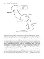
being apart at other times. These are also difficult to see by radiography. Figure 16.1
shows failure of epicardial patches with the conductor broken where it meets the
Fig. 16.1. (opposite page) Radiograph of a patch failure. Epicardial screw in leads are noted as well as the
patches. Note the discontinuity between the lead and the patch (arrows). This system was nonfunctional
and required replacement with a transvenous system.
131Evaluation of Defibrillator Malfunction
16
Ta b le 16.1. Failure to shock
Undersensing
Lead malposition
Lead dislodgment
Lead perforation
Lead fracture
Lead insulation failure
Lead to device connector problem
Sensitivity set too low (i.e., insensitive)
Poor electrogram amplitude due to change in myocardial substrate
Myocardial infarction
Drug therapy
Metabolic imbalance
“Fine” ventricular fibrillation
Primary circuit failure
Battery failure
Shock therapy turned off (by programming or magnet)
Magnet placed over the device
Strong magnetic field present
Detection rate set too high
Failure to meet additional detection criteria
Rate stability
Sudden onset
Morphology criteria
Slowing of tachycardia below detection rate
Electrolyte changes
Drug therapy changes
Interaction with permanent pacemaker
↑
↑
132 Handbook of Cardiac Pacing
16
patch. This is a common site of patch failure and should be looked for when evalu-
ating the X-ray. Several views may be required to visualize the failure. An
overpenetrated film is better for seeing wire failures. We find it best to ask the
radiology department to use a “thoracic spine technique” when determining the
exposure settings on our chest films.
If the undersensing problem develops within 30 days after implant then lead
malposition, dislodgment or myocardial perforation should be suspected. Frac-
tures and insulation failures are more likely to occur after one or more years. As
the transvenous defibrillation leads, are substantially thicker than pacing leads
they are exposed to more forces under the clavicle when placed by the popular
subclavian access technique. This is the area to pay special attention to when re-
viewing the x-ray. Observation of the connector pins within the connector block
of the ICD will reveal an obvious loose connection. Though most ICDs are very
reliable, there have been several alerts on different devices. Failures of circuitry,
lock up of software and other problems are known to occur. Interrogation of the
ICD will usually fail if any of these situations are present. If the patient does not
have routine clinic evaluations, then the battery may become depleted and the
device will become either nonfunctional or will not have sufficient power to charge
the capacitors to the required voltage for discharge. Occasionally the detection
rate is simply set too high. This occurs most commonly after a new drug such as
amiodarone or sotalol has been started, but may also be the result of inappropri-
ate programming. The result of the drug therapy may be a slowing of the ven-
tricular tachycardia rate such that it falls below the programmed detection rate.
Significant metabolic or electrolyte abnormalities may affect not only the tachy-
cardia rate, but also the amplitude of the signals resulting in undersensing or fail-
ure to detect. As noted previously, the addition of extra detection criteria may
delay or prevent the ICD from delivering therapy. Use of these modifiers must be
done cautiously. Occasionally the patient will sustain a myocardial infarction that
will result in a significant change of the intracardiac electrogram. The new
electogram may not be a sufficient signal to sense for the purpose of detection.
One might also see asynchronous pacing if the bradycardia backup pacing is turned
on. Be aware that a patient in close contact with a magnet might have the device
deactivated if this feature is present. We have a patient who carried a stereo speaker
(they have big magnets inside) and deactivated his ICD.
Though many patients have ICDs that are capable of providing backup pacing
support, frequent use of this feature may result in a significant reduction in device
longevity. Thus, patients with ICDs often have a separate pacemaker inserted. This
does not usually cause a problem unless the ICD can sense the output pulses of
the pacemaker. The worst case scenario for this situation is a patient who develops
ventricular fibrillation that is not sensed by the pacemaker. The pacemaker would
then continue to deliver pacing pulses into the fibrillating myocardium thinking
that asystole was present. If the ICD senses these pacing pulses it will interpret
them to be QRS complexes and preferentially detect the pacing rate rather than
the rate of the fibrillating heart. Therapy would be indefinitely withheld and the
133Evaluation of Defibrillator Malfunction
16
patient would never be rescued. For this reason, special care is exercised during
the implant of an ICD in a pacemaker patient or a pacemaker in an ICD patient.
Be aware that the ICD can also oversense an atrial pacemaker output with similar
results.
C
ORRECTIVE ACTION
Lead related problems virtually always require a surgical procedure. Most phy-
sicians feel that if the lead has failed it should be removed due to its large size and
the potential interaction with a new lead. A recently implanted lead that has moved
or simply has poor sensing performance may be repositioned if its integrity is
without question. A failed ICD or one with a depleted battery must be replaced.
Reprogramming of the device will resolve issues due to the rate of the tachycardia
or if the ICD is withholding therapy due to additional criteria being applied. In-
teraction with a permanent pacemaker may be eliminated by programming the
output and pulse of the pacemaker to a lower value if this can be done safely.
Bipolar pacemakers are virtually mandatory if a separate pacemaker is to be used
with an ICD. In addition the pacemaker should be either a dedicated bipolar de-
vice or one that has bipolar pacing as the “power on reset” polarity. The latter
allows the device to begin pacing in the bipolar mode if its power is temporarily
interrupted rather than being reset to the unipolar polarity (see below).
FAILURE TO CONVERT ARRHYTHMIA
The tachycardia may be detected and therapy delivered without conversion to
a more stable rhythm. As with failure to deliver therapy, this may be lethal to the
patient. Table 16.2 lists many of the problems that result in delivered therapy that
Ta b le 16.2. Failure to convert the arrhythmia
High defibrillation threshold
Poor cardiac substrate
Acute myocardial infarction
Metabolic abnormality
Electrolyte abnormality
Drug therapy
Drug proarrhythmia
High voltage lead fracture
High voltage lead insulation failure
High voltage lead migration
Inappropriate device programming
Low shock energy
Ineffective polarity
Suboptimal pulse duration (“tilt”)
Ineffective pacing sequence
Pacemaker polarity switch
Atrial arrhythmias
134 Handbook of Cardiac Pacing
16
fails to restore normal rhythm. Although the ICD may perform properly when
implanted, the patient may experience additional changes in substrate that may
make rhythm conversion difficult or impossible. If an acute myocardial infarction
occurs or a severe electrolyte or metabolic imbalance is present, the heart may not
respond to either shock or pacing therapy. Some drugs may increase the defibril-
lation threshold. Amiodrone is one drug that is very commonly given to patients
with life threatening arrhythmias that has the potential to make defibrillation more
difficult. Other drugs may be proarrhythmic to the point that the arrhythmia will
not convert or resumes immediately after conversion. Fractures or insulation fail-
ures related to the lead system will reduce the amount of energy that actually
reaches the heart with a marked reduction in efficacy. If a lead has moved it may
not be in an optimal position to cause the current to flow through the heart. Pro-
gramming of the shock energy output to a value below maximum is often done to
conserve battery life, allow delivery of therapy more quickly and cause less pain to
the patient. However, if an insufficient safety margin is allowed the probability of
a successful conversion is reduced. The pulse width of the shock is programmable
on some devices and automatic on others. If this is set too short or too long then
defibrillation will not be successful. The optimal duration of the pulse is some-
what controversial and will vary based on the resistance of the system. The dura-
tion of the positive and negative phase of the shock wave may be programmable
as well and can significantly affect the efficiency of the therapy. In some situations
anti-tachycardia pacing therapy or a low energy cardioversion meant to convert a
relatively stable ventricular tachycardia may accelerate it or lead to ventricular
fibrillation. If this occurs the device is usually capable of defibrillating the patient.
An anti-tachycardia pacing sequence that is not aggressive enough will not be able
to convert ventricular tachycardia either. Finally, it may be that the tachycardia is
due to an atrial arrhythmia with a rapidly responding ventricle rather than a pri-
mary ventricular arrhythmia. In this situation repeated therapies may be deliv-
ered due to a fast ventricular rate caused by an atrial arrhythmia. Though conver-
sion of the atrial arrhythmia may occur from a shock, this is not always the case
and the patient may receive multiple shocks. A pacemaker can indirectly cause a
failure to convert an arrhythmia. As noted in the previous section on failure to
shock, a pacemaker pulse can be interpreted by the pacemaker as a normal QRS.
Some pacemakers that have a programmable pacing polarity can have their pro-
gramming “reset” by the delivery of an external or internal patient shock. The
result in the situation of an ICD could cause the pacemaker to change from bipo-
lar to unipolar. If the first shock did not convert the arrhythmia and the pace-
maker did not sense the fibrillation, asynchronous pacing would occur. The ICD
could sense the unipolar pacing pulses and think that it had terminated the ar-
rhythmia. Unfortunately this would result in the patient continuing to fibrillate
with the ICD failing to detect and convert the rhythm.
C
ORRECTIVE ACTION
If the failure to shock is caused by a reversible metabolic, drug or electrolyte
problem these should be corrected. Lead and device issues will likely require a
135Evaluation of Defibrillator Malfunction
16
Ta b le 16.3. Inappropriate delivery of therapy
Oversensing
Electromagnetic interference
Interaction with another implanted device
Lead fracture
Lead insulation failure
Loose connections
Myopotentials
Permanent pacemaker
Detection rate set too low
Supraventricular arrhythmias
Paroxysmal supraventricular tachycardia
Atrial fibrillation
Atrial flutter
Sinus tachycardia
surgical revision. Device programming may be evaluated and corrected if the shock
energy is set too low, or if the ATP sequence is not appropriate. Atrial arrhythmias
may require drug therapy, ablation therapy aimed at the cause, or ablation of the
AV-node. Appropriate pacemaker selection and programming are mandatory. In
some cases the pacemaker may require replacement with a new model.
INAPPROPRIATE DELIVERY OF THERAPY
Far more common than failure to convert or failure to deliver therapy, inap-
propriate shock is present. Often the shock may have been thought by the patient
to be inappropriate, but on evaluation of the telemetry data and stored electro-
grams it is apparent that an arrhythmia was actually present. However, once a
determination is made that a shock was not due to an actual ventricular arrhyth-
mia the cause must be found and corrected quickly. Patients do not tolerate re-
peated shocks as they are quite painful. The patient will often become angry, frus-
trated and might even demand that the device be removed. Though an inappro-
priate shock is less likely to result in a patient death than the previous two situa-
tions, it must be addressed quickly as in many cases a failure of one or more
components is present.
The most common cause of inappropriate shocks is the presence of an atrial
arrhythmia and has already been discussed above. Atrial fibrillation is by far the
most common arrhythmia leading to spurious shock in our clinic. Many patients
who receive ICD implants have enlarged hearts predisposing them to atrial
arrhythmias. Another common situation is a patient with a slow ventricular ta-
chycardia who has sinus rates that overlap with the tachycardia rate. Patients who
exercise or become emotionally aroused may get their sinus rates into the tachy-
cardia detection zone and receive a shock. Oversensing may also lead to inappro-
priate detections. Strong electromagnetic interference or myopotentials may cause