Báo cáo y học: "Sepsis-associated microvascular dysfunction measured by peripheral arterial tonometry: an observational study" pdf
Bạn đang xem bản rút gọn của tài liệu. Xem và tải ngay bản đầy đủ của tài liệu tại đây (382.57 KB, 9 trang )
Open Access
Available online />Page 1 of 9
(page number not for citation purposes)
Vol 13 No 5
Research
Sepsis-associated microvascular dysfunction measured by
peripheral arterial tonometry: an observational study
Joshua S Davis
1,2
, TsinWYeo
1
, Jane H Thomas
3
, Mark McMillan
1
, Christabelle J Darcy
1
,
Yvette R McNeil
1
, Allen C Cheng
1,2
, David S Celermajer
4
, Dianne P Stephens
3
and
Nicholas M Anstey
1,2
1
International Health Division, Menzies School of Health Research and Charles Darwin University, Rocklands Drive, Darwin, NT 0810, Australia
2
Division of Medicine, Royal Darwin Hospital, Rocklands Drive, Darwin, NT 0810, Australia
3
Intensive Care Unit, Royal Darwin Hospital, Rocklands Drive, Darwin, NT 0810, Australia
4
Department of Medicine, University of Sydney and Department of Cardiology, Royal Prince Alfred Hospital, Missenden Road, Sydney, NSW 2006,
Australia
Corresponding author: Nicholas M Anstey,
Received: 20 Apr 2009 Revisions requested: 30 Jun 2009 Revisions received: 6 Aug 2009 Accepted: 25 Sep 2009 Published: 25 Sep 2009
Critical Care 2009, 13:R155 (doi:10.1186/cc8055)
This article is online at: />© 2009 Davis et al.; licensee BioMed Central Ltd.
This is an open access article distributed under the terms of the Creative Commons Attribution License ( />),
which permits unrestricted use, distribution, and reproduction in any medium, provided the original work is properly cited.
Abstract
Introduction Sepsis has a high mortality despite advances in
management. Microcirculatory and endothelial dysfunction
contribute to organ failure, and better tools are needed to
assess microcirculatory responses to adjunctive therapies. We
hypothesised that peripheral arterial tonometry (PAT), a novel
user-independent measure of endothelium-dependent
microvascular reactivity, would be impaired in proportion to
sepsis severity and related to endothelial activation and plasma
arginine concentrations.
Methods Observational cohort study in a 350-bed teaching
hospital in tropical Australia. Bedside microvascular reactivity
was measured in 85 adults with sepsis and 45 controls at
baseline and 2-4 days later by peripheral arterial tonometry.
Microvascular reactivity was related to measures of disease
severity, plasma concentrations of L-arginine (the substrate for
nitric oxide synthase), and biomarkers of endothelial activation.
Results Baseline reactive hyperaemia index (RH-PAT index),
measuring endothelium-dependent microvascular reactivity;
(mean [95% CI]) was lowest in severe sepsis (1.57 [1.43-
1.70]), intermediate in sepsis without organ failure (1.85 [1.67-
2.03]) and highest in controls (2.05 [1.91-2.19]); P < 0.00001.
Independent predictors of baseline RH-PAT index in sepsis
were APACHE II score and mean arterial pressure, but not
plasma L-arginine or markers of endothelial activation. Low
baseline RH-PAT index was significantly correlated with an
increase in SOFA score over the first 2-4 days (r = -0.37, P =
0.02).
Conclusions Endothelium-dependent microvascular reactivity
is impaired in proportion to sepsis severity and suggests
decreased endothelial nitric oxide bioavailability in sepsis.
Peripheral arterial tonometry may have a role as a user-
independent method of monitoring responses to novel
adjunctive therapies targeting endothelial dysfunction in sepsis.
Introduction
Mortality from severe sepsis remains high, despite advances in
its management [1]. Organ failure commonly occurs despite
the achievement of normal haemodynamics in response to
fluid resuscitation, vasopressors and the treatment of infec-
tion. This may be due to impaired vasomotor regulation of the
microcirculation [2]. In sepsis, the endothelium has key roles
in regulating vascular tone and permeability and its activation
is pivotal in initiating both the inflammatory and coagulation
cascades [3].
Endothelial function is assessed clinically by the ability of
blood vessels to vasodilate in response to pharmacological
stimuli or to shear stress, and is primarily dependent on
endothelial nitric oxide (NO) production [4]. As a result, many
clinical studies investigating the endothelium in sepsis have
APACHE: Acute Physiology and Chronic Health Evaluation; CI: confidence interval; ELISA: enzyme-linked immunosorbent assay; ICAM-1: intra-cel-
lular adhesion molecule-1; ICU: intensive care unit; IL: interleukin; MAP: mean arterial pressure; NIRS: near infrared spectroscopy; NO: nitric oxide;
NOS: nitric oxide synthase; NS: not significant; OR: odds ratio; RH-PAT: reactive hyperaemia peripheral arterial tonometry; SOFA: Sequential Organ
Failure Assessment.
Critical Care Vol 13 No 5 Davis et al.
Page 2 of 9
(page number not for citation purposes)
measured circulating endothelial activation markers, as a sur-
rogate for endothelial function. Current techniques for meas-
urement of endothelial function, such as laser Doppler,
plethysmography and flow-mediated dilatation of the brachial
artery, require skilled operators and are technically difficult to
perform at the bedside. Some studies have assessed
endothelial function by measuring reactive hyperaemia in
human sepsis using these operator-dependant techniques [5-
10]. These studies have generally shown normal baseline
blood flow and impaired reactive hyperaemic responses in
sepsis, but have been small (n = 8 to 45) and have not corre-
lated reactive hyperaemia with L-arginine or circulating mark-
ers of endothelial activation. More recently, investigators using
dynamic near-infrared spectroscopy (NIRS) have found
impaired microvascular responses in sepsis; however, the
nature of the relation between NIRS and endothelial NO activ-
ity is unclear [11].
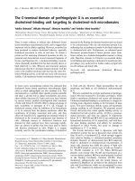
Reactive hyperaemia peripheral arterial tonometry (RH-PAT) is
a novel, simple and user-independent bedside technique used
to measure microvascular endothelial function [12] (Figure 1).
It is increasingly being used to measure endothelial function as
a cardiovascular risk assessment tool in ambulatory patients
[12-16], including in the third-generation Framingham Heart
Study cohort [17]. RH-PAT has been shown to be at least
50% dependent on endothelial NO activity [18]. RH-PAT uses
finger probes to measure digital pulse wave amplitude
detected by a pressure transducer, and has been validated
against the operator-dependent flow-mediated dilatation
method [19,20] and with endothelial function in other vascular
beds, including the coronary arteries [13]. Using RH-PAT, we
have demonstrated endothelial dysfunction in subjects with
severe malaria [21] but it has not previously been evaluated in
subjects with sepsis.
Vasodilatory shock in sepsis has been hypothesized to reflect
a state of NO excess. However, several recent isotope studies
have shown no net increase in NO synthesis in humans with
sepsis [22-24]. To explain this, it has been proposed that sep-
sis may be a state of imbalance between the NOS isoforms
inducible NOS and endothelial NOS in the microvasculature
[25]. This could lead to a relative deficiency of endothelial NO,
which is required to maintain the microvascular endothelium in
a healthy, quiescent state.
Another possible reason for endothelial NO deficiency is
decreased availability of L-arginine, the substrate for NOS and
the precursor for NO [26]. Sepsis has been hypothesised to
be an arginine-deficient state [27], although plasma L-arginine
levels in humans with sepsis have been variably reported to be
high [28], normal [29,30] or low [22,31,32]. Decreased
Figure 1
Representative normal and abnormal peripheral arterial tonometry tracesRepresentative normal and abnormal peripheral arterial tonometry traces. The tracings represent the pulse wave amplitude from a fingertip over a
15-minute period. The y axis is pulse wave amplitude in arbitrary units (derived from millivolts). The top trace was taken from a control subject whose
reactive hyperaemia peripheral arterial tonometry; (RH-PAT) index was 1.98, and the bottom from a severe sepsis subject whose RH-PAT index was
1.16. The horizontal axis is time. The first shaded section is averaged as a baseline signal. The middle section is arterial occlusion, with consequent
loss of the pulse wave signal. The final section is the pulse wave amplitude following release of the cuff. The random vertical spikes are movement
artefacts. In the top trace there is reactive hyperaemia, with an increase in average pulse wave amplitude. The shaded post-occlusion section is com-
pared with the shaded baseline section to give a ratio the RH-PAT index.
Available online />Page 3 of 9
(page number not for citation purposes)
plasma L-arginine has been linked to decreased NO produc-
tion in animal and in vitro models [33].
We hypothesised that RH-PAT would be a feasible technique
to measure microvascular reactivity in sepsis and that microv-
ascular reactivity would be impaired in subjects with sepsis in
proportion to disease severity. Our secondary hypotheses
were that microvascular reactivity would correlate with plasma
L-arginine and measures of endothelial activation, and that
plasma L-arginine concentrations would be decreased in
sepsis.
Materials and methods
Study design and setting
We performed a prospective observational cohort study in a
350-bed teaching hospital in tropical northern Australia, with
an 18-bed mixed intensive care unit (ICU). Approval was
obtained from Human Research Ethics Committee of the Men-
zies School of Health Research and the Department of Health
and Community Services, Darwin. Written informed consent
was obtained from all participants or next of kin.
Participants
Between March 2006 and November 2007, all adult subjects
(≥ 18 years) admitted to the hospital were screened regarding
eligibility for the study. Inclusion criteria for sepsis subjects
were: suspected or proven infection; presence of two or more
criteria for the systemic inflammatory response syndrome
within the past four hours [34]; and admission to ICU within
the preceding 24 hours or to the wards within the preceding
36 hours. Exclusion criteria were coagulopathy (platelets ≤ 20
× 10
9
/L, activated partial thromboplastin time ≥ 70 seconds,
international normalized ratio ≥ 2.0); smoking of tobacco
within the preceding four hours; and current administration of
intravenous nitrates. Control subjects were recruited from hos-
pital patients with no clinical or laboratory evidence of inflam-
mation or infection, and who had not met systemic
inflammatory response syndrome criteria within the preceding
30 days. Severe sepsis was defined as sepsis with organ dys-
function or shock at the time of enrolment according to Amer-
ican College of Chest Physicians/Society of Critical Care
Medicine consensus criteria [34,35].
Measurement of microvascular reactivity
Sepsis subjects underwent standardised demographic and
clinical data collection, bedside RH-PAT measurement
(Endopat 2000, Itamar Medical, Caesarea, Israel), and blood
collection at days 0 and 2 to 4. All studies were performed
after resuscitation and at least one hour of hemodynamic sta-
bility (defined as no change in vasopressor dose or need for
fluid boluses) in a quiet room at 25°C, with the patient recum-
bent. Control subjects had the same assessment at a single
time point.
In this study, probes were placed on the index fingers of both
hands of all patients, or on other fingers if the index fingers
were not suitable. Digital pulse wave amplitude was recorded
from both hands for a resting baseline period of five minutes
and then a blood pressure cuff was rapidly inflated on the
study arm up to 200 mmHg, or 50 mmHg above systolic blood
pressure, whichever was greater. After five minutes ± 10 sec-
onds, the cuff was deflated. Pulse wave amplitude was then
recorded for a further five minutes. An automated computer-
ised algorithm provided by the manufacturer (Endo-PAT 2000
software version 3.1.2, Itamar Medical, Caesarea, Israel) was
used to calculate a post occlusion-pre occlusion ratio (RH-
PAT index), thus making the measurements user independent.
The software also normalises the RH-PAT index to the control
arm to correct for changes in systemic vascular tone (Figure
1).
There was no systematic difference between RH-PAT indices
generated by different observers. We have previously exam-
ined the reproducibility of RH-PAT measurements by repeat-
ing them after 0.5 to 0.75 hours in 37 healthy adults [21].
Reproducibility was acceptable according to the method of
Bland and Altman [36], and was comparable with previous
reproducibility results for RH-PAT [37] and with those
obtained with the flow-mediated dilatation method [38].
Laboratory assays
Blood was collected in lithium heparin tubes at each time point
and the plasma was frozen. Plasma arginine concentrations
were determined using high-performance liquid chromatogra-
phy, with a method modified from van Wandelen and Cohen
[39]. To assess circulating measures of endothelial activation,
intra-cellular adhesion molecule-1 (ICAM1) and E-selectin
were measured by ELISA (R&D Systems, Minneapolis, Min-
nestoa, USA). Plasma IL-6 was measured by flow cytometry
using a cytokine bead array (BD Biosciences, San Jose, Cali-
fornia, USA). Ex vivo plasma arginase activity causes signifi-
cant degradation of L-arginine at room temperature [40], thus
only L-arginine levels derived from blood frozen within 30 min-
utes of collection were included in the analysis.
Statistical methods
Predefined groups for analysis were sepsis without organ fail-
ure, severe sepsis and controls. Continuous variables were
compared using Student's t-test and analysis of variance or
Mann Whitney U test for parametric and non-parametric varia-
bles, respectively. Categorical variables were compared using
Fisher's exact test. Correlates with baseline RH-PAT index
were determined using Pearson's (parametric) or Spearman's
(non-parametric) coefficient for univariate analysis. For multi-
variate analysis, linear regression with backward selection was
used. To examine longitudinal correlations, linear mixed-effects
models were used. A two-sided P value of < 0.05 was consid-
ered significant. All analyses were performed using Stata ver-
sion 10 (Stata Corp, College Station, Texas, USA).
Critical Care Vol 13 No 5 Davis et al.
Page 4 of 9
(page number not for citation purposes)
Results
Participants
Over the 19-month study period, 85 subjects with sepsis and
45 control subjects were enrolled. Of the sepsis subjects, 54
had organ failure due to sepsis at baseline (severe sepsis
group) and 31 did not (sepsis without organ failure). The three
groups were well matched in terms of risk factors for endothe-
lial dysfunction and other baseline characteristics (Table 1). Of
the 85 sepsis subjects, 92% had community-acquired sepsis,
with no preceding trauma or surgery, and pneumonia was the
most common focus of infection.
Baseline microvascular reactivity
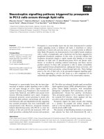
Baseline microvascular reactivity was impaired in sepsis sub-
jects compared with controls (P < 0.0001; Table 2). Mean
RH-PAT index was lowest in the severe sepsis group (1.57,
95% confidence interval (CI): 1.43 to 1.70), intermediate in
the sepsis without organ failure group (1.85, 95% CI: 1.67 to
2.03), and highest in the control group (2.05, 95% CI: 1.91 to
2.19; P < 0.00001; Figure 2). Subjects with severe sepsis
were more likely to have endothelial dysfunction than control
subjects (odds ratio (OR) 9.4, 95% CI: 3.5 to 25.0). This rela-
tion persisted after controlling for known associations with and
risk factors for endothelial dysfunction (diabetes, smoking,
ischaemic heart disease, chronic renal disease, hypercholes-
terolaemia, hypertension, statin use and age; adjusted OR
17.0, 95% CI: 5.0 to 58.0). Within the severe sepsis group,
mean RH-PAT index was not significantly different in the 27
subjects requiring vasopressors (1.48, 95% CI: 1.30 to 1.66)
than in those not requiring vasopressors (1.64, 95% CI: 1.39
to 1.89; P = not significant (NS)). In those receiving noradren-
aline (n = 25), there was no correlation between RH-PAT index
and noadrenaline dose (r = 0.19, P = NS). There was also no
relation between body temperature and RH-PAT index. Males
Table 1
Baseline characteristics of participants
Severe sepsis Sepsis without organ failure Control P value
a
N 54 31 45
Age
b
52.4 (48.3-56.5) 50.8 (46.5-55.2) 47.2 (43.1-51.4) NS
Male n (%) 33 (61) 21 (68) 30 (67) NS
Diabetic n (%) 18 (33) 7 (23) 14 (31) NS
Smoker n (%) 28 (57) 12 (39) 18 (41) NS
IHD n (%) 9 (17) 6 (19) 6 (13) NS
On statin n (%) 13 (24) 9 (29) 13 (29) NS
APACHE II
c
19.0 (15-23) 7.5 (5-11) < 0.0001
SOFA score
c
6 (3-9) 1 (0-2) < 0.0001
Focus of infection n (%)
Pleuropulmonary n (%) 26 (48) 16 (52)
Skin/soft tissue n (%) 9 (17) 9 (29)
Intra-abdominal n (%) 6 (11) 1 (3)
Urinary n (%) 4 (7) 3 (10)
Other n (%) 9 (17) 2(6)
Causative organism
None cultured n (%) 25 (46) 20 (65)
Gram positive bacterium n (%) 15 (28) 5 (16)
Gram negative bacterium n (%) 14 (26) 6 (19)
Origin of sepsis
Community-acquired n (%) 47 (87) 30 (97)
Nosocomial n (%) 7 (13) 1 (3)
a. For difference between all three groups by one way analysis of variance
b. Mean (95% confidence interval)
c. Median (interquartile range)
APACHE II = Acute Physiology and Chronic Health Evaluation II; IHD = ischaemic heart disease; NS = not significant; SOFA = Sequential Organ
Failure Assessment score
Available online />Page 5 of 9
(page number not for citation purposes)
(1.76, 95% CI: 1.62 to 1.89) had higher baseline microvascu-
lar reactivity than females (1.50, 95% CI: 1.32 to 1.68; P =
0.02).
RH-PAT was well tolerated by all subjects. In 18 of 227 meas-
urements (8%), a result was not obtainable. This occurred in
15 of 182 measurements (8%) in sepsis subjects and 3 of 45
(7%) in controls and was due either to inability to obtain a
baseline pulse wave reading, or failure to completely occlude
forearm blood flow due to oedema.
Plasma markers of endothelial activation (ICAM-1 and E-selec-
tin) were both significantly raised in sepsis subjects compared
with controls (Table 2); however, they did not correlate with
RH-PAT index. Blood lactate levels were routinely measured
only in subjects with severe sepsis, in whom the baseline
median lactate was 1.6 mmol/L (range 0.5 to 12.7; interquar-
tile range (IQR) 1.0 to 2.3). Among severe sepsis subjects,
lactate correlated inversely with RH-PAT index, but this was
not statistically significant (r = -0.28, P = 0.06).
Among all sepsis subjects, baseline RH-PAT index correlated
with mean arterial pressure (MAP; r = 0.55, P < 0.0001) and
serum albumin (r = 0.27, P = 0.03), and was inversely related
Figure 2
Baseline microvascular reactivity is impaired in sepsis, in proportion to disease severityBaseline microvascular reactivity is impaired in sepsis, in proportion to
disease severity. Solid circles represent mean values, with error bars
representing 95% confidence intervals (CI). P values indicate pairwise
comparisons between groups. RH-PAT = reactive hyperaemia periph-
eral arterial tonometry.
Table 2
RH-PAT index and related variables at time of initial measurement
Severe sepsis Sepsis without organ
failure
Control P value pooled sepsis
v control
P value severe sepsis
vs SWOF
N 54 31 45
RH-PAT index
a
1.57 (1.43-1.70) 1.85 (1.67-2.03) 2.05 (1.91-2.19) < 0.00001 0.01
Plasma L-arginine
(μmol/L)
35.8 (30.2-41.4) 40.9 (33.5-48.3) 80.4 (72.3-88.6) < 0.00001 NS
MAP (mmHg)
a
77 (74-81) 89 (83-95) 83 (79-87) NS 0.0006
Receiving vasopressors
n (%)
27 (50) 0
Noradrenaline dose
(μg/kg/min)
b, c
0.08 (0.03-0.42)
Receiving assisted
ventilation n (%)
20 (37) 0
CVP (cmH20)
a
12.2 (10.3-14.1)
Plasma ICAM-1 (ng/
ml)
b
811 (500-1502) 507 (368-673) 323 (252-397) < 0.00001 0.003
Plasma E-selectin (ng/
ml)
b
329 (138-502) 90 (51-164) 38 (26-63) < 0.00001 0.0003
Plasma IL 6 (pg/ml)
b
385 (124-996) 148 (46-315) 5 (2-8) < 0.00001 0.009
White blood cell count
a
16.7 (14.2-19.2) 15.5 (13.3-17.7) 8.4 (6.9-9.8) < 0.00001 NS
C-reactive protein
b
190 (131-255) 102 (84-234) 7 (3-24) < 0.00001 NS
a. mean (95% confidence interval)
b. Median (interquartile range)
c. Of 27 patients receiving vasopressors, 25 were receiving noradrenaline
d. Severe sepsis n = 30, sepsis without organ failure n = 26, control n = 2.
CVP = central venous pressure; ICAM = intra-cellular adhesion molecule-1; NS = not significant; MAP = mean arterial pressure; RH-PAT =
reactive hyperaemia peripheral arterial tonometry; SWOF = sepsis without organ failure.
Critical Care Vol 13 No 5 Davis et al.
Page 6 of 9
(page number not for citation purposes)
to Acute Physiology and Chronic Health Evaluation
(APACHE) II score (r = -0.36, P = 0.002), C-reactive protein
(r = -0.30, P = 0.02) and the cardiovascular component of the
Sequential Organ Failure Assessment (SOFA) score (r = -
0.29, P = 0.01), but not with total SOFA score. Independent
predictors of baseline RH-PAT index on multivariate analysis
were APACHE II score (β = -0.014, P = 0.03) and MAP (β =
0.012, P < 0.0001).
Baseline plasma L-arginine
In the subjects whose blood samples were processed within
30 minutes of collection, baseline mean plasma L-arginine
concentration was significantly lower in sepsis subjects (38.6
μmol/L, 95% CI: 34.2 to 43.1; n = 56) than in controls (80.3
μmol/L, 95% CI: 72.5 to 88.1; n = 27; P < 0.0001). There was
no significant difference in L-arginine levels between severe
sepsis and sepsis without organ failure (Figure 3). When all
subjects including controls were considered, baseline plasma
L-arginine correlated with baseline RH-PAT index (r = 0.32, P
= 0.007); however, this association was no longer significant
when stratified by disease severity.
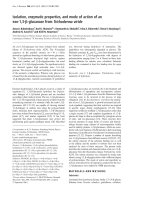
Longitudinal changes in RH-PAT and L-arginine
Longitudinal RH-PAT readings were only available in 70% of
subjects. There was no difference in disease severity, as
measured by APACHE II score, in those with (median 14, IQR
8 to 23) and without (median 15.5, IQR 8.5 to 20.5; P = NS)
longitudinal data. In sepsis subjects, there was no statistically
significant change in mean RH-PAT index from baseline to day
2 to 4 (95% CI: 1.67 to 1.85, P = NS; Figure 3). The same
was true in the severe sepsis subgroup (95% CI: 1.57 to 1.76,
P = NS). In contrast, mean plasma L-arginine concentrations
significantly increased from baseline to day 2 to 4 (95% CI:
38.2 to 49.9 μmol/L, P = 0.01). In a mixed-effects linear
regression model, change in microvascular reactivity over the
first 2 to 4 days of treatment correlated significantly with
increasing MAP and decreasing C-reactive protein, but not
with change in plasma L-arginine.
Subject outcomes
Low baseline RH-PAT index was significantly correlated with
an increase in SOFA score over the first 2 to 4 days (r = -0.37,
P = 0.02). In subjects whose SOFA score worsened over the
first 2 to 4 days, the median RH-PAT index was 1.54, com-
pared with 1.74 in those whose SOFA score improved or did
not change (P = 0.01). At both hospital discharge and 28-day
follow-up, 8 of 85 (9%) subjects with sepsis had died. Among
those with septic shock at baseline, 6 of 29 (21%) had died at
28-day follow-up. The mean baseline RH-PAT index was 1.67
among survivors and 1.60 among non-survivors (P = NS). The
strongest baseline predictors of death on univariate analysis
were APACHE II score (P = 0.008), SOFA score (P = 0.002)
and IL-6 level (P = 0.004).
Discussion
To the authors' knowledge, this is the largest published study
to date assessing reactive hyperaemia in human sepsis and
the first to use peripheral arterial tonometry. We have found
that endothelium-dependent microvascular reactivity is
impaired in sepsis, in proportion to disease severity, even after
controlling for known associations with endothelial dysfunc-
tion, suggesting that sepsis itself is the explanation for the
observed impairment in microvascular reactivity, rather than
traditional cardiovascular risk factors. Furthermore, the degree
of impairment of baseline microvascular reactivity predicted
subsequent deterioration in organ function.
RH-PAT proved to be a practical and feasible method of meas-
uring microvascular reactivity at the bedside in critically ill sep-
tic subjects, with a low proportion of technical failures, which
were no more common in sepsis subjects than in controls, and
which showed no relation with noradrenaline dose. The find-
ings of this study are generally consistent with those of the
previous small studies of reactive hyperaemia in adult subjects
with sepsis using other methods, which were generally user-
dependant and of limited availability.
Figure 3
Longitudinal change in microvascular reactivity and plasma arginine in sepsis subjectsLongitudinal change in microvascular reactivity and plasma arginine in
sepsis subjects. Solid circles represent mean values, with error bars
representing 95% confidence intervals (CI). RH-PAT = reactive hyper-
aemia peripheral arterial tonometry.
Available online />Page 7 of 9
(page number not for citation purposes)
Plethysmographic measures of forearm blood flow in sepsis
have found a post occlusion-pre occlusion ratio of 1.6 [9] and
forearm skin laser Doppler studies have found a ratio of 1.4
[5]. These results are very similar to our observed ratio of 1.57,
suggesting that the finding of impaired reactive hyperaemia in
adults with sepsis is a true phenomenon, which is independent
of the method used to measure it.
Compared with laser Doppler flowmetry, venous plethysmog-
raphy and flow-mediated dilatation of the brachial artery, PAT
requires less staff training and simpler equipment, has less
potential for inter-observer variability, and is easier to perform
on uncooperative patients. PAT has also been validated with
regards to accuracy [13,19,20] and reproducibility [37,41].
Disadvantages of PAT include the expense of disposable fin-
ger probes.
Because RH-PAT is at least 50% NO-dependent [18],
impaired RH-PAT responses in sepsis suggest reduced
endothelial NO bioavailability. Our results are in accord with
increasing data from radiolabelled arginine flux studies sug-
gesting that NO synthesis is decreased in sepsis [22-24].
Impaired RH-PAT has been demonstrated to be reversible
with L-arginine infusion in malaria caused by Plasmodium fal-
ciparum, providing direct evidence for NO dependence in
acute inflammatory states [21]. However, we cannot exclude
contributions by other mechanisms, including impaired pro-
duction of prostacyclin and endothelium-derived hyperpolariz-
ing factor [42,43].
There was a significant correlation between plasma L-arginine
and microvascular reactivity when all subjects were consid-
ered together, but this was not significant within groups. Fur-
thermore, the improvement of plasma L-arginine over the first
2 to 4 days was not significantly correlated with change in
microvascular reactivity. These findings suggest that NO pro-
duction and endothelial function in sepsis are influenced by
other factors in addition to circulating L-arginine. Such factors
may include an increase in competitive inhibitors of NOS, such
as asymmetric dimethylarginine [44]; deficiency of NOS
cofactors such as tetrahydrobiopterin; NO quenching by
microvascular reactive oxygen intermediates [45]; and the
enhanced local expression and activity of endothelial cell argi-
nase [46]. The observation of higher microvascular reactivity in
males compared with females is an unexpected finding; previ-
ous studies have found better microvascular function in
females than males, both in non-inflammatory states [47] and
in response to infusion of lipopolysaccharide [48]. However,
gender-specific microvascular function has not previously
been reported in sepsis.
The marked hypoargininaemia, which we found in subjects
with sepsis, supports the hypothesis that L-arginine is
decreased in sepsis, independent of trauma [27]. This finding
is strengthened by the fact that we only included subjects
within 24 to 36 hours of admission, with standardised sepsis
criteria and with more than 90% having community-acquired
sepsis.
Targeting tissue oxygen delivery [49] or the splanchnic micro-
circulation [50] as resuscitation goals in sepsis have not been
shown to improve outcomes. What, then, is the significance of
monitoring the microvascular endothelium in sepsis? Endothe-
lial cells have multiple roles in sepsis pathophysiology, includ-
ing the regulation of microcirculatory vasomotor tone and the
regulation of coagulation, immune and inflammatory
responses and microvascular barrier function. Preliminary
studies aimed at increasing endothelial NO bioavailability in
sepsis have shown promising results [51] and the interven-
tions which have been demonstrated to improve outcomes in
sepsis (activated protein C [52], early goal directed therapy
[53] and intensive insulin therapy [54]) could all potentially be
mediated, at least in part, via attenuation of endothelial cell
dysfunction [55]. Thus, monitoring of microvascular and
endothelial function are likely to be important components of
future trials of adjunctive treatments in sepsis.
Our study has several potential limitations. Baseline blood flow
measurements were not available, and it is possible that the
apparent decrease in reactive hyperaemia in sepsis is an arte-
fact of marked baseline vasodilatation. This could potentially
limit the subjects' ability to respond to ischaemia by increased
blood flow, because they already have near-maximal vasodila-
tation. This is unlikely to be the case because baseline forearm
blood flow in septic subjects has been found to be normal or
decreased by multiple investigators [6,7,10,56]. Furthermore,
skeletal muscle has the capacity to increase blood flow by up
to 10-fold [57], which greatly exceeds the increase seen in
both healthy and septic subjects in this and other studies.
Although we controlled for the major factors influencing
endothelial function, we cannot exclude minor influences of
altered thyroid or adrenal function. Due to variations in sample
processing time, we were unable to determine accurate
plasma arginine values for all subjects. Thus the reported
arginine values may not be fully representative of the groups as
a whole. Of the subjects who had an initial measurement of
RH-PAT index, 70% had a repeat measurement 2 to 4 days
later. Although those who were not followed up had a similar
baseline APACHE II score to those who were followed up, this
may not have been a representative population, because sub-
jects who rapidly improved and were discharged home did not
have repeat measurements. Thus the observed degree of
recovery in microvascular reactivity is likely to be an
underestimate.
The mortality rate in this cohort was low (hospital and 28-day
mortality 9% overall and 21% among those with septic shock).
Although this is consistent with the relatively low mortality rate
in severe sepsis previously documented in our ICU [35], it
Critical Care Vol 13 No 5 Davis et al.
Page 8 of 9
(page number not for citation purposes)
does mean that the study may have been underpowered to
detect associations of measured variables with mortality.
Conclusions
In summary, we have found that peripheral arterial tonometry is
a feasible tool for measuring microvascular reactivity in sepsis,
and that it is impaired in sepsis in proportion to disease sever-
ity, suggesting reduced endothelial function and decreased
endothelial NO bioavailability. Baseline RH-PAT was useful in
predicting subsequent deterioration in organ dysfunction,
although this should be reproduced by other investigators
before its clinical utility can be confirmed. Given the growing
interest in HMG CoA reductase inhibitors [58] and other
potential adjunctive therapies targeting the endothelium in
sepsis [55], better tools for monitoring the response of the
endothelium in clinical trials are needed. RH-PAT is an attrac-
tive option for such studies, as other current methods are user-
dependent and have limited availability.
Competing interests
DC has received research support (as equipment) from Itamar
Medical, the manufacturer of the RH-PAT device, and has
received speaker's fees (less than US$1000 per year) for
speaking at Itamar-sponsored educational events. The other
authors have no competing interests.
Authors' contributions
Study design was performed by JSD, NMA, TWY, DPS and
DSC. Patient recruitment was carried out by JHT, MM, JSD
and DPS. The data was processed by JSD and MM, and was
analysed by JSD with help from ACC, TWY and NMA. Labo-
ratory sample processing and HPLC assays were performed
by CJD and YRM. The manuscript was drafted by JSD and
NMA. All authors had access to all data and contributed to the
final draft of the paper. All authors read and approved the final
manuscript.
Acknowledgements
We would like to thank Kim Piera, Tonia Woodberry, Barbara Mac-
Hunter and Catherine Jones for laboratory assistance; Karl Blenk,
Antony Van Asche, Steven Tong and Paulene Kittler for RH-PAT meas-
urements; Craig Boutlis for help with initial study design; Ric Price and
Joseph McDonnell for statistical advice; and the medical and nursing
staff of the Royal Darwin Hospital Intensive Care and Hospital in the
Home units.
Funding sources: The study was funded by the National Health and
Medical Research Council of Australia (NHMRC Program Grants
290208, 496600; Practitioner Fellowship to NMA, Scholarship to JSD).
The funding source played no role in the design or conduct of the study,
nor in the drafting of the manuscript or the decision to submit it for
publication.
References
1. Angus DC, Pereira CA, Silva E: Epidemiology of severe sepsis
around the world. Endocr Metab Immune Disord Drug Targets
2006, 6:207-212.
2. Ince C, Sinaasappel M: Microcirculatory oxygenation and
shunting in sepsis and shock. Crit Care Med 1999,
27:1369-1377.
3. Aird WC: The role of the endothelium in severe sepsis and
multiple organ dysfunction syndrome. Blood 2003,
101:3765-3777.
4. Deanfield JE, Halcox JP, Rabelink TJ: Endothelial function and
dysfunction: testing and clinical relevance. Circulation 2007,
115:1285-1295.
5. Young JD, Cameron EM: Dynamics of skin blood flow in human
sepsis. Intensive Care Med 1995, 21:669-674.
6. Neviere R, Mathieu D, Chagnon JL, Lebleu N, Millien JP, Wattel F:
Skeletal muscle microvascular blood flow and oxygen trans-
port in patients with severe sepsis. Am J Respir Crit Care Med
1996, 153:191-195.
7. Kubli S, Boegli Y, Ave AD, Liaudet L, Revelly JP, Golay S, Broccard
A, Waeber B, Schaller MD, Feihl F: Endothelium-dependent
vasodilation in the skin microcirculation of patients with septic
shock. Shock (Augusta, Ga) 2003, 19:274-280.
8. Hartl WH, Gunther B, Inthorn D, Heberer G: Reactive hyperemia
in patients with septic conditions. Surgery 1988, 103:440-444.
9. Astiz ME, DeGent GE, Lin RY, Rackow EC: Microvascular func-
tion and rheologic changes in hyperdynamic sepsis. Crit Care
Med 1995, 23:265-271.
10. Vaudo G, Marchesi S, Siepi D, Brozzetti M, Lombardini R, Pirro M,
Alaeddin A, Roscini AR, Lupattelli G, Mannarino E: Human
endothelial impairment in sepsis. Atherosclerosis 2007,
197:747-752.
11. Creteur J, Carollo T, Soldati G, Buchele G, De Backer D, Vincent
JL: The prognostic value of muscle StO(2) in septic patients.
Intensive Care Med 2007, 33:1549-1556.
12. Celermajer DS: Reliable endothelial function testing: at our
fingertips? Circulation 2008, 117:2428-2430.
13. Bonetti PO, Pumper GM, Higano ST, Holmes DR Jr, Kuvin JT, Ler-
man A: Noninvasive identification of patients with early coro-
nary atherosclerosis by assessment of digital reactive
hyperemia. J Am Coll Cardiol 2004, 44:2137-2141.
14. Chenzbraun A, Levin G, Scheffy J, Keren A, Stern S, Goor D: The
peripheral vascular response to exercise is impaired in
patients with risk factors for coronary artery disease. Cardiol-
ogy 2001, 95:126-130.
15. Haller MJ, Stein J, Shuster J, Theriaque D, Silverstein J, Schatz DA,
Earing MG, Lerman A, Mahmud FH: Peripheral artery tonometry
demonstrates altered endothelial function in children with
type 1 diabetes. Pediatr Diabetes 2007, 8:193-198.
16. Kuvin JT, Mammen A, Mooney P, Alsheikh-Ali AA, Karas RH:
Assessment of peripheral vascular endothelial function in the
ambulatory setting. Vasc Med 2007, 12:13-16.
17. Hamburg NM, Keyes MJ, Larson MG, Vasan RS, Schnabel R,
Pryde MM, Mitchell GF, Sheffy J, Vita JA, Benjamin EJ: Cross-sec-
tional relations of digital vascular function to cardiovascular
risk factors in the Framingham Heart Study. Circulation 2008,
117:2467-2474.
18. Nohria A, Gerhard-Herman M, Creager MA, Hurley S, Mitra D,
Ganz P: Role of nitric oxide in the regulation of digital pulse
volume amplitude in humans. J Appl Physiol 2006,
101:545-548.
19. Kuvin JT, Patel AR, Sliney KA, Pandian NG, Sheffy J, Schnall RP,
Karas RH, Udelson JE: Assessment of peripheral vascular
Key messages
• Current tools for assessing endothelial function in
patients with sepsis are generally user dependant and
are not widely available.
• Peripheral arterial tonometry, a simple, user-independ-
ent technique for measuring endothelium-dependent
microvascular reactivity is feasible in patients with
sepsis.
• Endothelium-dependent microvascular reactivity is
impaired in sepsis, in proportion to disease severity, and
may predict subsequent deterioration in organ function.
Available online />Page 9 of 9
(page number not for citation purposes)
endothelial function with finger arterial pulse wave amplitude.
Am Heart J 2003, 146:168-174.
20. Dhindsa M, Sommerlad SM, DeVan AE, Barnes JN, Sugawara J,
Ley O, Tanaka H: Interrelationships among noninvasive meas-
ures of postischemic macro- and microvascular reactivity. J
Appl Physiol 2008, 105:427-432.
21. Yeo TW, Lampah DA, Gitawati R, Tjitra E, Kenangalem E, McNeil
YR, Darcy CJ, Granger DL, Weinberg JB, Lopansri BK, Price RN,
Duffull SB, Celermajer DS, Anstey NM: Impaired nitric oxide bio-
availability and L-arginine reversible endothelial dysfunction in
adults with falciparum malaria. J Exp Med 2007,
204:2693-2704.
22. Luiking YC, Poeze M, Ramsay G, Deutz NE: Reduced citrulline
production in sepsis is related to diminished de novo arginine
and nitric oxide production. Am J Clin Nutr 2009, 89:142-152.
23. Kao CC, Bandi V, Guntupalli KK, Wu M, Castillo L, Jahoor F:
Arginine, citrulline, and nitric oxide metabolism in sepsis. Clin
Sci (Lond) 2009, 117:23-30.
24. Villalpando S, Gopal J, Balasubramanyam A, Bandi VP, Guntupalli
K, Jahoor F: In vivo arginine production and intravascular nitric
oxide synthesis in hypotensive sepsis. Am J Clin Nutr 2006,
84:197-203.
25. McGown CC, Brookes ZL: Beneficial effects of statins on the
microcirculation during sepsis: the role of nitric oxide. Br J
Anaesth 2007, 98:163-175.
26. Hecker M, Sessa WC, Harris HJ, Anggard EE, Vane JR: The
metabolism of L-arginine and its significance for the biosyn-
thesis of endothelium-derived relaxing factor: cultured
endothelial cells recycle L-citrulline to L-arginine. Proc Natl
Acad Sci USA 1990, 87:8612-8616.
27. Luiking YC, Poeze M, Dejong CH, Ramsay G, Deutz NE: Sepsis:
an arginine deficiency state? Crit Care Med 2004,
32:2135-2145.
28. Chiarla C, Giovannini I, Siegel JH, Boldrini G, Castagneto M: The
relationship between plasma taurine and other amino acid lev-
els in human sepsis. J Nutr 2000, 130:2222-2227.
29. Ochoa JB, Udekwu AO, Billiar TR, Curran RD, Cerra FB, Simmons
RL, Peitzman AB: Nitrogen oxide levels in patients after trauma
and during sepsis. Ann Surg
1991, 214:621-626.
30. Askanazi J, Carpentier YA, Michelsen CB, Elwyn DH, Furst P,
Kantrowitz LR, Gump FE, Kinney JM: Muscle and plasma amino
acids following injury. Influence of intercurrent infection. Ann
Surg 1980, 192:78-85.
31. Sprung CL, Cerra FB, Freund HR, Schein RM, Konstantinides FN,
Marcial EH, Pena M: Amino acid alterations and encephalopa-
thy in the sepsis syndrome. Crit Care Med 1991, 19:753-757.
32. Druml W, Heinzel G, Kleinberger G: Amino acid kinetics in
patients with sepsis. Am J Clin Nutr 2001, 73:908-913.
33. Hallemeesch MM, Lamers WH, Deutz NE: Reduced arginine
availability and nitric oxide production. Clin Nutr 2002,
21:273-279.
34. Bone RC, Balk RA, Cerra FB, Dellinger RP, Fein AM, Knaus WA,
Schein RM, Sibbald WJ: Definitions for sepsis and organ failure
and guidelines for the use of innovative therapies in sepsis.
The ACCP/SCCM Consensus Conference Committee. Ameri-
can College of Chest Physicians/Society of Critical Care
Medicine. Chest 1992, 101:1644-1655.
35. Stephens DP, Thomas JH, Higgins A, Bailey M, Anstey NM, Currie
BJ, Cheng AC: Randomized, double-blind, placebo-controlled
trial of granulocyte colony-stimulating factor in patients with
septic shock. Crit Care Med 2008, 36:448-454.
36. Bland JM, Altman DG: Statistical methods for assessing agree-
ment between two methods of clinical measurement. Lancet
1986, 1:307-310.
37. Bonetti PO, Barsness GW, Keelan PC, Schnell TI, Pumper GM,
Kuvin JT, Schnall RP, Holmes DR, Higano ST, Lerman A:
Enhanced external counterpulsation improves endothelial
function in patients with symptomatic coronary artery disease.
J Am Coll Cardiol 2003, 41:1761-1768.
38. Jarvisalo MJ, Jartti L, Marniemi J, Ronnemaa T, Viikari JS, Lehtimaki
T, Raitakari OT: Determinants of short-term variation in arterial
flow-mediated dilatation in healthy young men. Clin Sci (Lond)
2006, 110:475-482.
39. van Wandelen C, Cohen SA: Using quaternary high-perform-
ance liquid chromatography eluent systems for separating 6-
aminoquinolyl-N-hydroxysuccinimidyl carbamate-derivatized
amino acid mixtures. J Chromatogr A 1997, 763:11-22.
40. Nuttall KL, Chen M, Komaromy-Hiller G: Delayed separation and
the plasma amino acids arginine and ornithine. Ann Clin Lab
Sci 1998, 28:354-359.
41. Yeo TW, Lampah DA, Gitawati R, Tjitra E, Kenangalem E, Piera K,
Price RN, Duffull SB, Celermajer DS, Anstey NM: Angiopoietin-2
is associated with decreased endothelial nitric oxide and poor
clinical outcome in severe falciparum malaria. Proc Natl Acad
Sci USA 2008, 105:17097-17102.
42. Bellien J, Thuillez C, Joannides R: Contribution of endothelium-
derived hyperpolarizing factors to the regulation of vascular
tone in humans. Fundam Clin Pharmacol 2008, 22:363-377.
43. Mitchell JA, Ali F, Bailey L, Moreno L, Harrington LS: Role of nitric
oxide and prostacyclin as vasoactive hormones released by
the endothelium. Exp Physiol 2008, 93:141-147.
44. O'Dwyer MJ, Dempsey F, Crowley V, Kelleher DP, McManus R,
Ryan T: Septic shock is correlated with asymmetrical dimethyl
arginine levels, which may be influenced by a polymorphism in
the dimethylarginine dimethylaminohydrolase II gene: a pro-
spective observational study. Crit Care 2006, 10:R139.
45. Xia Y, Roman LJ, Masters BS, Zweier JL: Inducible nitric-oxide
synthase generates superoxide from the reductase domain. J
Biol Chem 1998, 273:22635-22639.
46. Argaman Z, Young VR, Noviski N, Castillo-Rosas L, Lu XM, Zura-
kowski D, Cooper M, Davison C, Tharakan JF, Ajami A, Castillo J:
Arginine and nitric oxide metabolism in critically ill septic pedi-
atric patients. Crit Care Med 2003, 31:591-597.
47. Kneale BJ, Chowienczyk PJ, Brett SE, Coltart DJ, Ritter JM: Gen-
der differences in sensitivity to adrenergic agonists of forearm
resistance vasculature. J Am Coll Cardiol 2000, 36:1233-1238.
48. van Eijk LT, Dorresteijn MJ, Smits P, Hoeven JG van der, Netea
MG, Pickkers P: Gender differences in the innate immune
response and vascular reactivity following the administration
of endotoxin to human volunteers. Crit Care Med 2007,
35:1464-1469.
49. Hayes MA, Timmins AC, Yau EH, Palazzo M, Hinds CJ, Watson D:
Elevation of systemic oxygen delivery in the treatment of criti-
cally ill patients. New Engl J Med 1994, 330:1717-1722.
50. Palizas F, Dubin A, Regueira T, Bruhn A, Knobel E, Lazzeri S, Bare-
des N, Hernandez G: Gastric tonometry versus cardiac index as
resuscitation goals in septic shock: a multicenter, randomized,
controlled trial. Crit Care 2009, 13:R44.
51. Spronk PE, Ince C, Gardien MJ, Mathura KR, Oudemansvan
Straaten HM, Zandstra DF: Nitroglycerin in septic shock after
intravascular volume resuscitation. Lancet 2002,
360:1395-1396.
52. Bernard GR, Vincent JL, Laterre PF, LaRosa SP, Dhainaut JF,
Lopez-Rodriguez A, Steingrub JS, Garber GE, Helterbrand JD, Ely
EW, Fisher CJ: Efficacy and safety of recombinant human acti-
vated protein C for severe sepsis. New Engl J Med 2001,
344:699-709.
53. Rivers E, Nguyen B, Havstad S, Ressler J, Muzzin A, Knoblich B,
Peterson E, Tomlanovich M: Early goal-directed therapy in the
treatment of severe sepsis and septic shock. New Engl J Med
2001, 345:1368-1377.
54. Berghe G van den, Wouters P, Weekers F, Verwaest C, Bruyn-
inckx F, Schetz M, Vlasselaers D, Ferdinande P, Lauwers P, Bouil-
lon R: Intensive insulin therapy in the critically ill patients. New
Engl J Med 2001, 345:1359-1367.
55. Aird WC: Endothelium as a therapeutic target in sepsis. Curr
Drug Targets 2007, 8:501-507.
56. Astiz ME, Tilly E, Rackow ED, Weil MH: Peripheral vascular tone
in sepsis. Chest 1991, 99:1072-1075.
57. Hudlicka O: Regulation of muscle blood flow. Clin Physiol
1985, 5:201-229.
58. Terblanche M, Almog Y, Rosenson RS, Smith TS, Hackam DG:
Statins: panacea for sepsis? Lancet Infect Dis 2006,
6:242-248.