Báo cáo y học: "The relationship between CD4+CD25+CD127regulatory T cells and inflammatory response and outcome during shock states" potx
Bạn đang xem bản rút gọn của tài liệu. Xem và tải ngay bản đầy đủ của tài liệu tại đây (1.28 MB, 11 trang )
RESEARC H Open Access
The relationship between CD4
+
CD25
+
CD127
-
regulatory T cells and inflammatory response and
outcome during shock states
François Hein
1
, Frédéric Massin
2
, Aurélie Cravoisy-Popovic
1
, Damien Barraud
1
, Bruno Levy
3
,
Pierre-Edouard Bollaert
1
, Sébastien Gibot
1,3*
Abstract
Introduction: Although regulatory T lymphocytes (Tregs) have a pivotal role in preventing autoimmune diseases
and limiting chronic inflammatory conditions, they may also block beneficial immune responses by preventing
sterilizing immunity to certain pathogens.
Methods: To determine whether naturally occurring Treg cells have a role in inflammatory response and outcome
during shock state we conducted an observational study in two adult ICUs from a university hospital. Within 12
hours of admission, peripheral whole blood was collected for the measurement of cytokines and determination of
lymphocyte count. Sampling was repeated at day three, five and seven. Furthermore, an experimental septic shock
was induced in adult Balb/c mice through caecal ligation and puncture.
Results: Forty-three patients suffering from shock (26 septic, 17 non septic), and 7 healthy volunteers were
included. The percentage of Tregs increased as early as 3 days after the onset of shock, while their absolute
number remained lower than in healthy volunteers. A similar pattern of Tregs kinetics was found in infected and
non infected patients. Though there was an inverse correlation between severity scores and Tregs percentage, the
time course of Tregs was similar between survivors and non survivors. No relation between Tregs and cytokine
concentration was found. In septic mice, although there was a rapid increase in Treg cells subset among
splenocytes, antibody-induced depletion of Tregs before the onset of sepsis did not alter survival.
Conclusions: These da ta argue against a determinant role of Tregs in inflammatory response and outcome during
shock states.
Introduction
Sepsis syndrome remains the most common cause of
mortality in ICUs, causing about 750,000 deaths
annually in the USA [1,2].
Despite this, our knowledge of the underlying process
of sepsis is still unclear, and this is particularly obvious
from an immunological standpoint. Indeed, after having
considered for years the host response to severe infection
only as a stormy inflammatory reaction, it appears now
that anti-inflammatory components are released at the
same tim e as pr o-inflammatory mediators and the net
result of these two arms of immune response determines
the patient ’s phenotype: overwhelming inflammation
with early death, delicate balance between pro- and anti-
inflammation with progressive recovering, or exaggerated
anti-inflammation predisposing to superinfections [3,4].
The later phenotype has been called ‘immune paraly-
sis’ or ‘leukocyte reprogramming’ and is characterized
by a lymphocytes’ anergy, apoptosis, and a reduced
capacity to present antigens [5]. Several mechanisms
may partly explain this phenomenon such as an
increased production of anti-inflammatory cytokines IL-
10 and transforming growth factor (TGF)-b [3].
More recently, several groups have implicated an
active lymphoid suppressor cell population: the naturally
occurring regulatory T cells (Tregs) [5,6]. Altho ugh
these CD4
+
CD25
+
cells make up only a small fraction of
* Correspondence:
1
Service de réanimation médicale, Hôpital Central, CHU de Nancy, 29 Bld du
Mal de Lattre de Tassigny, Nancy, 54000, France
Hein et al. Critical Care 2010, 14:R19
/>© 2010 Hein et al.; licensee BioMed Central Ltd. T his is an open a ccess article distrib uted under t he terms of the Creative Commons
Attribution License (http://c reativecommons.org/licenses/by/2.0), which permits unrestricted use, distribution, and reproduction in
any medium, provided the original work is properly cited.
the T-lymphocyte populat ion, they are potent inhibitors
of immune response [7], and are now widely regarded
as the primary mediators of peripheral tolerance, able to
maintain immune homeostasis, to prevent autoimmu-
nity, and to modulate inflammation induced by patho-
gens and environmental insults [5].
From a functional perspective, the suppressive
mechanisms used by Tregs can be divided into four
groups: suppression by inhibito ry cytokines (such as IL-
10 and TGF-b) and cell-to-cell contact, suppression by
cytolysis through the secretion of granzymes, suppres-
sion by metabolic disruption, and suppression by the
modulation of dendritic cells maturation or function
(through cell-surface molecules such as cytotoxic T lym-
phocyte antigen-4, IL-10 and TGF-b) [8].
Although Tregs have a pivotal role in preventing auto-
immune diseases and limiting chronic inflammatory dis-
orders, they may also block beneficial im mune responses
by preventing sterilizing immunity to certain pathogens
[3,9]. As an elevated perc entage of Tregs was observed
during septic shock, a role of this cell subset has been
suspected during this syndrome [10]. Indeed, data are
scarce and conflicting: whereas Heuer and colleagues
demonstrated that adoptive tr ansfer treatment with ex
vivo-stimulated Tre gs improved sepsis mortality [11],
Scumpia and colleagues found that endogenous Tregs
did not play a role in sepsis mortality in mice [12].
In humans, Venet and colleagues showed that,
although the percentage of Tregs increased during sepsis,
absolute cell count was decreased [10]. Moreover, these
authors recently found an inverse correlation between
Tregs and lymphocyte proliferation capacity [13].
In the present study we report that the relative
increase in Tregs appea rs lately during septic shock and
show that this phenomenon is not specific to sepsis but
also occurs during non-septic shock. We also observe
that the percentage of Tregs is inversely correlated with
severity at admission, although with no relation to cyto-
kine plasma concentrations. Finally, we demonstrate
that Tregs do not influence survival or cytokine produc-
tion in a polymicrobial model of septic shock in mice.
Materials and methods
Study population
All consecutive patients newly admitted to the medical
ICUs of two French teaching hospital between January
2007 and June 2007 were prospectively enrolled in the
study if they were suffering from shock whatever its
etiology. Septic shock was defined by the standard cri-
teria of the American College of Chest Physicians/
Society of Critical Care Medicine consensus conference
[14], and non-septic shock states were defined according
to definitions used by Antonelli and colleagues [15].
Patients who w ere immunocompromised were not
enrolled (treatment with corticosteroids > 0.5 mg/kg
equivalent prednisolone, bone marrow or organ trans-
plant recipients, leukopenia (white blood cell count <
1000/mm
3
) or neutropenia (polymorphonuclear granulo-
cyte count < 500/mm
3
), hematological malignancy or
AIDS). Patients who presented with early death or dis-
charge (within 12 h ours after admission) were also
excluded. Seven healthy volunteers from the laboratory
served as controls: four men, three women, mean age
37.5 ± 3.6 years, without chronic or acute diseases.
Approvals from the Comité de Protection des Per-
sonnes-EST III and the ethical committee of the Société
de Réanimation de Langue Française were obtained, and
the informed consents of the patients and volunteers
were sought prior to inclusion.
Upon admission to the ICU, the following data were
recorded for each patient: age; sex; severity of underly-
ing medical condition according to the criteria of
McCabe and Jackson [16]; Simplified A cute Physiology
Score (SAPS) II [17]; Sepsis-related Organ Failure
Assessment (SOFA ) score [18]; reason for admission
into the ICU; principal diagnosis; vital signs; respiratory
parameters; routine blood tests and microbiological cul-
ture results. Survival or death in the ICU was assessed
during a follow-up period of up to 28 days.
Blood sampling
Within 12 hours upon admission, peripheral whole
blood was collected on citrated tubes. Sampling was
repeated at day three, five and seven provided that the
patient was still in the ICU.
Flow cytometry
Flow cytometry analyses were conducted on a Coulter
Cytomics FC500 cytometer (Beckman-Coulter, Hialeah,
FL, USA). All the monoclonal antibodies and the isoty-
pic controls (except anti-F oxp3) used were from Immu-
notech (Marseille, France): PE-Texas Red (ECD)-labelled
anti-CD3, fluorescein isothiocyanate (FITC)-labelled
anti-CD14, phycoerythrin (PE)-labelled anti-HLA-DR
(clone IM 1639), FITC-labelled anti-CD3, PC5-labeled
anti-CD56, PC5-labelled anti-natural killer (NK) G2D,
PC5-labeled anti-CD4, FITC-labelled anti-CD25 and PE-
labelled anti-CD127; the Foxp3 monoclonal antibody
was from eBiosciences (San Diego, CA, USA). After red
blood cells lysis (Q-prep system, Beckman Coulter, Hia-
leah, FL, USA), naturally occurring Tregs were charac-
terized by the expression of CD4 and CD25 and the
lack of expression of CD127. We also characterized the
number of monocytes and NK cells based upon the
expression of the usual markers (CD45, CD14, HLA-DR,
CD16, and NKG2D, respectively). Results are expressed
as percentages of the CD4
+
lymphocyte population and
as number of cells per microliter of whole blood.
Hein et al. Critical Care 2010, 14:R19
/>Page 2 of 11
Cytokines plasma concentrations
Plasma concentrations of IL-2, IL-4, IL-5, IL-10, inter-
feron (INF) g and TNFa were quantified through fluor-
escence activated cell sorting (FACS) by the use of
human Th1/Th2 CBA kit (BD Biosciences, San Diego,
CA, USA) according to the recommendations of the
manufacturer. Intra- and inter-assay coefficients of var-
iation were less than 8%. Concentration of TGFb was
determined by ELISA (R&D Systems, Lille, France).
Polymicrobial sepsis in mice
All experiments were approved by the Institutional Ani-
mal Care and Use Committee. Adult (5 to 6 weeks, 20
to 23 g) male Balb/c mice were purchased from the
Centre d’élevage Dépré (Saint-Doulchard, France).
For induction of polymicrobial sepsis, mice were
anesthetized by intraperitoneal administration of keta-
mine and xylazine in 0.2 ml sterile pyrogen-free saline.
The cecum was exposed through a 1.0 cm abdominal
midline incision and subjected to ligation of the distal
half followed by two punctures with a 21G needle. A
small amount of stool was expelled from the punctures
to ensure patency. The cecum was replaced into the
peritoneal cavity and the abdominal incision closed in
two layers. After surgery, all mice were injected subcuta-
neously with 0.5 ml of ph ysiologic saline solution for
fluid resuscitation and subcutaneously every 12 hours
with 1.25 mg (i.e., 50 μg/g) of imipenem. A sham opera-
tion was performed by isolating the cecum without liga-
tion or puncture.
When specified, animals were given either an intraper-
itoneal injection of 500 μg of affinity purified CD25
(PC61 hybridoma)-depleting antibody (BioExpress, West
Lebanon, NH, USA) or an identical volume (250 μl) of
sterile normal saline 72 hours before the caecal ligation
and puncture (CLP). Administr ation of the antibody
(250 μg) or normal saline was repeated two hours before
the procedure.
Animals were observed for up to seven days to deter-
mine survival. For certain experiments, 4 to 6 mice were
euthanized at 24, 48 or 96 hours after surgery to harvest
blood or splenocytes. Among splenocytes, the Tregs
were considered to be the CD4
+
CD25
+
Foxp3
+
cells on
FACS.
At indicated times, plasma concentrations of TNF- a ,
IL-1b and IL-6 were measured by ELISA.
Statistical analysis
Changes in time or between groups were analyzed by
one-way analysis of varia nce. Comparisons between
patients with s eptic shock, patients with no n-septic
shock and/or healthy volunteers were performed b y
using Student’s t test as normally distributed values
(Kolmogoroff-Smirnov test), Mann-Whitney U test, or
Kruskal Wallis test when indicated. Data are expressed
as mean ± standard deviation or median ± interquartile
range when appropriate. Correlations were established
using Spe arman’s test. Survival analysis (mice) was per-
formed using the log rank test after drawing the Kaplan
Meier survival curves. Analysis was completed with the
Statview software (Abacus Concepts, Berkeley, CA,
USA), with a two-tailed P value less than 0.05 consid-
ered as statistically significant.
Results
From January to June 2007, 43 consecutive patients suf-
fering from shock were included. There were 26 septic
shocks (10 of pulmonary origin, 4 peritonitis, 2 endocar-
ditis, 3 urinary tract infections and 7 bacteremia of
unknown origin) and 17 non-septic shocks (10 cardio-
genic, 3 hemorrhagic, and 4 toxic). Characteristics of the
patient population are shown in Table 1. The high mor-
tality (42%) was expected regarding the SOFA and SAPS
II severity scores.
The septic shock patients had lower neutrophils and
lymphocytes counts, higher plasma concentrations of C-
reactive protein and procalcitonin as compared with
non-septic patients.
Table 1 Characteristics of the studied population
Characteristic* Septic shock
patients (n = 26)
Non-septic
shock patients
(n = 17)
P
value
Age, years 67 (6) 59 (17) NS
Sex
†
NS
Male 19 (73) 11 (65)
Female 7 (27) 6 (35)
McCabe and Jackson
criteria
1 (1) 1 (1) NS
SAPS II score 70 (22) 73 (24) NS
SOFA score 13 (5) 11 (5) NS
Mean arterial pressure,
mmHg
72 (12) 71 (12) NS
Catecholamines
†
26 (100) 17 (100) NS
Mechanical ventilation
†
26 (100) 16 (93) NS
Corticosteroids
†
16 (62) 3 (18) 0.01
Extra-renal support
†
10 (38) 7 (40) NS
Arterial lactate (mmol/L) 5 (4) 6 (4) NS
C-reactive protein, mg/L 178 (110) 54 (49) 0.001
Procalcitonin (ng/mL) 34 (42) 1.4 (1.8) < 0.001
Neutrophils (cells/μL) 9806 (6891) 15077 (4178) 0.009
Lymphocytes (cells/μL) 684 (647) 2305 (2719) 0.001
Monocytes (cells/μL) 894 (862) 1000 (772) NS
Mortality rate
†
11 (42) 7 (41) NS
*Values are expressed as mean (standard deviation). P values are for the
comparison of septic shock vs non-septic shock patients.
NS, not significnat; SAPS, Simplified Acute Physiology Score; SOFA, Sepsis-
related Organ Failure Assessment.
†
Values are expressed as number (percentage).
Hein et al. Critical Care 2010, 14:R19
/>Page 3 of 11
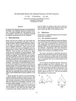
Kinetics of Tregs
Naturally occurring Tregs were defined as CD4
+
CD25
+
CD127
-
cells (Figure 1a).
At day one , the absolute number of Tregs was lower
in patients than in healthy volunteers (Figure 1b and
Table 2) without any difference between the two groups
of patients. When expressed as a percentage of CD4
+
lymphocytes, no differences were observed betw een
patients and volun teers on admission (Figure 1c and
Table 2).
By day three, both Treg numbers and percentages
progressively increased in both groups of pati ents, espe-
cially the septic ones, although there w as no statistical
difference between the two groups of patients (Figures
1b and 1c). At day seven, although the percentage of
Tregs was higher in the septi c patients than in the non-
septic patients and healthy volunteers, there were no dif-
ferences in terms of absolute count, reflecting the per-
sistence of the CD4
+
lymphopenia in the septic shock
group.
Figure 1 Treg cells kinetics during shock. Regulatory T lymphocytes (Treg) cells are de fined as CD4
+
CD25
+
CD127
-
lymphocytes. Percentages
of Tregs among CD4
+
lymphocytes, as well as absolute count are determined in shock patients within 12 hours of admission, and then at days
three, five and seven. Seven healthy volunteers served as controls. (a) Examples of Tregs percentage determination in a healthy volunteer (left
panel) and a septic shock patient on admission (right panel). (b) Time-course of Tregs absolute count and (c) Percentage in patients with shock.
* P < 0.03 patients vs. healthy volunteers.
Hein et al. Critical Care 2010, 14:R19
/>Page 4 of 11
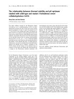
We next sought to evaluate the relation between Treg
kinetics and survival. Considering the whole group of
patients, or those suffering from a non-septic shock,
there were no differences between survivors and non-
survivors in terms of percentage or number of Tregs. By
contrast, within the septic shock patients, those surviv-
ing had constantly higher Tregs counts and percentages
than non-survivors (Figure 2).
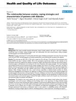
Correlation between Tregs, severity and cytokines plasma
concentrations
Considering the correlation between Tregs and out-
come, at least in septic patients, we evaluated the rela-
tion between these cells and severity. Indeed, there was
an inverse correlation between the percentage of Tregs
and SAPS II (not shown), SOFA score or arterial lactate
level (Figure 3).
Table 2 The sub-populations of lymphocytes on admission
Septic shock
(n = 26)
Non septic shock
(n = 17)
Healthy volunteers
(n = 7)
P value
CD4
+
lymphocytes (cells/μL)* 343 (230) 760 (926) 890 (384) NS
Tregs (cells/μL)
†
27 (13-33) 43 (22-66) 65 (45-72) 0.03
Tregs (% of CD4+) 8,2 (1) 8.1 (1) 8 (2) NS
NK cells (cells/μL)
†
55 (32-85) 94 (27-228) 213 (115-294) 0.03
NK cells (% of lymphocytes) 5.4 (0.6) 10.2 (0.8) 11.6 (0.8) NS
*Values are expressed as mean (standard deviation).
†
Values are expressed as median (interquartile range).
P values are for the comp arison between the three groups.
NK, natural killer; NS, not significant; Tregs, regulatory T lymphocytes.
Figure 2 Treg cells kinetics according to outcome in septic shock patients.*P = 0.02 septic survivors vs. septic non survivors. Treg,
regulatory T lymphocytes.
Hein et al. Critical Care 2010, 14:R19
/>Page 5 of 11
Bycontrast,wewereunabletofindanycorrelation
between Tregs and plasma cytokine (IL-2, IL-4, IL-5, IL-
10, INFg,TNFa and TGF-b) concentrations, either
taken individually or expressed as Th1/Th2 r atios (data
not shown). This absence of correlation was found in
septic as well as non-septic patients.
NK cells kinetics
NK cells have been advocated as an important compo-
nent influencing outcome during septic shock [18].
We observed that, although the percentage of NK cells
was not different between patients and healthy volun-
teers throughout the study period, patients with shock
presented with constantly decreased NK absolute
numbers (Figure 4). As we observed for the Treg counts,
there were no differences between septic and non-septic
shock patients, or between survivors and non-survivors.
There was no correlation between the percentages or
absolute counts of Tregs and NK cells.
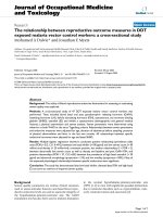
Polymicrobial sepsis in mice
To get further insight in to the role of Tregs during sep-
tic shock, we used a CLP-induced model of peritonitis
in mice. We first observed tha t as early as 24 hours
after surger y, there was an increase in the percentage of
Tregs among sple nocytes (CD4
+
CD25
+
Foxp3
+
cells) of
septic mice as compared with the sham-operated ones
(Figure 5).
Figure 3 Correlation between Tregs and s everity on admissio n. Regulatory T lymphocytes (Tregs) percentages are inversely correlated to
arterial lactate concentration (P = 0.01) and Sepsis-related Organ Failure Assessment (SOFA) score (P = 0.0001).
Hein et al. Critical Care 2010, 14:R19
/>Page 6 of 11
We next s ought to investigate the influence of Tregs
on survival. We depleted animals of CD25
+
cells by
using repeated injection of anti-CD25 antibody. Deple-
tion was highly effective with less than 0.2% of CD25
+
cells left 24 hours after the last antibody injection (Fig-
ure 6). This Treg depletion persisted at day four but
was not checked thereafter.
Animal survival was not affected by the CD25
+
cells
status with 40% of CD25-depleted mice surviving as
compared with 31% for the non-depleted group (P =
0.62; Figure 7).
Finally, plasma concentrations of TNF-a (Figure 8),
IL-1b or IL-6 (not shown) were not different between
the two groups of mice.
Discussion
There is now enough evidence that morbidity and mor-
tality occurring during septic, as well as non-septic,
shock may be due to the effect of distinct mechanisms
over time: in addition to an overwhelming pro-inflam-
matory immune response, concomitant counter-inflam-
matory mechanisms develop that may be responsible for
‘immune paralysis’ [3]. Although naturally occurring
Tregs have been found to play major roles in a wide
range of disorders, such as tumour progression, inflam-
mation or transplantation tolerance [4,7,9], their impli-
cation during shock states is unclear.
In agreement with the findings of Monneret and col-
leagues [10,19], we observed t hat the percentage of
Tregs increased as early as three days after the onset of
shock, while their absolute number remained lower than
in healthy volunteers. This phenomenon has been
explained by Vene t and colleagues by a decrease of the
CD4
+
CD25
-
lymphocytes subset [10], most probably
through apoptosis. Adding to the similarities that exist
between septic and non-septic shock in terms of
Figure 4 NK cells kinetics during shock. Time-course of natural killer (NK) cells absolute count and percentage (among total lymphocytes) in
patients with shock. * P < 0.01 patients vs. healthy volunteers.
Hein et al. Critical Care 2010, 14:R19
/>Page 7 of 11
Figure 5 Sepsis increases Tregs percentage in mice. Twenty-four hours after caecal ligation and puncture (CLP), proportion of regulatory T
lymphocytes (Treg) cells among splenocytes (CD4
+
CD25
+
Foxp3
+
cells) is determined (n = 6 per group) (a). In some experiments, Tregs were
depleted by using anti-CD25 antibody prior to sepsis induction. Depletion was highly effective with less than 1% of Tregs remaining 24 hours
after the (b, B) last antibody injection as compared with (b, A) control animals.
Hein et al. Critical Care 2010, 14:R19
/>Page 8 of 11
immune derangement, we found similar patterns of
Treg kinetics in infected and non-infected patients.
Indeed, immune paralysis is also observed during
trauma/haemorrhage or cardiogenic shock as demon-
strated by a reduced ability of antigen presentation or ex
vivo pro-inflammatory cytokine production [20]. Never-
theless, this apparent absence of difference between sep-
tic and non-septic patients may also, at least in part, be
expl ained by the small sample size and the variability in
each of the measurements.
Although we observed an inverse correlation between
severity, assessed by SOFA score or arterial lactate con-
centration, and percentage of Tregs, the time course of
the percentage or absolute number of Tregs was similar
between survivors and non-survivors. Most probably,
this finding may also be explained by the negative corre-
lation between severity and CD4
+
CD25
-
lymphocyte
number (data not shown). Nevertheless, only consider-
ing the group of septic shock patients, we observed that
survivors had higher percentage and absolute numbers
Figure 6 Survival according to the regulatory T lymphocytes status in septic mice. Antibody-mediate d depletion of CD25
+
cells does not
alter survival during caecal ligation and puncture (n = 20 per group, P = 0.62).
Figure 7 TNF- a plasma concentration according to the regulatory T lymphocytes status in septic mice. Antibody-mediated depletion of
CD25
+
cells does not influence TNF-a plasma concentration during caecal ligation and puncture (n = 6 per group).
Hein et al. Critical Care 2010, 14:R19
/>Page 9 of 11
of Tregs by day five than non-survivors. Although this
finding may suggest a specific effect of Tregs in sepsis
outcome, the low number of non-survivors studied at
days five and seven preclude any definite conclusion.
The influence of cytokines on Treg proliferation or
activation is unknown during shock states, as is the
effect of Tregs on cytokine production. Here we were
unable to show any correlation between percentage or
absolute count of Tregs and plasma cytokine concentra-
tion (IL-2, IL-4, IL-5, IL-10, INFg,TNFa and TGF-b),
taken individually or expressed as a Th1/Th2 balance.
Therefore, Tregs do not seem to modulate the inflam-
matory response during shock in vivo.Ofnote,this
absence of relation was found in septic as well as in
non-septic patients.
As NK cells have recently been advocated to play a
role in sepsis outcome, we also investigated the
kinetics of this cell’s subset. In contrast to the findings
of Giamarellos-Bourboulis and colleagues [21], we
observed that the percentage of NK cells was not dif-
ferent between patients and healthy volunteers
throughout the study period. This discrepancy may be
explained by the unexpectedly low NK percentage
(4.12%) found in the six healthy volunteers, and the
old age of the patients (77 years) e nrolled in the Greek
study. Patients with shock presented with a constantly
decreased NK absolute count, without any differences
between septic and non-septic shock. Finally, NK cell
kinetics was similar between survivors and non-survi-
vors. Taken together, these data suggest a marginal
role of NK cells on shock outcome, although the rela-
tively small number of patients enrolled precludes defi-
nite conclusion.
To further evaluate the role of Tregs on sepsis out-
come, we used a well characterized CLP model of poly-
microbial sepsis in mice. In accordance with Scumpia
and colleagues [12], we observed a rapid increase in
Tregs subset among splenocytes in septic animals. How-
ever, despite their increased proportion, antibody-
induced depletion of Tregs before the onset of sepsis
did not alter survival. Again, these data are in line with
those of Scum pia and colleagues [12]. The origina lity of
the present study stems from the septic shock model we
used: mice were fluid resuscitated and antibiotics gi ven,
further approaching the complex physiopathology of
human sepsis. Therefore, our data are more able to be
extrapolated to human septic shock. In accordance with
what we observed in humans, Tregs did not appear to
influence cytokine production (TNF-a,IL-1b, IL-6) in
septic animals.
In contrast, our animal data seem in discordance with
those reported by Heuer and colleagues [11]. Several
important differences between these two st udies need to
be underlined. First, Heuer and colleagues performed an
adoptive transfer of in vitro prestimulated Tregs and
then it is not clear whether the protective role on survi-
val was due to Tregs themselves or to the artificial
induction of membrane/soluble mediators. Second, the
model of CLP we used here appears to be more strin-
gent and thus, a potential protective role of Tregs may
have been masked by the severity of the model. Finally,
although we used the classical and well-accepted anti-
CD25 antibody approach to deplete Tregs, we must
acknowledge that anti-CD25 is not specific for Tregs
and leads to the depletion of other important cells (acti-
vated T cells). Moreover, anti-CD25 antibody may not
have completely eliminated CD25
low
Tregs.
Conclusions
Taken together, these data argue against a determinant
role of naturally occurring Tregs on inflammatory
response and outcome during shock states.
Key messages
• Percentage of Tregs increases early after the onset
of shock whatever its etiology, while their absolute
number remains lower than in healthy volunteers.
• Percentage of Tregs i s i nversely correlated to
severity of shock, althou gh the time course of per-
centage or absolute number of Tregs is similar
between survivors and non-survivors.
• There is no correlation between percentage or
absolute number of Tregs and plasma cytokine con-
centrations taken individually or expressed as a Th1/
Th2 balance.
• In a mouse model of septic shock, antibody-
induced depletion of Tregs before the onset of sepsis
does not alter survival.
Abbreviations
ELISA: enzyme-linked immunosorbent assay; FACS: fluorescent activated cell
sorting; FITC: fluorescein isothiocyanate; IFN: interferon; IL: interleukin; NK:
natural killer; PE: phycoery thrin; SAPS: Simplified Acute Physiology Score;
SOFA: Sepsis-related Organ Failure Assessment; TGF: transforming growth
factor; TNF: tumor necrosis factor; Tregs: regulatory T cells.
Acknowledgements
This work has been supported by a Contrat de Programme Recherche
Clinique (CPRC) 2007 from Nancy University’s hospital.
Author details
1
Service de réanimation médicale, Hôpital Central, CHU de Nancy, 29 Bld du
Mal de Lattre de Tassigny, Nancy, 54000, France.
2
Laboratoire
d’immunologie, Hôpital Brabois, CHU de Nancy, Avenue de la Foret de Haye,
Vandoeuvre-les-Nancy, 54500, France.
3
Groupe Choc, contrat AVENIR INSERM,
Faculté de Médecine, Nancy Université, Avenue de la Foret de Haye,
Vandoeuvre-les-Nancy, 54500, France.
Authors’ contributions
FH and SG designed the study, enrolled patients, performed experiments,
and wrote the manuscript; FM performed experiments; ACP, DB, BL and PEB
enrolled patients and analyzed data. All authors read and approved the final
manuscript.
Hein et al. Critical Care 2010, 14:R19
/>Page 10 of 11
Competing interests
The authors declare that they have no competing interests.
Received: 23 September 2009 Revised: 7 December 2009
Accepted: 15 February 2010 Published: 15 February 2010
References
1. Angus DC, Linde-Zwirble WT, Lidicker J, Clermont G, Carcillo J, Pinsky MR:
Epidemiology of severe sepsis in the United States; analysis of
incidence, outcome and associated costs of care. Crit Care Med 2001,
29:1303-1310.
2. Brun-Buisson C, Meshaka P, Pinton P, Vallet B, EPISEPSIS Study Group:
EPISEPSIS: a reappraisal of the epidemiology and outcome of severe
sepsis in French intensive care units. Intensive Care Med 2004, 30:580-588.
3. Hotchkiss RS, Karl IE: The pathophysiology and treatment of sepsis. N Engl
JMed2003, 348:138-150.
4. Hotchkiss RS, Swanson PE, Cobb JP, Jacobson A, Buchman TG, Karl IE:
Apoptosis in lymphoid and parenchymal cells during sepsis: findings in
normal and T- and B-cell-deficient mice. Crit Care Med 1997, 25:1298-1307.
5. Sakaguchi S, Ono M, Setoguchi R, Yagi H, Hori S, Fehervari Z, Shimizu J,
Takahashi T, Nomura T: Foxp3+ CD25+ CD4+ natural regulatory T cells in
dominant self-tolerance and autoimmune disease. Immunol Rev 2006,
212:8-27.
6. Fehérvari Z, Sakaguchi S: CD4+ Tregs and immune control. J Clin Invest
2004, 114:1209-1217.
7. Shevach EM: Regulatory T cells in autoimmmunity. Annu Rev Immunol
2000, 18:423-449.
8. Vignali DA, Collison LW, Workman CJ: How regulatory T cells work. Nat
Rev Immunol 2008, 8:523-532.
9. Monneret G, Venet F, Pachot A, Lepape A: Monitoring immune
dysfunctions in the septic patient: a new skin for the old ceremony. Mol
Med 2008, 14:64-78.
10. Venet F, Pachot A, Debard AL, Bohé J, Bienvenu J, Lepape A, Monneret G:
Increased percentage of CD4+CD25+ regulatory T cells during septic
shock is due to the decrease of CD4+CD25- lymphocytes. Crit Care Med
2004, 32:2329-2331.
11. Heuer JG, Zhang T, Zhao J, Ding C, Cramer M, Justen KL, Vonderfecht SL,
Na S: Adoptive transfer of in vitro-stimulated CD4+CD25+ regulatory T
cells increases bacterial clearance and improves survival in polymicrobial
sepsis. J Immunol 2005, 174:7141-7146.
12. Scumpia PO, Delano MJ, Kelly KM, O’Malley KA, Efron PA, McAuliffe PF,
Brusko T, Ungaro R, Barker T, Wynn JL, Atkinson MA, Reeves WH, Salzler MJ,
Moldawer LL: Increased natural CD4+CD25+ regulatory T cells and their
suppressor activity do not contribute to mortality in murine
polymicrobial sepsis. J Immunol 2006, 177:7943-7949.
13. Venet F, Chung CS, Kherouf H, Geeraert A, Malcus C, Poitevin F, Bohé J,
Lepape A, Ayala A, Monneret G: Increased circulating regulatory T cells
(CD4(+)CD25 (+)CD127 (-)) contribute to lymphocyte anergy in septic
shock patients. Intensive Care Med 2009, 35:678-686.
14. Bone RC, Balk RA, Cerra FB, Dellinger RP, Fein AM, Knaus WA, Schein RM,
Sibbald WJ: Definitions for sepsis and organ failure and guidelines for
the use of innovative therapies in sepsis. The ACCP/SCCM Consensus
Conference Committee. American College of Chest Physicians/Society of
Critical Care Medicine. Chest 1992,
101:1644-1655.
15. Antonelli M, Levy M, Andrews PJD, Chastre J, Hudson LD, Manthous C,
Meduri GU, Moreno RP, Putensen C, Stewart T, Torres A: Hemodynamic
monitoring in shock and implications for management. International
consensus conference, Paris, France, 27-28 April 2006. Intensive Care Med
2007, 33:575-590.
16. McCabe WR, Jackson GG: Gram negative bacteremia: I. Etiology and
ecology. Arch Intern Med 1962, 110:845-847.
17. Le Gall JR, Lemeshow S, Saulnier F: A new Simplified Acute Physiology
Score (SAPS II) based on a European/North American multicenter study.
JAMA 1993, 271:2957-2963.
18. Vincent JL, de Mendonça A, Cantraine F, Moreno R, Takala J, Suter PM,
Sprung CL, Colardyn F, Blecher S: Use of the SOFA score to assess the
incidence of organ dysfunction/failure in intensive care units: results of
a multicenter, prospective study. Working group on “sepsis-related
problems” of the European Society of Intensive Care Medicine. Crit Care
Med 1998, 26:1793-1800.
19. Monneret G, Debard AL, Venet F, Bohe J, Hequet O, Bienvenu J, Lepape A:
Marked elevation of human circulating CD4+CD25+ regulatory T cells in
sepsis-induced immunoparalysis. Crit Care Med 2003, 31:2068-2071.
20. Oberholzer A, Oberholzer C, Moldawer LL: Sepsis syndromes:
understanding the role of innate and acquired immunity. Shock 2001,
16:83-96.
21. Giamarellos-Bourboulis EJ, Tsaganos T, Spyridaki E, Mouktaroudi M,
Plachouras D, Vaki I, Karagianni V, Antonopoulou A, Veloni V, Giamarellou H:
Early changes of CD4-positive lymphocytes and NK cells in patients with
severe Gram-negative sepsis. Crit Care 2006, 10:R166.
doi:10.1186/cc8876
Cite this article as: Hein et al.: The relationship between CD4
+
CD25
+
CD127
-
regulatory T cells and inflammatory response and outcome
during shock states. Critical Care 2010 14:R19.
Submit your next manuscript to BioMed Central
and take full advantage of:
• Convenient online submission
• Thorough peer review
• No space constraints or color figure charges
• Immediate publication on acceptance
• Inclusion in PubMed, CAS, Scopus and Google Scholar
• Research which is freely available for redistribution
Submit your manuscript at
www.biomedcentral.com/submit
Hein et al. Critical Care 2010, 14:R19
/>Page 11 of 11