Báo cáo y học: "Renal hypoperfusion and impaired endothelium-dependent vasodilation in an animal model of VILI: the role of the peroxynitrite-PARP pathwa" ppt
Bạn đang xem bản rút gọn của tài liệu. Xem và tải ngay bản đầy đủ của tài liệu tại đây (914.47 KB, 10 trang )
Vaschetto et al. Critical Care 2010, 14:R45
/>Open Access
RESEARCH
© 2010 Vaschetto et al.; licensee BioMed Central Ltd. This is an open access article distributed under the terms of the Creative Commons
Attribution License ( which permits unrestricted use, distribution, and reproduction in
any medium, provided the original work is properly cited.
Research
Renal hypoperfusion and impaired
endothelium-dependent vasodilation in an animal
model of VILI: the role of the peroxynitrite-PARP
pathway
Rosanna Vaschetto*1,2,3,4, Jan W Kuiper
2,4
, René JP Musters
4,5
, Etto C Eringa
4,5
, Francesco Della Corte
1
,
Kanneganti Murthy
6
, AB Johan Groeneveld
3,4
and Frans B Plötz
2,4
Abstract
Introduction: Mechanical ventilation (MV) can injure the lungs and contribute to an overwhelming inflammatory
response, leading to acute renal failure (ARF). We previously showed that poly(adenosine diphosphate-ribose)
polymerase (PARP) is involved in the development of ventilator-induced lung injury (VILI) and the related ARF, but the
mechanisms underneath remain unclear. In the current study we therefore tested the hypothesis that renal blood flow
and endothelial, functional and tissue changes in the kidney of rats with lipopolysaccharide (LPS)-induced lung injury
aggravated by MV, is caused, in part, by activation of PARP by peroxynitrite.
Methods: Anesthetized Sprague Dawley rats (n = 31), were subjected to intratracheal instillation of lipopolysaccharide
at 10 mg/kg followed by 210 min of mechanical ventilation at either low tidal volume (6 mL/kg) with 5 cm H
2
O positive
end-expiratory pressure or high tidal volume (19 mL/kg) with zero positive end-expiratory pressure in the presence or
absence of a peroxynitrite decomposition catalyst, WW85 or a PARP inhibitor, PJ-34. During the experiment,
hemodynamics and blood gas variables were monitored. At time (t) t = 0 and t = 180 min, renal blood flow was
measured. Blood and urine were collected for creatinine clearance measurement. Arcuate renal arteries were isolated
for vasoreactivity experiment and kidneys snap frozen for staining.
Results: High tidal volume ventilation resulted in lung injury, hypotension, renal hypoperfusion and impaired renal
endothelium-dependent vasodilation, associated with renal dysfunction and tissue changes (leukocyte accumulation
and increased expression of neutrophil gelatinase-associated lipocalin). Both WW85 and PJ-34 treatments attenuated
lung injury, preserved blood pressure, attenuated renal endothelial dysfunction and maintained renal blood flow. In
multivariable analysis, renal blood flow improvement was, independently from each other, associated with both
maintained blood pressure and endothelium-dependent vasodilation by drug treatment. Finally, drug treatment
improved renal function and reduced tissue changes.
Conclusions: The peroxynitrite-induced PARP activation is involved in renal hypoperfusion, impaired endothelium-
dependent vasodilation and resultant dysfunction, and injury, in a model of lung injury.
Introduction
Mechanical ventilation (MV) remains the cornerstone of
treatment in patients with acute lung injury (ALI) [1]. Ani-
mal and clinical studies show that MV can further injure the
lungs, causing ventilator-induced lung injury (VILI) and
can contribute to a systemic inflammatory response and
development of multiple organ dysfunction syndrome [2-5].
The kidney is one of the organs most commonly involved
[6,7]. There are few experimental studies addressing the
role of MV in the development of acute renal failure (ARF)
[2,5,8-10]. Multiple mechanisms could link VILI with ARF
but specific contributions are difficult to ascertain [11].
There is increasing evidence that renal endothelial dysfunc-
* Correspondence:
1
Department of Clinical and Experimental Medicine, University of Eastern
Piedmont "Amedeo Avogadro", Corso Mazzini 18, 28100, Novara, Italy
Vaschetto et al. Critical Care 2010, 14:R45
/>Page 2 of 10
tion plays a significant role in the development of ARF [12-
14]. With injury, the endothelial cell loses its ability to mod-
ulate vasomotor and inflammatory responses [12-14].
In previous experimental studies, we described a fall in
renal blood flow during injurious MV of normal lungs [10],
and benefits of poly(ADP-ribose) polymerase (PARP)
inhibitor given as pre-treatment on renal function and tissue
integrity in lipopolysaccharide (LPS)-induced lung injury
with superimposed MV [5], but their relation remains
unclear. Indeed, the PARP pathway is activated both in
VILI and ARF [5,15-18].
Oxygen and nitrogen-derived reactive species, such as
peroxynitrite, induce oxidative DNA damage and conse-
quent activation of the nuclear enzyme PARP. PARP over-
activation is detrimental by depleting cellular ATP stores,
resulting in cell dysfunction and death [19,20]. Thereby,
activation of the pathway leads to endothelial dysfunction,
as described in a wide variety of models [21-23]. Although
PJ-34 is a pharmacological inhibitor of PARP independent
on the activating stimuli [5,16], WW85 is a novel metal-
loporphyrinic peroxynitrite decomposition catalyst, releas-
ing of NO
3
. The compound thus blocks peroxynitrite and
thereby reduces PARP activation [24-26].
Peroxynitrite formation and PARP activation in lungs of
animals with VILI have been demonstrated before
[5,16,27]. To our knowledge, renal mechanisms involved in
VILI-associated ARF and in particular related to the activa-
tion of PARP by peroxynitrite have not been studied before.
Our current study extends previous observations [5] by fur-
ther exploring the route of PARP inhibition involved in
renal hemodynamic during LPS-induced lung injury aggra-
vated by MV. We tested the hypothesis that renal blood
flow and endothelial, functional and tissue changes in the
kidney of rats with LPS-induced lung injury aggravated by
MV, is caused, in part, by activation of PARP by peroxyni-
trite. We demonstrated that inhibition of PARP activation
by peroxynitrite attenuates VILI and renal hypoperfusion
and dysfunction, by maintaining endothelium-dependent
vasodilation and decreasing inflammation and tissue injury.
Materials and methods
Animal preparation
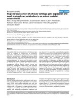
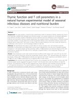
The experimental setup is shown in Figure 1. Animals were
treated according to national guidelines and with permis-
sion of the Institutional Animal Care and Use Committee
(Amsterdam, The Netherlands). A total of 31 male Sprague
Dawley rats (Harlan CPB, Zeist, The Netherlands) with a
mean weight of 310 ± 10 g, were anesthetized with a bolus
of 60 mg/kg pentobarbital sodium (Nembutal; CEVA Santa
Animale BV, Maassluis, The Netherlands) given intraperi-
toneally (ip) and 70 mg/kg ketamine (Alfasan, Woerden,
The Netherlands) intramuscularly. Anesthesia was main-
tained with pentobarbital at 15 mg/kg every 30 minutes
through an ip catheter and ketamine intravenously (iv) 20
mg/kg/h via tail vein; muscle relaxation was achieved by iv
administration of pancuronium bromide 0.6 mg/kg/h. Rats
were placed in the supine position on a heating pad, main-
taining body temperature at 37°C. A tracheostomy was per-
formed and a cannula (14 gauge) was inserted into the
trachea. The right jugular vein, right carotid artery, and left
femoral artery were cannulated with polyethylene tubing.
The right jugular vein catheter and the left femoral artery
catheter were connected to pressure transducers. Central
venous pressure, mean arterial pressure (MAP) and heart
rate were continuously monitored during the experiment.
An acetone-stripped pulmonary artery catheter leaving only
the thermistor was placed in the thoracic aorta via the right
femoral artery. The bladder was catheterized for urine sam-
pling using a transabdominal approach. Blood gas analysis
was performed using a pH/blood-gas analyzer (ABL 50;
Radiometer, Copenhagen, Denmark).
Experimental protocol
PJ-34 was purchased from Alexis Biochemicals, Lausen,
Switzerland. WW85 was kindly provided by Inotek Phar-
maceuticals Corporation, Beverly, MA, USA. The rats were
initially ventilated at a tidal volume (Vt) of 6 mL/kg and
positive end-expiratory pressure (PEEP) of 5 cmH
2
O
(AVEA Ventilator, Viasys Healthcare, Yorba Linda, CA,
USA). Rats were randomly allocated into four groups: Vt 6
ml/kg and PEEP 5 cmH
2
O or Vt 19 ml/kg, no PEEP treated
with either vehicle, PJ-34 or WW85 (Figure 1). For the con-
trol group, we adopted a relatively low Vt (6 ml/kg) plus
PEEP following current clinical practice to minimize VILI.
A second group was ventilated with high Vt (19 ml/kg) and
zero PEEP, which is known to induce VILI [28,29] but has
been used in the past years to maintain adequate oxygen-
ation and normocapnia [30].
After a one-hour period, during which the animal was
prepared and invasive monitoring was placed, drugs or
vehicle bolus infusion was started: PJ-34 was administered
iv as a loading dose of 10 mg/kg over 30 minutes, WW85
was administered 0.8 mg/kg ip. After one hour, a baseline
arterial blood gas was measured to confirm similar gas-
exchange conditions in all rats. LPS (055:B5, Sigma-
Aldrich, St Louis, MO, USA) at 10 mg/kg in 0.5 ml normal
saline was administered by using an intratracheal aero-
solizer (PennCentury Inc, Philadelphia, PA, USA). Five
minutes later, a recruitment manoeuvre was performed by
increasing PEEP level to 25 cmH
2
O for five breaths, fol-
lowed by 10 minutes of stabilization under the ventilator
settings described above. Thereafter ventilation setting was
changed according to the randomization and continued for
3.5 hours. PJ-34 was administered iv as a continuous infu-
sion at 2 mg/kg/h for the remainder of the experiments [31].
Partial pressure of arterial carbon dioxide (PaCO
2
) was
maintained at 40 ± 5 mmHg by adjusting the respiratory
rate. The inspiration to expiration ratio was set to 1:2 and
Vaschetto et al. Critical Care 2010, 14:R45
/>Page 3 of 10
the fraction of inspired oxygen (FiO
2
) was kept at 0.45 for
the whole experiment. Only in the case of a partial pressure
of arterial oxygen (PaO
2
)/fraction of inspired oxygen
(FiO
2
) inferior to 150 was FiO
2
increased to 0.60. Adminis-
tration of fluids was kept to a minimum, and did not differ
between the groups. Approximately 1.5 mL/h normal saline
per animal was infused to replace blood samples and flush
intravascular catheters. Upon completion of the MV, the
animals were sacrificed with an overdose of anesthetic.
Right kidneys were snap frozen and stored at -80°C for his-
tological examination. Left kidneys were immediately pro-
cessed to isolate renal arcuate arteries. Plasma and urine
were stored at -80°C until assayed. Lungs and heart were
removed en-bloc. The right middle lobe was used to esti-
mate wet/dry weight ratio.
Cardiac output and renal blood flow measurements
Cardiac output (CO) (Cardiac Output Computer 9520A,
Edwards Lifesciences, Irvine, CA, USA) was obtained
every 60 minutes using the thermodilution method; 200 μl
of cold saline was injected via the right jugular vein cathe-
ter as described previously [32]. Renal blood flow was
measured at the randomization and at the end of the experi-
ments using FluoSpheres polystyrene microspheres (15 μm
scarlet fluorescent (645/680) and 15 μm blue-green fluores-
cent (430/465), Molecular Probes Europe, Leiden, The
Netherlands). Renal blood flow in the left and right kidneys
was calculated using a reference blood sample as previ-
ously described in detail, [33] and is expressed as the mean
renal blood flow. The blood flow from the left and right tri-
ceps muscles was used to assess microsphere distribution.
Renal functional parameters
Urine samples were collected from the 120
th
to the 180
th
minute after randomization, after emptying the urine tube.
Arterial blood sample was collected at the 180
th
minute.
The samples were analyzed for sodium, creatinine, and urea
(Modular Analytics, Roche Diagnostics, Mannheim, Ger-
many). In rats with preserved urinary production, creatinine
clearance was calculated using the formula U
Cr
× V/P
Cr
. In
this formula U
Cr
represents the urine creatinine concentra-
tion (mg/mL), V is the urine flow (mL/min) and P
Cr
is the
plasma creatinine concentration.
Vasoreactivity experiments
To elucidate the contribution of endothelial damage via the
peroxynitrite-PARP pathway, renal arcuate arteries were
isolated (n= 6/group) and mounted in a pressure myograph.
The mean arterial diameter was not different among groups
(320 ± 20 μm). Diameter reponses of arteries to various
stimuli under 37°C were measured as previously described
[34]. 3-(N-morpholino)propanesulfonic (MOPS) buffer was
used (in mM: 145 NaCl, 5 KCl, 2 CaCl, 1 MgSO
4
, 1
NaH
2
PO
4
, 3 MOPS, 2 pyruvate, 10 glucose, and 0.02
EDTA, pH 7.4) to fill the arteriole and pressure column.
The organ chamber was filled with Krebs buffer (in mM:
110 NaCl, 5 KCl, 2.5 CaCl, 1 MgSO
4
, 1 KH
2
PO
4
, 10 glu-
cose, 0.02 EDTA, and 24 NaHCO
3
, gassed with 95% air 5%
CO
2
, pH 7.4). Vascular smooth muscle contractile function
was studied by performing a cumulative concentration-
response curve to determine norepinephrine sensitivity.
As a measure of norepinephrine sensitivity, we deter-
mined the -log EC50 value; this is the norepinephrine con-
centration at which the artery is constricted by 50%. This
Figure 1 Timeline of the protocol. Animals were anesthetized, a tracheotomy was performed and animals were connected to a ventilator and ven-
tilated in volume-controlled mode at 6 ml/kg, 5 cmH
2
O positive end-expiratory pressure. Arterial and venous catheters were inserted. One hour before
lipopolysaccharide intratracheal injection, vehicle control or WW85 or PJ-34 were infused. At t = 0 minute, mechanical ventilation setting was changed
according to the randomization and renal blood flow was measured. From t = 120 minute to t = 180 minutes urine was collected and blood samples
were taken. At time t = 180 minutes renal blood flow was measured with different fluorescence microspheres. At the end of the experiment, at t =
210 minutes, blood samples were taken, animals were sacrificed, organs were harvested and arcuate renal arteries were isolated. Vt, tidal volume.
Vaschetto et al. Critical Care 2010, 14:R45
/>Page 4 of 10
norepinephrine constriction level was used to test the
endothelium-dependent vasodilatation with acetylcholine.
The arteries were exposed to concentrations of acetylcho-
line ranging from 10
-8.5
to 10
-5.5
mol/L. Diameter changes
were recorded until a steady state was reached. Dilations
are expressed as a percentage of basal diameter (dia) =
[(dia
acetylcholine
- dia
norepinephrine
)/(dia
basal
- dia
norepinephrine
)] ×
100.
Kidney staining
Kidney cryosections (5 μm; duplicate of n = 4/group) were
fixed in formaldehyde 4% (Sigma-Aldrich, St. Louis, MO,
USA). Common leukocyte antigen CD45 (AbD Serotec,
Düsseldorf, Germany) or neutrophil gelatinase-associated
lipocalin (NGAL) (Santa Cruz Biotechnology, Inc., Santa
Cruz, CA, USA) antibody was incubated 1: 25 in PBS over-
night at 4°C and washed three times in PBS with 0.05%
Tween (PBST, Sigma-Aldrich, St. Louis, MO, USA) for
five minutes. Thereafter, the sections were incubated for
one hour with Alexa Fluor 488 conjugated anti-mouse or
anti-rabbit depending on the primary antibody (Molecular
Probes Europe, Leiden, The Netherlands) 1:100 in PBS. As
a negative control a section with no primary antibody was
used. After staining, sections were rinsed three times in
PBST and incubated with rhodamine-conjugated wheat
germ agglutinin (WGA, Molecular Probes Europe, Leiden,
The Netherlands) for 20 minutes. Finally after five minutes
washes in PBST, the sections were mounted on standard
glass slide using Vectashield™ hard set mounting medium
(Vector Laboratories, Burlingame, CA, USA) containing
DAPI nuclear staining. Kidney sections were examined
with Zeiss Axiovert 200 M Marianas™ inverted micro-
scope (Carl Zeiss, Jena, Germany). Microscopy was per-
formed with a 10 × air lens. The microscope, camera, and
data were controlled by SlideBook™ software (Slide-
Book™ version 4.0.8.1 (Intelligent Imaging Innovations,
Denver, CO, USA). SlideBook software was used to deter-
mine the mean fluorescence intensity.
Statistics
Results are reported as median ± interquartile range. Data
were analyzed in non-parametric tests by using Prism
Graphpad 4.0 software package (Prism, San Diego, CA,
USA). Comparison among groups was performed using
Kruskal-Wallis test. When an overall P < 0.05, a Dunn's
multiple-comparison post hoc analysis was conducted. A P
value less than 0.05 was considered statistically significant.
To assess the relative contribution of MAP, CO, acetylcho-
line responses and treatment, in the prediction of renal
blood flow by these factors, we performed generalized esti-
mating equations, taking repeated measures in the same
animals into account. A P value less than 0.05 was consid-
ered significant.
Results
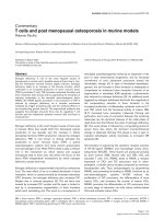
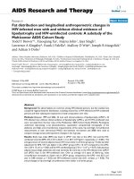
Lung injury by LPS and MV
The experimental setup is shown in Figure 1. Mean values
of PaO
2
/FiO
2
ratio were similar in all animals until the 120
th
minute of MV when the PaO
2
/FiO
2
started decreasing in the
high Vt+Vehicle group compared with the other groups
(Figure 2a). There were no differences in the levels of
PaCO
2
and pH among groups (data not shown). The lung
wet/dry ratio was higher in the high Vt+Vehicle than in the
low Vt+Vehicle group, and the treatment with the peroxyni-
trite decomposition catalyst or PARP inhibitor attenuated
lung edema (Figure 2b).
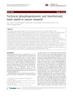
Hemodynamics variables
MAP at baseline was similar among groups. After 180 min-
utes, MAP decreased in the high Vt+Vehicle group com-
pared with the low Vt+Vehicle group (Figure 3a). WW85 or
PJ-34 both attenuated the drop in MAP in the high Vt
Figure 2 Effects of WW85 or PJ-34 on respiratory mechanic and lung edema. n = 8/group in low tidal volume (Vt)+Vehicle, high Vt+Vehicle, high
Vt+WW85, n = 7/group in high Vt+PJ-34. (a). Partial pressure of arterial oxygen (PaO
2
)/fraction of inspired oxygen (FiO
2
) ratio over time. * P < 0.05 high
Vt+Vehicle vs. others.(b) Lung wet to dry weight ratio. * P < 0.05 high Vt+Vehicle vs. all. Values represent median (interquartile range).
Vaschetto et al. Critical Care 2010, 14:R45
/>Page 5 of 10
groups. There were no differences in CO among groups
(Figure 3b).
Renal blood flow did not differ among groups at t = 0.
After 180 minutes, the renal blood flow was 6.6 ml/min/g
tissue (3.3 to 8.2 ml/min/g tissue) in the high Vt+Vehicle
group, which was approximately 68% lower (P < 0.05)
compared with the low Vt+Vehicle group, 20.4 ml/min/g
tissue (13.5 to 23.2 ml/min/g tissue). WW85 or PJ-34 treat-
ments preserved renal blood flow at 10.6 (7.6 to 14.3 ml/
min/g tissue) and 13.2 ml/min/g tissue (11.5 to 15.1 ml/min/
g tissue), respectively (Figure 3c).
Endothelium-dependent vasodilation of renal arteries ex
vivo
Endothelium-dependent vasodilation of renal arcuate arter-
ies, as indicated by the acetylcholine response, was
decreased in high Vt+Vehicle group compared to low
Vt+Vehicle control group. The acetylcholine response was
conserved in high Vt groups treated with WW85 or PJ-34
(Figure 4a). The norephineprine-induced vasoconstriction
response did not differ among the groups (Figure 4b).
Renal function
The serum creatinine increased in the high Vt+Vehicle
compared with the low Vt+Vehicle (Figure 5a) and creati-
nine clearance decreased in the former compared with the
latter (Figure 5b). Treatment with either WW85 or PJ-34
preserved the increase in serum creatinine and prevented
the fall in creatinine clearance. Blood urea nitrogen and
fractional excretion of sodium did not differ among groups
(data not shown).
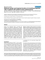
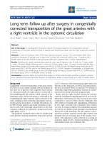
Leukocyte accumulation and NGAL expression in renal
tissue
The quantitative analysis of fluorescence intensity of
CD45, a leukocyte marker, shows that the total amount of
CD45-positive cells, mainly localized in corticomedullary
area, was increased in the high Vt+Vehicle as compared
with the low Vt+Vehicle group. Treatment with WW85 or
PJ-34, in the former, decreased leukocyte infiltration to a
level comparable with that of the latter (Figure 6a). We
found an increase in NGAL tubular expression in rats venti-
lated with high Vt+Vehicle compared with those ventilated
with low Vt+Vehicle, which was blunted by the administra-
tion of WW85 or PJ-34 (Figure 6b). Histological sections
did not reveal other signs of injury (data not shown), as
often happens in these short-time double hit models
[35,36].
Multivariable analyses
Although MAP (and not CO) was a major contributor to
predict renal blood flow in time (P = 0.003), incorporating
acetylcholine responses revealed that acetylcholine
responses independently (P = -0.006) contributed to predic-
tion of renal blood flow, together with MAP and drug treat-
ments (P < 0.001). Conversely, the acetylcholine response
was, independently of MAP (P = 0.006), predicted by drug
treatment (P < 0.001).
Discussion
Our current study suggests that hypoperfusion, impaired
endothelial vasodilation, and associated functional and tis-
sue changes in the kidney of rats with LPS-induced lung
injury aggravated by MV, are caused, in part, by activation
of PARP by peroxynitrite.
In our model, we instilled LPS intratracheally to induce
pulmonary inflammation, followed by a high Vt and zero
PEEP as injurious MV as conducted before [5]. VILI was
characterized by diffuse alveolar lung injury as shown by a
fall in PaO
2
/FiO
2
ratio and lung edema compared with low
Vt ventilation plus PEEP. However, severe hypoxemia
(PaO
2
<40 mm Hg) never occurred and PaCO
2
was kept in a
normal range in order to avoid alterations in renal blood
flow due to changes in gas exchange [11]. Furthermore, to
avoid the hemodynamic consequences of increased thoracic
Figure 3 Effects of WW85 or PJ-34 on hemodynamics. Rats received lipopolysaccharide (10 mg/kg) intratracheally at time 0, followed by mechan-
ical ventilation. n = 8/group in low tidal volume (Vt)+Vehicle, high Vt+Vehicle, high Vt+WW85, n = 7/group in high Vt+PJ-34. (a) Mean arterial pres-
sures. * P < 0.05 high Vt+Vehicle vs. all at time 180 and 210 minutes. (b) Cardiac output over time. (c) Renal blood flow at time t = 0 and t = 180 minutes.
† P < 0.05 high Vt+Vehicle vs. low Vt+Vehicle and high Vt+Vehicle vs. high Vt+PJ-34. n = 5/group. Values represent median (interquartile range).
Vaschetto et al. Critical Care 2010, 14:R45
/>Page 6 of 10
pressures, we applied the same mean airway pressures in
the ventilated groups. As a result, the CO was similar
among the groups.
Peroxynitrite formation and PARP activation in lungs of
animals with VILI have been demonstrated before [5,16,27]
and our current study extends previous observations [5] by
further exploring the route of PARP inhibition involved in
renal hemodynamics during LPS-induced lung injury
aggravated by MV. Only a few studies explored vascular
dysfunction in VILI, in particular norepinephrine- and ace-
tylcholine-induced impaired aortic vascular responses [37-
40] and impaired acetylcholine-induced pulmonary micro-
vascular responses [40]. In these animal models, very large
Vt of 35 ml/kg were applied to healthy rats to induce VILI
during one hour of MV, leading to hypotension and micro-
vascular hyperpermeability. The mechanism involved in
these vascular alterations seems to be the consequence of
intracellular reactive oxygen species and peroxynitrite for-
mation, reversed, in vitro, by free-radical scavengers [37].
Other studies using lower Vt to injure the lung (15 to 17 ml/
kg) in both healthy [10] or pre-injured animals [2,5,8,9]
failed to show a decrease in blood pressure.
To our knowledge our study is the first to address renal
microvascular responses during VILI. The renal changes
evoked in our model were characterized by renal hypoper-
fusion, impaired endothelium-dependent vasodilation and
associated dysfunction and tissue changes.
Figure 4 Concentration-response curves. (a) Concentration-response curves for norepinephrine (NE) of isolated renal arcuate arterioles. n = 5/
group. (b) Concentration-response curves for acetylcholine (Ach) of isolated renal arcuate arterioles. ACh responses were tested in a pressure myo-
graph after 50% preconstriction with NE. n = 5/group. * P < 0.05 high Vt+Vehicle vs. all. Vt, tidal volume. Values represent median (interquartile range).
Figure 5 Renal function. (a) Serum creatinine at t = 180 minutes. (b) Creatinine clearance was measured over t = 120 minutes to t = 180 minutes.
Creatinine clearance = U
Cr
× V/P
Cr
, where U
Cr
represents the creatinine concentration in urine (mmol/L), V the urine flow (mL/min), and P
Cr
the creati-
nine concentration in plasma (mmol/L). n = 8/group in low tidal volume (Vt)+Vehicle, high Vt+Vehicle, high Vt+WW85, n = 7/group in high Vt + PJ-
34. * P < 0.05 high Vt+Vehicle vs. all. Values represent median (interquartile range).
Vaschetto et al. Critical Care 2010, 14:R45
/>Page 7 of 10
These observations may warrant a discussion of potential
cause-effect relations in a complex model of inter-organ
crosstalk. The model was characterized by global systemic
vasodilation, in which release of soluble factors may be
involved, and this may have directly contributed to the fall
in renal blood flow. The data suggest that impaired endothe-
lium-dependent vasodilation also contributed to this fall.
However, we cannot definitively ascertain whether the ben-
eficial effect of the two drugs on endothelium-dependent
vasodilation and renal blood flow was caused by a direct
protective effect on renal endothelium rather than by an
anti-inflammatory effect preserving renal blood flow inde-
pendent of endothelial changes. Our multivariable analysis
suggests a direct protective effect on renal endothelium was
the cause. It remains therefore unclear how the endothe-
lium-dependent vasodilation is impaired. One possibility is
that factors derived from the lung spill over into the sys-
temic circulation, reach the kidney and evoke endothelial
changes, but factors generated in the kidney and sensitive to
the peroxynitrite-PARP pathway may also play a role
[41,42]. Together with positive effects on MAP, acetylcho-
line response and, thereby, renal blood flow, drug treatment
to inhibit the peroxynitrite-PARP pathway also inhibited
inflammatory and tissue changes in the kidneys that may
have contributed to the observed fall in renal function
judged by creatinine clearance. Leukocyte accumulation
and NGAL expression, detected predominantly in proximal
tubule cells in response to tubular epithelial damage, are
commonly observed in models of renal injury and dysfunc-
tion [43,44]. Indeed, in our study, we can not exclude also
an endothelial expression of NGAL.
Figure 6 Quantitative analysis. (a) CD45. (b) Neutrophil gelatinase-associated lipocalin (NGAL) staining. Duplicate of n = 4/group. * P < 0.05 high
tidal volume (Vt)+Vehicle vs. all. Values represent median (interquartile range). Representative kidney sections (10× air lens). Red staining: wheat germ
agglutinin; blue staining: nuclei; green staining: (c) CD45, (d) NGAL.
Low Vt
+Vehicle
High Vt
+Vehicle
High Vt
+PJ-34
High Vt
+WW85
D
Low Vt
+Vehicle
High Vt
+Vehicle
High Vt
+ PJ-34
High Vt
+WW85
0
100
200
300
400
500
600
Mean Fluorescece Intensity [NGAL]
*
B
C
Low Vt
+Vehicle
High Vt
+Vehicle
High Vt
+PJ-34
High Vt
+WW85
0
20
40
60
80
100
Mean Fluorescece Intensity [CD45]
Low Vt
+Vehicle
High Vt
+Vehicle
High Vt
+PJ-34
High Vt
+WW85
*
A
Vaschetto et al. Critical Care 2010, 14:R45
/>Page 8 of 10
Few limitations of the study should be taken into account.
First, we studied the peroxynitrite-PARP pathway in an
experimental rat model of VILI, often employed in this
contest [5,8,45-48]. Further research in humans is needed
before these results can be translated to human medicine
[49]. Second, taking into account the possible gender differ-
ences with respect to PARP activation found in animal
models of stroke and LPS-induced inflammation, the
results discussed previously might be applicable only to
males [50-52]. Finally, although unlikely according to the
literature, we can not exclude that WW85 or PJ-34 affect
microcirculatory hemodynamics with other mechanisms
other than through catalysation of peroxynitrite decomposi-
tion and PARP inhibition, respectively.
Conclusions
In conclusion, our data suggest that inhibition of PARP acti-
vation by peroxynitrite attenuates VILI and renal hypoper-
fusion and dysfunction, by maintaining endothelium-
dependent vasodilation and decreasing inflammation and
tissue injury, in the rat kidney during LPS-induced lung
injury aggravated by MV.
Key messages
• VILI complicating ALI remains associated with high
mortality rates and with the development of multiple
organ failure. The kidney is one of the first organs to
fail. The mechanisms that link MV with kidney failure
are only speculated.
• The PARP pathway is activated in different models of
ALI and ARF.
• In an animal model of lung injury, the pharmacologi-
cal inhibition of peroxynitrite or PARP attenuated lung
injury, preserved blood pressure, attenuated renal
endothelial dysfunction and maintained renal blood
flow, improving kidney function and reducing tissue
changes.
• Renal blood flow improvement was, independently
from each other, associated with both maintained blood
pressure and endothelium-dependent vasodilation by
drug treatment.
Abbreviations
ALI: acute lung injury; ARF: acute renal failure; CO: cardiac output; FiO
2
: fraction
of inspired oxygen; ip: intraperitoneally; iv: intravenously; LPS: lipopolysaccha-
ride; MAP: mean arterial pressure; MV: mechanical ventilation; NGAL: neutrophil
gelatinase-associated lipocalin; PaCO
2
: partial pressure of carbon dioxide; PaO
2
:
partial pressure of oxygen; PARP: poly(adenosine diphosphate-ribose) poly-
merase; PBS: phosphate-buffered saline; PBST: phosphate-buffered saline and
Tween; PEEP: positive end-expiratory pressure; Vt: tidal volume; VILI: ventilator-
induced lung injury.
Competing interests
Kanneganti Murthy has stock options and employment with Inotek Pharma-
ceuticals Corporation. All other authors declare that they have no competing
interests.
Authors' contributions
RV, FDC, JWK, ABJG and FBP have made substantial contributions to concep-
tion and design, acquisition of data, analysis and interpretation of data. RJPM
and ECE have made substantial contributions to acquisition and analysis of
data. RV, FDC, ABJG, KM and FBP have been involved in drafting the manuscript
and revising it critically for important intellectual content. All authors read and
approved the final manuscript.
Acknowledgements
Rosanna Vaschetto was supported by the European Society of Intensive Care
Medicine, Basic Science Award 2006. WW85 (INO-4885) was kindly donated by
Inotek Pharmaceuticals Corporation.
Author Details
1
Department of Clinical and Experimental Medicine, University of Eastern
Piedmont "Amedeo Avogadro", Corso Mazzini 18, 28100, Novara, Italy,
2
Department of Pediatric Intensive Care, Vrije Universiteit Medical Center, De
Boelelaan 1117, 1081 HV, Amsterdam, The Netherlands,
3
Department of
Intensive Care, Vrije Universiteit Medical Center, De Boelelaan 1117, 1081 HV,
Amsterdam, The Netherlands,
4
Institute for Cardiovascular Research, Vrije
Universiteit Medical Center, De Boelelaan 1117, 1081 HV, Amsterdam, The
Netherlands,
5
Department of Physiology, Vrije Universiteit Medical Center, De
Boelelaan 1117, 1081 HV, Amsterdam, The Netherlands and
6
Inotek
Pharmaceuticals Corporation, 33 Hayden Avenue, 0242, Lexington, MA, USA
References
1. Ware LB, Matthay MA: The acute respiratory distress syndrome. N Engl J
Med 2000, 342:1334-1349.
2. Imai Y, Parodo J, Kajikawa O, de PM, Fischer S, Edwards V, Cutz E, Liu M,
Keshavjee S, Martin TR, Marshall JC, Ranieri VM, Slutsky AS: Injurious
mechanical ventilation and end-organ epithelial cell apoptosis and
organ dysfunction in an experimental model of acute respiratory
distress syndrome. JAMA 2003, 289:2104-2112.
3. Ranieri VM, Suter PM, Tortorella C, De TR, Dayer JM, Brienza A, Bruno F,
Slutsky AS: Effect of mechanical ventilation on inflammatory mediators
in patients with acute respiratory distress syndrome: a randomized
controlled trial. JAMA 1999, 282:54-61.
4. Ranieri VM, Giunta F, Suter PM, Slutsky AS: Mechanical ventilation as a
mediator of multisystem organ failure in acute respiratory distress
syndrome. JAMA 2000, 284:43-44.
5. Vaschetto R, Kuiper JW, Chiang SR, Haitsma JJ, Juco JW, Uhlig S, Plotz FB,
Corte FD, Zhang H, Slutsky AS: Inhibition of poly(adenosine
diphosphate-ribose) polymerase attenuates ventilator-induced lung
injury. Anesthesiology 2008, 108:261-268.
6. Uchino S, Kellum JA, Bellomo R, Doig GS, Morimatsu H, Morgera S, Schetz
M, Tan I, Bouman C, Macedo E, Gibney N, Tolwani A, Ronco C: Acute renal
failure in critically ill patients: a multinational, multicenter study. JAMA
2005, 294:813-818.
7. Hoste EA, Clermont G, Kersten A, Venkataraman R, Angus DC, De Bacquer
D, Kellum JA: RIFLE criteria for acute kidney injury are associated with
hospital mortality in critically ill patients: a cohort analysis. Crit Care
2006, 10:R73.
8. Crimi E, Zhang H, Han RN, Sorbo LD, Ranieri VM, Slutsky AS: Ischemia and
reperfusion increases susceptibility to ventilator-induced lung injury in
rats. Am J Respir Crit Care Med 2006, 174:178-186.
9. Gurkan OU, O'Donnell C, Brower R, Ruckdeschel E, Becker PM: Differential
effects of mechanical ventilatory strategy on lung injury and systemic
organ inflammation in mice. Am J Physiol Lung Cell Mol Physiol 2003,
285:L710-L718.
10. Kuiper JW, Versteilen AM, Niessen HW, Vaschetto RR, Sipkema P, Heijnen
CJ, Groeneveld AB, Plotz FB: Production of endothelin-1 and reduced
blood flow in the rat kidney during lung-injurious mechanical
ventilation. Anesth Analg 2008, 107:1276-1283.
11. Kuiper JW, Groeneveld AB, Slutsky AS, Plotz FB: Mechanical ventilation
and acute renal failure. Crit Care Med 2005, 33:1408-1415.
12. Brodsky SV, Yamamoto T, Tada T, Kim B, Chen J, Kajiya F, Goligorsky MS:
Endothelial dysfunction in ischemic acute renal failure: rescue by
Received: 28 October 2009 Revised: 10 January 2010
Accepted: 26 March 2010 Published: 26 March 2010
This article is available from: 2010 Vaschetto et al.; licensee BioMed Central Ltd. This is an open access article distributed under the terms of the Creative Commons A ttribution License ( which permits unrestricted use, distribution, and reproduction in any medium, provided the original work is properly cited.Critical Care 2010, 14:R45
Vaschetto et al. Critical Care 2010, 14:R45
/>Page 9 of 10
transplanted endothelial cells. Am J Physiol Renal Physiol 2002,
282:F1140-F1149.
13. Lieberthal W, Wolf EF, Rennke HG, Valeri CR, Levinsky NG: Renal ischemia
and reperfusion impair endothelium-dependent vascular relaxation.
Am J Physiol 1989, 256:F894-F900.
14. Molitoris BA, Sutton TA: Endothelial injury and dysfunction: role in the
extension phase of acute renal failure. Kidney Int 2004, 66:496-499.
15. Hammerschmidt S, Sandvoss T, Gessner C, Schauer J, Wirtz H: High in
comparison with low tidal volume ventilation aggravates oxidative
stress-induced lung injury. Biochim Biophys Acta 2003, 1637:75-82.
16. Kim JH, Suk MH, Yoon DW, Kim HY, Jung KH, Kang EH, Lee SY, Lee SY, Suh
IB, Shin C, Shim JJ, In KH, Yoo SH, Kang KH: Inflammatory and
transcriptional roles of poly (ADP-ribose) polymerase in ventilator-
induced lung injury. Crit Care 2008, 12:R108.
17. Liaudet L, Pacher P, Mabley JG, Virag L, Soriano FG, Hasko G, Szabo C:
Activation of poly(ADP-Ribose) polymerase-1 is a central mechanism
of lipopolysaccharide-induced acute lung inflammation. Am J Respir
Crit Care Med 2002, 165:372-377.
18. Stone DH, Al-Badawi H, Conrad MF, Stoner MC, Entabi F, Cambria RP,
Watkins MT: PJ34, a poly-ADP-ribose polymerase inhibitor, modulates
renal injury after thoracic aortic ischemia/reperfusion. Surgery 2005,
138:368-374.
19. Ha HC, Snyder SH: Poly(ADP-ribose) polymerase is a mediator of
necrotic cell death by ATP depletion. Proc Natl Acad Sci USA 1999,
96:13978-13982.
20. Virag L, Szabo C: The therapeutic potential of poly(ADP-ribose)
polymerase inhibitors. Pharmacol Rev 2002, 54:375-429.
21. Helyar SG, Patel B, Headington K, El-Assal M, Chatterjee PK, Pacher P,
Mabley JG: PCB-induced endothelial cell dysfunction: role of poly (ADP-
ribose) polymerase. Biochem Pharmacol 2009, 78(8):959-965.
22. Mathews MT, Berk BC: PARP-1 inhibition prevents oxidative and
nitrosative stress-induced endothelial cell death via transactivation of
the VEGF receptor 2. Arterioscler Thromb Vasc Biol 2008, 28:711-717.
23. Tasatargil A, Dalaklioglu S, Sadan G: Inhibition of poly(ADP-ribose)
polymerase prevents vascular hyporesponsiveness induced by
lipopolysaccharide in isolated rat aorta. Pharmacol Res 2005,
51:581-586.
24. Cuzzocrea S, Misko TP, Costantino G, Mazzon E, Micali A, Caputi AP,
Macarthur H, Salvemini D: Beneficial effects of peroxynitrite
decomposition catalyst in a rat model of splanchnic artery occlusion
and reperfusion. FASEB J 2000, 14:1061-1072.
25. Lancel S, Tissier S, Mordon S, Marechal X, Depontieu F, Scherpereel A,
Chopin C, Neviere R: Peroxynitrite decomposition catalysts prevent
myocardial dysfunction and inflammation in endotoxemic rats. J Am
Coll Cardiol 2004, 43:2348-2358.
26. Pieper GM, Nilakantan V, Chen M, Zhou J, Khanna AK, Henderson JD Jr,
Johnson CP, Roza AM, Szabo C: Protective mechanisms of a
metalloporphyrinic peroxynitrite decomposition catalyst, WW85, in rat
cardiac transplants. J Pharmacol Exp Ther 2005, 314:53-60.
27. Peng X, Abdulnour RE, Sammani S, Ma SF, Han EJ, Hasan EJ, Tuder R,
Garcia JG, Hassoun PM: Inducible nitric oxide synthase contributes to
ventilator-induced lung injury. Am J Respir Crit Care Med 2005,
172:470-479.
28. Dos Santos CC, Slutsky AS: Invited review: mechanisms of ventilator-
induced lung injury: a perspective. J Appl Physiol 2000, 89:1645-1655.
29. Dos Santos CC, Slutsky AS: The contribution of biophysical lung injury to
the development of biotrauma. Annu Rev Physiol 2006, 68:585-618.
30. Pontoppidan H, Geffin B, Lowenstein E: Acute respiratory failure in the
adult. 3. N Engl J Med 1972, 287:799-806.
31. Goldfarb RD, Marton A, Szabo E, Virag L, Salzman AL, Glock D, Akhter I,
McCarthy R, Parrillo JE, Szabo C: Protective effect of a novel, potent
inhibitor of poly(adenosine 5'-diphosphate-ribose) synthetase in a
porcine model of severe bacterial sepsis. Crit Care Med 2002,
30:974-980.
32. Nijveldt RJ, Prins HA, van Kemenade FJ, Teerlink T, van Lambalgen AA,
Boelens PG, Rauwerda JA, van Leeuwen PA: Low arginine plasma levels
do not aggravate renal blood flow after experimental renal ischaemia/
reperfusion. Eur J Vasc Endovasc Surg 2001, 22:232-239.
33. Raab S, Thein E, Harris AG, Messmer K: A new sample-processing unit for
the fluorescent microsphere method. Am J Physiol 1999,
276:H1801-H1806.
34. Versteilen AM, Korstjens IJ, Musters RJ, Groeneveld AB, Sipkema P: Rho
kinase regulates renal blood flow by modulating eNOS activity in
ischemia-reperfusion of the rat kidney. Am J Physiol Renal Physiol 2006,
291:F606-F611.
35. Dhanireddy S, Altemeier WA, Matute-Bello G, O'Mahony DS, Glenny RW,
Martin TR, Liles WC: Mechanical ventilation induces inflammation, lung
injury, and extra-pulmonary organ dysfunction in experimental
pneumonia. Lab Invest 2006, 86:790-799.
36. O'Mahony DS, Liles WC, Altemeier WA, Dhanireddy S, Frevert CW, Liggitt
D, Martin TR, Matute-Bello G: Mechanical ventilation interacts with
endotoxemia to induce extrapulmonary organ dysfunction. Crit Care
2006, 10:R136.
37. Martinez-Caro L, Lorente JA, Marin-Corral J, Sanchez-Rodriguez C,
Sanchez-Ferrer A, Nin N, Ferruelo A, de PM, Fernandez-Segoviano P,
Barreiro E, Esteban A: Role of free radicals in vascular dysfunction
induced by high tidal volume ventilation. Intensive Care Med 2009,
35:1110-1119.
38. Nin N, Valero JA, Lorente JA, de PM, Fernandez-Segoviano P, Sanchez-
Ferrer A, Esteban A: Large tidal volume mechanical ventilation induces
vascular dysfunction in rats. J Trauma 2005, 59:711-716.
39. Nin N, Penuelas O, de PM, Lorente JA, Fernandez-Segoviano P, Esteban A:
Ventilation-induced lung injury in rats is associated with organ injury
and systemic inflammation that is attenuated by dexamethasone. Crit
Care Med 2006, 34:1093-1098.
40. Nin N, Lorente JA, de PM, El AM, Vallejo S, Penuelas O, Fernandez-
Segoviano P, Ferruelo A, Sanchez-Ferrer A, Esteban A: Rats surviving
injurious mechanical ventilation show reversible pulmonary, vascular
and inflammatory changes. Intensive Care Med 2008, 34:948-956.
41. Piepot HA, Groeneveld AB, van Lambalgen AA, Sipkema P: Tumor
necrosis factor-alpha impairs endothelium-dependent relaxation of rat
renal arteries, independent of tyrosine kinase. Shock 2002, 17:394-398.
42. Piepot HA, Groeneveld AB, van Lambalgen AA, Sipkema P: Endotoxin
impairs endothelium-dependent vasodilation more in the coronary
and renal arteries than in other arteries of the rat. J Surg Res 2003,
110:413-418.
43. Chen X, Liu X, Wan X, Wu Y, Chen Y, Cao C: Ischemic Preconditioning
Attenuates Renal Ischemia-Reperfusion Injury by Inhibiting Activation
of IKKbeta and Inflammatory Response. Am J Nephrol 2009, 30:287-294.
44. Kinsey GR, Li L, Okusa MD: Inflammation in acute kidney injury. Nephron
Exp Nephrol 2008, 109:e102-e107.
45. Chiumello D, Pristine G, Slutsky AS: Mechanical ventilation affects local
and systemic cytokines in an animal model of acute respiratory
distress syndrome. Am J Respir Crit Care Med 1999, 160:109-116.
46. Dahlem P, Bos AP, Haitsma JJ, Schultz MJ, Meijers JC, Lachmann B:
Alveolar fibrinolytic capacity suppressed by injurious mechanical
ventilation. Intensive Care Med 2005, 31:724-732.
47. Dahlem P, Bos AP, Haitsma JJ, Schultz MJ, Wolthuis EK, Meijers JC,
Lachmann B: Mechanical ventilation affects alveolar fibrinolysis in LPS-
induced lung injury. Eur Respir J 2006, 28:992-998.
48. Haitsma JJ, Schultz MJ, Hofstra JJ, Kuiper JW, Juco J, Vaschetto R, Levi M,
Zhang H, Slutsky AS: Ventilator-induced coagulopathy in experimental
Streptococcus pneumoniae pneumonia. Eur Respir J 2008,
32:1599-1606.
49. Gattinoni L, Carlesso E, Cadringher P, Valenza F, Vagginelli F, Chiumello D:
Physical and biological triggers of ventilator-induced lung injury and
its prevention. Eur Respir J Suppl 2003, 47:15s-25s.
50. Hagberg H, Wilson MA, Matsushita H, Zhu C, Lange M, Gustavsson M,
Poitras MF, Dawson TM, Dawson VL, Northington F, Johnston MV: PARP-1
gene disruption in mice preferentially protects males from perinatal
brain injury. J Neurochem 2004, 90:1068-1075.
51. Mabley JG, Horvath EM, Murthy KG, Zsengeller Z, Vaslin A, Benko R, Kollai
M, Szabo C: Gender differences in the endotoxin-induced inflammatory
and vascular responses: potential role of poly(ADP-ribose) polymerase
activation. J Pharmacol Exp Ther 2005, 315:812-820.
52. McCullough LD, Zeng Z, Blizzard KK, Debchoudhury I, Hurn PD: Ischemic
nitric oxide and poly (ADP-ribose) polymerase-1 in cerebral ischemia:
male toxicity, female protection. J Cereb Blood Flow Metab 2005,
25:502-512.
Vaschetto et al. Critical Care 2010, 14:R45
/>Page 10 of 10
doi: 10.1186/cc8932
Cite this article as: Vaschetto et al., Renal hypoperfusion and impaired
endothelium-dependent vasodilation in an animal model of VILI: the role of
the peroxynitrite-PARP pathway Critical Care 2010, 14:R45